Original Article
Over Expression of Influenza Virus M2 Protein in Prokaryotic
System
Alavi-Esfahani MA1, Fotouhi-Chahooki F1*, Saleh M1, Tavakoli R1, Farahmand B1, Ghaemi A2, Tavassoti-Kheiri M1
1. Influenza Research Lab, Pasteur Institute of Iran, Tehran, Iran.
2. Department of Medical Virology and Immunology, School of Medicine, Golestan University of Medical Sciences, Gorgan, Iran.
Abstract
Background and Aims: Influenza A virus of Orthomyxoviridae family is able to create pandemic influenza. Vaccination is the most effective way to prevent influenza virus infection. Matrix protein 2 (M2) is a homotetramer ion channel with 97 amino acids length and highly conserved among influenza viruses and is considered for development of a universal influenza vaccine.
Materials and Methods: We present here cloning and expression of influenza A virus M2 protein as a fusion with 6-His tag in Escherichia coli BL21 strain. The gene was amplified by PCR and ligated into the prokaryotic expression vector pET28a. The expression of M2 protein was induced by IPTG and confirmed by SDS-PAGE and western blotting. The desired protein was purified with affinity chromatography on a Ni-TED resin column and has to be evaluated in animal models for further studies.
Results: The results of sequencing showed that M2 gene was cloned in pET28a properly in frame to histidine tag and the product was confirmed by xpreimmune reaction of monoclonal anti-M2 antibody to recombinant M2 in western blotting.
Conclusion: This study might provide a basis for production of a universal and broad-spectrum human influenza vaccine.
Keywords: M2 Protein; Influenza Virus; Vaccine; pET28
Introduction
nfluenza A viruses are capable of infecting human and causing pandemic flu (1). With regard to the mortality and complications for the elderly and vulnerable people, influenza is potentially a dangerous viral infection (2, 3). Statistical analysis in The United States of America indicate that Influenza A virus causes 20000 to 40000 death and over 100000
hospitalization every year (4). Influenza A virus genome consists of 8 single strand segments of RNA with negative polarity producing structural and nonstructural proteins of the virus. Structural proteins such as haemagglutinin (HA), neuraminidase (NA) and M2 are located in the virus envelope (1). M2 protein is a membrane-bound homotetramer with pH-inducible proton transfer activity (5). It enables hydrogen ions entry from the endosome, causing dissociation of the viral matrix protein M1 from the ribonucleoprotein RNP (5, 6). M2 is a small viral protein, composed of 97 amino acids including a portion of the 54 cytoplasmic
I
*Corresponding author: Fotouhi-Chahooki F, Ph.D. Influenza Research Lab, Pasteur Institute of Iran, Tehran, Iran.
Email: [email protected]
amino acids, 19 transmembrane amino acids and 23 ectodomain amino acids(M2e) (7). In contrast to HA, M2 is highly conserved among all influenza A subtypes (8). It increases immune response and protects against lethal challenge with influenza A virus, therefore it is considered as a suitable target for development of vaccine with broad-spectrum protection (5-9). Recent studies have shown that antibodies against the influenza A virus M2e has reduced viral load in the challenged mouse lungs (10). In addition, several studies have demonstrated efficiency of different tandem repeats of M2e sequence (11). Eliasson et al demonstrated that immunization with three repeats of M2e and CTA1-DD adjuvant induced effective mucosal immunity and T-cell stimulation in mice against lethal challenge with influenza virus (12).
Different strategies have been used to design M2 protein based vaccines. Since M2 does not induce enough immune responses to protect against infection, therefore M2 vaccine has been conjugated with different carriers like
Neisseria meningitidis outer membrane protein complex (OMPC), hepatitis B virus core antigen (HBVc), or keyhole limpet hemocyanin (KLH). Reports have demonstrated that these improvements can induce antigen-specific antibody responses and protect mice against influenza virus lethal challenge (13).
We previously constructed recombinant pQE30-M2 plasmid and expressed the M2 protein in E.coli host strain M15 (14). In this report, we describe the over expression of M2 protein in E.coli host strain BL21 using pET28a vector and compared protein production efficacy in these different hosts. This is the first step of our project focusing on production of an efficient universal subunit vaccine against different influenza viruses.
Methods
M2 gene amplification and subcloning into pET28a
Full length of the M2 gene has previously been amplified by PCR using influenza Virus mRNA, cloned into pGEMTeasy vector and
sequenced (15). A specific primer set previously used for amplification of M2 DNA was modified to incorporate desired restriction enzyme sites (BamHI and Hind III) in the 5' ends of the gene. The PCR product was cleaned up and annealed by ligation to the pET28a expression vector using T4 DNA ligase and transformed into competent E.coli Top10F' cells. Colony PCR and restriction enzyme analysis were conducted to monitor transformants. Recombinant pET28a-M2 plasmid was purified using Mini preparation kit (iNtRON Biotechnology, Inc.) and finally confirmed by sequencing.
Expression of M2 protein in E. coli
Recombinant plasmid pET28a-M2 was transferred into E. coli BL21 strain and cultured in LB agar plate containing 50μg/ml kanamycin and 50μg/ml tetracycline. Then the selected colonies were grown in LB broth at 37°C with shaking at 185 rpm. Expression of influenza M2 protein was induced by addition of 0.5mM of isopropylthiogalactoside (IPTG) at OD600 = 0.6.
Samples were collected by centrifugation at 9000 g for 3 min at 4°C before induction at different time point after.
Protein expression analysis
M2 expression was determined by sodium dodecyl sulphate polyacrylamide gel electrophoresis (SDS-PAGE) using the method of Laemmli. Bacterial pellet was mixed with 2x sample buffer (100mM Tris HCl pH 8, 20% glycerol, 4% SDS, 2% 2-ME 0.2% bromo phenol blue), and boiled for 5 minutes. Following electrophoresis, the gel was stained with coomassie brilliant blue R-250.
Western blot analysis/Immunoblotting
Recombinant protein was electrophoresed on 12% polyacrylamide gel and then transferred to nitrocellulose membrane (Sartorius, Germany). The expected M2 protein was detected using monoclonal antibody (Antibody 5416, Abcam, Cambridge, MA) as primary antibody and Peroxidase-conjugated goat anti-mouse immunoglobulin (Dako) as secondary antibody. Diamino benzoic acid (DAB, Amersham Bioscience, Piscataway, NJ, USA) was used as the chromogenic substrates in final step.
One-step Protein purification of His-tagged M2
Recombinant proteins expressed in E.coli, usually accumulated in insoluble aggregated, called inclusion bodies. In this study inclusion bodies solubilized using buffers containing8M urea. Cell pellet from an E.coli BL21
expression culture, were disrupted under denaturing condition and sonicated (10 × 15s bursts with a 15 s cooling period between each burst). For better dissolution, the inclusion bodies were stirred on ice for 60min, and then centrifuged at 10000g for 30min. The cleared lysate loaded on the Ni-TED packed column (MACHEREY-NAGEL/www.mn-net.com). LEW Buffer (Lysis-Equilibration-Wash Buffer) containing 50mM NaH2PO4, 300mM NaCl and 8M urea (pH 8) was used for lysis, equilibration, and washing column. Bound polyhistidine-tagged protein eluted by Elution Buffer containing 50mM NaH2PO4, 300mM NaCl, 8M urea and 250mM imidazole (pH 8). Eluted fractions were analyzed by SDS-PAGE. The purified protein was dialyzed in Phosphate Buffer Saline (pH 7.2) and finally ultrafiltrated using Vivaspin (Sarterious).
Results
Insilico analysis
The full length of human influenza M2 gene (A/H1N1) was cloned in plasmid pET28a in frame with histidine residue at both C- and N-terminal.
According to the Expasy online database surveys, the M2 protein produced in E.coli BL21 contains 144 amino acids with about 16.12kD molecular weight. It encodes M2 protein (97aa) plus 46 extra amino acids at both sides. The related isoelectric pH was around 6.58. This information was used in protein purification process. M2 gene sequence with 6xHIS tagged sequence at both side (underlined) are shown as follow:
M G S S H HHHHH S S G L V P R G S H M A S M T G G Q Q M G R G S M S L L T E V E T P I R N E W G C R C N D S S D P L V V A A S I I G I V H L I L W I I D R L F S K S I Y R I F K H G L K R G P S T E G V P E S M
R E E Y R E E Q Q N A V D A D D G H F V S I E L K K L A AA L E H HHHHH Stop
Construction of pET28a- M2 Recombinant plasmid
H1N1 human influenza virus M2 gene (300bp) was amplified by PCR and purified for integrating in pET28a vector. Construction of recombinant pET28a-M2 was determined by colony PCR, restriction enzymes analysis and sequencing (data not shown). Analysis of sequencing confirmed the correctness of PCR amplification and cloning of the gene of interest downstream of T7 promoter and in frame with 6xHis tags.
Expression and confirmation of recombinant M2
Recombinant pET28a-M2 was transferred into
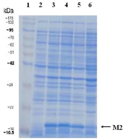
E.coli host strain BL21(DE3) and samples were collected at 2,3,4 and 20 hours after IPTG induction and disrupted by lysis buffer. Protein bands were visualized on 12% polyacrylamide gel (Fig. 1) and confirmed by Western-blotting using monoclonal anti-M2 antibody (Fig. 2). SDS-PAGE analysis demonstrated an approximately 17KD band as expected. Protein
Fig. 1. Expression analysis of M2 protein (17 kDa) by SDS-PAGE.
Lane 1: Protein marker; Lane2: Lysate before induction; Lane 3-6: Lysate collected at the time of 2h, 3h, 4h and 20h after IPTG induction.
expression was detectable after one hour and reached to highest amount after two hours. Overnight incubation at 37°C decreased the
protein expression. The yield of M2 protein production was analyzed using Quantity One software. The results showed that the recombinant M2 protein was about 21% of the total BL21 proteins.
We previously constructed recombinant pQE30-M2 plasmid and expressed the M2 protein in E.coli host strain M15 (15). M2 protein production in BL21 and M15 cells was compared using Quantity One software (Fig. 3). The results showed that the yield of M2 protein in BL21 cells was at least 3 times more than its production in M15.
Purification of M2 protein
The recombinant protein was purified with
Ni-TED column chromatography and
electrophoresed on 12% SDS-PAGE (Fig. 4). The results of SDS-PAGE showed that the purification of M2 protein using this system was very specific.
Discussion
A comprehensive vaccine that can induce protection against different influenza viruses has become a research emphasis. A limitation of current vaccines is that the major vaccine targets, the antigenic regions of HA and NA, are highly susceptible to continuous mutation
Fig. 2. Western blot analysis: The recombinant protein was electrophoresed on 12% SDS-PAGE, transferred to a nitrocellulose membrane and probed by using anti- M2 monoclonal antibody.Lane 1: Protein marker; Lane 2: Lysate collected at the time of 4h after IPTG induction; Lane3: Cell lysate before induction.
Fig. 3. Comparison of M2 protein production percentage in BL21 and M15 host strain before and after IPTG induction up to 4 hours.
Fig. 1. Analysis of purification M2 protein by 12% SDS-PAGE. Lane 1: Washing fraction, Lane2~4: Elution fractions, Lane 5: Protein marker, Lane6: Cleared lysate.
in circulating epidemic virus strains. There are various projects in the context of different universal influenza vaccines at the base of conserved sequences of viral proteins (1). Developing universal influenza vaccines that have protection against different types of influenza virus and even pandemic strains needs to identify conserved areas in influenza virus proteins. In influenza A viruses, the M2 protein is highly conserved among and within different subtypes and is therefore an attractive target antigen for vaccines that could provide cross-strain protection. Therefore, recombinant vaccine technologies based on M2 protein has been investigated widely (1). External domain of highly conserved M2 protein has been proposed to develop universal influenza vaccines. Recent studies with induced protective immune responses and broad spectrum protection has created hopes on further development of safe universal influenza vaccines against seasonal and pandemic influenza in the near future )14-20(.
James W. Huleat et al produced recombinant protein (STF2.4xM2e) in E.coli by fusion of four copy tandem of human influenza virus M2e protein sequence to the TLR5-specific ligand of Salmonella typhimurium flagellin fljB (STF2) gene. The study revealed that the (STF2.4xM2e) has high immunogenicity to induce T cell and B cell responses )11(.
Denisa et al used plant virus protein, papaya mosaic virus (PapMV) with the ability to carry M2e (papmV-cp-M2e) as a peptide delivery system and also as an adjuvant to enhance influenza vaccine efficacy. They reported production of antibodies against M2e using this construct in animal models )18(.
According to Jae-Min Song et al, the influenza M2 protein virus-like Particle (M2 VLPs) has high immunogenicity in mice. It was found that M2e VLPs causes the induction of T cell specific responses and the production of antibodies against different virus strains. Also, it could confer long-lasting cross protection against heterologous and heterosubtypic influenza viruses )19(.
Stephen Mark Tompkins et al investigated prime-boost vaccination method. In this experiment M2-DNA was used as the prime
vaccine and recombinant adenovirus expressing M2 (M2-Ad) as the booster vaccine. Results showed that this kind of vaccination induced T cell responses and increased antibodies against the human and birds viruses and finally protected against H5N1 lethal challenge )20(.
M2 protein has been expressed in various eukaryotic and prokaryotic cells. Esghaei et al
compared M2 protein expression in different kinds of eukaryotic cells including COS-7, Hela and MDCK. A higher level expression of M2 protein in COS-7 cells was confirmed using ELISA technique, SDS-PAGE and western blot analysis )15).
Several studies have been conducted for M2 protein production in prokaryotic cells as well. Ebrahimi et al reported M2 protein expression in Escherichia Coli BL21 (DE3) strain using pAED4 expression vector to produce universal recombinant vaccine (21).
In the present study, full-length M2 gene of the seasonal human influenza virus A/NewCaledonia/20/99 (H1N1) was subcloned into pET28a under the control of T7 promoter. T7 RNA polymerase activity is around five times more than E.coli RNA polymerase (22). So it has been expected overexpression of the genes under control of T7 promoter. E.coli
strain BL21 (DE3) is highly used to produce cloned proteins in plasmid vectors containing T7 promoter. In comparison to M2 expression in M15 cells using pQE30 expression vector in our previous study (14), we showed that the M2 expression using this system is much more valuable and cost effective for M2 production in prokaryotic system than the previous one. The expressed M2 protein showed to have an apparent molecular weight around 17kD as estimated by Expasy online database and reacted specifically with commercial anti-M2 monoclonal antibody in Western blot analysis. As the recombinant M2 protein prepared here has histidine residue at both terminals, it was successfully purified in one-step purification procedure.
Conclusion
In summary, our study showed successful use of pET28a plasmid for cloning and expression of influenza virus M2 protein as a fusion with 6-His tag in Escherichia coli BL21strain. In order to design an effective and protective vaccination regimen, we are going to evaluate immunogenicity of the purified M2 protein with different adjuvant and/or M2-DNA vaccine in animal model.
References
1. Saluja V, Visser MR, van Roosmalen ML, Leenhouts K, Huckriede A, Hinrichs WL, Frijlink HW. Gastro-intestinal delivery of influenza subunit vaccine formulation adjuvanted with Gram-positive enhancer matrix (GEM) particles .European Journal of Pharmaceutics and Biopharmaceutics. 2010;(76):470–474.
2. Fiers W, De Filette M, El Bakkouri K, Schepens B, Roose K, Schotsaert M. M2e-based universal influenza A vaccine. Vaccine. 2009;(45):6280-3. 3. Wang R, Song A, Levin J, Dennis D, Zhang NJ, Yoshida H, et al. Therapeutic potential of a fully human monoclonal antibody against influenza A virus M2 protein. Antiviral Research. 2008;(80):168–177.
4. Du L, Zhou Y, Jiang S. Research and development of universal influenza vaccines. Microbes and Infection. 2010;(12):280–286. 5. Betakova T. M2 Protein–A Proton Channel of Influenza A Virus. Current Pharmaceutical Design. 2007;3231-3235.
6. Hatta Y, Hatta M, Bilsel P, Neumann G, Kawaoka Y.An M2 cytoplasmic tail mutant as alive attenuated influenza vaccine against pandemic (H1N1) 2009 influenza virus. Vaccine 2011;(29):2308–2312.
7. Fu TM, Freed DC, Horton MS, Fan J, Citron MP, Joyce JG, et al. Characterizations of four monoclonal antibodies against M2 protein ectodomain of influenza A virus. Virology. 2009;385(1):218-26.
8. Zhao G, Du L, Xiao W, Sun S, Lin Y, Chen M, et al. Induction of protection against divergent H5N1 influenza viruses using a recombinant fusion protein linking influenza M2e to Onchocerca volvulus activation associated protein-1 (ASP-1) adjuvant. Vaccine. 2010;(44):7233-40.
9. Wu F, Yuan XY, Li J, Chen YH. The co-administration of CpG-ODN influenced protective
activity of influenzaM2e vaccine .Vaccine. 2009;(27):4320–4324.
10. Kitikoon P, Vincent AL, Janke BH, Erickson B, Strait EL, Yu S. Swine influenza matrix 2 (M2) protein contributes to protection against infection with different H1 swine influenza virus (SIV) isolates. Vaccine. 2010;(28):523–531.
11. Huleatt JW, Nakaar V, Desai P, Huang Y, Hewitt D, Jacobs A. Potent immunogenicity and efficacy of a universal influenza vaccine candidate comprising a recombinant fusion protein linking influenza M2e to the TLR5 ligand flagellin. Vaccine. 2008;(26):201-214.
12. Eliasson DG, El Bakkouri K, Schön K, Ramne A, Festjens E, Löwenadler B. CTA1-M2e-DD: A novel mucosal adjuvant targeted influenza vaccine. Vaccine. 2008;(26):1243-1252.
13. Fu TM, Grimm KM, Citron MP, Freed DC, Fan J, Keller PM. Comparative immunogenicity evaluations of influenza A virus M2 peptide as recombinant virus like particle or conjugate vaccines in mice and monkeys. Vaccine. 2009;(27):1440–1447.
14. Alavi Esfahani MA, Fotouhi F, Ghaemi A, Saleh M, Chalabiani S, Farahmand B, Tavasoti Kheiri M. Expression of M2 Protein of Human Influenza Virus in Escherichia Coli. J Isfahan Med Sch. 2013;30(216):2091-102.
15. Esghaei M, Monavari SH, Tavassoti-Kheiri M, Shamsi-Shahrabadi M, Heydarchi B, Farahmand B, Saleh M, Fotouhi F. Expression of the influenza M2 protein in three different eukaryotic cell lines. Journal of Virological Methods. 2012;(179):161– 165.
16. Wang TT, Tan GS, Hai R, Pica N, Ngai L, Ekiert DC. Vaccination with a synthetic peptide from the influenza virus hemagglutinin provides protection against distinct viral subtypes. Proc Natl Acad Sci USA. 2010;(44):18979–18984.
17. Ameiss K, Ashraf S, Kong W, Pekosz A,Wu WH, Milich D. Delivery of woodchuck hepatitis virus-like particle presented influenza M2e by recombinant attenuated Salmonella displaying a delayed lysis phenotype.Vaccine. 2010;(28):6704– 6713.
18. Denis J, Acosta-Ramirez E, Zhao Y, Hamelin ME, Koukavica I, Baz M, et al. Development of a universal influenza A vaccine based on the M2e peptide fused to the papaya mosaic virus (PapMV) vaccine platform. Vaccine. 2008;(26):3395–3403. 19. Song JM, Wang BZ, Park KM, Van Rooijen N, Quan FS, Kim MC. Influenza virus-like particles containing M2 induce broadly cross protective immunity. PLoS One. 2011;(1):e14538.
20. Tompkins SM, Zhao ZS, Lo CY, Misplon JA, Liu T, Ye Z. Matrix protein 2 vaccination and protection against influenza viruses, including subtype H5N1. Emerg Infect Dis. 2007 (3):426-35. 21. Ebrahimi SM, Aghaiypour K, Nili H, Esmailzadeh M, Mirjalili A. Construction of a pAED4-M2 vector for expressing avian influenza
A (H9N2) virus M2 gene as a universal recombinant vaccine model. Turk. J. Vet. Anim. Sci. 2009;33:401-406.
22. Lussier FX, Denis F, Shareck F. Adaptation of the highly productive T7 expression system to Streptomyces lividans. Appl Environ Microbiol. 2010;76(3):967-70.