DETECTION OF URINARY TRACT ANOMALIES USING
ULTRASONOGRAPHY DURING ANTENATAL SCREENING
1
Vishva Chauhan,2Chetan Mehta,3Harshad Wankhade,4Dhruti Maisuri,
5
Ayaz Dabiwala
1&3
3rdYear Resident,2Associate Professor & Head, 2ndYear Resident, 1stYear Resident, Radiology Department, SSG Hospital & Medical College, Baroda, Gujarat
Corresponding Author: Dr Vishva Chauhan, 3rdYear Resident, Radiology Department, SSG Hospital & Medical College, Baroda, Gujarat. Mail: [email protected]
INTRODUCTION
Urinary tract anomaly is very common in occurrence, with the rate of 1.08 %, i.e. approximately 1 urinary tract anomaly is encountered in every 100 ante-natal
patients scanned. A systematic approach to the prenatal diagnosis of urinary tract abnormalities includes assessment of amniotic fluid volume, localization and characterization of urinary tract abnormalities, assessment of fetal gender, ABSTRACT
Background & Aim: To study the pattern and prevalence of urinary tract anomalies, which can be diagnosed by USG during the antenatal period and to look for any associated anomaly. Material & Method: This study was carried out on a total of 1750 patients in SSG Hospital Vadodara. All the patients had come for their routine ante-natal screening. All USGs were done using curvilinear low frequency (3 -5 MHz) transducers. Results: Out of the 1750 patients studied over a period of 8 months, 19 patients were found to be having urinary tract anomalies. The prevalence of urinary tract anomaly in our study was 10.86 per 1000 fetuses (1.08%). In our study of the 19 fetuses with urinary tract anomalies, 2 had unilateral absent kidney, 2 fetuses were of Cystic renal dysplasia, 2 had ectopic kidneys and 1 patient, each of PUJ obstruction, vesicoureteric reflux, megaureter and posterior urethral valve giving the typical key hole appearance of the bladder. There were 2 fetuses with bilateral absent kidneys, oligohydramnios and pulmonary hypoplasia (Potter's sequence) which had to be terminated. Associated anomalies detected were of prune belly syndrome in association with megaureter. There were 2 fetuses with bilateral echogenic kidneys associated with absent radius. A fetus with bilateral cystic renal dysplasia also had associated hemi-vertebra and pulmonary hypoplasia. In our study, 26 % of the patients reflected changes in liquor be it oligohydramnios or polyhydramnios and 21 % of the patients had to be terminated prematurely as the anomalies wouldn’t have
sustained in the late gestation. Conclusion: Urinary tract anomalies can be precisely diagnosed and classified in the antenatal period using ultrasonography imaging and it can be helpful in planning immediate postnatal care.
and search for associated abnormalities.1,2 Normal AFV in the second half of pregnancy implies at least one functioning kidney and a patent urinary conduit to the amniotic cavity.2In the setting of a urinary tract abnormality, normal AFV indicate a good prognosis. Occasionally and paradoxically, polyhydramnios may occur, especially with unilateral obstructive uropathy, with meroblastic nephroma, or when there are concomitant abnormalities of the central nervous system (CNS) or gastrointestinal (GI) tract.3 A detailed anatomic scan to search for associated abnormalities, which may indicate the presence of a syndrome or chromosomal abnormality. Renal anomalies may be part of the VATER association (vertebral defects, anal atresia, trachea-esophageal fistula, radial defects and renal anomalies). An expansion of this syndrome, VACTERL, includes cardiac and non-radial limb defects.3The present study was undertaken with the aim to study the pattern and prevalence of urinary tract anomalies, which can be diagnosed by USG during the antenatal period and to look for any associated anomaly.
MATERIAL & METHOD
This prospective longitudinal study was conducted in the Department of Radiology, SSG Hospital & Medical College, Baroda, Gujarat during the period of one year. All cases, which were brought to the department of Radiology for antenatal screening were taken into the consideration. Ethical clearance was obtained from the Institutional Ethical Committee before commencement of the study. All the patients who came for their routine ante-natal screening were examined by Ultrasonography using
curvilinear low frequency (3-5 MHz) transducers after taking informed consent. Detailed examination was conducted and data were collected in a pre-designed proforma and analyzed after comparing with other authors.
RESULT
One thousand and fifty cases of pregnancy were screened out by USG abdomen to diagnose any urinary tract anomaly and out of them 19 patients were found to be having urinary tract anomalies. The prevalence of urinary tract anomaly in our study was 10.86 per 1000 births. The anomalies we encountered were ranging from simple ones like single kidney to complicated ones like mega-ureter in prune-belly syndrome and urinoma in a posterior urethral valve. The most common anomaly was dilation of the renal pelvis, which was compatible with life and
didn’t require any intervention post -natally. Single absent, dysplastic and ectopic kidneys had uneventful course, while the bilaterally absent kidney was found to be lethal anomaly. VUR, PUJ obstruction, mega-ureter, urinoma, PUV and bilateral echogenic kidneys required intervention post-natally. 26 % of the patients reflected changes in liquor be it oligohydramnios or polyhydramnios. 21 % of the patients had to be terminated
prematurely as the anomalies wouldn’t
have sustained in the late gestation.
DISCUSSION
trimester scans was found to be 11.4 per 1000 fetuses (1.14%). In our study of the 19 fetuses with urinary tract anomalies, 2 had unilateral absent kidney, 2 fetuses were of cystic renal dysplasia, 2 had ectopic kidneys and 1 patient each of PUJ obstruction. Vesicoureteric reflux, megaureter and posterior urethral valve giving the typical key hole appearance of the bladder. There were 2 fetuses with bilateral absent kidneys, oligohydramnios and pulmonary hypoplasia (Potter's sequence) which had to be terminated. Associated anomalies detected were of Prune Belly Syndrome in association with megaureter. There were 2 fetuses with bilateral echogenic kidneys associated with absent radius. A fetus with bilateral cystic renal dysplasia also had associated hemi-vertebra and pulmonary hypoplasia. In our study, 26 % of the patients reflected changes in liquor be it oligohydramnios or polyhydramnios. 21 % of the patients had to be terminated prematurely as the
anomalies wouldn’t have sustained in the
late gestation, which is comparable to the data concluded by Rasmussen et al,4where 127 of the 412 fetuses (30.8%) underwent termination and to the perinatal mortality as found by Taguchi K et al where 7 lethal anomalies were detected of the 56 fetuses (12.5%) with urinary tract anomalies. Lethal anomalies in our study were four, including- Bilateral absent kidneys in 2, bilateral cystic renal dysplasia in 1, and bilateral echogenic kidneys with absent radius in 1. In a retrospective study conducted by Gloor et al5 in Mayo clinic, Rochester, out of 56 children with urinary tract anomalies, they found 38 children (68%) having the most common anomaly as isolated hydronephrosis and multicystic renal dysplasia. In study by Taguchi et al,
most common anomaly detected was hydronephrosis (33.9%), which is comparable to our study, where the most common anomaly was dilation of the renal pelvis found in (21.05%), which was
compatible with life and didn’t require any intervention postnataly. Single absent, dysplastic and ectopic kidneys had uneventful course. The bilaterally absent kidney was lethal anomaly. VUR, PUJ obstruction, mega-ureter, urinoma, PUV and bilateral echogenic kidneys required intervention post-natally. Gloor et al5 found that 4 of the 32 newborn (12.5%) in their study, who underwent VCUG, had reflux. While in our study, 2 neonates underwent VCUG (10.5 %).
1) UNILATERAL RENAL
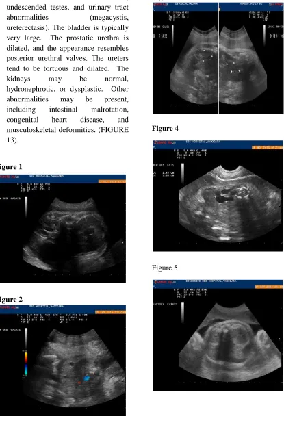
AGENESIS: Unilateral renal agenesis is three to four times more common than bilateral renal agenesis. It may be difficult to diagnose prenatally because AFV is normal and the bladder appears normal. If a kidney is not found in the renal fossa, most are either congenitally absent or ectopic. The contra-lateral kidney may be enlarged because of compensatory hypertrophy. (FIGURE 1).
2) BILATERAL RENAL AGENESIS: ultrasound findings include severe oligohydramnios and non-visualization of the kidneys and bladder.4 Distinctive, flattened appearance of the adrenal gland on longitudinal
sonogram (“lying down” adrenal sign)
arising from the aorta (Au) in a fetus with bilateral renal agenesis.7 Before
16 weeks’ gestation, AFV is not dependent on urine production and may be normal despite the absent renal function. (FIGURE 2).
3) RENAL ECTOPIA: Renal fossa is empty, careful scanning may demonstrate the ectopic kidney adjacent to the bladder or iliac wing. The ectopic kidney is located on the opposite side of the abdomen relative to its ureteral insertion into the bladder, resulting in crossed renal ecotopia with or without fusion. In most cases the crossed kidney fuses with the normally located kidney (cross-fused ecotopia), and an enlarged bilobed kidney is seen. Renal ecotopia is associated with a high incidence of urologic abnormalities, most often VUR. (FIGURE 3).3
4) MCKD: Malformed kidney is usually enlarged but may be normal or small. There are multiple cysts of varying sizes that do not communicate with each other and are randomly distributed. Large peripheral cysts distort the retiform contour. Pelvis and ureter are usually atretic and not visible. On color Doppler evaluation, the renal artery is either absent or very small.8Occasionally, a MCDK with a large central cyst and small peripheral cysts can mimic hydro nephrosis from ureteropelvic junction (UPJ) obstruction. In hydro nephrosis, however, the dilated calyces are of uniform size and anatomically aligned and communicate with the dilated renal pelvis. The kidney usually maintains the reniform contour, with renal parenchyma present peripherally. On
serial examinations, the kidney and its cysts may increase or decrease in size or may initially enlarge and later involute. (FIGURE 4).8
5) ARPKD: Sonography reveals bilateral retiform enlargement of the kidneys. There is poor delineation of the intrarenal structures. The numerous tiny cysts are usually smaller than the limit of sonographic resolution, but they create multiple acoustic interfaces, accounting for the characteristic increased renal echogenicity and loss of corticomedullary differentiation. The kidney demonstrates increased echogenicity, with no visible cysts. There is loss of corticomedullary differentiation. When renal function is abnormal, there is oligohydramnios, and the bladder is small to absent. (FIGURE 5).12,13
7) MESOBLASTIC NEPHROMA is
indistinguishable from Wilms’ tumor.
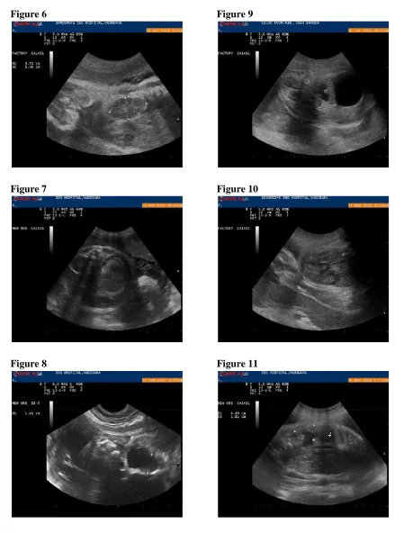
Mesoblastic nephroma is usually seen as a moderately echogenic, solid mass completely replacing the kidney or localized to the part of the kidney. The mass may demonstrate increased vascularity and cystic components. Polyhydramnios is a frequent association84 and may lead to preterm labor and preterm birth. Perinatal complications are likely, including acute fetal distress, neonatal hypertension, and hypercalcemia. (FIGURE 7).14
8) PUJO: Dilated renal pelvis with or without cablecasts is identified. The ureter and the bladder are not dilated. Severe chronic obstruction leads to the effacement of the calyces and thinning of the renal cortex (SFU grade 4). Rarely, the renal pelvis may be extremely dilated, presenting as a large, unilocular cystic mass. Rupture of the collecting system results in the development of a perirenal urinoma. (FIGURE 9). This “pop-off”
mechanism may protect the obstructed kidney from further prenatal damage and may diminish the degree of hydro nephrosis. The AFV is usually normal but may be increased paradoxically. When unilateral hydronephrosis is accom- panied by oligohydramnios, a search for contralateral renal pathology is warranted. (FIGURE 8).15
9) VUJO: The affected kidney may demonstrate dilation of the ureter and renal pelvis. serpiginous cystic segments must be traced to the renal pelvis and bladder. Megaureters are classified into three types per their morphologic appearance. Type I
megaureter dilation involves only the distal ureter, with a normal- appearing upper tract. Type II extends to both ureters and pelvis. Type III is associated with severe hydro-nephrosis and ureteric tortuosity. To differentiate the dilated ureter from the bowel is important. Bowel contents are usually more echogenic than urine. In addition, the ureter generally comes into close contact with the spine, but the small bowel does not. (FIGURE 10).15
10) HN and VUR: The most common prenatal sonographic diagnosis is hydronephrosis, which may be unilateral or bilateral. The ureter may be dilated. Intermittent dilation of the collecting system favors VUR. Fluctuation or variation in the RPD (changing by more than 3 mm) during an obstetric sonogram was strongly associated with high-grade VUR (grades IV-V). (FIGURE 11).16
11) Fetal megacystis has been reported as early as 10 to 14 weeks gestation when the longitudinal bladder diameter is 7 mm or more. Severe megacystis (bladder length >15 mm) Posterior urethral valves are the most common cause of lower urinary tract obstruction, followed by urethral atresia or stricture. Posterior urethral valves are seen exclusively in males and may cause total, intermittent, or partial obstruction, with variable prognosis. Back pressure causes a persistently dilated urinary bladder, with a dilated proximal urethra (keyhole sign) (FIGURE 12)15, 16,17 12) Prune Belly Syndrome is
absent abdominal musculature, undescended testes, and urinary tract abnormalities (megacystis, ureterectasis). The bladder is typically very large. The prostatic urethra is dilated, and the appearance resembles posterior urethral valves. The ureters tend to be tortuous and dilated. The kidneys may be normal, hydronephrotic, or dysplastic. Other abnormalities may be present, including intestinal malrotation, congenital heart disease, and musculoskeletal deformities. (FIGURE 13).
Figure 1
Figure 2
Figure 3
Figure 4
Figure 6
Figure 7
Figure 8
Figure 9
Figure 10
Figure 12
Figure 13
CONCLUSIONS
The prevalence of different types of urinary tract anomalies is higher than that reported in developed countries. Urinary tract anomalies can be precisely diagnosed and classified in the antenatal period using ultrasonography imaging and it can be helpful in planning immediate postnatal care.
Source of Funding : Nil. Conflict of Interest : None. REFERENCES
1. Grandjean H, Larroque D, Levi S. The performance of routine ultra
sonographic screening of pregnancies in the Eurofetus Study. Am J Obstet Gynecol 1999; 181:446-454.
2. Goldstein RB, Filly RA. Sonographic estimation of amniotic fluid volume: subjective assessment versus pocket measurements. J Ultrasound Med 1988; 7:363-369.
3. Wiesel A, Queisser-Luft A, Clementi M, et al. Prenatal detection of congenital renal malformations by fetal ultra sonographic examination: an analysis of 709,030 births in 12 European countries. Eur J Med Genet 2005; 48:131-144.
4. Rasmussen K, Nielsen J, Dahl G. The Prevalence of Chromosome Abnormalities Among Mentally Retarded Persons in a Geographically Delimited Area of Denmark Clin Genet 1982;22(5):244-255.
5. Gloor JM, Ogburn PL JR., Breckle RJ, Morgenstern BZ, Milliner DS. Urinary Tract Anomalies Detected by Prenatal Ultrasound Examination at Mayo Clinic Rochester. 1995;70(6):526-531. 6. Deshpande C, Hennekam RCM.
Genetic syndromes and prenatally detected renal anomalies. Semin Fetal Neonatal Med 2008;13: 171-180. 7. Potter EL. Bilateral absence of ureters
and kidneys: a report of 50 cases. Obstet Gynecol 1965; 25:3-12.
8. DeVore GR. The value of colour Doppler sonography in the diagnosis of renal agenesis. J Ultrasound Med 1995; 14:443-449.
associations and management. Prenat Diagn 2001; 21:924-935.
10. Van Eijk L, Cohen-Overbeek TE, den Hollander NS, et al. Unilateral multicystic dysplastic kidney: a combined pre- and postnatal assessment. Ultrasound Obstet Gynecol 2002; 19:180-183.
11. Estroff JA, Mandell J, Benacerraf BR. Increased renal parenchymal echogenicity in the fetus: importance and clinical outcome. Radiology 1991; 181:135-139.
12. Twining P. Genitourinary malformations. In: Nyberg DA, McGahn JP, Pretorius DH, Pilu G, editors. Diagnostic imaging of fetal anomalies. Philadelphia: Lippincott–
Williams & Wilkins; 2003.
13. Tsatsaris V, Gagnadoux MF, Aubry MC, et al. Prenatal diagnosis of bilateral isolated fetal hyperechogenic kidneys. Is it possible to predict long term outcome? BJOG 2002; 109:1388-1393.
14. Chaumoitre K, Brun M, Cassart M, et al. Differential diagnosis of fetal hyperechogenic cystic kidneys unrelated to renal tract anomalies: a multicentre study. Ultrasound Obstet Gynecol 2006; 28:911-917.
15. Chen WY, Lin CN, Chao CS, et al. Prenatal diagnosis of congenital mesoblastic nephroma in mid-second trimester by sonography and magnetic resonance imaging. Prenat Diagn 2003; 23:927-931.
16. Glazer GM, Filly RA, Callen PW. The varied sonographic appearance of the urinary tract in the fetus and new born
with urethral obstruction. Radiology 1982; 144:563-568.