Original Article
Learning-dependent LTP and synaptic ultrastructural
modification after physical exercise in rats
with middle cerebral artery occlusion:
relevance for learning and memory
Yamei Li1, Qian Yu1, Jing Fu1, Yufeng Li1, Bo Peng1, Guo Huang2, Zongxin Xiao1
Departments of 1Rehabilitation, 2Laboratory Medicine, Sichuan Academy of Medical Sciences & Sichuan
Provin-cial People’s Hospital, University of Electronic Science and Technology of China, Chengdu, Sichuan, China Received May 23, 2018; Accepted August 3, 2018; Epub December 15, 2018; Published December 30, 2018
Abstract: Objective: The goal of this study was to investigate the effect of physical exercise on the abilities of learning and memory, as well as long-term potentiation (LTP) and synaptic ultrastructural modification in the contralateral hippocampal CA3 region of rat treated with right middle cerebral artery occlusion (MCAO). Methods: Thirty-two rats after right MACO procedure were randomly divided into two groups with 16 respectively. Physical exercise was given to the rehabilitation group, while the control group were raised in their original living state. The abilities of learning and memory were tested by one-trial passive avoidance response and Y-maze test. With the electrophysi-ological techniques, LTP in left CA3 region was recorded after physical exercise, 60 times of Y-maze learning and the rats had acquired the Y-maze test. Finally, this region was processed for electron microscopy and stereological techniques were used to assess the plasticity of synaptic ultrastructure. Results: LTP in the CA3 region of the reha-bilitation group emerged earlier than that of the control group (P<0.05), and the same LTPs presented when rats in both groups had acquired the test. The synaptic curvature and thickness of post-synaptic density (PSD) as well as percentage of perforated synapses were significantly increased in the rehabilitation group comparing with the con -trol group (P<0.05). Furthermore, the abilities of learning and memory in the rehabilitation group were significantly superior to that in the control group (P<0.05). Conclusion: There is a widespread assumption that the improvement of learning and memory in MACO rats following physical exercise are likely to be related to the synaptic ultrastruc-tural modification and the earlier emergence of LTP in contralateral CA3 region.
Keywords: Middle cerebral artery occlusion (MCAO), exercise, learning-dependent potentiation (LTP), synaptic ultrastructure modification, learning-memory ability
Introduction
Post-stroke cognitive impairments including learning and memory dysfunction are major problems that affects up to 70% of stroke survi-vors [1, 2]. Patients with learning and memory dysfunctions after stroke may be also troubled by residual motor, perceptual, and lingual defi -cits, so it is considered that post-cognitive impairments not only weaken patients’ ability to restore motor skills due to memory problems or poor judgment, but also obviously decline the activities of daily living (ADL) and quality of life (QOL) even if the physical functions have recovered to a certain extent [3]. Therefore, how to improve the post-stroke learning and
-tive effect on various cogni-tive domains includ-ing learninclud-ing and memory abilities [5-7]. However, the related neurobiological funda-mentals are still not absolutely clear, and in previous studies it has been said that physical exercise may increase neurogenesis, synapto-genesis, long-term potentiation (LTP), and cere-bral blood volume (CBV). Additionally, the neu-roplasticity of structures and functions are promoted by various growth factors such as brain-derived neurotrophic factor (BDNF), insu-lin-like growth factor-I (IGF-I), and vascular endothelial growth factor (VEGF) [8, 9]. Even so, there are a limited number of studies on the exercise-related changes of learning and mem-ory dysfunctions caused by stroke, as well as changes of synaptic ultrastructure and function at the same time.
Synaptic plasticity, which refers to changes in the strength of connections between neurons in response to many different rehabilitation strategies, is one of the functional aspects of hippocampus which is closely related to learn-ing and memory [10]. LTP, a persistent increase in synaptic efficacy, is a leading cellular model of learning and memory. It could be detected by electrophysiological methods as lasting modifi -cations of synaptic transmission, and could be accompanied by morphological changes in the synapses undergoing plasticity [11]. The hippo-campus is extremely sensitive to cerebral isch-emia and may ultimately lead to cognitive impairment [12]. Therefore, the aim of this study was to investigate the effects of physical exercise on the abilities of learning and memo-ry, as well as LTP and synaptic ultrastructural modification in contralateral hippocampal CA3 region of rats that underwent right middle cere-bral artery occlusion (MCAO). Preliminarily eval-uation of the effect of physical exercise on the central nervous system, especially synaptic plasticity of substructure and morphology in status of cerebral infarction, is reported here. Materials and methods
Animals and grouping
Thirty-two male Wistar rats (8 weeks, 250±50 g, purchased from the Experimental Animal Center of Army Medical University, Chongqing, China) were randomly divided into two groups with 16 rats in each. The control group was
allowed free movement after cerebral infarc-tion, while the rehabilitation group is subjected to behavioral training after cerebral infarction. All rats were group housed under a 12 hour light/dark cycle with food and water provided at will. This study was carried out in strict accor-dance with the recommendations in the Guide for the Care and Use of Laboratory Animals of the National Institutes of Health. The animal experimentation protocol was reviewed and approved by the Institutional Animal Care and Use Committee (IACUC) of the Army Medical University (Chongqing, China).
Surgical procedure
Implantation of the electrode: Electrodes were implanted into the rats for inducing LTP [13]. All rats were anesthetized by intraperitoneally injecting of 25% urethane (Shanghai Hengyuan Biotechnology Co., Ltd.) with a dosage of 5 ml/ kg and mounted in a brain stereotaxic instru-ment (Shanghai Mobiledatum Co., Ltd., RD16- 17-1ss). Then they were subjected to implanta-tion of bipolar stimulating electrodes in the left entorhinal cortex that comprised the perfo-rant path (PP) (AP6.8~7.0 mm, L4.4~4.5 mm, H5.5~6.2 mm) and monopolar recording elec-trodes in the left pyramidal cell layer of CA3 region of the hippocampus (Ap3.0~3.4 mm, L3.3~3.5 mm, H3.8~4.2 mm) with a micro- electrode propeller (Beijing Sunny Instruments Co. Ltd., model: XX11-WK-III), which were used to monitor the evoked potentials at the perfo-rant path-CA3 synapses. The stimulating elec-trodes were constructed from two twisted insu-lated nichrome wires (0.14 mm diameter), and recording electrodes were made of insulated metal wire (0.2 mm diameter, tip resistance <5 kQ). The electrodes were lowered into the brain through the holes drilled on the skull of hippo-campus and PP respectively. The reference electrode was fixed in the skull with a small anchor screw. Test-pulse recordings during sur-gery aided the depth adjustment of the elec-trodes in order to record the population spike (PS) with maximum amplitude, which was later verified by postmortem histology. The wires were stabilized on the skull using dental cement.
Establishment of animal models
procedure by thread-inserting method [14]. During the surgery, a heating water bag was used maintain the rectal temperature at 37°C. The animals were anesthetized by intraperito-neally injecting of 25% urethane (Shanghai Hengyuan Biotechnology Co., Ltd.) with the dos-age of 5 ml/kg and fixed onto the operating table in a supine position. A right paramedian incision (25 mm) was made in the neck and the right common carotid artery (CCA), internal carotid artery (ICA) and external carotid artery (ECA) were surgically exposed. The distal sec-tion of the ECA was ligated by a vascular clip. A small puncture opening (Φ 0.2 mm) was made in the proximal ECA. A length of monofilament (Φ 0.22 mm) covered by a layer of polyurethane varnish at the extremity (Φ <0.25 mm) was inserted through the opening. Then the mono-filament was gently advanced from the lumen of the ECA, passing the bifurcation of CCA and entering into the ICA, moving and stopping when met with resistance, for a distance of 17 to 19 mm from the bifurcation of CCA. Skin on the neck incision was sutured and the rats were allowed to recover. After awaking, four of the following signs indicated that the model was successful: the left forelimb adduction and flex -ion when lifting the tail, Horner syndrome in the right side, circled to the left when crawling, and inclined to the left when standing. If the rats died, other healthy rats were timely involved to supplement. After surgery, rats were housed individually and given 4 days to recovery before interventions.
Physical exercise
Exercise training, designed according to previ-ous studies with modifications [15, 16], were performed on the rats in the rehabilitation group on the fifth day after the MCAO proce -dure, including rolling-cage training, balance training and screen training, to improve the walking, grabbing, and balancing functions, with the frequency of twice a day, 10 minutes each time for 4 weeks with 6 days in each week.
Rolling-cage training: the rats were set to run passively in a circular mesh instrument (length = 100 cm, diameter = 60 cm, made at the College of Basic Medical Sciences, Army Medical University, Chongqin) manually rotated by the researcher, to improve the abilities of grabbing, rotation, and walking.
Balance training: the rats were placed on a square stick (height = 5 cm, length = 150 cm, width = 2 cm, made at the College of Basic Medical Sciences, Army Medical University, Chongqin) and induced to walk by food temptation.
Screen training: the rats were stayed on a screen (mesh: 1 cm × 1 cm, length = 50 cm, width = 40 cm, height = 80 cm, made at the College of Basic Medical Sciences, Army Medical University, Chongqin), with 12 cm-thick sponge on the ground under it. During the screen moving gradually from horizontal posi-tion to vertical posiposi-tion and maintaining 5 sec-onds, rats were observed whether falling down or holding the screen by forepaw. The longer time that the rat stayed on the screen reflected better muscle force. This training was taken to improve the grabbing ability and muscle force. Rats in the control group were allowed to move freely, and were given no special physical exercise.
Cognition testing
Rats in the two groups were given cognition tests on the fifth week after operation. Two tests were used as previously described [17]: one-trial passive avoidance response and Y-maze test.
One-trial passive avoidance response
Y-maze test
The Y-maze contained three arms, a start arm and two choice arms (Zhangjiagang Biomedical Instrument Factory, China). On the top of each arm there was a signal lamp, which was used as an indicator of dangerous region when the lamp lightened for 6 seconds. There should be a dark arm without current as a safe region and two light arms as dangerous regions all the time during the training with the safe region being changed irregularly. The rats were stimu-lated to run from the light arm to the dark arm by the energization of 36 V alternating current. If the rat ran from one light arm to another light one, it was recorded as wrong. Otherwise, if the rat ran to the dark one and it was recorded as correct. The rats were trained for 10 consecu-tive sessions within one day, 10 times per ses-sion, with a 2-minute rest between sessions. The number of correct times in the first 60 training times and total times needed for the rats to get hold of the test (9 correct times out of 10 continuous tests were set as acquire-ment) were recorded. The fewer the total times, the greater the learning ability of the rats had.
LTP recording in the hippocampal CA3 region
The synaptic transmission efficacy in left hip -pocampal CA3 area was assessed in both groups 3 hours after physical exercise (that was before cognition tests), 60 times of Y-maze learning, and the rats had acquired the Y-maze test. The data for rats in the control group before cognition tests was regarded as base-line for comparison. Rats were kept in a self-made cage in a quiet state, and the test stimu-lation (monophasic square pulses, 0.7 Hz, 0.1 ms, with constant current intensity that elicited 50% of the maximum amplitude of PS after model made and before behavioral training) was delivered via stimulating electrodes to the perforant path. The pyramidal cell layer in CA3 region received perforant path inputs, and evoked potentials called population spikes (PS) were produced at the perforant path-pyramidal cell synapses. The recording electrodes were allowed for recording of the PS. Then these evoked responses were amplified by three amplifiers step by step (microelectrode amplifi -er WF-I (Model: ME-200A, Chengdu Taimeng Science & Technology Co. Ltd., China), physio- logical amplifier DSJ-F (Changzhou No. 2 Radio
Factory, China), and A/D amplifier (Model No.: UA352, Beijing Priority monitoring and control technology Co., Ltd., China). Finally, singles were acquired, processed, and analyzed by computer. The peak latency and peak ampli-tude of PS were used as indicators. The peak amplitude was measured as the vertical dis-tance between the line of the two lowest points and the peak of PS. The relative ratio of the increased amplitude of PS over the amplitude before exercise therapy was used as an indica-tor of synaptic transmission enhancement, that was, also as an indicator of LTP effect. At the end of the experiment, the positions of record-ing and stimulatrecord-ing electrodes were checked. Rats with electrodes not in the right position were removed.
Quantitative measurements of synaptic ultra-structure
After all the former steps, rats from each group were anaesthetized (0.4% sodium pentobarbi-tal, 1 μl/g, intraperitoneal) and decapitated. Then a 2 mm × 2 mm × 5 mm slice from their left hippocampal CA3 area (according to Woolsey’s new cortical functional zoning and marginal system anatomy) was quickly removed into a fixative solution (3% glutaraldehyde and 4% paraformaldehyde). The preparation under-went routine embedding and counterstaining with uranyl acetate and lead citrate. Sub- sequently, ultrathin sections were cut serially with an ultramicrotome and collected onto two copper grids per rat. With the HU-12A transmis-sion electron microscope (TEM) moving from the top left to the lower right corner of the cop-per grids, a total of 5 electron micrographs were obtained randomly from each rat with the magnification of 15,000 times, and then mag -nified to 30,000 times by optical microscopy.
[19]. The width of synaptic cleft was counted with a multi-point-average determination. For each group, a total of 40 micrographs were used for quantification.
Statistical analysis
Data were analyzed in SPSS (version 19.0). STL are expressed as median, and Nonparametric Mann-Whitney U tests were used to analyze the differences of STL between the two groups. The other data are expressed as mean ± SD, and the statistical differences of those data were determined by Student’s t-test except that the differences of the percentage of perforated synapses between the two groups was ana-lyzed by chi-square test. The level of signifi -cance was considered to be below 0.05.
Results
Cognition tests
The median of the STL in one-trial passive avoidance response for the rehabilitation group was significantly longer than that for the control group (286.7 seconds in the rehabilitation group v.s. 126.7 seconds in the control group; Z = -4.264, P<0.05) (Figure 1). The mean correct times for rats in the previous 60 times of Y-maze learning was 46.07±8.17 in rehabilita-tion group compared to the mean of 25.04±3.78 in control group. The difference was statistical-ly significant (t-test; T (t value) = 9.345, P<0.05). The mean times for the rats to get hold of the Y-maze test in rehabilitation group was less than that in the control group (68.02±11.67 for rehabilitation group, 107.07±16.32 for control group; T (t value) = -7.785, P<0.05) (Figure 2). Therefore, rats in the rehabilitation group had better abilities of learning and memory than rats in the control group.
LTP recording in the hippocampal CA3 region
[image:5.612.90.290.73.185.2]With respect to the relative ratio of increased amplitude, the amplitude of PS in the rehabili-tation group was significantly increased than that in the control group after 60 times of Y-maze learning (78.57±16.32% for the reha-bilitation group v.s. 43.12±11.21% for the con-trol group; T (t value) = 7.162, P<0.05), showing that the LTP effect was more distinct than that in the control group. However, no statistically significant difference between the two groups was observed after the rats had acquired the Y-maze test (238.58±18.96% for the rehabilita-tion group v.s. 235.16±16.46% for the control group; T (t value) = 0.545, P > 0.05) (Figure 3). This implied that same LTPs emerged in rats when the behavior had been acquired.
[image:5.612.90.289.250.373.2]Figure 1. The median of the step-through latency for rehabilitation group was significantly longer than that for control group (*P<0.05).
Figure 2. Rehabilitation group had more correct times in the previous 60 times of Y-maze test and needed less times to acquire the Y-maze test com-pared with control group (*P<0.05). A. correct times
in the previous 60 times of Y-maze test; B. times needed to acquire the Y-maze test.
Figure 3. Amplitude of population spike for rehabili-tation group was significantly increased than that for the control group after 60 times of Y-maze learning (*P<0.05), no statistically significant difference be
-tween the two groups was observed after all rats had acquired the Y-maze test (#P>0.05). A. after 60 times
[image:5.612.90.288.461.586.2]The mean peak latency of PS for the rehabilita-tion group was 3.08±0.41 ms before physical exercise versus 2.62±0.18 ms after the rats had acquired the Y-maze test (T (t value) = 4.109, P<0.05) (Figure 4). Furthermore,
com-parison of the mean peak latency between the two groups after cognition tests revealed a sig-nificantly shorter time in the rehabilitation group (2.62±0.18 ms for the rehabilitation group v.s. 3.09±0.48 ms for the model group; T (t value) = -3.667, P<0.05) (Figure 4).
After physical exercise, the peak amplitude of PS for rats in the rehabilitation group increased by 66.7% compared to the mean amplitude of rats in control group before cognition tests, the latter was regarded as baseline for compari-son. This illustrated that physical exercise had already induced learning-dependent long-term potentiation (LTP) in the left hippocampal CA3 region of rats with cerebral infarction, and the LTP effect could be further enhanced by the behavior-like cognition tests (one-trial passive avoidance response and Y-maze test). There- fore, improvement of synaptic efficacy was observed. These results were paralleled by improvement of learning and memory ability demonstrated in the following cognition tests. Therefore, physical exercise on rats with cere-bral infarction could improve their ability of learning and memory.
Quantitative analysis of synaptic ultrastructure
[image:6.612.90.285.73.195.2]On the electron micrographs, Gray’s type I syn-apses were identified by the presence of asym -metric interfaces with obviously thicker post-synaptic membrane comparing with prepost-synaptic membrane, prominent post-synaptic density (PSD) in the postsynaptic membrane, and clus-tering of vesicles near the presynaptic mem-brane. All of them accorded with the morpho-logical characteristic of excitatory synapse. Synapses in the control group mostly showed flat synaptic interface. Therefore, presynaptic and postsynaptic membranes were close to two lines and being parallel, with other few syn-apses curving slightly to form slightly concave or slightly convex type of presynaptic and post-synaptic membrane. Usually a single one active zone, less vesicles, severely degenerated mito-chondria were observed in these synapses. In contrast with the control group, trend toward an increase in the number of concave synapses was observed in the rehabilitation group. In other words, the surface of the postsynaptic membrane facing the axon terminal is obvious-ly concave, and the axon terminal also called presynaptic membrane protruded apparently to
Figure 4. Peak latency of population spike for the rehabilitation group after cognition tests got short-er comparing with that before physical exshort-ercise (*P<0.05). Peak latency of PS for the rehabilitation
[image:6.612.90.285.287.417.2]group after cognition tests was shorter than that for the control group after cognition tests (#P<0.05).
Figure 5. The rehabilitation group was superior in synaptic curvatures, PSD thickness and percentage of perforated synapses than control group (*P<0.05).
[image:6.612.92.289.479.594.2]fill the cavity, forming “U”-shaped configuration in the longitudinal section. The “U”-shaped syn -apses exhibited larger overall dimensions, at least two active zones, more vesicles clustering to active zones and the mitochondria of these synapses in cytoplasm showed integrated bilayer structure and clear cristae. Perforated synapses could be found which were character-ized as discontinuous PSD profiles in serial sections.
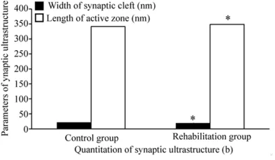
The synapse quantitation showed that the difference of synaptic curvatures, PSD thick-ness and percentage of perforated synapses between the two groups were statistically significant (1.049±0.038, 70.26±10.47 nm, 24.31% for rehabilitation group vs 1.020± 0.029, 61.32±11.74 nm, 7.98% for control group; T (t value) = 2.427, T (t value) = 2.278, χ2 = 9.524, P<0.05) (Figure 5) but no statistically significant difference was observed in the width of synaptic cleft and the length of active zone between the two groups (18.68±3.24 nm, 348.56±92.32 nm for rehabilitation group vs 20.98±8.72 nm, 341.32±87.39 nm for control group; T (t value) = -1.419, T (t value) = 0.228, P
> 0.05) (Figure 6).
Discussion
The hippocampus, which belongs to limbic sys-tem, is the important central nervous system for learning and memory, and is characterized by high vulnerability to any noxious input rang-ing from ischemia and hypoxia [10, 12]. Previous studies have demonstrated that the hippocampus has functional and structural plasticity. Therefore, its functional status is closely related to learning and memory ability, and it is now widely accepted that synaptic plasticity in hippocampus is an important com-ponent of the neural mechanisms underlying improvement of learning and memory dysfunc-tion [20]. The CA3 region in the hippocampus is considered to be closely related to spatial dis-crimination tasks of learning and memory [21]. Previous studies have shown that physical exer-cise or physical activity may have a significant positive effect on cognition impairments like learning and memory disfunctions [5-7], but the related neurobiological mechanisms are still not absolutely clear. Therefore, physical exercise may bring improvement of learning and memory abilities after cerebral infarction
and the accompanied mechanisms may be related to the synaptic ultrastructural modifica -tion and the earlier emergence of LTP in contra-lateral CA3 region.
In this study, physical exercise including rolling-cage exerciser, screen exerciser, and balance exerciser were performed on the rats with learning and memory dysfunctions after cere-bral infarction, and then cognition testing was used to assess their learning and memory abili-ties, LTP in the contralateral CA3 region, and synaptic ultrastructure in this region. The results showed that the learning and memory abilities of rats in rehabilitation group after physical exercise were significantly better than that of rats in control group. This was in line with earlier induction of learning-dependent long-term potentiation (LTP), higher peak ampli-tude, and shorter peak latency of PS for rats in the rehabilitation group. Furthermore, the syn-aptic curvature, thickness of post-synsyn-aptic den-sity (PSD) as well as percentage of perforated synapses were significantly increased in the rehabilitation group in comparing with the con-trol group. All of these results are in accordance with the former hypothesis.
synapsin-I, VEGF, caveolin-1, and post-synaptic density protein 95 (PSD-95), increasing phos-phorylation of CAMP-response element binding protein (CREB), microtubule-associated protein 2 (MAP2), newborn cell survival and cerebral blood flow, reducing the lipoperoxidation in the hippocampus through an increase of antioxi-dant capacity, also involving LTP within hippo-campus (dentate gyrus, CA1 or CA3 areas) [27-29]. Additionally, there are some limitations in these studies, such as small sample sizes, lack of standardization procedure of exercise inten-sity and timing, small samples, and different measurements of the outcome. However, me- chanisms which are at the origin of exercise-induced effects on cognitive recovery in stroke patients are still unknown. Therefore, it is important to firmly establish the link between cognitive changes and functional and structur-al responses in brain due to physicstructur-al exercise. In the present study, synaptic functional and structural plasticity was observed to evaluate underlying changes in brain for the improving of learning and memory performance following physical exercise. The benefits of learning and memory are accompanied by long-term enhan- cement of synaptic transmission efficacy and synaptic ultrastructural modification in contra -lateral CA3 region of hippocampus. This can enrich the neurological mechanisms of phy- sical exercise affecting global cognitive impair-ments, and providing a theoretical basis for clinical research or intervention.
LTP refers to a long-term enhancement of syn-aptic transmission efficacy induced by condi -tioned stimulus to the neural pathways, to be more specific, the excitatory postsynaptic potential caused by the same test stimulation is significantly increased, so it is thought to be one kind of functional plasticity of synapse [30, 31]. LTP was discovered in the dentate gyrus of hippocampus following several seconds of 10-100 Hz stimulation of the perforant path (PP), and a number of investigations had dem-onstrated that LTP in the hippocampus medi-ates the plasticity of synaptic function that underlies spatial learning and memory, as a result, it has been thought to be an ideal model for studying synaptic events during learning and memory [30, 31]. The formation of LTP con-sists of induction phase and maintenance phase, the latter is also known as the expres-sion phase. The mechanism for its production
Based on these findings, LTP was further enhanced by the cognition testing (one-trial passive avoidance response and Y-maze test). This phenomenon is called superimposed effect of LTP. Since there is “saturation” phe -nomenon for PS change, in other words, once the amplitude of PS reaches the highest level it will no longer continue to increase [34], so same LTPs were observed when the rats in the two groups had acquired the Y-maze test only that the rats in the control group needs more test times. Therefore, physical exercise may be able to accelerate the induction of LTP, signifi -cantly increase the peak amplitude of PS, and shorten the peak latency, which results in enhancement of synaptic transmission efficacy in hippocampus, that is, functional plasticity has occurred at hippocampal synapses. Any functional change must have been accom-panied by morphological basis, therefore such a long time of enhancement of synaptic trans-mission efficacy could be attached to changes in the structures of the central nervous system. Research on the correlation between LTP and structural plasticity of hippocampal synaptic connection began with Fifkova’s work more than 30 years ago [35]. Currently, there has accumulated a lot of data supporting that the inducing and maintaining of LTP were accompa-nied by changes in the morphology and number of synapses [36]. However, it is still not clear that whether physical exercise could improve learning and memory ability, as well as produc-ing changes in synaptic physiology like behav-ioral LTP and structural synaptic modifications. Therefore, in this study the synaptic ultrastruc-ture in the left hippocampal CA3 region was observed after rats had undergone physical exercise and cognition tests, exactly 3 hours after the formation of LTP, to explore the main morphological features related to maintaining of LTP following physical exercise. Consistent with our prediction, these results represent that the synaptic curvatures and PSD thickness as well as percentage of perforated synapses were significantly increased in the rehabilita -tion group in comparing with the control group. The former three are important parameters for the structural plasticity of synapses and closely related to synaptic efficacy.
More recent studies showed that changes in the synaptic curvature may play an important
part in the synaptic efficacy relating to learning and memory improvement [37]. That is to say, the increase of synaptic curvature may lead to larger synaptic junctional area, meanwhile more excitatory neurotransmitter such as glu-tamic acid may be released by the presynaptic membrane [38] and the surface of postsynap-tic membrane with a large degree of curvature presented as “U”-shaped configuration (horse -shoe-shaped synapses) looks like a pouch, which can facilitate glutamic acid reaching the target site rather than diffusing to the surround-ing gap. This improves its bindsurround-ing with the NMDA and AMPA receptors, and activates a series of intracellular reactions to induce LTP formation [38]. Dyson found that synapses with curved junctions had more mitochondria than that with flat junctions, it means that the syn -apses with curved junctions were in a more active state [39]. There have already been stud-ies which demonstrated that the concave syn-apses like horseshoe-shaped synsyn-apses increa- sed in the hippocampus following the induction of LTP [38, 40]. Therefore, we are able to show that physical exercise may cause increase of synaptic curvature and promote the LTP induc-tion by presynaptic and postsynaptic mecha-nisms in the rehabilitation group, which results in an enhancement of synaptic transmission efficacy, while in behavioral performance it rep -resented better learning and memory abilities than the control group.
recep-tors library) but also lateral movement along the postsynaptic membrane between synaptic and nonsynaptic sites, thereby causing chang-es in the number and composition of receptors in the PSD, which may also manifest as the changes of its shape and size morphologically. Therefore, the thickness and length of PSD may change in response to its high degree of activity [43-45]. In our study, the results were consis-tent with the earlier studies which showed strongest evidence supporting a connection between LTP induction and increase of PSD thickness [46]. Therefore, there is a widespread assumption that physical exercise may make synaptic receptors move and cluster at synap-tic sites resulting in an increase of PSD thick-ness then the increased neurotransmitter receptor bindings bring LTP induction by acti-vating a series of intracellular cascade reaction which is followed by increasing efficacy of syn -aptic transmission, ultimately improving perfor-mance on the learning and memory abilities. Perforated synapses are such a subtype char-acterized by a discontinuous PSD along the postsynaptic membrane with multiple and com-pletely partitioned contact zones [38]. Previous findings support the idea that perforated syn -apses are especially important for synaptic plasticity because they have a higher efficacy of impulse transmission [47]. Geinisman have also done a lot of works on the relationship between perforated synapses and synaptic efficacy and catch the point that perforated synapse may be the intermediate of synapse reconstruction and transformation [48], and partitioned zones of PSD may be involved in the realization of a high-performance synaptic transmission [49]. Their quantitative analysis of synapses in dentate gyrus of rats after LTP induction also showed an increase of the num-ber of perforated synapses [50]. Thus, it is pre-sumably believed that perforated synapses, with more active zones and more contact areas, may be an important morphological feature for prominent enhancement of synaptic transmis-sion following LTP induction in response to physical exercise.
There are several limitations to this study. For one, just one type of strategy for learning and memory impairments after stroke was tested in this study, however, there is no recommenda-tion about the use of one single type of mea-sure for cognitive impairment after stroke, and
growing evidence has shown that a hybrid com-bination of physical activity and cognitive train-ing may promote more benefits in cognitive per -formance [22]. Second, cognition includes global domains, such as processing speed, attention, intelligence, spatial learning, novel object recognition memory, cognitive flexibility, and also vocabulary learning [51]. In the pres-ent study, learning and memory abilities were only analyzed, so it is impossibly to make a comprehensive measurement of the exercise-induced cognitive changes. Third, only on one outcome of learning and memory was observed. Post-cognitive impairments not only weaken patients’ ability to relearn motor skills due to memory problems or poor judgment, but also decrease the activities of daily living (ADL) and quality of life (QOL) [3]. Hence, in further stud-ies, higher emphasis on hybrid combinations of physical activity and other strategies, measure-ments of more cognitive domains, and more aspects of the outcomes are needed, to increase our limited knowledge regarding effects on post-stroke patients with cognitive impairments induced by physical exercise. In conclusion, this study presents that physical exercise can bring a significant improvement of the learning and memory abilities to rats with cerebral infarction. The probable mechanism may lie in that behavioral training is able to pro-mote the induction of learning-dependent LTP in the left hippocampal CA3 region, significantly increasing the peak amplitude of PS, and short-ening the peak latency of PS, which results in the enhancement of synaptic transmission effi -cacy. Furthermore, the changes of synaptic ultrastructure including increase of synaptic curvatures, PSD thickness, and percentage of perforated synapses are the morphological basis for the maintenance of LTP. LTP and changes of synaptic ultrastructure are physical exercise-induced plasticity of synaptic function and structure respectively, as well as the neural basis of learning and memory improvement.
Acknowledgements
This study was supported by Foundation of Universal Application Projects of the Heath Commission of Sichuan Province (No. 17PJ422). Disclosure of conflict of interest
Address correspondence to: Qian Yu, Department of Rehabilitation, Sichuan Academy of Medical Sciences & Sichuan Provincial People’s Hospital, University of Electronic Science and Technology of China, Chengdu 610072, Sichuan, China. Tel: +86 18981838959; E-mail: [email protected]
References
[1] Wang S, Sun H, Liu S, Wang T, Guan J and Jia J. Role of hypothalamic cannabinoid receptors in post-stroke depression in rats. Brain Res Bull 2016; 121: 91-97.
[2] Qu Y, Zhuo L, Li N, Hu Y, Chen W, Zhou Y, Wang J, Tao Q, Hu J, Nie X and Zhan S. Prevalence of post-stroke cognitive impairment in china: a community-based, cross-sectional study. PLoS One 2015; 10: e0122864.
[3] Pinter MM and Brainin M. Rehabilitation after stroke in older people. Maturitas 2012; 71: 104-108.
[4] Bordet R, Ihl R, Korczyn AD, Lanza G, Jansa J, Hoerr R and Guekht A. Towards the concept of disease-modifier in post-stroke or vascular cognitive impairment: a consensus report. BMC Med 2017; 15: 107.
[5] Moriya M and Sakatani K. Effects of motor im-agery on cognitive function and prefrontal cor-tex activity in normal adults evaluated by NIRS. Adv Exp Med Biol 2017; 977: 227-231. [6] Forbes D, Thiessen EJ, Blake CM, Forbes SC
and Forbes S. Exercise programs for people with dementia. Cochrane Database Syst Rev 2015; 4: CD006489.
[7] Oberlin LE, Waiwood AM, Cumming TB, Marsland AL, Bernhardt J and Erickson KI. Ef-fects of physical activity on poststroke cogni-tive function: a meta-analysis of randomized controlled trials. Stroke 2017; 48: 3093-3100. [8] Ambrogini P, Lattanzi D, Ciuffoli S, Betti M,
Fanelli M and Cuppini R. Physical exercise and environment exploration affect synaptogene-sis in adult-generated neurons in the rat den-tate gyrus: possible role of BDNF. Brain Res 2013; 1534: 1-12.
[9] Pereira AC, Huddleston DE, Brickman AM, So-sunov AA, Hen R, McKhann GM, Sloan R, Gage FH, Brown TR and Small SA. An in vivo corre-late of exercise-induced neurogenesis in the adult dentate gyrus. Proc Natl Acad Sci U S A 2007; 104: 5638-5643.
[10] Dolatabadi LK and Reisi P. Acute effect of cho-lecystokinin on short-term synaptic plasticity in the rat hippocampus. Res Pharm Sci 2014; 9: 331-336.
[11] Navakkode S, Chew KC, Tay SJ, Lin Q, Behnisch T and Soong TW. Bidirectional modulation of hippocampal synaptic plasticity by
dopaminer-gic D4-receptors in the CA1 area of hippocam-pus. Sci Rep 2017; 7: 15571.
[12] Ye Y, Li H, Yang JW, Wang XR, Shi GX, Yan CQ, Ma SM, Zhu W, Li QQ, Li TR, Xiao LY and Liu CZ. Acupuncture attenuated vascular dementia-induced hippocampal long-term potentiation impairments via activation of D1/D5 recep-tors. Stroke 2017; 48: 1044-1051.
[13] Chen HX, Otmakhov N and Lisman J. Require-ments for LTP induction by pairing in hippo-campal CA1 pyramidal cells. J Neurophysiol 1999; 82: 526-532.
[14] Chelluboina B, Klopfenstein JD, Gujrati M, Rao JS and Veeravalli KK. Temporal regulation of apoptotic and anti-apoptotic molecules after middle cerebral artery occlusion followed by reperfusion. Mol Neurobiol 2014; 49: 50-65. [15] Zhang L, Zheng H, Luo J, Li L, Pan X, Jiang T,
Xiao C, Pei Z and Hu X. Inhibition of endothelial nitric oxide synthase reverses the effect of ex-ercise on improving cognitive function in hyper-tensive rats. Hypertens Res 2018; 41: 414-425.
[16] Zhang C, Zou Y, Li K, Li C, Jiang Y, Sun J, Sun R and Wen H. Different effects of running wheel exercise and skilled reaching training on corti-cofugal tract plasticity in hypertensive rats with cortical infarctions. Behav Brain Res 2018; 336: 166-172.
[17] Chou W, Liu YF, Lin CH, Lin MT, Chen CC, Liu WP, Chang CP and Chio CC. Exercise rehabilita-tion attenuates cognitive deficits in rats with traumatic brain injury by stimulating the cere-bral HSP20/BDNF/TrkB signalling axis. Mol Neurobiol 2018; 55: 8602-8611.
[18] Güldner FH and Ingham CA. Increase in post-synaptic density material in optic target neu-ron of the rat suprachiasmatic nucleus after bilateral enucleation. Neurosci Lett 1980; 17: 27-31.
[19] Jones DG. Synaptic plasticity perforated syn-apses: their relevance for an understanding of abnormal synaptic organization. APMIS Suppl 1993; 40: 25-34.
[20] Penn AC, Zhang CL, Georges F, Royer L, Breillat C, Hosy E, Petersen JD, Humeau Y and Cho-quet D. Hippocampal LTP and contextual learn-ing require surface diffusion of AMPA recep-tors. Nature 2017; 549: 384-388.
[21] Kesner RP and Rolls ET. A computational theo-ry of hippocampal function, and tests of the theory: new developments. Neurosci Biobehav Rev 2015; 48: 92-147.
randomized controlled trial. Trials 2017; 18: 405.
[23] Vanderbeken I and Kerckhofs E. A systematic review of the effect of physical exercise on cog-nition in stroke and traumatic brain injury pa-tients. Neuro Rehabilitation 2017; 40: 33-48. [24] Callisaya ML, Daly RM, Sharman JE, Bruce D,
Davis TME, Greenaway T, Nolan M, Beare R, Schultz MG and Phan T. Feasibility of a multi-modal exercise program on cognition in older adults with Type 2 diabetes - a pilot ran-domised controlled trial. BMC Geriatr 2017; 17: 237.
[25] Wogensen E, Malá H and Mogensen J. The ef-fects of exercise on cognitive recovery after acquired brain injury in animal models: a sys-tematic review. Neural Plast 2015; 2015: 830871.
[26] El-Tamawy MS, Abd-Allah F, Ahmed SM, Dar-wish MH and Khalifa HA. Aerobic exercises en-hance cognitive functions and brain derived neurotrophic factor in ischemic stroke pa-tients. NeuroRehabilitation 2014; 34: 209-213.
[27] Gao Y, Zhao Y, Pan J, Yang L, Huang T, Feng X, Li C, Liang S, Zhou D, Liu C, Tu F, Tao C and Chen X. Treadmill exercise promotes angiogen-esis in the ischemic penumbra of rat brains through caveolin-1/VEGF signaling pathways. Brain Res 2014; 1585: 83-90.
[28] Shih PC, Yang YR and Wang RY. Effects of exer-cise intensity on spatial memory performance and hippocampal synaptic plasticity in tran-sient brain ischemic rats. PLoS One 2013; 8: e78163.
[29] Cechetti F, Worm PV, Elsner VR, Bertoldi K, Sanches E, Ben J, Siqueira IR and Netto CA. Forced treadmill exercise prevents oxidative stress and memory deficits following chronic cerebral hypoperfusion in the rat. Neurobiol Learn Mem 2012; 97: 90-96.
[30] Biliss TV and Collingridge GL. A synaptic model of memory: long term potentiation in the hip-pocampus. Nature 1993; 361: 31-39. [31] Lynch MA. Long-term potentiation and
memo-ry. Physiol Rev 2004; 84: 87-136.
[32] Stroebel D, Casado M and Paoletti P. Trihetero-meric NMDA receptors: from structure to syn-aptic physiology. Curr Opin Physiol 2018; 2: 1-12.
[33] Gupta R. Self-crowding of AMPA receptors in the excitatory postsynaptic density can effec-tuate anomalous receptor sub-diffusion. PLoS Comput Biol 2018; 14: e1005984.
[34] Schubert M, Siegmund H, Pape HC and Al-brecht D. Kindling-induced changes in plastic-ity of the rat amygdala and hippocampus. Learn Mem 2005; 12: 520-526.
[35] Fifková E and Van Harreveld A. Long-lasting morphological changes in dendritic spines of dentate granular cells following stimulation of the entorhinal area. J Neurocytol 1977; 6: 211-230.
[36] Han YY, Wang XD, Liu L, Guo HM, Cong W, Yan WW, Huang JN, Xiao P and Li CH. L-type VDCCs participate in behavioral-LTP and memory re-tention. Neurobiol Learn Mem 2017; 145: 75-83.
[37] Medvedev NI, Popov VI, Dallérac G, Davies HA, Laroche S, Kraev IV, Rodriguez Arellano JJ, Do-yère V and Stewart MG. Alterations in synaptic curvature in the dentate gyrus following induc-tion of long-term potentiainduc-tion, long-term de-pression, and treatment with the N-methyl-D-aspartate receptor antagonist CPP. Neuro- science 2010; 171: 390-397.
[38] Santuy A, Rodríguez JR, DeFelipe J and Mer-chán-Pérez A. Study of the size and shape of synapses in the juvenile rat somatosensory cortex with 3D electron microscopy. eNeuro 2018; 5.
[39] Dyson SE and Jones DG. Quantitation of termi-nal parameters and their inter-relationships in maturing central synapses: a perspective for experimental studies. Brain Res 1980; 183: 43-59.
[40] Liepert J. Transeranial magnetic stimulation in neurorehabilitaion. Acta Neurochir Suppl 2005; 93: 71-74.
[41] Sturgeon M, Davis D, Albers A, Beatty D, Austin R, Ferguson M, Tounsel B and Liebl FL. The notch ligand E3 ligase, mind bomb1, regulates glutamate receptor localization in drosophila. Mol Cell Neurosci 2016; 70: 11-21.
[42] Li TP, Song Y, Mac Gillavry HD, Blanpied TA and Raghavachari S. Protein crowding within the postsynaptic density can impede the escape of membrane proteins. J Neurosci 2016; 36: 4276-4295.
[43] Borgdorff AJ and Choquet D. Regulation of AMPA receptor lateral movements. Nature 2002; 417: 649-653.
[44] Ehlers MD, Heine M, Groc L, Lee MC and Cho-quet D. Diffusional trapping of GluR1 AMPA receptors by input-specific synaptic activity. Neuron 2007; 54: 447-460.
[45] Heine M, Groc L, Frischknecht R, Béïque JC, Lounis B, Rumbaugh G, Huganir RL, Cognet L and Choquet D. Surface mobility of postsynap-tic AMPARs tunes synappostsynap-tic transmission. Sci-ence 2008; 320: 201-205.
[47] Ganeshina O, Berry RW, Petralia RS, Nicholson DA and Geinisman Y. Differences in the expres-sion of AMPA and NMDA receptors between axospinous perforated and nonperforated syn-apses are related to the configuration and size of postsynaptic densities. J Comp Neurol 2004; 468: 86-95.
[48] Geinisman Y, Detoledo-Morrell L, Morrell F, Per-sina IS and Beatty MA. Synapse restructuring associated with the maintenance phase of hip-pocampal long-term potentiation. J Comp Neu-ral 1996; 368: 413-423.
[49] Ganeshina O, Berry RW, Petralia RS, Nicholson DA and Geinisman Y. Synapses with a seg-mented, completely partitioned postsynaptic density express more AMPA receptors than other axospinous synaptic junctions. Neurosci-ence 2004; 125: 615-623.
[50] Geinisman Y, deToledo-Morrell L, Morrell F, Persina IS and Rossi M. Structural synaptic plasticity associated with the induction of long-term potentiation is preserved in the dentate gyrus of aged rats. Hippocampus 1992; 2: 445-456.