PREPARATION AND PROPERTIES OF BINUCLEAR SCHIFF BASE
COMPLEXES OF Fe (II) Zn(II)AND Mn(II)INTER –COMPLEX
REACTION
Dr. Mahananda A. Raut*1 and Dr. Archana A. Kachare2
Department of Chemistry, Prathisthan Mahaviadhylaya Paithan. Dist Aurangabed (M.S.)
India.
ABSTRACT
Homo and hetero binuclear Schiff base complexes of Fe(II),Zn (II) and
Mn (II) were prepared by inter-complex reaction between the
corresponding metal complexes of hydroxy1- napthaldehyde and
2-amino 3-hydroxy pyridine. The complexes were characterized by
elemental analysis and estimation of metals. Thermal behavior of the
complexes was studied by TG-DTA analysis. Structures of the
complexes were elucidated by spectroscopic methods like, infrared
spectroscopy, UV-visible spectroscopy, mass spectrometry and
1
HNMR spectroscopy. The powder X-ray diffraction study suggested
crystalline nature of the complexes with tetragonal geometry. Magnetic
moments and electronic spectra reveal tetrahedral structure of the complexes. Antibacterial
activity of complexes were studied against Gram-positive bacteria, Staphylococcus aureus,
Bacillus subtilis and Gram-negative bacteria, Salmonella typhi, Escherishia coli by agar cup
method. Their antifungal activity was also tested against Aspergillus niger, Penicillium
chrysogenum, Fusarium moneliforme and Aspergillus flavus by poison plate method using
potato dextrose agar medium at one percent concentration. All complexes show considerable
antimicrobial activity.
KEYWORDS: Schiff base, inter-complex reaction, binuclear complex, biological activity.
INTRODUCYION
Mixed metal complexes differ from traditional complexes in the sense that they are having at
least two different or same metals associated with two different ligands (metal organic
Volume 7, Issue 15, 434-444. Research Article ISSN 2277– 7105
Article Received on 12 June 2018,
Revised on 03 July 2018, Accepted on 23 July 2018
DOI: 10.20959/wjpr201815-12768
*Corresponding Author
Dr. Mahananda A. Raut
Department of Chemistry,
Prathisthan Mahaviadhylaya
Paithan. Dist Aurangabed
ligands) the presence of more than one type of ligands in a complex increases chances of
variation in properties expected for the complex. this makes the researcher interested in the
synthesis of mixed metal complexes with varying properties.[1] Synthesis and characterization
of mixed metal complexes is gaining importance day by day. The increased interest in this
research area has motivated many researchers to get involved in this field. In recent years
many publication are devoted to synthesis and characterization of mixed metal as well as
ligands complexes.[2-6] The Schiff base complexes were also used as drugs and they possess a
wide variety of antimicrobial activity against bacteria, fungi and it also inhibits the growth of
certain type of tumors.[7-8] The complexes formed by coordination with metal ions, have the
tendency to coordinate further or react with other complexes, then they may act as metal
organic ligand (MOL). The donor atoms are unable to coordinate with the same metal ions
duo to steric factors. This unutilized functionality is drawn on another metal ion forming poly
nuclear complex.[9-10]
Here a complex containing some unutilized functionality in ligand is considered as a ligand
and named as metal organic ligand (MOL). This MOL when allowed to react with metal ions
result in the formation of mixed metal complexes. We report here, a novel approach of
synthesizing mixed metal complexes. It has been hypothesized that the coordinated ligands of
two metal chelates can be reacted to obtain a new metal chelate. In the present work, we have
allowed to react two such complexes under the conditions that permit coordinated NH2 to
react with the coordinated CHO group. Here an ionic bonds of the precursor do not dissociate
and metal-ligand bonding in both the complexes remained intact.[11] Due to the reaction
between coordinated amino and aldehyde groups, Schiff base were formed. The imine
nitrogen of the Schiff base was allowed to coordinate with the metal nearby while the
deficiency created at the metal ion on aldehyde end was sufficed by aquo-ligands liberated
during imine formation. The resultant binuclear complex thus has one of the metal ions in di
aquo form. When the metal ion in the reacting complexes was different, the resultant complex
was mixed metal complex.[12]
MATERIALS AND METHOD
Reagents: 2-amino 3-hydroxy pyridine and 3-ethoxy salicyaldehyde (>99.0%) were
purchased from S.D. Fine Chemicals. Nickel acetate, copper acetate, sodium hydroxide and
solvents (>99.0%) were purchased from E-Marck Ltd, Mumbai (India).The purification was
Measurements: Elemental analysis (C, H, N & O) was done using Perkin Elmer, series II,
2400 CHNS/O Analyzer. The metal content of the complexes were determined by EDTA
titration method after decomposition of the metal complexes with an acid mixture of HCLO4,
H2SO4 and HNO3 (1:1.5:2.5) in case of Fe2(SB)2(H2O)2. The amount of Fe(II)from homo
dinuclear complex of Fe(II) Viz Fe2(SB)2(H2O)2 was determined by by EDTA titration
method FeZn(SB)2(H2O)2was done by separating the iron from zinc. Solution containing a
mixture of metal ions. before precipitating iron as hydroxide, add 5grms of NH4Cl to retain
zinc in the solution. Now estimate the iron as iron oxide gravimetrically. Reserve the filtrate
and washings for volumetric estimation of zinc by titrating against standard EDTA solution
volumetrically. In same manner separation and estimation metals of FeMn(SB)2(H2O)2 can be
done by separating iron as iron oxide gravimetrically and Mn(II) volumetrically by titrating
against standard EDTA solution volumetrically. All chemicals used were of analytical grade
and used without purification. All metal salts were purchased from SD fine chemicals.
Elemental analysis (C, H, N, O) were performed on Perkin Elmer-2400. IR spectra were
recorded on FTIR spectrophotometer model RZXC Perkin-Elmer in the range (4000-400
cm-1), 1H NMR spectra were recorded on BruckerAvance II at 400 MHz using tetramethylsilane
as an internal standard. Electronic spectra was recorded on Shimadzu 1800
spectrophotometer using DMSO as solvent. Mass spectra were recorded on Waters, Q-TOF
Micro Mass (LC-MS). Magnetic data at room temperature were collected on Guoy balance.
Mercury (II) tetrathiocynato cobalt acetate was used as a calibrant. Diamagnetic contributions
were calculated for each compound by Pascal’s constants. TGA/DT analysis was performed
in an inert nitrogen atmosphere on Perkin Elmer STA 6000. Heating rate was 10ο/min. x-ray
diffractogram was scanned on Bruker AXC Ds.
Synthesis of Metal Complexes
The method used in the synthesis of metal complexes consists of following three steps. In the
first step, 2-amino-3-hydroxy pyridine (2A-3OH-PYR), (0.404gm) in absolute alcohol
(~20mL) was prepared and a solution of iron/Manganese/Zinc acetates (0.399g/0.497g) in
rectified spirit (20mL),were mixed, stirred for an hour to obtain a four coordinated complex,
M(2A-3OH-PYR)2 in solution as shown in equation-1,
M+2(2A-3OH-PYR) M(2A-3OH-PYR)2 1
In the second step,3-ethoxy salicylaldehyde(3E-SAL),(0.665 g) in absolute alcohol (~20ml)
spirit(~20ml), were mixed and stirred for an hour to obtain a four coordinated complex,
M’(3E-SAL)2 in solution. The reaction is shown in equation 2.
M’+ (3E-SAL) 2 M’ (3E-SAL) 2 2
In third step, a solution of M (2A-3OH-PYR) 2 was added to the refluxing solution of M’
(3E-SAL)2. The reaction mixture was refluxed for 6-hours in a water bath to obtain the product
under slightly alkaline condition created by sodium hydroxide. The precipitate was then
filtered, washed with ethanol and dried over fused CaCl2. The third step of the reaction is
depicted in equation 3.
M (2A-3OH-PYR) 2 +M’ (3E-SAL) 2 MM’(SB)2(H2O)2 3
All complexes were prepared by the above discussed method. The heterodinuclear complex,
whereas homobinuclear complex, Fe2 (SB)2(H2O)2, FeZn(SB)2(H2O)2 and FeMn(S B)2(H2O)2
were obtained when M=Fe and M’=Zn (II),Mn (II) respectively in heterodinuclear complexes
and M& M’=Fe in mononuclear complex. The melting points of all the complexes were
found to be higher than 3000C.
RESULTS AND DISCUSSION
IR Spectra: The IR Spectra of reactant complexes and dinuclear complexes displayed some
similarities and dissimilarities, Significant IR bands are shown in Table 1. The spectra of the
reactant complex M(2H-3AP)2 Showed a strong absorption at 1551 cm-1 frequency 1 which
was assigned to coupled vibrations of NH2 bending and stretching[13] absorptions at 3330 cm-1
were attributed to NH2 asymmetric and symmetric stretching frequency respectively. A weak
band at 556 cm-1 was observed in the complex which was assigned to the M-N stretching.
IR spectra of reactant complex M ’(3E-S)2 exhibited a broad band and strong peak at 1530
cm-1 which was assigned to C=O stretching in the complex. A weak band at 456 cm-1
‘observed in the spectra was due to M-O stretching frequency. Both the complexes showed a
band in the region of 3330 cm-1 & 3365 cm-1 arising due to aromatic ring vibrations the
spectra of both the reactant complexes did not show a broad band in the region of 3400 cm-1
which indicated the absence of any coordinated water molecule.
In the spectra of resulting dinuclear complexes viz MM’(SB)2 (H2O)2 peak due to C=O
stretching (1530 cm-1) NH2 bending and NH2 stretching (1551 cm-1) was found to be absent
M-N stretching frequencies. A broad band in the region 3400 show presence of two
coordinated water molecules and a sharp and strong peek between 1600-1597 cm-1 which
may be attributed to C=N stretching was in accordance with proposed structure of the
complex.
Table 1: FT-IR Spectral frequencies of Complexes.
System VC=N
cm-1
VO-H cm-1
VM-O cm-1
VM-N cm-1
M’(3E-S)2 557
M(3H-2AP)2 551 457
Fe2(SB) (H2O)2 1597 3405 547 401
FeZn (SB)2(H2O)2 1612 3415 582 466
FeMn(SB)2 (H2O)2 1612 3403 562 430
Electronic Spectra and Magnetic Studies
All the complexes showed absorption peaks in the near UVregion and these high intensity
bands were due to * transition in the aromatic group of ligand. The spectra of the
homodinuclear complex Fe2 (SB) (H2O) is characterized by two weak bands at region,
437--435nm, 376-378 nm assigned to spin forbidden 5T2g→5Eg transitions respectively. The
effective magnetic moment at room temperature for Fe2 (SB) (H2O)2 was found to be 4.55
BM for each Fe(II) ion that was less than the suggested magnetic moments for the tetrahedral
geometry of iron[14] The spectra of Hetero nuclear complexes complex FeZn (SB)2(H2O)2is
characterized by two weak bands at region, 391-388 nm, 376-373 nm assigned to spin
forbidden 6A1g4A1g, transitions respectively. The effective magnetic moment at room
temperature for FeZn (SB)2(H2O)2 was found to be 5.1 BM for each Fe(II)) & Zn(II) ion
which was found to be less than the expected value of tetrahedral geometry of
heterometals.[15] The spectra of hetero dinuclear complex FeMn(SB)2 (H2O)2 is characterized
by two weak bands at region, 409-403nm and 387-380 nm assigned to spin forbidden
5
T2g→5Eg, transitions respectively. For the heterodinuclear complex FeMn (SB) 2 (H2O) 2, It
was difficult to find the effective magnetic moment per each ion whereas the total effective
magnetic moment was high. The higher value of the effective magnetic moment suggest the
presence of some ferromagnetic interaction at room temperature. On the basis
N N O O N N O O Fe OH2 OH2 Fe
[image:6.595.13.596.273.435.2]Fig 1: Proposed structure for the complexes.
Table 2: Physicochemical and analytical data of metal complexes.
System Mol.Wt
g/mole Color
% Yield µeff per ion B.M.
Elemental Analysis % Found (Calculated)
C H N O Fe
(II)
Zn (II)
Mn (II)
Fe2 (SB)2(H2O)2
668
grm Brown 76 4.55
29.60 (29.69) 2.70 (2.72) 5.28 (5.29) 31.80 (31.89) 16.60 (16.66) FeZn
(SB)2(H2O)2
678
grm Brown 77 5.1
56.52 (56.55) 2.52 (2.64) 8.20 (8.24) 14.10 (14.12) 9.60 (9.62) 8.20 (8.21) 8.10 (8.20) FeMn(SB)2
(H2O)2
669 grm
Yello wish brown
75 * 56.40 (56.43) 2.60 (2.68) 8.35 (8.37) 14.30 (14.34) 8.30 (8.34) 8.10 (8.20)
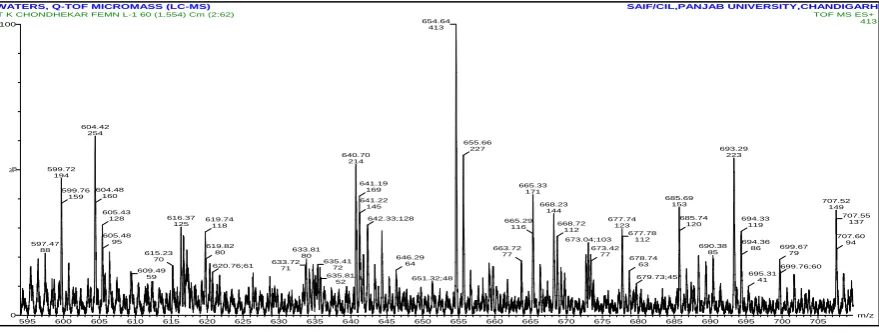
Mass and 1H-NMR Spectra of the Complexes
In the mass spectrum, the molecular ion peak of complex FeMn (SB)2(H2O)2is observed at
m/z 690(M+1) which confirms the molecular weight of the binuclear complex as 689 which
is in good agreement with calculated theoretically from the proposed structure. These results
are further supported by the conclusions drawn from the elemental analysis which agree with
the molecular formula assigned to these complexes.
WATERS, Q-TOF MICROMASS (LC-MS) SAIF/CIL,PANJAB UNIVERSITY,CHANDIGARH
m/z 595 600 605 610 615 620 625 630 635 640 645 650 655 660 665 670 675 680 685 690 695 700 705
%
0 100
T K CHONDHEKAR FEMN L-1 60 (1.554) Cm (2:62) TOF MS ES+
413 654.64 413 604.42 254 599.72 194 597.47 88 599.76 159 640.70 214 604.48 160 605.43 128 616.37 125 605.48 95 615.23 70 609.49 59 619.74 118 633.81 80 619.82 80 633.72 71 620.76;61 635.4172
635.81 52 641.19 169 641.22 145 642.33;128 646.29 64 651.32;48 655.66 227 693.29 223 665.33 171 665.29 116 663.72 77 685.69 153 668.23 144 677.74 123 668.72 112 673.04;103 673.42 77 677.78 112 678.74 63 679.73;45 685.74 120 690.38 85 707.52 149 694.33 119 694.36 86 699.67 79 695.31 41 699.76;60 707.55 137 707.60 94
[image:6.595.79.519.582.748.2]1
H NMR Spectra
1
HNMR Spectrum of Fe2(SB)2 (H2O)2 dimethyl sulfoxide‘, (400 MHZ) showed two
characteristic absorptions (singlet at =10.286ppm) attributed to coordinated imine proton[16]
(Figure:2). Multiplates were observed between values 6.921-8.307ppm attributed to
aromatic protons. Spectrum of FeZn (SB)2(H2O)2 and FeMn (SB)2(H2O)2 showed similar
types of spectra exhibited the characteristic imine proton peak and the multiplates for
aromatic protons between values6.921-8.307ppm.
Fig: 2 1HNMR Spectrum of Fe2(SB)2 (H2O)2.
Thermal analysis
Thermogram of the complexes shown in fig exhibited weight loss was observed below 150 C
this was attributed to the presence of small amounts of lattice water. The weight loss in the
first step above 3000C was found to be around (Obs.= 5.2%, Calc.=5.8) which accounts for
two coordinated water molecules. The complexes exhibited Thermal stability up to 600 C
after which an accelerated weight loss was observed in the region 300 C to 600 C which was
attributed to ligand decomposition, with mass loss(Obs.=75%, Calc.=75.50%).
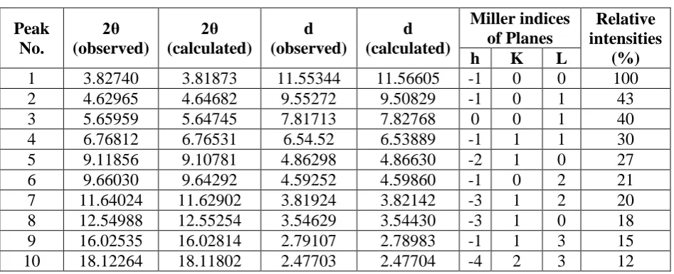
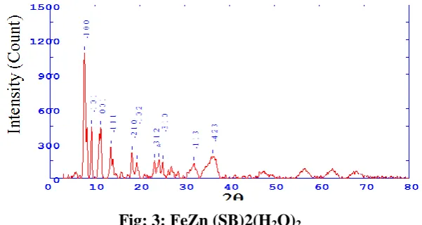
Powder X-ray diffraction data
FeZn (SB)2 (H2O)2 Complex.
The FeZn (SB)2(H2O)2 complex was used to study the X-ray powder diffraction.
Diffractogram is presented in Fig. 3 The indexing in the powder diffraction was done
independently by trial and error method. The crystallographic data and the indexed powder
diffraction data is presented in Table 3. The standard deviation observed is within the
calculated density from Z value and unit cell volume for complex is 0.9988gcm-3
respectively. The porosity percentage calculated from the observed and calculated densities
was found to be 0.12.The crystal system was found to be monoclinic with 2 molecules per
unit cell having probable space group P.[17-18]
Table 3: Indexed X-ray Diffraction Data of FeZn(SB)2(H2O)2 Complex of Ligand L5&L6
Peak No.
2θ (observed)
2θ (calculated)
d (observed)
d (calculated)
Miller indices of Planes
Relative intensities
(%)
h K L
1 3.82740 3.81873 11.55344 11.56605 -1 0 0 100 2 4.62965 4.64682 9.55272 9.50829 -1 0 1 43
3 5.65959 5.64745 7.81713 7.82768 0 0 1 40
4 6.76812 6.76531 6.54.52 6.53889 -1 1 1 30 5 9.11856 9.10781 4.86298 4.86630 -2 1 0 27 6 9.66030 9.64292 4.59252 4.59860 -1 0 2 21 7 11.64024 11.62902 3.81924 3.82142 -3 1 2 20 8 12.54988 12.55254 3.54629 3.54430 -3 1 0 18 9 16.02535 16.02814 2.79107 2.78983 -1 1 3 15 10 18.12264 18.11802 2.47703 2.47704 -4 2 3 12
Unit cell data and crystal lattice parameters
a (A˚) =14.13822 Volume (V) = 998.84A˚3
b (A˚) =9.02213 Density (obs.) =1 gcm-3
c (A˚) =9.57245 Density (cal.) =0.9988 gcm-3
α =90˚ Z = 9
β =125.271586˚ Crystal system= Monoclinic
γ=90˚ Space group = P
Standard deviation (%) = 0.088 Porosity = 0.12%
[image:8.595.62.536.184.377.2]Fig: 3: FeZn (SB)2(H2O)2.
Table 4: Report for Antibacterial Testing.
Medium-Nutrient Agar
Method –Agar cup method Dose of compound -1%
Cup size-10mm
Sr. No. Test
Compound
Inhibition Zone (nm)
Escherishia coli Salmonella typhi
Staphylococcus aureus
Bacillus subtilis
Penicillin 14 mm 20 mm 36 mm 28 mm
1 Fe2 (SB)2 (H2O)2
16 -ve -ve -ve
2 FeZn (SB)
(H2O)2 -ve -ve 16 -ve
3 FeMn(SB)2 (H2O)2
-ve 16 -ve 13
Table 5: Report for Antifungal Testing.
Test compound
Inhibit Aspergillus
niger
Penicillium chrysogenum
Fusarium moneliforme
Aspergillus flavus
Griseofrin -ve -ve -ve -ve
Fe2 (SB)2 (H2O)2 RG RG -ve RG
FeZn (SB)2 (H2O)2 RG RG -ve RG
FeMn(SB)2(H2O)2 RG RG -ve RG
Complex: +ve growth = Antifungal activity absent -ve growth = Antifungal activity present
RG = reduced growth (more than 50% reduction in growth observed).
Antimicrobial activity of the complexes
Escherishia coli, Salmonella typhi, Staphylococcus aureus and Bacillus subtilis by agar cup
method at fixed concentration of 1%[19] and compared with known antibiotic viz Penicillium
(Table 4). For fungicidal activity, compounds were screened in Vitro against Aspergillus
niger, penicillin chrysogenum, Fusarium moneliforme, Aspergillus flavus by poison plate
method with potato dextrose agar media. The complexes were tested at 1% concentration in
DMSO and compared with control (Table 3).
The complexes individually show varying degrees of inhibiting effects on the growth of the
bacterial species. Some complexes show activity against Gram-negative bacteria. Escherishia
coli, salmonella typhi & Bacillus subtilis. The some complexes show activity against
Gram-positive bacteria Escherishia coli & Bascillus. The metal complex FeZn (SB) (H2O)2 show
better activity for Escherishia coli however the activity of these complexes is considerably
less than that of standard drug. The complex of Femn (SB)2 (H2O) 2 is found to be active
against Bacillus subtilis bacterium. However the activity of these complex is higher than that
of standard drug.
Result of antifungal testing indicate that the all the bimetallic complexes show moderate to
high antifungal activity.
CONCLUSION
The preparation of dinuclear complexes by a novel synthetic route is strongly supported by
analytical data. The formation of precursor complexes as well as imine in dinuclear
complexes confirmed by existant and missing peaks in infrared spectra. The effective
magnetic moment and electronic spectral deta supported the tetrahedral environment in the
metal ion. The presence of two coordinated water molecules was detected both from
elemental analysis and thermogravimetric anaiysis. The molecular ion peak in the mass
spectra also supported the formation of dinuclear complexes. Finally; the molecular
mechanical method used for energy minimization corroborated the proposed structure of the
complexes. The novel method to synthesize the dinuclear complexes is capable of opening a
new area in the preparation of complexes with a lot more variations.
ACKNOWLEDGEMENT
We are thankful for Department of Biology N.S.B. College Nanded and C.I.L. Chandigrah
REFERENCES
1. Journal of Current Chemical and pharmaceutical Sciences, 2014; 4(3): 135-141.
2. S.V. Sanap and R. M Patil, J. Pharma. Sci., 2013; 2: (1,1).
3. L. Mrinalini and A.K. Manihar singh, J Chem. Sci., 2014; 2(1): 45.
4. Y.S. Malghe, R. C Prabhu and R. W. Raut,Acta Polo. Pharma and drug Res., 2009;
66(1): 45.
5. N.B. Ndosiri, M.O. Agwara, A.G. Paboudam, P.T. Ndifon, D.M. Yufanyi and C.Amah,
Res. J. Pharma., Bio Chem. Sci., 2013; 4(1): 386.
6. S.A. Shaker, Y. Farina and A.A. Salleh, Europ. J. Sci. Res., 2009; 33(4): 702.
7. Kovacs J. A. chem, Rev., 2004; 104: 825.
8. Fontecilla-camps, J.C; Volbeda A; Cavazza, C.; Nicolet, Y. chem. Rev., 2007; 107: 4273.
9. Greatti A, Scarpellini M Peralta R A, Casellato A, Bortoluzzi A J, Xavier F R, Jovito R,
Bsito M A, Szpoganicz B, Tomkowicz Z, Rams M, Haase W and Neves A, Inorg chem.,
2008; 47(3): 1107-1119.
10.Oliveira E, Costa SPG, Raposo MM, FaZaon and Lodeiro C, Inorgchim Acta, 2011;
366(1): 154-160.
11.Dobrokhotova Z, Emelina A Sidorov A, et. al .Synthesis and characterization of Li (1)- M
(ii) (M=Co, Ni) heterometallic complexes as molecular precursors for LiMO2.
polyhedron, 2011; 30: 132-141.
12.V.D. Bhatt, SR Ram Chemical Sciences Journal, 2012; CSJ-63.19.
13.M. Islam, B. Hossain and M. Reza. Antimicrobial studies of mixed ligand transition metal
complexes of maleic acid and heterocyclic amine bases .journal of Medical science, 2003;
3: 289-293.
14.V.D. Bhatt, S.R. Ram, Chemical Science Transactions, 2013; Accepted.
15.R. Boca, M. Gembicky M, Herchel R, et al, 2003; Ferromagnetism in a dinuclear nickel
(ii) complex containing triethylenetetramine and tricyanomethanide. Inorganic.
16.Bhatt V.D. and Ray A, Synth met., 1998; 92(2): 115-120.
17.V.D. Bhatt, SR Ram Chemical Sciences Journal, 2012; CSJ-63.