Original Article
Elevated expression of hypoxia
inducible factor-1 alpha is correlates to
recurrence and poor outcome in gastric cancer
Wei-Jie Zhang1,2*, Guo-Ming3*, Liu-Qing Yang4, Qi Sun5,Hong-Yu Wu5, Xin-Yun Xu5, Wen-Xian Guan1,2, Gui-Fang Xu4
Departments of 1Gastrointestinal Surgery, 4Gastroenterology, 5Pathology, Affiliated Drum Tower Hospital of Nanjing University, School of Medicine, Nanjing, China; 2Department of Gastrointestinal Surgery, Drum Tower Clinical College of Nanjing Medical University, Nanjing, China; 3Department of Gastroenterology, People’ s Hospital of Anji, Huzhou, China. *Equal contributors.
Received October 18, 2015; Accepted January 10, 2016; Epub April 15, 2016; Published April 30, 2016
Abstract: Background: Hypoxia-inducible factor-1 alpha (HIF-1α) is a transcription factor that plays a central role
in biologic processes under hypoxic conditions, especially concerning tumor angiogenesis. In this study, we
inves-tigated the correlation of HIF-1α expression with clinicopathological characteristics, tumor recurrence and progno -sis in gastric cancer (GC) after curative resection. Methods: The clinical data of 196 GC patents who underwent
curative resection were analyzed retrospectively. The expressions of HIF-1α in recurrent GC tissues compared to non-recurrent GC tissues were examined, and the relationship between HIF-1α expression and clinicopathological
characteristics was evaluated. In addition, these patients were followed up to investigate the relationship between
HIF-1α expression and the survival time. Results: Immunohistochemical staining demonstrated that 114 of 196 GC samples (58.2%) were positive for HIF-1α. The positive rate of HIF-1α expression was significantly higher in recurrent
GC tissue, than that in non-recurrent GC tissues (80.9%, 51.4%, respectively, P < 0.05). There was a close
relation-ship between HIF-1α expression and TNM stage (P = 0.009), lymph node status (P = 0.004), differentiation (P = 0.042), vascular invasion (P = 0.019), T stage (P = 0.013) and VEGF expression (P = 0.030). Furthermore, patients
with HIF-1α positive showed significantly higher recurrence and poorer prognosis than those with HIF-1α negative. Multivariate analysis showed that HIF-1α expression was a significant independent factor for tumor recurrence and overall survival. Conclusion: The results of the present study suggest that HIF-1α may be used as an unfavorable
indicator in predicting tumor recurrence and prognosis for with GC after curative surgery. This study also suggests
that HIF-1α might be a potential therapeutic target for GC.
Keywords: Gastric cancer, hypoxia-inducible factor-1 alpha, recurrence, tumor markers, immunohistochemistry
Introduction
Gastric cancer remains one of the most com-mon causes of cancer-related deaths world-wide [1]. In spite of progress in the surgical treatment and chemotherapy, the prognosis of gastric cancer patients remains poor [2]. Recurrence and metastasis are the main causes of death, and recurrence after curative intent resection is relatively common, occurring in 20% to 50% of patients [3, 4]. However, the underlying molecular mechanisms responsible for metastasis and tumor recurrence have not been fully elucidated, and the specific tumor markers in detection of tumor recurrence have
In this study, HIF-1α expression in human gas -tric cancer was examined by immunohisto-chemistry. Its correlation with clinical charac-teristics and prognosis was evaluated to deter-mine whether HIF-1α expression level could be used to predict recurrence and prognosis in patients with gastric cancer after curative surgery.
Materials and methods
Tumor samples
This study included 196 patients with histologi-cal confirmed primary gastric cancer, all of whom underwent gastrectomy between 2006 and 2007 at the Department of gastrointesti-nal surgery, the Affiliated Drum tower Hospital of Nanjing University Medical School, Nanjing, China. They included 122 men and 74 women, ranging from 27 to 81 years of age (mean, 59.0 years). Patients lost during follow up or who died within one year of surgery was excluded. All specimens were pathologically reassessed independently by two gastrointestinal patholo-gists according to the 7th edition of the American Joint Committee on Cancer (AJCC) of gastric cancer [9].
Clinicopathological variables including age, sex, location, tumor size, tumor differentiation, Lauren type, T stage; and vascular, lymphatic, and perineural invasion were collected for each patient. Tumor size was defined as the longest
diameter according to the pathology report. None of these patients received any preopera-tive anticancer treatment. All specimens were obtained from patients with written informed consent was obtained from all patients or their families and approved by the Clinical Research Ethics Committee of Drum Tower hospital.
Immunohistochemistry
[image:2.629.100.532.79.279.2]Four micrometer thick sections were cut from archival formalin-fixed paraffin-embedded tis -sue blocks. The samples were deparaffinized and dehydrated using a graded series of etha-nol solutions. For HIF-1α antigen retrieval, sec -tions were then irradiated by a domestic micro-wave oven at 99°C in 10 mM citrate buffer (pH 9.0) for 30 min, and cooled to room tempera-ture. After microwave irradiation, the slides were washed with phosphate-buffered saline (PBS), treated with 0.3% hydrogen peroxide in methanol for 30 min to block endogenous per-oxidase, and then incubated with the primary antibody in a humidified chamber at 48°C overnight. As the primary antibody, the rabbit polyclonal antibody H206 (Santa Cruz Bio- technology, Santa Cruz, CA, USA) for HIF-1α, diluted at 1:200 was used. Sections were washed three times with PBS, then incubated with biotinylated horse anti-mouse/anti-rabbit immunoglobulin G antibody for 30 min, washed again three times with PBS, and then incubated with avidin-biotinylated peroxidase complex for
30 min. After three additional washings with PBS, staining was developed by incubating the sections in 3-amino-9-ethylcarbazole (Vector) for 10 min. The sections were then counter-stained with hematoxylin and mounted.
Assessment of HIF-1α
The HIF-1α expression was defined as positive if nuclear staining was observed in ≥ 5% of the tumor cells. Concomitant cytoplasmic staining was not counted because HIF-1α in the nucleus determines the functional activity of the HIF-1α complex (Figure 1). In regard to overall survival curve, the HIF-1α expression was classified as one of four categories, depending on the per-centage of tumor cells stained: - (0-5%), 1+ (5-10%), 2+ (10-15%), 3+ (≥ 15%). The HIF-1α expression through nuclear staining of positive cells was predominant at the invading edge of the tumor margin and at the periphery of necrotic regions within tumors.
Follow-up
No major perioperative complications occurred in patients, and all were discharged from the hospital. The closing date for follow-up was March 31, 2014. As a protocol for follow-up, all patients were checked every 3 months during the first 2 years and every 6 months thereafter. Recurrence were confirmed by tumor markers levels including CEA, AFP, CA199 and CA125, and imaging including chest radiography, bari-um meal, abdominal ultrasonography (US), computed tomography (CT), and endoscopy according to the clinical situation after gastrec-tomy. The locations and times of tumor recur-rence were recorded. The follow-up time had the day of surgery as a starting point, the time of tumor recurrence and death were recorded, and these two points were evaluated for prog-nostic analysis. If the follow-ups were incom-plete, patients or their families were contacted by telephone. The median follow-up period after surgery was 39.8 months (range from 8 to 91 months).
Statistical analysis
All statistical analyses were performed with SPSS software, version 17.0 (SPSS Inc., Chicago, IL, USA). The correction between HIF-1α expression and clinicopathological features was analyzed by the χ2 test and Fisher’s exact
test. Survival curves were estimated using the Kaplan-Meier method, and differences in sur-vival distributions were evaluated by the log-rank test. Cox’s proportional hazards modeling of factors potentially related to survival was performed to identify factors that might have a significant influence on survival. Differences with a p value of 0.05 or less were considered statistically significant.
Results
Patient characteristics
The detailed clinicopathological characteristics of patients after curative resection are shown in Table 1. When follow-up was over, a total of 67 patients died because of tumor progression and 8 due to other causes. Overall survival (OS) was defined as the interval between surgery and last visit or death. The mean OS of 196 gastric cancer patients was 39.92 ± 19.52 months (range from 8 to 91 months), and the 5-year survival rate of all enrolled patients was 19.39%.
HIF-1α expression in gastric cancer tissues
The patterns of HIF-1α expression in the tumor cells were mixed nuclear/cytoplasmic staining, and expression of HIF-1α in gastric cancer tis -sue was significantly higher than that in the nor -mal gastric tissue. HIF-1α expression through nuclear staining of positive cells was predomi-nant at the invading edge of the tumor margin and at the periphery of necrotic regions within tumors (Figure 1). Of the 196 total cases, 114 (58.2%) were HIF-1α positive. The positive rate of HIF-1α expression in gastric cancer tissues with recurrence was 80.9% (72/89), signifi -cantly higher than that without recurrence 51.4% (55/107). HIF-1α expression was associ -ated with depth of invasion, T stage, TNM stage, LN metastasis, venous invasion, lymphatic invasion, and VEGF expression. There was no correlation between HIF-1α and other patho -logical parameters, such as age, gender, lauren type, serum CEA, perineural invasion, tumor location, Borrmann type, or tumor size (Table 1).
Time to recurrence and recurrence pattern
recur-rence was 18.0 months (range from 6 to 78 months). Among patients with recurrence, 63 (70.7%) had recurrence within 2 years (Table 2). Of the 89 patients with recurrence, 34 (38.2%) were diagnosed with loco-regional re- lapse, which was the most preva-lent, 28 (31.5%) patients had hematogenous metastases (16 in liver, 5 in lung, and 6 in bone). Other recurrences were peritoneal recurrence (n = 19, 21.3%), dis-tant lymph node metastases (n = 5, 5.6%), or at multiple sites (n = 3, 3.4%).
HIF-1α expression association with tumor recurrence and poor
overall survival
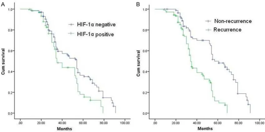
Compared with patients without recurrence, patients with recur-rence showed advanced tumor stages (P = 0.003), longer tumor size (P = 0.0018), depth of sion (P = 0.035), lymph node inva-sion (P = 0.008), positive HIF-1α (P = 0.001) and VEGF expression (P = 0.028), and vascular invasion (P = 0.027) (Table 3). Multivariate model identified that expression of HIF-1α, advanced TNM stage, and lymph node metastases were independent predictive factors for tumor recurrence (Table 4). Survival curves according to posi-tive or negaposi-tive HIF-1α staining are shown in Figure 2. Respec- tively, survival rates for patients with HIF-1α-positive staining were significantly lower than that of HIF-1α negative (P = 0.016). Compared with patients without recurrence, survival rates for patients with recurrence were significantly low-er (P < 0.001). Multivariate Cox’s proportional hazard analyses of clinicopathological factors reveal- ed HIF-1α expression (hazard ratio (HR) 2.289; 95% CI 1.208-4.339; P = 0.011) and advanced TNM stage (HR 2.406; 95% CI 1.278-Table 1. The relationship between expression of HIF-1α, tumor
recurrence and clinicopathological features
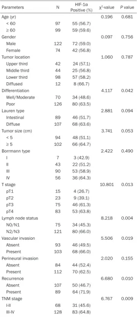
Parameters N Positive (%)HIF-1α χ2-value P value
Age (yr) 0.196 0.681
< 60 97 55 (56.7)
≥ 60 99 59 (59.6)
Gender 0.097 0.756
Male 122 72 (59.0)
Female 74 42 (56.8)
Tumor location 1.060 0.787
Upper third 42 24 (57.1)
Middle third 44 25 (56.8)
Lower third 98 57 (58.2)
Diffused 12 8 (66.7)
Differentiation 4.117 0.042
Well/Moderate 70 34 (48.6)
Poor 126 80 (63.5)
Lauren type 2.881 0.094
Intestinal 89 46 (51.7)
Diffuse 107 68 (63.6)
Tumor size (cm) 3.741 0.053
< 5 94 48 (51.1)
≥ 5 102 66 (64.7)
Borrmann type 2.422 0.490
I 7 3 (42.9)
II 43 22 (51.2)
III 90 53 (58.9)
IV 56 36 (64.3)
T stage 10.801 0.013
pT1 15 4 (26.7)
pT2 23 9 (39.1)
pT3 75 46 (61.3)
pT4 83 53 (63.8)
Lymph node status 8.218 0.004
N0/N1 75 34 (45.3)
N2/N3 121 80 (66.0)
Vascular invasion 5.506 0.019
Absent 93 46 (49.5)
Present 103 68 (66.0)
Perineural invasion 2.020 0.155
Absent 84 44 (52.4)
Present 112 70 (62.5)
Recurrence 6.680 0.010
Absent 107 50 (46.7)
Present 89 64 (71.9)
TNM stage 6.767 0.009
I-II 68 31 (45.6)
[image:4.629.98.368.104.726.2]4.531; P = 0.007) as independent prognostic indicators of poor sur-vival for gastric cancer after surgery.
Discussion
Gastric cancer is one of the most common malignancies worldwide and still the second leading cause of cancer-related deaths [2]. The treatment of gastric cancer in- cludes a combination of surgery, chemotherapy, and radiation ther-apy. However, the majority of patients develop local or distant recurrence after gastrectomy and adjuvant chemotherapy, and the dismal prognosis of gastric cancer is due principally to the frequency of recurrence and metastasis. Therefore, the identification of diagnostic and prognostic bio-markers is needed for optimizing management and treatment stra- tegies.
Tumor hypoxia is well recognized in oncology to be a key factor resulting in treatment resistance and poor prognosis. Hypoxia can increase HIF-1α protein stability via altered ubiquitination and then lead to overexpression of HIF-1α. There is a considerable body of data supporting the notion that HIF-1α has an important role in invasion and metastasis of malig-nant tumors, including stomach, brain, oropharynx, cervix, ovary and breast [10, 11].
It has been showed that the degree of intratumoral hypoxia is positively correlated with the ability of tumor invasion, metasta-sis, and drug resistance [12]. Furthermore, hypoxia and HIF-1α over-expression are implicated in the tumor aggressiveness of many cancers, including gastric cancer [13]. Our study suggests that over-expression of HIF-1α is a common feature in gastric cancer, and might represent a novel predictive
VEGF 4.724 0.030
Negative 85 42 (47.1)
Positive 111 72 (66.7)
Serum CEA level (μg /L) 1.177 0.278
< 5 101 55 (54.4)
≥ 5 95 59 (62.1)
HIF-1α: hypoxia Inducible Factor-1 alpha; TNM: Tumor Node Metastasis; VEGF:
[image:5.629.98.368.233.736.2]vascular endothelial growth factor; CEA: carcinoembryonic antigen.
Table 2. Characteristics of gastric cancer with recurrence com-pared with patients without recurrence
Parameters Recurrence (n = 89) No recurrence (n = 107) valueχ2- valueP
Age (yr) 0.090 0.764
< 60 43 54
≥ 60 46 53
Gender 0.504 0.478
Male 53 69
Female 36 38
Tumor location 2.478 0.479
Upper third 19 23
Middle third 23 21
Lower third 40 58
Diffused 7 5
Differentiation 3.318 0.076
Well/Moderate 25 43
Poor 64 64
Lauren type 0.556 0.456
Intestinal 43 46
Diffuse 46 61
Tumor size (cm) 5.562 0.018
< 5 34 60
≥ 5 54 48
Borrmann type 3.845 0.279
I 2 5
II 15 28
III 43 47
IV 29 27
T stage 4.731 0.193
pT1 5 10
pT2 9 14
pT3 30 45
pT4 46 38
Lymph node status 7.145 0.008
N0/N1 25 50
N2/N3 64 57
Vascular invasion 4.869 0.027
Absent 35 59
recurrence and survival after curative rese- ction.
However, how HIF-1α functions in these pro -cesses is not entirely clear; further studies are needed to elucidate the molecular mechanisms by which HIF-1α participates in the develop -ment and progression of GC. Accumulating evi-dence has indicated that HIF-1α as a novel and key regulator that integrates EMT [18-21], which is an important mechanism during the early steps of tumor progression and metasta-sis, when neoplastic cells disseminate from the primary tumor.
between HIF-1α and human gas -tric cancer [16], and also the expression of HIF-1α in patients with recurrence was obviously more frequently detected than that of no-recurrence. However, HIF-1α expression was not corre -lated with age, gender, differentia-tion status, tumor site or Lauren classification. Recent studies sug -gested that the hypoxic activation of NF-κB seems to contribute to the expression of HIF-1α protein at the translational level, shRNA-mediated downregulation of HIF-1α expression reduced the cell viability of SNU-668, SNU-484, and SNU-216 gastric cancer cells in vitro under hypoxic conditions [17], which support our results demonstrating the association of HIF-1α with gastric cancer inva -sion and metastasis.
In this study, HIF-1α expression was found to be associated with a malignant behavior category, including postoperative progno-sis. We found that high HIF-1α expression was associated with poor prognosis and unfavorable clinical outcome, and patients who had tumors with high HIF-1α expression had remarkably poor overall survival as compared with patients who had low HIF-1α expression. Furthermore, multi-variate analysis revealed that HIF-1α expression level was an inde -pendent, significant risk factor for marker for the clinical outcome of the disease.
Therefore, therapeutic agents that inhibit func-tion have the potential to improve the outcome of patients with metastasis [14, 15].
In this study, we investigated HIF-1α protein expression in gastric cancer and its association with recurrence and disease-free survival. Our results showed HIF-1α protein expression was stronger positive staining of HIF-1α gastric cancer with deep invasion, differentiation, lymph node metastasis, vascular invasion, and advanced TNM stage, which was consis-tent with previous studies on the relationship
Perineural invasion 3.171 0.075
Absent 32 52
Present 57 55
TNM stage 8.863 0.003
I-II 21 47
III-IV 68 60
VEGF 4.836 0.028
Negative 31 54
Positive 58 53
HIF-1α 11.838 0.001
Negative 17 53
Positive 72 55
Serum CEA level (μg /L) 0.286 0.593
< 5 44 57
≥ 5 45 50
HIF-1α: hypoxia Inducible Factor-1 alpha; TNM: Tumor Node Metastasis; VEGF:
[image:6.629.98.370.347.403.2]vascular endothelial growth factor; CEA: carcino-embryonic antigen.
Table 3. Multivariate analysis of risk factors for recurrence in gastric cancer
Hazard ratio 95% CI P value
HIF-1α positive 2.345 (1.261-4.360) 0.007
Lymph node status 1.063 (0.754-1.499) 0.047
TNM stage 1.986 (1.009-3.911) 0.043
CI: confidence interval; HIF-1α: hypoxia-inducible factor-1α; TNM: Tumor Node
[image:6.629.99.368.473.514.2]Metastasis.
Table 4. Multivariate analysis of significant prognostic factors for survival in patients with gastric cancer
Parameters Hazard ratio 95% CI P value
TNM stage 2.406 (1.278-4.531) 0.007
HIF-1α 2.289 (1.208-4.339) 0.011
CI: confidence interval; HIF-1α: hypoxia-inducible factor-1α; TNM: Tumor Node
In addition, HIF-1α is crucial for the formation and maturation of the vasculature through interaction with the signals that regulating angiogenesis, such as vascular endothelial growth factor (VEGF) and Notch pathway [22, 23]. Previously study had shown that inhibition of HIF-1α function in human gastric cancer cells resulted in the reduction of VEGF secretion in vitro, the inhibition of tumor growth and angio-genesis in vivo, and alterations in tumor vessel morphology and maturation in vivo [24].
In conclusion, HIF-1α expression was increased in clinical gastric cancer specimens, and it was associated with tumor invasion and metasta-sis. Our study also provides the evidence that HIF-1α is an independent prognostic factor of overall survival, and a predictive factor of can-cer recurrence for patients with gastric cancan-cer. Therefore, HIF-1α could serve as a potential predictive marker for predicting malignant behavior, and it is an independent prognostic factor for tumor recurrence and prognosis for patients with gastric cancer.
Acknowledgements
This work was supported by grants from the National Natural Science Foundation of China (Nos. 81201909 and 81572338), the Fund- amental Research Funds for the Central Universites (No. 021414380031), Nanjing
Medical Science and Technology Development program (Nos. QYK11166, YKK12072).
Disclosure of conflict of interest
None.
Address correspondence to: Wen-Xian Guan, De-
partment of Gastrointestinal Surgery, Affiliated
Drum Tower Hospital of Nanjing University, School of Medicine, Nanjing, China. E-mail: Guan-wenxian@ 163.com; Gui-Fang Xu, Department of Gastroentero-
logy, Affiliated Drum Tower Hospital of Nanjing
University, School of Medicine, Nanjing, China. E-mail: [email protected]
References
[1] Ferlay J, Shin HR, Bray F, Forman D, Mathers C, Parkin DM. Estimates of worldwide burden of cancer in 2008: GLOBOCAN 2008. Int J Cancer 2010; 127: 2893-2917.
[2] Siegel RL, Miller KD, Jemal A. Cancer statistics, 2015. CA Cancer J Clin 2015; 65: 5-29. [3] Sakar B, Karagol H, Gumus M, Basaran M,
Kaytan E, Argon A, Ustuner Z, Bavbek SE, Bugra D, Aykan FN. Timing of death from tumor recurrence after curative gastrectomy for gas-tric cancer. Am J Clin Oncol 2004; 27: 205-209.
[image:7.629.101.526.81.293.2][4] Huang KH, Chen JH, Wu CW, Lo SS, Hsieh MC, Li AF, Lui WY. Factors affecting recurrence in node-negative advanced gastric cancer. J Gastroenterol Hepatol 2009; 24: 1522-1526.
[5] Miao ZF, Zhao TT, Wang ZN, Xu YY, Mao XY, Wu
JH, Liu XY, Xu H, You Y, Xu HM. Influence of dif -ferent hypoxia models on metastatic potential of SGC-7901 gastric cancer cells. Tumour Biol 2014; 35: 6801-8.
[6] Mizokami K, Kakeji Y, Oda S, Irie K, Yonemura T, Konishi F, Maehara Y. Clinicopathological
significance of hypoxia-inducible factor 1 alpha
overexpression in gastric carcinomas. J Surg Oncol 2006; 94: 149-154.
[7] Kung AL, Wang S, Klco JM, Kaelin WG, Livingston DM. Suppression of tumor growth through disruption of hypoxia inducible tran-scription. Nat Med 2000; 6: 1335-1340. [8] Isobe T, Aoyagi K, Koufuji K, Shirouzu K,
Kawahara A, Taira T, Kage M. Clinicopathologi-
cal significance of hypoxia-inducible factor-1 alpha (HIF-1α) expression in gastric cancer. Int
J Clin Oncol 2013; 18: 293-304.
[9] Edge SB, Byrd DR, Compton CC, Fritz AG, Greene FL, Trotti A. AJCC cancer staging manu-al. 7th edition. New York: Springer; 2010. pp. 381-5.
[10] Semenza GL. Cancer-stromal cell interactions mediated by hypoxia-inducible factors promote angiogenesis, lymphangiogenesis, and metas-tasis. Oncogene 2013; 32: 4057-63.
[11] Lu X, Kang Y. Hypoxia and hypoxia-inducible factors: master regulators of metastasis. Clin Cancer Res 2010; 16: 5928-5935.
[12] Wilson WR, Hay MP. Targeting hypoxia in can-cer therapy. Nat Rev Cancan-cer 2011; 11: 393-410.
[13] Chen L, Shi Y, Yuan J, Han Y, Qin R, Wu Q, Jia B, Wei B, Wei L, Dai G, Jiao S. HIF-1 alpha overex-pression correlates with poor overall survival and disease-free survival in gastric cancerpa-tients post-gastrectomy. PLoS One 2014; 9: e90678.
[14] Miyake K, Yoshizumi T, Imura S, Sugimoto K, Batmunkh E, Kanemura H, Morine Y, Shimada M. Expression of hypoxia-inducible factor-1al-pha, histone deacetylase 1, and metastasis-associated protein 1 in pancreatic carcinoma correlation with poor prognosis with possible regulation. Pancreas 2008; 36: e1-e9 [15] Semenza GL. Targeting HIF-1 for cancer
thera-py. Nat Rev Cancer 2003; 3: 721-732.
[16] Qiu MZ, Han B, Luo HY, Zhou ZW, Wang ZQ, Wang FH, Li YH, Xu RH. Expressions of
hypoxia-inducible factor-1α and hexokinase-II in gastric
adenocarcinoma: the impact on prognosis and correlation to clinicopathologic features. Tumour Biol 2011; 32: 159-166.
[17] Nam SY, Ko YS, Jung J, Yoon J, Kim YH, Choi YJ, Park JW, Chang MS, Kim WH, Lee BL. A hypox-ia-dependent upregulation of
hypoxia-induc-ible factor-1 by nuclear factor-κB promotes
gastric tumour growth and angiogenesis. Br J Cancer 2011; 104: 166-174.
[18] Zhu GH, Huang C, Feng ZZ, Lv XH, Qiu ZJ. Hypoxia-induced snail expression through
tran-scriptional regulation by HIF-1α in pancreatic
cancer cells. Dig Dis Sci 2013; 58: 3503-3515. [19] Liu Y, Liu Y, Yan X, Xu Y, Luo F, Ye J, Yan H, Yang
X, Huang X, Zhang J, Ji G. HIFs enhance the migratory and neoplastic capacities of hepato-cellular carcinoma cells by promoting EMT. Tumour Biol 2014; 35: 8103-8114.
[20] Velpula KK, Dasari VR, Tsung AJ, Dinh DH, Rao JS. Cord blood stem cells revert glioma stem cell EMT by down regulating transcriptional ac-tivation of Sox2 and Twist1. Oncotarget 2011; 2: 1028-1042.
[21] Joseph JV, Conroy S, Pavlov K, Sontakke P, Tomar T, Eggens-Meijer E, Balasubramaniyan V, Wagemakers M, den Dunnen WF, Kruyt FA. Hypoxia enhances migration and invasion in glioblastoma by promoting a mesenchymal
shift mediated by the HIF1α-ZEB1 axis. Cancer
Lett 2015; 359: 107-116.
[22] Yu S, Sun J, Zhang J, Xu X, Li H, Shan B, Tian T, Wang H, Ma D, Ji C. Aberrant expression and association of VEGF and Dll4/Notch pathway molecules under hypoxia in patients with lung cancer. Histol Histopathol 2013; 28: 277-284. [23] Bai X, Zhi X, Zhang Q, Liang F, Chen W, Liang C,
Hu Q, Sun X, Zhuang Z, Liang T. Inhibition of protein phosphatase 2A sensitizes pancreatic cancer to chemotherapy by increasing drug
perfusion via HIF-1α-VEGF mediated angiogen -esis. Cancer Lett 2014; 355: 281-287. [24] Stoeltzing O, McCarty MF, Wey JS, Fan F, Liu W,