Original Article
Correlation between imageological characteristics and
pathological patterns of mini-nodule lung cancer
Hai-Yun Ren1, Peng Ji2, Liu Liu1, Wei Yan1, She-Tian Jiang1
1Department of Ultrasound, Zhumadian Central Hospital, Zhumadian, China; 2Department of Radiotherapy,
Zhumadian Central Hospital, Zhumadian, China
Received July 9, 2015; Accepted January 7, 2016; Epub February 15, 2016; Published February 29, 2016
Abstract: This study aims to explore the relationship between the imageological characteristics and the pathological patterns in patients with mini-nodule lung cancer, and to investigate the threshold value for predicating the patho-logical patterns with invasive characteristic from the imageopatho-logical aspect. A total of 137 lung cancer patients were selected to record the characteristics of the maximum diameter, density, blood-supply, vacuole and mini-lobules and analyze their relationships with pathological patterns. Receiver Operating Characteristic (ROC) analysis was applied to predicate the threshold value of lung cancer invasiveness. There were significant differences in the maximum diameter, density, blood supply, sublobe, burr and pleural indentation syndrome of the nidi in lung cancer patients with different pathological patterns (P<0.05 or P<0.01). Further analysis on the relationships between the above characteristics and lung cancer invasiveness showed that lung cancer invasiveness was in moderate correlation with the nodular density (r=0.589, P=0.000), in low connection with nodular size (r=0.368, P=0.000) and in ex-tremely weak association with blood supply, sublobe, burr and pleural indentation syndrome (r<0.3). ROC analysis on the nodular size and density indicated that the area under ROC curve, sensitivity and specificity of nodular size were 0.700, 0.861, and 0.431 respectively, with threshold value being 12.50 mm, while those of nodular density were 0.831, 0.806 and 0.769, with threshold value being 37.50%, respectively. Imageological characteristics, es-pecially the nodular size and density, have certain predictable value in the invasiveness of mini-nodule lung cancer.
Keywords: Lung cancer, lung nodule, imageological characteristics, pathological pattern
Introduction
Lung cancer, which is also called bronchial lung cancer because most of it is derived from the bronchial epithelium, is a kind of malignant tumor with the highest morbidity across the whole world [1, 2]. Because of the absence of specific clinical manifestation in the early stage of lung cancer and effective early diagnostic methods, 80% patients are in middle and advanced stage when diagnosed, thus leading to huge therapeutic cost but slight survival ben-efits [3]. It was reported that lung cancer-relat -ed death account-ed for 23.8% of all cancer-associated death, and the 5~10-year survival rate of patients with lung cancer was only 8~14% [4]. In another study, the 5-year survival rate of the patients with lung cancer received early diagnosis and treatment was increased from 14% to 49%, which was even >70% in stage I A patients, and if treatment could be
given in the early micro-damage of primary can-cer, the recovery rate could be up to 100% [5]. Therefore, the early detection and cognition of lung cancer is of critical significance in improv -ing the patients’ survival rate [6].
objective misdiagnosis. In recent years, with the continuous update and improvement of CT devices, imaging software and scanning tech-niques, the accuracy and reliability are signifi -cantly promoted, especially the popularization of low-dose spiral CT that markedly increases the detection rate of lung nodules, the mini-nodules in particular [8]. To further distinguish the property of lung nodules, analyze the rela-tionships between the morphological charac-teristics of mini-nodule nidi and the pathologi-cal patterns and to seek a reliable threshold value to predicate the low- and high-invasive-ness lung cancer, this study diagnosed the ima-geology of patients with lung cancer and com-pared them with the pathological diagnostic results.
Materials and methods
General data
This study was approved by the Ethnic Committee of Zhumadian Central Hospital and all informed consent forms were signed by the patients and (or) their families. A total of 137 patients with lung cancer admitted in Zhumadian Central Hospital from October, 2009 to February, 2015 were selected as study objects. All patients were diagnosed as non-small cell lung cancer (NSCLC) and treated with surgical resection. The lung nodules ≤20 mm and all patients had complete CT thin-layer three-dimensional imageological data before operation. There were 91 males and 46 females; aged 34~79 years, with median age of 57 years; clinical stage: 129 in stage I, 2 in stage II and 6 in stage III; 4 were with atypical adenomatous hyperplasia (AAH), 39 with ade-nocarcinoma insitu (AIS), 22 with microinvasive adenocarcinoma (MIA), 68 with invasive adeno-carcinoma (IAC), 3 with squamous (SQ) and 1 with large cell carcinoma (LC); 69 with single nidus and 68 with multiple nidi; and 12 were with multiple primary tumor.
Methods
Siemens 64-layer high-resolution spiral CT was used to perform thin scanning (1 mm) before operation and re-construct three-dimension. According to the pre-established image-reading sequence, the maximum diameter, density, blood supply, vacuole, mini-nodules, morpholo-gy, sublobe, burr, pleural indentation syndrome
and the presence of multiple nidi were record-ed. The images were read jointly by 2 chief phy-sicians from Department of Internal Medicine, 2 from Department of Radiotherapy and 1 from Department of Pathology who were unknown of the pathological results.
Evaluation criteria
The maximum diameter of nidi was defined as the maximum diameter of the measuring nidi on the maximum plane and was then divided into 3 grades (≤10 mm, 10~15 mm and 15~20 mm). Nodular density was defined as the ratio of parenchymal component in the mini-nodular lesion and divided into 5 grades (0%: pure ground-glass opacity; 100%: pure parenchymal nodules; 25%, 50% and 75%: the ratio of paren-chymal component accounted for 25%, 50% and 75%, respectively). Mini-nodules were the regional clustered granules with increased den-sity in nodules. Round and quasi-circular forms were defined as regular morphology and others as irregular morphology. Sublobe was defined as the nodule border with obvious depression. Multiple nidi meant there were multiple nodular nidi in the lung, including benign and malignant lesions and nidi with unknown property. AAH, AIS and MIA were defined as low-invasiveness lung cancer while IAC, LC and SQ as high-inva -siveness lung cancer.
Statistical analysis
SAS 9.3 and SPSS 17.0 were applied for all data analysis. Enumeration data was expressed by percentage (%) and detected with χ2 test.
The relationship between invasiveness and imageological characteristics was analyzed by Spearman correlation analysis and Receiver Operating Characteristic (ROC) was used to analyze the optimal diagnostic threshold value. Results
Relationships between imageological char-acteristics and pathological patterns in lung cancer
Figure 1. Comparison of imageological characteristics of mini-nodule lung cancer with different pathological pat-terns. Note: A: IAC, imageology showed mixed glass nodular images; B: MIA, imageology showed ground-glass ingredient-based partial parenchymal nodules, and the parenchymal ingredients were located in the center of the lesions; C: AIS, imageology showed pure ground-glass nodular images.
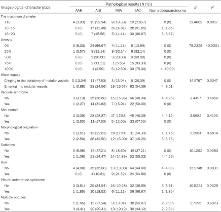
Table 1. Relationships between pathological patterns and imageological characteristics of mini-nodule lung cancer
Imageological characteristics Pathological results [N (%)] χ2 P
AAH AIS MIA IAC Non-adenocarcinoma
The maximum diameter
≤10 4 (3.93) 15 (52.94) 9 (18.58) 10 (2.857) 0 (0) 31.4802 0.0017
10~15 0 (0) 17 (31.48) 8 (14.81) 28 (51.85) 1 (1.85)
15~20 0 (0) 7 (15.56) 5 (11.11) 30 (66.67) 3 (6.67)
Density
0% 3 (8.33) 24 (66.67) 4 (11.11) 5 (13.89) 0 (0) 78.2325 <0.0001
25% 1 (3.57) 9 (32.14) 9 (32.14) 9 (32.14) 0 (0)
50% 0 (0) 3 (20.00) 3 (20.00) 9 (60.00) 0 (0)
75% 0 (0) 2 (11.11) 1 (5.56) 15 (83.33) 0 (0)
100% 0 (0) 1 (2.50) 5 (12.50) 30 (75.00) 4 (10.00)
Blood supply
Clinging to the periphery of nodular vessels 3 (13.04) 11 (47.83) 3 (13.04) 6 (26.09) 0 (0) 14.9767 0.0047 Entering into nodular vessels 1 (0.88) 28 (24.56) 19 (16.67) 62 (54.39) 4 (3.51)
Vacuole syndrome
No 3 (3.19) 25 (26.60) 15 (15.96) 46 (48.94) 4 (4.26) 3.4497 0.4856 Yes 1 (2.27) 14 (31.82) 7 (15.91) 22 (50.00) 0 (0)
Mini-nodule
No 3 (3.09) 28 (28.87) 17 (17.53) 45 (46.39) 4 (4.12) 3.9682 0.4103 Yes 1 (2.50) 11 (27.50) 5 (12.50) 23 (57.50) 0 (0)
Morphological regulation
No 2 (3.51) 13 (22.81) 10 (17.54) 31 (54.39) 1 (1.75) 2.2964 0.6814 Yes 2 (2.50) 26 (32.50) 12 (15.00) 37 (46.25) 3 (3.75)
Sublobes
No 3 (6.98) 16 (37.21) 8 (18.60) 16 (37.21) 0 (0) 10.1292 0.0383
Yes 1 (1.06) 23 (24.47) 14 (14.89) 52 (55.32) 4 (4.26) Burr
No 4 (4.00) 35 (35.00) 13 (13.00) 44 (44.00) 4 (4.00) 15.9748 0.0031
Yes 0 (0) 4 (10.81) 9 (24.32) 24 (64.86) 0 (0)
Pleural indentation syndrome
No 3 (3.61) 29 (34.94) 16 (19.28) 32 (38.55) 3 (3.61) 10.5221 0.0325 Yes 1 (1.85) 10 (18.52) 6 (11.11) 36 (66.67) 1 (1.85)
Multiple nodules
[image:3.612.89.525.319.737.2]ground-glass nodules in round or quasi-circular
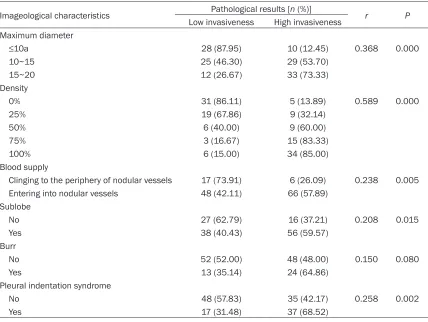
shape, with clear broader; and IAC was mostly and lung cancer invasiveness showed that lung cancer invasiveness was in moderate correla-Table 2. Relationships between imageological characteristics and mini-nodule lung cancer invasive-ness
Imageological characteristics Pathological results [n (%)] r P
Low invasiveness High invasiveness Maximum diameter
≤10a 28 (87.95) 10 (12.45) 0.368 0.000
10~15 25 (46.30) 29 (53.70)
15~20 12 (26.67) 33 (73.33)
Density
0% 31 (86.11) 5 (13.89) 0.589 0.000
25% 19 (67.86) 9 (32.14)
50% 6 (40.00) 9 (60.00)
75% 3 (16.67) 15 (83.33)
100% 6 (15.00) 34 (85.00)
Blood supply
Clinging to the periphery of nodular vessels 17 (73.91) 6 (26.09) 0.238 0.005 Entering into nodular vessels 48 (42.11) 66 (57.89)
Sublobe
No 27 (62.79) 16 (37.21) 0.208 0.015
Yes 38 (40.43) 56 (59.57)
Burr
No 52 (52.00) 48 (48.00) 0.150 0.080
Yes 13 (35.14) 24 (64.86)
Pleural indentation syndrome
No 48 (57.83) 35 (42.17) 0.258 0.002
Yes 17 (31.48) 37 (68.52)
Figure 2. ROC curve of nodular size and density in diagnosis of mini-nodule lung cancer.
defined as density-mixed ground-glass nodules with irregular shapes. χ2 test showed that from
AAH, AIS and MIA to IAC, the maxi-mum diameter, density, sublobe, burr, pleural indentation syn-drome and the ratio of blood sup-ply entering into nodular vessels increased subsequently, and the-re wethe-re significant diffethe-rences (P<0.05 or P<0.01). The compari-son of imageological diagnosis and pathological patterns are shown in Figure 1 and Table 1. Relationships between imageo-logical characteristics and mini-nodule lung cancer invasiveness
[image:4.612.93.521.95.417.2]tion with the nodal density, in low connection with nodular size and in extremely weak asso-ciation with blood supply, sublobe, burr and pleural indentation syndrome (Table 2).
Diagnostic value of nodal size and density in mini-nodule lung cancer
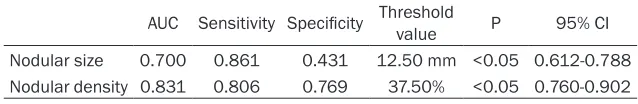
ROC analysis on the nodular size and density indicated that the area under ROC curve, sensi-tivity and specificity of nodular size were 0.700 (95% CI; 0.612~0.788), 0.861, and 0.431, with threshold value being 12.50 mm, while those of nodular density were 0.831 (95% CI: 0.760~0.902), 0.806 and 0.769, with thresh-old value being 37.50%, respectively (Figure 2 and Table 3). The area under the ROC curve is a popular summary measure of the accuracy of a test. Since the area under ROC curve of nodular density was higher than nodular size, nodal density had higher predictable value in the invasiveness of mini-nodule lung cancer. Discussion
With the development of CT imageological tech-nique, the detection rate of mini-nodules in lung has been significantly increased. According to the clinical statistics, the detection rate of nodular lesion by conventional chest X-ray was 0.2%, but was 40%~60% by high-resolution CT [9]. Dabrowska et al. [10] applied contrast-enhancement CT and 18-FDG positron emis-sion computed tomography (PECT) to identify the malignant isolated lung nodules, in which the optimal diagnostic point of contrast-enhancement CT was 19 hounsfield units of enhancement value, whose sensitivity, specific -ity, positive predictive value (PPV), negative predictive value (NPV) and accuracy were 100%, 37%, 32%, 100% and 58% respectively, whereas on the optimal diagnostic point of 18-FDG PECT, the sensitivity, specificity, PPV, NPV and accuracy were 77%, 92%, 83%, 89% and 90% respectively, showing that the two had their own advantages and disadvantages in
ules with the maximum diameter ≥5 mm were found, and the measured CT value of lung nod-ules could be applied for distinguishing the pri-mary tumors.
Early detection and intervention are of great significance in reducing the mortality of patients with lung cancer [12, 13]. American National Lung Screening Trial (NLST) has proved that low-dose spiral CT examination once a year can greatly reduce the relevant death risk by 20% in high-risk patients with lung cancer [14]. It was illustrated in a study that AAH, AIS and MIA were tumors with favorable prognosis in adeno-carcinoma, which showed scale-like growth or mainly in scale-like growth, with tumor size ≤3 cm and postoperative survival rate near to 100% after radical surgery, whereas IAC was mainly in scale-like, acinar, palillary and solid growth, and the 5-year survival rate of IAC patients with stage I was 70%~80%, with evi-dently lower prognosis than that of AIS and MIA [15]. Therefore, preoperative effective diagno-sis of the risk of mini-nodule lung cancer, which can guide the physicians to conduct different interventional measures, is of great signifi -cance in clinic. However, due to the facts that there lacks gold standard for the diagnosis in clinic and the emphasis of the previous studies is on the differentiation of benign and malig-nant nodules, the difference between mini-nod-ule lung cancer is ignored, thus leading to the deficiency or excess of interventions in clinic [16].
This study analyzed the relationships between the CT imageological characteristics and path-ological patterns in patients with mini-nodule lung cancer, aiming to use imageological char-acteristics to reflect the lung cancer subtypes with high-invasiveness. The results of this study revealed that the imageological characteristics changed in mini-nodule lung cancer patients with different pathological patterns, especially in adenocarcinoma subtypes, and the maxi-Table 3. ROC related parameters of nodular size and density in diagnosis
of mini-nodule lung cancer
AUC Sensitivity Specificity Threshold value P 95% CI Nodular size 0.700 0.861 0.431 12.50 mm <0.05 0.612-0.788 Nodular density 0.831 0.806 0.769 37.50% <0.05 0.760-0.902
[image:5.612.88.407.98.150.2]nod-mum diameter and density of mini-nodules increased along with the aggravation of infiltra -tion severity, indicating that the size and densi-ty of mini-nodules might reflect the develop -mental process of lung cancer. Additionally, although blood supply, sublobe, burr and pleu-ral indentation syndrome were in weak associa-tion with lung cancer invasiveness, there was no significant difference in different pathologi -cal patterns, which could provide references for the diagnosis of the pathological patterns of mini-nodule lung cancer. In this study, ROC analysis was applied to further analyze the size and density of nodules related to moderate and poor differentiation, whose results demonstrat-ed that as to highly-suspectdemonstrat-ed mini-nodules in clinic, if the maximum diameter was >12.50 mm and the ratio of parenchymal component in ground-glass opacity was 37.50%, the patient could be diagnosed as high-invasiveness lung cancer.
To sum up, if the size and density of mini-nod-ules reached to the optimal diagnostic points and one of the imageological characteristics (blood supply, sublobe, burr and pleural inden-tation syndrome) was accompanied, the patients could be diagnosed as high-invasive-ness lung cancer, for which positive interven-tional measures, like standard radical surgery was recommended. And if the above conditions were not achieved, surgical method should be further studied to reduce the range of surgical resection and avoid excessive intervention. Acknowledgements
This research received no specific grant from any funding agency in the public, commercial, or not-for-profit sectors.
Disclosure of conflict of interest
None.
Address correspondence to: Hai-Yun Ren, Depart- ment of Ultrasound, Zhumadian Central Hospital, No. 747 Zhonghua Road, Zhumadian 463000, Henan, China. Tel: +86-13583578692, E-mail: ren- [email protected]
References
[1] Zhang Y, Yu LK and Xia N. Effect of brucea ja-vanica oil emulsion combined with GP regimen on the immune function of patients with
ad-vanced non-small cell lung cancer. J Int Transl Med 2014; 2: 262-265.
[2] Shen D and Li CH. The influence of compound shougong powder on JAK2-STAT3 signaling pathway in mice with lewis lung cancer. J Int Transl Med 2014; 2: 476-481.
[3] Yan Y, Zhang YX, Fang WF, Kang SY, Zhan JH, Chen N, Hong SD, Liang WH, Tang YN, He DC, Wu X and Zhang L. Roles of immunohisto-chemical staining in diagnosing pulmonary squamous cell carcinoma. Asian Pac J Cancer Prev 2015; 16: 551-557.
[4] Ma L, Wu H and Sun J. The expression of ezrin and its significance in non-small cell lung can -cer. J Int Transl Med 2014; 2: 408-412. [5] Lee HY, Choi YL, Lee KS, Han J, Zo JI, Shim YM
and Moon JW. Pure ground-glass opacity neo-plastic lung nodules: histopathology, imaging, and management. AJR Am J Roentgenol 2014; 202: W224-233.
[6] Wang HQ, Zhao L, Zhao J and Wang Q. Analysis on early detection of lung cancer by PET/CT Scan. Asian Pac J Cancer Prev 2015; 16: 2215-2217.
[7] Lee HY and Lee KS. Ground-glass opacity nod-ules: histopathology, imaging evaluation, and clinical implications. J Thorac Imaging 2011; 26: 106-118.
[8] Nagatani Y, Takahashi M, Murata K, Ikeda M, Yamashiro T, Miyara T, Koyama H, Koyama M, Sato Y, Moriya H, Noma S, Tomiyama N, Ohno Y, Murayama S; investigators of ACTIve study group. Lung nodule detection performance in five observers on computed tomography (CT) with adaptive iterative dose reduction using three-dimensional processing (AIDR 3D) in a Japanese multicenter study: Comparison be-tween ultra-low-dose CT and low-dose CT by receiver-operating characteristic analysis. Eur J Radiol 2015; 84: 1401-1412.
[9] Kim HY, Shim YM, Lee KS, Han J, Yi CA and Kim YK. Persistent pulmonary nodular ground-glass opacity at thin-section CT: histopatholog-ic comparisons. Radiology 2007; 245: 267-275.
[10] Dabrowska M, Krenke R, Korczynski P, Maskey-Warzechowska M, Zukowska M, Kunikowska J, Orłowski T and Chazan R. Diagnostic accuracy of contrast-enhanced computed tomography and positron emission tomography with 18-FDG in identifying malignant solitary pulmo-nary nodules. Medicine (Baltimore) 2015; 94: e666.
[12] Wu XY and Huang XE. Screening for patients with non-small cell lung cancer who could sur-vive long term chemotherapy. Asian Pac J Cancer Prev 2015; 16: 647-652.
[13] Veronesi G. Lung cancer screening: the euro-pean perspective. Thorac Surg Clin 2015; 25: 161-174.
[14] National Lung Screening Trial Research Team, Aberle DR, Adams AM, Berg CD, Black WC, Clapp JD, Fagerstrom RM, Gareen IF, Gatsonis C, Marcus PM and Sicks JD. Reduced lung-cancer mortality with low-dose computed to-mographic screening. N Engl J Med 2011; 365: 395-409.
[15] Liu H, Zhang CM, Su ZY, Wang K and Deng K. Research on a pulmonary nodule segmenta-tion method combining fast self-adaptive FCM and classification. Comput Math Methods Med 2015; 2015: 185726.