Original Article
Auditory function and inner ear pathology in
guinea pigs with peripheral nerve demyelination
Cen Zhang, Xuhong Zhou, Kaiyu Sun, Tingting Wu, Jing Huang, Zhe Chen, Shuo Huang, Shuang Li, Peng Song
Department of Otorhinolaryngology-Head and Neck Surgery, Zhongnan Hospital, Wuhan University, Wuhan, Hubei, China
Received March 6, 2017; Accepted June 27, 2017; Epub August 15, 2017; Published August 30, 2017
Abstract:The audiological or physiological data showed that auditory neuropathy was due to the demylination dis-ease in peripheral nerve, especially in auditory nerve. The aim of this study was to explore the changes of auditory function and auditory nerve pathology through establishing a guinea pig model with demyelinated peripheral nerve. The guinea pig model was established by immunizing guinea pigs with heterologous peripheral nerve myelin. Audi-tory brainstem response (ABR) and compound action potential (CAP) were tested weekly before and after immuniza-tion for a period of 7 weeks. Serum anti MBP-IgG levels, sciatic nerve conducimmuniza-tion velocity (NCV), the immunizaimmuniza-tion associated lesions in the inner ear, and pathological changes in the auditory nerve, sciatic nerve and cochlea were tested. In comparison with the control group, the experimental group showed the existence of demyelination
in sciatic nerve and auditory nerve, had a significant increase in serum anti MBP-IgG levels (P<0.01), ABR, CAP thresholds and the latencies of wave I, III, V and N1 (P<0.01) with no noticeable changes in the inter-peak latencies of I-III, III-V (P>0.05), while had a marked decrease in sciatic NCV (P<0.01). Immunohistochemistry revealed that
the antibodies to inner ear from the MBP-sensitized animals distributed in cochlear nerve, nerve fiber of inner ear,
and spiral ganglion cells. Cochlear scanning electron microscopy indicated the pathological changes of stereociliary disorder and cytoplasmic protrusions in the inner hair cell. In the present experiment, the EAN animal model which has been proved a reliable model with peripheral nerve demyelination demonstrated auditory nerve demyelination and auditory malfunction. The results suggest that this model can be an effective one for the study of the auditory dysfunction due to demyelination disorders.
Keywords: Myelin basic protein, auditory neuropathy, demyelination, inner ear
Introduction
Auditory neuropathy is a recently recognized sensorineural hearing loss with special pattern of electrophysiological findings[1]. The etiolo-gies and pathogenesis of this disorder are still unclear. From the most widely accepted view, one possibility of this disorder results from an impaired function of the auditory nerve due to demyelination [2, 3]. Experimental autoimmune neuritis (EAN) is an acute demyelinating inflam -matory polyradiculoneuropathy. In this experi-ment, myelin basic protein (MBP) from bovine sciatic nerve was adopted to develop a guinea pig model of EAN with a well-defined immuniza -tion [4, 5]. The purpose of this study was to observe the pathological changes in the audi-tory nerve, and to determine the audiaudi-tory elec-trophysiological alterations.
Materials and methods
Experimental animals
Healthy adult hartley guinea pigs weighing 250 to 350 g, with a normal Preyer’s reflex, supplied by the Experimental Animal Center of Tongji Medical College, Huazhong University of Sci- ence and Technology, were used in this study. For all experimental procedures, the animals were anesthetized by intramuscular injection of ketamine (30 mg/kg body weight) and chlor-promazine (15 mg/kg).
Antigen preparation
Animal immunization
All animal treatments were strictly in accor-dance with international ethical guidelines and the National Institutes of Health Guide con-cerning the Care and Use of Laboratory Ani- mals, and the experiments were carried out with the approval of the Animal Experimentation Ethics Committee of Zhongnan Hospital, Wu- han University. Forty-eight guinea pigs were randomly divided into an experimental group (n=36) and a control group (n=12). The EAN was induced by the modified protocol of Hall [4]. The guinea pigs were subjected to a subcu-taneous injection of 300 µg MBP with equal volume of complete Freund’s adjuvant (Sigma, USA) on the back. The animals in the control group received the same amount of the mixture of Freund’s adjuvant and 0.9% saline.
Immunoassay
Sera were collected from the heart before ani-mals were sacrificed, and the levels of anti-MBP antibody were determined by the indirect enzyme-linked immunosorbent assay (ELISA).
Auditory brainstem response
The auditory brainstem response (ABR) was determined before and after immunization weekly for a period of 6 weeks, and recorded using a Nicolet SpiritTM evoked potential
sys-tem. The response signals were recorded using subcutaneous needle electrodes. The active electrode was inserted subcutaneously into the vertex, and the reference electrode was placed postauricularly and the ground placed on the nose. The sound stimulus consisted of 10-ms clicks at a repetition rate of 11.1/s. The evoked auditory potentials were bandpass-filtered between 100 and 3000 Hz, and averaged for 1000 stimuli. The hearing threshold were deter-mined as the lowest intensity at which a wave III was observable, and the peak latencies and interpeak latencies were measured at a stimu-lus level of 100 dB peSPL.
Auditory nerve compound action potential
The auditory nerve compound action potential (CAP) was recorded at the same time points and by the same system as the ABR. The active electrode was inserted subcutaneously into the post-superior corner of the external auditory
meatus for about 4 mm with a direction vertical to the sagittal plane of the head, and stopped when the resistance was evident, and the refer-ence electrode was placed postauricularly and the ground placed on the nose. The sound stim-ulus consisted of 10-ms clicks at a repetition rate of 11.1/s. The potentials were bandpass-filtered between 20 and 1500 Hz, and aver -aged for 1000 stimulis. The hearing thresholds were determined as the lowest intensity at which a wave N1 was observable. And the peak latencies, interpeak latencies of N1 and N2 and the amplitude of N1 were measured at a stimu-lus level of 100 dB peSPL.
Preparation of the specimens for transmission electron microscopy
The deeply anesthetized animals were per-fused transcardially with a fixative of 2.5% glu -taraldehyde in 0.1 M phosphate buffered solu-tion (pH 7.4) and decapitated. The labyrinths were rapidly dissected, and the intact auditory nerves were immersed in the same fixative above. The sciatic nerves from the hind legs were also immersed in the same fixative. These specimens were kept at 4°C for 4 h, and then were washed in phosphate buffer (pH 7.4) and post-fixed in 1% osmium tetroxide. Following osmification, the specimens were prepared for transmission electron microscopy. A Opton 10C scanning electron microscope (Germany) was used to examine the specimens.
Scanning electron microscopy
After the animals were sacrificed, the cochleas were prepared for the observation under Scanning Electron Microscopy (SEM). Cochleas were quickly removed and perfused with phos-phate buffered 2% glutaraldehyde, immersed in it for more than 4 h at 4°C, post-fixed with 1% OsO4, and dissected in 70% alcohol. After dissection, the specimens were prepared for SEM. A Opton 10C scanning electron micro-scope (Germany) was used to examine the specimens.
Immunohistochemistry
-tion overnight at 4°C, and then decalcified in 10% EDTA-Na2 for 2 weeks, dehydrated in a graded series of alcohols and embedded in paraffin. The sections were cut at a thickness of 7 µm. These sections were deparaffinized, treated with 3% H2O2 for 10 min to block intrin-sic peroxidase activity, digested with compound digestion solution to retrieve antigen and then treated with normal goat serum for 20 min. They were incubated overnight at 4°C with 1:50 sera from the animals of experimental group and from control group separately. Subsequent steps were performed at room temperature. The rinsed sections were incubated with 1:1000 goat polyclonal antibodies to guinea pig IgG (Sigma, American) for 20 min, followed by incubation with SABC (Boster, China) for 20 min. They were developed with 3,3’-diamino-benzidine for 10 min, dehydrated and mounted.
Statistical analysis
All data were expressed as mean ± standard deviation and were analyzed by analysis of vari-ance or Student’s t-test, as appropriate. A P
value of less than 0.05 was accepted as significant.
Results
Comparison of ABR and CAP thresholds be-tween experimental group and control group
According to our findings of the auditory stem response, MBP-immunized guinea pigs were divided into 2 subgroups: hearing loss group (group A), which demonstrated an increased threshold of more than 10 dB in 26 ears from 16 MBP-immunized guinea pigs (in 16 of 26
of ABR and CAP in groups B and C did not dis-play any change.
Comparison of peak latency and interpeak latency of ABR and CAP between control group and experimental groups
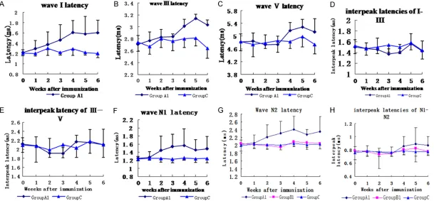
The animals in group A demonstrated pro-longed peak latencies of wave I, III, and V in a period of 1 week before immunization and 6 weeks after that. The interpeak latencies seemed to be normal as observed in control group. There were 4 animals (8 ears) in group B which had prolonged peak latencies, but the threshold of ABR and CAP was not elevated. Therefore, these animals served as group A1. The remaining animals in group B served as group B1. As shown in Figure 1, wave I latency was prolonged first at 2 weeks postimmuniza -tion, followed by changes in the wave III, and V at 3rd and 4th week respectively (all P<0.01, Student-Newman-Keuls test). No obvious pro-longations were observed in interpeak laten-cies of I-III, and III-V in groups A1, B1, or C. The peak latencies of waves N1 and N2 were also prolonged at 2nd week postimmunization, but at last two weeks some slowly decrease were observed (all P<0.01, Student-Newman-Keuls test). The interpeak latency of N1-N2 showed a little increase at 4th, 5th, and 6th week postim-munization, but the difference was not signifi -cant (all P>0.05, analysis of variance).
Transmission electron microscopic findings of the sciatic and cochlear nerves
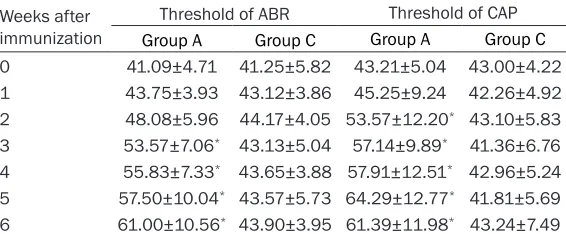
[image:3.612.93.376.85.202.2]In both nerves, demyelination was observed. The myelin sheath displayed malformation with splitting and vacuolization of consecutive lamellae, and the axoplasm seemed fine, but Table 1. Threshold of ABR and CAP (dB peSPL)
Weeks after immunization
Threshold of ABR Threshold of CAP
Group A Group C Group A Group C
0 41.09±4.71 41.25±5.82 43.21±5.04 43.00±4.22 1 43.75±3.93 43.12±3.86 45.25±9.24 42.26±4.92 2 48.08±5.96 44.17±4.05 53.57±12.20* 43.10±5.83
3 53.57±7.06* 43.13±5.04 57.14±9.89* 41.36±6.76
4 55.83±7.33* 43.65±3.88 57.91±12.51* 42.96±5.24
5 57.50±10.04* 43.57±5.73 64.29±12.77* 41.81±5.69
6 61.00±10.56* 43.90±3.95 61.39±11.98* 43.24±7.49
Data are shown as mean ± SD. *P<0.01 compared with group C (Student-Newman-Keuls test).
Figure 1. Changes of wave latency and interpeak latency in ABR and CAP. wave I latency was prolonged first at 2 weeks postimmunization, followed by changes in
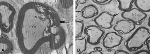
the dilation of the submyelinal space was observed especially in cochlear nerve (Figure 2).
Scanning electron microscopic findings of the hair cells
The scanning electron microscopy (SEM) of the organ of Corti showed the damage predomi-nantly on the inner hair cells (IHC). The IHC ste-reocilia disruption and swelling, and cytoplas-mic protrusions were observed. The outer hair cells appeared normal. Generally, the extent of stereocilia damage within each cochlea pro-gressed from apical to basal turns (Figure 3).
Immunohistochemical staining of anti-MBP antibody
The serum antibodies in experimental group bound mainly to cochlear nerves, spiral gangli-on cells and the nerve fibers in the osseous spi -ral lamina. No evident positive staining in the organ of Corti, stria vascularis and spiral liga-ment were detected. There was no immunos-taining in sections incubated with sera from animals of the control group (Figure 4).
Discussion
In 1996, Starr et al [1] reported 10 patients with auditory neuropathy. These investigators found evidence of peripheral neuropathy in 8 patients. Three of them were diagnosed as hav-ing Charcot-Marie-Tooth. Because demyelin-ation is a feature of this disease, they conclud-ed that, in these cases at least, the abnormal evoked potential findings were most likely the result of myelin damage. The exact pathology of
stem responses and the speech recognition deficits out of proportion to the puretone hear -ing loss [7, 8]. Some patients with auditory neu-ropathy were accompanied with limb peripheral nerve demyelinating impairment, such as/ slowed or blocked motor conduction velocity (MCV), sensory conduction velocity (SCV) and abnormal deep tendon reflexes. Such demyelin -ating disease as Guillain-Barre syndrome, HSMN type I (Charcot-Marie-Tooth disease) and primary demyelinating disorder in central ner-vous system (multiple sclerosis) might involve the auditory nerve resulting in the manifesta-tion of auditory neuropathy [1, 3, 9]. The diag-nosis of an “auditory neuropathy” is mainly based on the/audiological or physiological data. The clinical-pathologic correlation was not yet convincing as there was few autopsy studies carried out in auditory neuropathy patients diagnosed in life.
[image:5.612.91.381.73.178.2]At present, there was no such animal model as the demyelination was restricted to the audito-ry nerve for audiological study. Matsuoka [10] induced hearing loss of mice by peripheral nerve myelin protein P0 immunization. Peak latencies of wave I, III, and V and the interpeak latency I-III were prolonged in the BAEP study. Histopathologic findings showed the mononu -clear cell infiltration in the cochlear nerve region, and a reduced number of spiral gangli-on cells detected in the inner ear of the mice with hearing loss. This indicated that cochlear nerve was involved in this animal model with inflammatory demyelination in the peripheral nerve. Naito et al [11, 12] reported abnormal auditory evoked potential in mutant hamster ‘black tremor (bt)’ with significantly prolonged Figure 2. Transmission electron microscopy of nerves in experimental animals
with significant hearing loss. There are splitting and vacuolization of consecu -tive lamellae in myelin sheath, and the dilation in the submyelinal space of the
axoplasm (Original magnification ×5000). A. Sciatic nerve; B. Cochlear nerve (↑myelin sheath; ※axoplasm).
wave latencies or auditory brainstem respons-es and prolonged N1 latencies of compound
action potentials. Morphological observation revealed a myelin deficiency in the cochlear nerve. Experimental allergic neuritis (EAN) is an accepted animal model for human autoim-mune demyelinating disease of the peripheral nervous system (PNS), Guillain-Barre syndrome (GBS) [13]. EAN can be induced by immuniza-tion with myelin basic protein (MBP) or passive transfer of MBP-reactive T cell lines and clones. We established the guinea pig model of experi-mental allergic neuritis by immunization with whole bovine MBP. The animal demonstrated hunched posture, poor coat, loss of body weight, and abnormal walking with a waddling gait. The serum levels of antibody against MBP were increased and the peripheral nerve dis-played demyelination with electrophysiologic change of slowed motor conduction velocity. At the same time, the involvement of cochlear nerves was observed under transmission elec-tron microscopy and immunohistochemistry. To establish the dysmyelination model in this ani-mal for hearing research, it is important to assess the physiological status of the
periph-eral and brainstem auditory system in the guin-ea pigs. In this study, some similar findings as the mutant hamsters were observed [11, 12]. In myelinated nerve fibers, the ionic processes accounting for the generation of the action potential are restricted to the nodes of Ranvier, the junction points between adjacent myelin glial cells. The nodal membrane is rich in Na+
channels essential for the generation of the nerve action potential, whereas paranodal and internodal membrane sites have few Na+
chan-nels but are rich in K+ channels. Thus, the
gen-eration of Na+ current contributing to the
[image:6.612.94.521.72.176.2]devel-opment of the action potential in myelinated nerve is not continuous along the nerve but is restricted to the nodes of Ranvier. The restric-tion of the acrestric-tion potential to the nodes of Ranvier results in a discontinuity of conduction from node to node known as salutatory tion and accounts in part for the rapid conduc-tion velocity of myelinated fibers. Demyelinaconduc-tion results in an increased membrane capacitance and a decrease in membrane resistance, lead-ing to a delayed excitation, a reduction on the velocity of action potential propagation, and an
Figure 3. SEM from apical (A), middle (B) and basal (C) turn of Corti demonstrating the stereocilia damage to inner
hair cells (Original magnification A, C ×2500, B ×3500).
Figure 4. Anti-MBP antibody staining in cochlea (strepavidin–biotin peroxidase method, Original magnification ×400). A. There is intense immunostaining in cochlear nerves and spiral ganglion cells. B. There is staining in the nerve fibers between the organ of Corti and the spiral ganglion. C. There is no immunostaining in sections incubated
[image:6.612.97.522.227.332.2]increase in conduction vulnerability. The slowed internodal conduction velocity in a segmentally demyelinated axon varies with the extent of demyelination [14, 15]. So the slowed sciatic nerve conduction velocity was examined, and altered auditory function was found with the prolonged wave latencies of ABR and CAP in the guinea pigs of EAN we established. Wave I of the ABR generated in the Schwann cell-myelinated part of the VIII nerve in the cochlea, whereas wave II generated more proximally in the myelin of oligodendroglial origin, and wave III in the brainstem where it is unequivocally oli-godendroglial. Demyelination due to Schwann cell pathology will, therefore, prolong the wave I latency or I-III interval. In contrast, it is the wave III-V interval that will be prolonged by demyelin-ation due to oligodendroglial pathology affect-ing brain stem tracts [3]. Both N1 of the ECochG and wave I of the ABR arise from the distal por-tion of the cochlear nerve [3, 11]. Therefore, the electrophysiological findings of abnormal wave latency with relatively normal wave inter-val in these Guinea pigs indicate the damage to the distal cochlear nerves. Partial or complete loss of myelin has profound effects on the gen-eration and propagation of action potentials within auditory nerve fibers, resulting in the reductions in the temporal synchrony of the nerve fibers. The presence of ABR depends on the ability of auditory neurons to respond syn-chronously to an auditory stimulus. The findings of relatively normal wave intervals might indi-cate that the lesion in the animal was not enough severe to destroy more centrally audi-tory pathway, or the impairment in those guinea pigs was only Schwann cell origin. The deficits appear to involve the firing of the auditory nerve in the peripheral portions of the auditory path-way while sparing the signal conduction on the more central portions of the auditory pathway. The latencies of wave III and wave V might be secondary to the affect of wave I.
ABRI waveform disappears or shows obvious anomalies, which indicates that lesions begin in the distal end of auditory nerve. The normal appearance of otoacoustic emission in patients indicates that the lesions begin in the inner hair cells, inner hair cells and auditory nerve fiber tip, ganglion cells and nerve fibers [1]. As stated above, the auditory dysfunction of auditory neuropathy might be caused by a demyelin-ation of auditory nerve. We have demonstrated
an animal model of auditory nerve pathology due to demyelination, and discovered IHC ste-reocilia disruption and cytoplasmic swelling under SEM. We assumed that the disorders of IHC are subsequent to the primary injury of the VIII nerve. The abnormal electrophysiological findings in the peripheral component of ABR and CAP appeared to be consistent with the pathology. It appeared that the lesion may be at the site of ‘post-outer hair cells’ as the outer hair cells seems to be intact. More sufficient audiological data and further histology should be pursued in this animal model to clarify the underlying histological abnormalities and their relationship to altered auditory function. This animal model seems to be a promising model of auditory neuropathy due to demyelination disorders.
Acknowledgements
This work was supported by Natural Science Foundation of Hubei Province of China (2010CDB06106).
Disclosure of conflict of interest
None.
Address correspondence to: Dr. Peng Song, Depart- ment of Otorhinolaryngology-Head and Neck Sur- gery, Zhongnan Hospital, Wuhan University, 169 Donghu Road, Wuhan 430071, Hubei, China. Tel: 86-027-67813131; E-mail: pengsong15734@126. com
References
[1] Starr A, Picton TW, Sininger Y, Hood LJ, Berlin CI. Auditory neuropathy. Brain 1996; 119: 741-53.
[2] Starr A, Sininger YS, Pratt H. The varieties of auditory neuropathy. J Basic Clin Physiol Phar-macol 2000; 11: 215-30.
[3] Rapin I, Gravel J. “Auditory neuropathy”: physi-ologic and pathphysi-ologic evidence calls for more
diagnostic specificity. Int J Pediatr Otorhinolar -yngol 2003; 67: 707-28.
[4] Hall JI. Studies on demyelinated peripheral nerves in guinea-pigs with experimental aller-gic neuritis. A histoloaller-gical and electrophysio-logical study. Brain 1967; 90: 297-312. [5] Wu SJ, Chen J, Zhu GF. Establishment of
[6] London Y. Ox peripheral nerve myelin
mem-brane: purification and partial characterization
of two basic proteins. Biochim Biophys Acta 1971; 249: 188-96.
[7] Zeng FG, Oba S, Garde S, Sininger Y, Starr A.
Temporal and speech processing deficits in au -ditory neuropathy. Neuroreport 1999; 10: 3429-35.
[8] Kraus N, Bradlow AR, Cheatham MA, Cunning-ham J, King CD. Consequences of neural asyn-chrony: a case of auditory neuropathy. J Assoc Res Otolaryngol 2000; 1: 33-45.
[9] Xu J, Liu C, Liu B, Lian NJ, Gao YH, Zhao Y. Ves-tibular and limb peripheral nerve impairment in auditory neuropathy. Chin Arch Otolaryngol Head Neck Surg (Chinese) 2001; 8: 67-70. [10] Matsuoka H, Cheng KC, Krug MS, Yazawa Y,
Yoo TJ. Murine model of autoimmune hearing loss induced by myelin protein P0. Ann Otol Rhinol Laryngol 1999; 108: 255-64.
[11] Naito R, Murofushi T, Mizutani M, Kaga K. Audi-tory brainstem responses, electrocochleo-grams, and cochlear microphonics in the
my-elin deficient mutant hamster ‘bt’. Hear Res
1999; 136: 44-48.
[12] Naito R, Murofushi T, Mizutani M, Kaga K.
My-elin-deficiency in the cochlear nerve of the ‘bt’
mutant hamster. Hear Res 2003; 176: 17-24. [13] Gold R, Hartung HP, Toyka KV. Animal models
for autoimmune demyelinating disorders of the nervous system. Mol Med Today 2000; 6: 88-91.
[14] Starr A, Sininger Y, Winter M, Derebery MJ, Oba S, Michalewski HJ. Transient deafness due to temperature-sensitive auditory neuropathy. Ear Hear 1998; 19: 169-79.
[15] Rance G, Beer DE, Cone-Wesson B, Shepherd RK, Dowell RC, King AM, Rickards FW, Clark GM. Clinical findings for a group of infants and