warwick.ac.uk/lib-publications
Original citation:
Clifton, Rachel, Green, Laura E. and Purdy, Kevin J.. (2018) Development and validation of a
multiple locus variable number tandem repeat analysis (MLVA) scheme for Fusobacterium
necrophorum. Veterinary Microbiology, 213. pp. 108-113.
Permanent WRAP URL:
http://wrap.warwick.ac.uk/95437
Copyright and reuse:
The Warwick Research Archive Portal (WRAP) makes this work of researchers of the
University of Warwick available open access under the following conditions.
This article is made available under the Creative Commons Attribution 4.0 International
license (CC BY 4.0) and may be reused according to the conditions of the license. For more
details see: http://creativecommons.org/licenses/by/4.0/
A note on versions:
The version presented in WRAP is the published version, or, version of record, and may be
cited as it appears here.
Contents lists available atScienceDirect
Veterinary Microbiology
journal homepage:www.elsevier.com/locate/vetmic
Development and validation of a multiple locus variable number tandem
repeat analysis (MLVA) scheme for
Fusobacterium necrophorum
Rachel Clifton
⁎, Laura E. Green
1, Kevin J. Purdy
1School of Life Sciences, University of Warwick, Gibbet Hill Road, Coventry CV4 7AL, United Kingdom
A R T I C L E I N F O
Keywords:
Fusobacterium necrophorum Community
Strain typing MLVA
A B S T R A C T
Fusobacterium necrophorumis associated with various diseases in humans and animals. Reservoirs (sites where the pathogen persists in the absence of disease) ofF. necrophorumare believed to be present in healthy in-dividuals e.g. tonsillar epithelium, or their environment e.g. soil, but for most diseases the reservoir sites are unknown. Strain typing ofF. necrophorumwould facilitate linking specific reservoirs with a specific disease. The aim of this study was to develop multiple locus variable number tandem repeat analysis (MLVA) as a strain typing technique forF. necrophorum, and to test the use of this scheme to analyse both isolates and mixed communities of bacteria. Seventy-three tandem repeat regions were identified in theF. necrophorumgenome; three of these loci were suitable and developed as a MLVA scheme. The MLVA scheme was sensitive, specific, and discriminatory for both isolates and communities ofF. necrophorum. The MLVA scheme strain typed 46/52F. necrophorumisolates including isolates of both subspecies and from different countries, host species and sample sites within host. There were 12 unique MLVA strain types that clustered by subspecies. The MLVA scheme characterised theF. necrophorumcommunity in DNA from 32/49 foot- and 28/33 mouth swabs from sheep. There were 17 community types in total. In 31/32 foot swabs, single strains ofF. necrophorumwere detected while in the 28 mouth swabs there were up to a maximum of 8 strains ofF. necrophorumdetected. The results demonstrate the potential for this method to elucidate reservoirs ofF. necrophorum.
1. Introduction
Fusobacterium necrophorum is a Gram-negative, rod-shaped, anae-robic bacterium that is associated with a variety of diseases, termed necrobacilloses, in humans and animals. In humans, F. necrophorum causes Lemierre’s disease (Kuppalli et al., 2012; Lemierre, 1936; Riordan, 2007) and is associated with pharyngitis (Aliyu et al., 2004; Ludlam et al., 2009), periodontal disease (Enwonwu et al., 1999; Gomes et al., 2004; Jacinto et al., 2008) and appendicitis (Rogers et al., 2016). In animals, F. necrophorumcauses hepatic abscesses that occur in in-tensively reared beef cattle (Lechtenberg et al., 1988; Nagaraja and Chengappa, 1998; Narayanan et al., 1997) and it is associated with footrot in sheep (Egerton et al., 1969; Witcomb et al., 2014), foot in-fections in other ungulates (Clark et al., 1985; Edwards et al., 2001; Handeland et al., 2010), endometritis in cattle (Ruder et al., 1981), calf diphtheria (Panciera et al., 1989), respiratory disease in deer (Brooks et al., 2014) and periodontal disease in wallabies (Antiabong et al., 2013b).
Reservoirs of bacterial pathogens are sites in an organism or the
environment where the pathogen lives, and often multiplies (Krämer et al., 2010).F. necrophorumis considered to be an opportunistic pa-thogen (Langworth, 1977; Tan et al., 1996), consequently healthy in-dividuals and/or their environment are assumed to be reservoirs for the bacterium. However, there has been little research on the location of reservoir sites. In cattle, strain typing was used to identify the bovine rumen as the reservoir ofF. necrophorumthat causes hepatic abscesses (Narayanan et al., 1997). In humans,F. necrophorumwas thought to be part of the throat microflora of healthy individuals (Bartlett and Gorbach, 1976; Lemierre, 1936), however, it has only been detected in people aged 18–39, although Lemierre’s disease and otherF. necro-phoruminfections can occur at any age (Aliyu et al., 2004; Jensen et al., 2007; Ludlam et al., 2009). In sheep,F. necrophorumhas been isolated from the gingiva (Bennett et al., 2009; McCourtie et al., 1990) and detected on both healthy and footrot-diseased feet (Calvo-Bado et al., 2011; Frosth et al., 2015; Maboni et al., 2016; Witcomb et al., 2014), but the significance of these sites as reservoirs is unknown. WhilstF. necrophorumhas been widely assumed to be ubiquitous in sheep faeces and soil (Langworth, 1977; Marsh and Tunnicliff, 1934; Roberts and
https://doi.org/10.1016/j.vetmic.2017.11.017
Received 4 April 2017; Received in revised form 26 September 2017; Accepted 17 November 2017
⁎Corresponding author.
1Joint last author
E-mail address:[email protected](R. Clifton).
0378-1135/ © 2017 The Authors. Published by Elsevier B.V. This is an open access article under the CC BY license (http://creativecommons.org/licenses/BY/4.0/).
Egerton, 1969; Winter, 2004) this is unsubstantiated.
There are two subspecies ofF. necrophorum:necrophorumand fun-duliforme(Shinjo et al., 1991). These are distinguished by a PCR assay that detects a haemagglutinin-related gene that is present in subsp. necrophorumbut notfunduliforme(Narongwanichgarn et al., 2003). To confidently identify reservoirs associated with specific diseases, strain typing of F. necrophorum over time is needed, as exemplified by Narayanan et al. (1997). Multiple locus variable number tandem repeat analysis (MLVA) is an objective, repeatable, PCR-based strain typing method that has been used in a variety of epidemiological studies of bacterial pathogens (Eyre et al., 2013; Halkilahti et al., 2013; Mezal et al., 2014; Russell et al., 2013; Vranckx et al., 2011; Wada et al., 2007). MLVA was originally developed to analyse individual isolates but it can also be used to analyse samples that may contain a mixed community of strains within a species (Vranckx et al., 2011). In these cases, MLVA is used to produce a molecular“fingerprint”of the strains present and so identify similarities and differences between commu-nities.
The aim of the current study was to develop an MLVA typing scheme forF. necrophorum, and to demonstrate its potential to analyse isolates and community DNA. A selection of F. necrophorum isolates from a variety of host species and countries, together with DNA ex-tracted from swab samples from the feet and mouths of sheep, were used to develop and validate the scheme.
2. Materials and methods
2.1. Identification of tandem repeat regions for MLVA analysis
Seventy-three tandem repeat regions (Table S1) were identified from the whole genome shotgun sequence of F. necrophorum ATCC 51357 (GenBank Accession number AJSY00000000.1) using the Tandem Repeats Finder software v.4.08 (Benson, 1999). Nine regions were excluded due to insufficientflanking sequence to facilitate PCR primer design for amplification of the target region. There were 34 regions identified using blastn (Altschul et al., 1990), whereflanking sequences were present in all of the three publishedF. necrophorum genomes available (accessed March 2014). PCR primers targeting the 3′ and 5′flanking regions of these 34 repeat regions were designed using BatchPrimer3 v1.0 (You et al., 2008). EightF. necrophorumsubsp. ne-crophorumisolates (Table S2) were testedfirst for amplification of the target region and then for polymorphism at the tandem repeat region. Three loci (Fn13, Fn42 and Fn69; Table S1) showed good amplification and sufficient polymorphism for use in MLVA typing. PCR primers used to amplify the three selected MLVA targets and their tandem repeat sizes are given in Table S3.
2.2. MLVA PCR reactions and cycling conditions
PCR reactions were carried out in afinal volume of 25μl and con-tained 12.5μl Bioline MyTaq™Red Master Mix (2×; Bioline Reagents Ltd., London, UK), 1μl molecular biology grade bovine serum albumin (BSA; 100μg ml−1; Sigma-Aldrich Ltd., Gillingham, UK), 1μl each of forward and reverse primers (10μM), and 1μl template DNA. In reac-tions using mixed DNA, 1μl betaine (5 M; Sigma-Aldrich Ltd., Gillingham, UK) was also included to improve sensitivity. Cycling conditions were 95 °C for 5 min, followed by 32 cycles of 94 °C for 30 s, 55 °C (Fn13 and Fn69) or 62 °C (Fn42) for 30 s, 72 °C for 30 s, followed byfinal extension at 72 °C for 10 min. All PCR reactions were carried out on an Eppendorf Mastercycler ep gradient machine (Eppendorf, Hamburg, Germany) with DNA extracted fromF. necrophorumsubsp. necrophorumDSM 21784 as the positive control and nuclease free H2O as the reagent blank. PCR products were visualized after ethidium bromide-stained agarose gel electrophoresis and imaged using a Gene Flash imager (Syngene Bio Imaging, Cambridge, UK).
2.3. Validation of the MLVA typing scheme
PCR primer specificity was tested using DNA from a selection of non-target organisms (Fusobacterium gonidiaformans [DSM 19810], Fusobacterium nucleatum subsp. polymorphum [DSM 20482], Dichelobacter nodosus [VCS1703A], Mycobacterium bovis [BCG], Escherichia coli, Mannheimia sp.,Pseudomonas sp., Staphylococcus epi-dermidis, Staphylococcus intermedius, andStreptococcus uberis; all from University of Warwick).
The sensitivity of amplification for each loci was tested using a ten-fold dilution series from 106 to 101genome copiesμl−1of F. necro-phorumDSM 21784 DNA added to DNA extracted fromF. necrophorum negative sheep foot swabs (Witcomb et al., 2014). The number of genome copies in the stock DNA was calculated based on the genome size forF. necrophorumsubsp.funduliforme(2,088,497 bp;Calcutt et al., 2014). A blank containing DNA extracted fromF. necrophorumnegative foot swabs was run alongside the dilution series.
The stability of the MLVA scheme was tested by comparing the MLVA strain type of twoF. necrophorumisolates before and after ten passages of culture on Fusobacterium Agar, a selective medium based on that used byBrazier et al. (1991)(Wilkins-Chalgren Anaerobe Agar with Gram-negative Anaerobe Selective Supplement (both Oxoid Ltd., Altrincham, UK), 5% defibrinated sheep blood and josamycin (3μg ml−1)).
2.4. Determining PCR amplicon size using fragment analysis
The size, in base pairs, of PCR products was determined using fragment analysis: samples were submitted to DNA Sequencing and Services™(College of Life Sciences, University of Dundee, UK) and re-sults analysed with Peak Scanner 2 Software (Applied Biosystems, Warrington, UK). Sanger sequencing of the PCR products from each of the three assays forF. necrophorumDSM 21784 were used as a reference for the number of repeats to be calculated from the size in base pairs for each sample. A variation in expected size of PCR amplicon of ± 2 bp was tolerated.
2.5. MLVA typing of F. necrophorum isolates
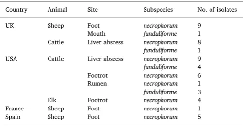
A total of 52 isolates, 43F. necrophorumsubsp.necrophorumand 9F. necrophorumsubsp.funduliforme,were used in this study. The country and sites of origin of the isolates are listed inTable 1.
[image:3.595.307.558.615.745.2]Isolates were cultured on Fusobacterium agar (as above) and then sub-cultured on Wilkins-Chalgren Anaerobe Agar (Oxoid Ltd., Altrincham, UK) with 5% defibrinated sheep blood. All incubations were carried out under anaerobic conditions (Don Whitley MACS-MG-1000 anaerobic workstation; 80% N2, 10% CO2 and 10% H2, Don Whitley Scientific Ltd., Shipley, UK) at 30 °C for 2–5 days. DNA was extracted from cultures using the Qiagen DNeasy Blood and Tissue Kit
Table 1
Country, animal host, site of sample and subspecies of 52Fusobacterium necrophorum isolates tested by MLVA.
Country Animal Site Subspecies No. of isolates
UK Sheep Foot necrophorum 9
Mouth funduliforme 1 Cattle Liver abscess necrophorum 8 funduliforme 1 USA Cattle Liver abscess necrophorum 9 funduliforme 4 Footrot necrophorum 6 Rumen necrophorum 1 funduliforme 3 Elk Footrot necrophorum 4 France Sheep Foot necrophorum 1 Spain Sheep Foot necrophorum 5
R. Clifton et al. Veterinary Microbiology 213 (2018) 108–113
(Qiagen Ltd., Manchester, UK) according to the manufacturer’s in-structions with a lysis time of 1 h. AF. necrophorumspecific standard PCR targeting the gyrase B gene (Antiabong et al., 2013a; Jensen et al., 2007) was used to confirm that isolates wereF. necrophorum,and am-plification of the haemagglutinin-related protein gene used to confirm isolates as subspeciesnecrophorumrather than subspeciesfunduliforme (Antiabong et al., 2013a; Narongwanichgarn et al., 2003).
The strain type ofF. necrophorumisolates was determined by the number of repeats at each of the three loci (Fn13, Fn42 and Fn69) after PCR and fragment analysis. Each strain type was assigned a unique number. The Hunter-Gaston Discriminatory Index (HGDI) for the strain typing scheme was calculated (Hunter and Gaston, 1988) with 95% confidence intervals (Grundmann et al., 2001). Minimum-spanning trees for the isolate strain typing data were created in PHYLOViZ-2.0 (Francisco et al., 2012) using the global optimal eBURST (goeBURST) distance algorithm with Euclidean distance (Francisco et al., 2009). The population was grouped on single locus variants (SLV).
2.6. MLVA typing of F. necrophorum communities from swab samples
Initially a model community was made by combining equal con-centrations of DNA from four isolates ofF. necrophorumwhich between them contained three variants at both Fn13 and Fn42, and two variants at Fn69. This was then tested to investigate whether, in a mixed com-munity, all the variants at each locus were detected using the MLVA typing scheme.
DNA was extracted from 82 swabs (33 mouth and 49 foot swabs) taken from sheep on six farms (A− F) in England, as described by Purdy (2005). TherpoBqPCR described byWitcomb et al. (2014)was used to detect and quantifyF. necrophorumin these samples, and those confirmed positive forF. necrophorum with a load > 103rpoBcopies swab−1were used for MLVA community analysis (Table 2). On Farm A, samples were collected as part of a longitudinal study: 10 sheep were sampled every 2 weeks for 8 weeks. On Farms B–F, 15 sheep were sampled per farm on one occasion.
For the swab samples, the number of MLVA variants within a locus was determined by fragment analysis. The minimum number of strains in a community was calculated as equal to the greatest number of MLVA variants at one locus. The maximum number of strains detected in a community was calculated by multiplying the number of variants at each locus together (e.g. if a sample contains 1, 2 and 3 variants for the three loci, the minimum number of strains is 3 and the maximum is 6
(1 × 2 × 3)). Each unique pattern of MLVA variants within these samples was assigned a unique“community type”number. The HGDI and associated confidence interval were calculated based on the fre-quency of detection of each community type.
3. Results
3.1. Validation of PCR amplification of the loci
The PCR assays for the three MLVA loci (Fn13, Fn42 and Fn69) were specific, with no PCR product produced from any of the non-target organisms tested. The detection limit was 104genome copiesμl−1of extracted DNA for the Fn13 assay, and 103genome copiesμl−1of ex-tracted DNA for the Fn42 and Fn69 assays. The MLVA scheme was stable; the MLVA type of the two isolates matched their original MLVA type after ten culture passages.
3.2. Population diversity of F. necrophorum isolates
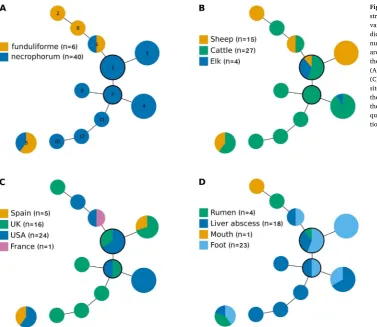
The three MLVA loci were characterised in 46/52 (88%)F. necro-phorumisolates. The 6 isolates that were not fully characterised were excluded from further analysis. In the fully characterised isolates there were three variants at locus Fn13,five at Fn42 and four at Fn69 (Table S4) giving 12 unique MLVA strain types (Table S5), 6 of which were detected only once. The HGDI for the strain typing scheme was 0.85 (95% CI 0.80–0.90), so that two distinct strains would be characterised as different on 85% of occasions.
Strain types varied within subspecies, country, host species and sample site. The goeBURST analysis detected 2 groups (Fig. 1): 11/12 strains were in a major group with strain types 3 (n = 9) and 7 (n = 2) the predicted ancestral strains. Strain type 5 (n = 5) was in an un-connected group by itself. Both subspecies were present in both groups and were clearly clustered within the major group (Fig. 1A); only 1/11 strain types in the major group contained both subspecies. There was no clear clustering of strains by host species (Fig. 1B), country of origin (Fig. 1C), or tissue site (Fig. 1D). The variation in strains indicates that analysis of a greater number of isolates could provide evidence of clustering if it exists.
3.3. Community diversity of F. necrophorum in DNA from swab samples
All expected locus variants were detected in the model community (data not shown) indicating that the MLVA scheme was able to detect strains in mixed communities ofF. necrophorum. All three MLVA loci were amplified from 28/33 (85%) mouth and 32/49 (65%) foot swab samples (Table 2). There were 17 unique community types (Table S6), 10 of which contained more than 1 strain, these ranged from a minimum of 2 to a maximum of 8 strains. TheF. necrophorum com-munities in mouth swabs were more complex than the comcom-munities in foot swabs. There were 16 community types in mouth swabs; the overall HGDI was 0.94 (95% CI 0.90 − 0.98). There were only 4 community types in foot swabs; 31/32 (97%) foot swabs had a single strain type (one of strain types 1, 3 and 6 fromFig. 1) consequently the HGDI was not calculated for foot swabs.
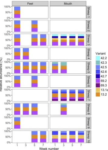
The locus variants from the 10 sheep from Farm A are presented in Fig. 2. The same strain was detected on feet over time and 24/25 foot swabs were a single strain (strain type 1 in the isolate analysis (Fig. 1)) rather than a community ofF. necrophorum. The three locus variants in this strain (13.2, 42.5 and 69.2) were also detected in mouths in sheep 5, 7 and 8, indicating that this strain was potentially present in mouths. There were, however, many more strain types in mouths than feet. In mouths, some locus variants and community types were stable over time for example, the same community type was detected at all four time points in sheep 3 and 7 and in sheep 5 and 10 the community types differed by one additional locus variant present in 50% of the samples. On Farms B-F, as with Farm A (Fig. 2), complex communities (up to 8 Table 2
Detection ofFusobacterium necrophorumand success of MLVA analysis by site of swab and farm.
Site and farm F. necrophoruma Community typeb
No. % No. %
Foot swabs
A 76/152 50 25/37 68
B 2/13 15 0/2 0
C 2/13 15 0/2 0
D 3/14 21 3/3 100
E 3/16 19 2/3 67
F 2/14 14 2/2 100
Mouth swabs
A 30/38 79 15/16 94
B 7/15 47 6/7 86
C 1/15 7 1/1 100
D 7/15 47 5/7 71
E 1/15 7 1/1 100
F 1/15 7 0/1 0
aNumber and percentage of samples positive forF. necrophorumout of total samples
collected.
bNumber and percentage of samples with an MLVA community type determined out of
strains) were present in mouth swabs whereas only single strains ofF. necrophorumwere detected in foot swabs. Community data from Farms B–F is presented in Fig. S1.
4. Discussion
The MLVA typing scheme developed forF. necrophorumwas specific and sensitive with the potential to strain type isolates and community DNA. Discriminatory ability, stability, epidemiological concordance, typeability and reproducibility are also used to evaluate typing schemes (van Belkum et al., 2007). A HGDI discriminatory value of≥0.95 is recommended for typing schemes (van Belkum et al., 2007). The dis-criminatory ability of this 3-loci scheme was 0.85 (95% CI 0.80–0.90) for isolates and 0.94 (95% CI 0.90–0.98) for communities ofF. necro-phorum. Ideally, we would have liked the scheme to be more dis-criminatory, however, there were no further suitable loci. The results from isolates and communities do suggest that the scheme is sufficiently discriminatory for these samples. The identification of the same strain type for two isolates after multiple passages through culture demon-strated the stability of the scheme. Finally, there was good epidemio-logical concordance for the scheme, for example, single strains were detected on the feet of sheep over time on Farm A, whilst more complex and varied communities were detected in mouths samples over the same time period.
A wide range ofF. necrophorumisolates from three ruminant hosts and four countries was used to develop the scheme. The MLVA scheme was sufficiently discriminatory to differentiate isolates from the same country, host, site and subspecies. There was no clustering of F. ne-crophorumstrain types by country, host or site from the isolates ana-lysed. This might be due to the relatively small number of isolates analysed or because provenance of the samples meant that there were no clusters in the dataset. Clusters might be detectable in a dataset specifically selected to investigate the host disease and its
complimentary reservoir, e.g. as reported byNarayanan et al. (1997) for liver abscesses and the rumen reservoir in the same host animal.
This is thefirst study of communities ofF. necrophorumin sheep and provides pilot data for further study. The communities in the mouth were more complex than on the feet. There were locus variants in mouths that were never detected on feet from sheep on the same farm, suggesting site-specificity for some strains. In contrast, the strain ofF. necrophorumdetected on feet was potentially (i.e. its 3 loci were pre-sent) in the mouths of some sheep on Farm A, suggesting that the mouth could be a reservoir or a spill-over site from feet. With the exception of one sample, only single strains ofF. necrophorumwere detected on feet. The consistency over time and the discriminatory power of the MLVA scheme suggest that this is likely to be a true reflection of the samples analysed. The generalisability of this pattern of very limited diversity on feet is unknown, however,Zhou et al. (2009) also reported the presence of single strains of F. necrophorumin 14 DNA samples ex-tracted from foot swabs from sheep.
It is likely that there were loci variants that were not detected in the community DNA samples in the current study because of the limit of detection of the PCR. This may have affected locus Fn13 more because detection of this locus is less sensitive than Fn42 and 69. This limits the use of the scheme for community samples to those withF. necrophorum loads of more than∼104copies perμl of extracted DNA. Improvements in the sensitivity of detection at this locus would enable the analysis of a wider range of samples.
5. Conclusions
[image:5.595.38.416.52.379.2]A sensitive, specific, stable and discriminatory MLVA typing scheme was developed and validated for both isolates and community DNA samples ofF. necrophorum. Using samples from sheep, the scheme is epidemiologically plausible and has potential to improve understanding of reservoirs of F. necrophorum and their association with Fig. 1.Analysis ofFusobacterium necrophorumMLVA strain type clustering using goeBurst. Single locus variants are connected by solid lines. Numbers in-dicate MLVA strain type, and size of circle represents number of isolates of each MLVA type. Types 3 and 7 are the suggested founder strain types, indicated by the black border. The shading indicates isolates of (A) different subspecies, (B) different host species, (C) different countries of origin, and (D) different sites of origin. Individual isolates are not always in the same position within a circle between the 4 trees, the coloured sections are placed with the most fre-quently representedfirst from the 12 o’clock posi-tion.
R. Clifton et al. Veterinary Microbiology 213 (2018) 108–113
necrobacilloses in both non-human and human animals.
Acknowledgements
Dr Rachel Clifton is funded by a Natural Environment Research Council (NERC) CASE studentship (NE/K007491/1) with AHDB Beef & Lamb as the industrial partner. Professor Laura Green and Dr Kevin Purdy are funded by BBSRC (BB/M012980/1). We gratefully ac-knowledge Professor T.G. Nagaraja of Kansas State University for sup-plying isolates ofF. necrophorum. We thank Dennis Homer of AHDB for assisting with collection of samples from bovine liver abscesses in the UK. We also thank Dr Ed Smith, Dr Emma Monaghan, Dr Kat Giebel, Jess Gaudy, Jess Taylor and Rebecca Jess for their assistance with sample collection from sheep and laboratory analysis.
Appendix A. Supplementary data
Supplementary data associated with this article can be found, in the online version, athttps://doi.org/10.1016/j.vetmic.2017.11.017.
References
Aliyu, S.H., Marriott, R.K., Curran, M.D., Parmar, S., Bentley, N., Brown, N.M., Brazier, J.S., Ludlam, H., 2004. Real-time PCR investigation into the importance of
Fusobacterium necrophorumas a cause of acute pharyngitis in general practice. J. Med. Microbiol. 53, 1029–1035.
Altschul, S.F., Gish, W., Miller, W., Myers, E.W., Lipman, D.J., 1990. Basic local alignment search tool. J. Mol. Biol. 215, 403–410.
Antiabong, J.F., Boardman, W., Smith, I., Brown, M.H., Ball, A.S., Goodman, A.E., 2013a. Cycliplex PCR confirmation ofFusobacterium necrophorumisolates from captive wallabies: a rapid and accurate approach. Anaerobe 19, 44–49.
[image:6.595.39.396.53.547.2]Antiabong, J.F., Boardman, W., Smith, I., Brown, M.H., Ball, A.S., Goodman, A.E., 2013b. A molecular survey of a captive wallaby population for periodontopathogens and the co-incidence ofFusobacterium necrophorumsubspeciesnecrophorumwith periodontal diseases. Vet. Microbiol. 163, 335–343.
Fig. 2.Relative abundance of locus variants in swab samples from Farm A. The ten sheep from Farm A are listed on the right of thefigure. Results from all sitive foot swabs from a sheep (sometimes > 1 po-sitive per sheep) are represented in the left-hand panels, and mouths in the right. Note, in all but one of the sheep (sheep 5, week 1) all positive foot swabs contained the same community type, which was re-presented by a single strain type (strain type 1 in
Bartlett, J.G., Gorbach, S.L., 1976. Anaerobic infections of head and neck otolaryngol. Clin. N. Am. 9, 655–678.
Bennett, G., Hickford, J., Sedcole, R., Zhou, H., 2009.Dichelobacter nodosus,Fusobacterium necrophorumand the epidemiology of footrot. Anaerobe 15, 173–176.
Benson, G., 1999. Tandem repeatsfinder: a program to analyze DNA sequences. Nucleic Acids Res. 27, 573–580.
Brazier, J.S., Citron, D.M., Goldstein, E.J.C., 1991. A selective medium for Fusobacterium spp. J. Appl. Bacteriol. 71, 343–346.
Brooks, J.W., Kumar, A., Narayanan, S., Myers, S., Brown, K., Nagaraja, T.G., Jayarao, B.M., 2014. Characterization of Fusobacterium isolates from the respiratory tract of white-tailed deer (Odocoileus virginianus). J. Vet. Diagn. Invest. 26, 213–220.
Calcutt, M.J., Foecking, M.F., Nagaraja, T.G., Stewart, G.C., 2014. Draft genome sequence of fusobacterium necrophorum subsp. funduliforme bovine liver abscess isolate B35. Genome Announc. 2, e00412–e00414.
Calvo-Bado, L.A., Oakley, B.B., Dowd, S.E., Green, L.E., Medley, G.F., Ul-Hassan, A., Bateman, V., Gaze, W., Witcomb, L., Grogono-Thomas, R., Kaler, J., Russell, C.L., Wellington, E.M.H., 2011. Ovine pedomics: thefirst study of the ovine foot 16S rRNA-based microbiome. ISME J. 5, 1426–1437.
Clark, B.L., Stewart, D.J., Emery, D.L., 1985. The role of Fusobacterium necrophorum and Bacteroides melaninogenicus in the etiology of interdigital necrobacillosis in cattle. Aust. Vet. J. 62, 47–49.
Edwards, J.F., Davis, D.S., Roffe, T.J., Ramiro-Ibanez, F., Elzer, P.H., 2001. Fusobacteriosis in captive wild-caught pronghorns (Antilocapra americana). Vet. Pathol. 38, 549–552.
Egerton, J.R., Roberts, D.S., Parsonson, I.M., 1969. The aetiology and pathogenesis of ovine foot-rot. I. A histological study of the bacterial invasion. J. Comp. Pathol. 79, 207–215.
Enwonwu, C.O., Falkler Jr., W.A., Idigbe, E.O., Afolabi, B.M., Ibrahim, M., Onwujekwe, D., Savage, O., Meeks, V.I., 1999. Pathogenesis of cancrum oris (noma): confounding interactions of malnutrition with infection. Am. J. Trop. Med. Hyg. 60, 223–232.
Eyre, D.W., Fawley, W.N., Best, E.L., Griffiths, D., Stoesser, N.E., Crook, D.W., Peto, T.E.A., Walker, A.S., Wilcox, M.H., 2013. Comparison of multilocus variable-number tandem-repeat analysis and whole-genome sequencing for investigation of clos-tridium difficile transmission. J. Clin. Microbiol. 51, 4141–4149.
Francisco, A.P., Bugalho, M., Ramirez, M., Carriço, J.A., 2009. Global optimal eBURST analysis of multilocus typing data using a graphic matroid approach. BMC Bioinf. 10, 1–15.
Francisco, A.P., Vaz, C., Monteiro, P.T., Melo-Cristino, J., Ramirez, M., Carriço, J.A., 2012. PHYLOViZ: phylogenetic inference and data visualization for sequence based typing methods. BMC Bioinf. 13, 1–10.
Frosth, S., Koenig, U., Nyman, A.-K., Pringle, M., Aspan, A., 2015. Characterisation of
Dichelobacter nodosusand detection ofFusobacterium necrophorumandTreponemaspp. in sheep with different clinical manifestations of footrot. Vet. Microbiol. 179, 82–90.
Gomes, B., Pinheiro, E., Gadê-Neto, C., Sousa, E., Ferraz, C., Zaia, A., Teixeira, F., Souza-Filho, F., 2004. Microbiological examination of infected dental root canals. Oral Microbiol. Immunol. 19, 71–76.
Grundmann, H., Hori, S., Tanner, G., 2001. Determining confidence intervals when measuring genetic diversity and the discriminatory abilities of typing methods for microorganisms. J. Clin. Microbiol. 39, 4190–4192.
Halkilahti, J., Haukka, K., Siitonen, A., 2013. Genotyping of outbreak-associated and sporadic Yersinia pseudotuberculosis strains by novel multilocus variable-number tandem repeat analysis (MLVA). J. Microbiol. Methods 95, 245–250.
Handeland, K., Boye, M., Bergsjo, B., Bondal, H., Isaksen, K., Agerholm, J.S., 2010. Digital necrobacillosis in norwegian wild tundra reindeer (Rangifer tarandus). J. Comp. Pathol. 143, 29–38.
Hunter, P.R., Gaston, M.A., 1988. Numerical index of the discriminatory ability of typing systems–an application of Simpson’s index of diversity. J. Clin. Microbiol. 26, 2465–2466.
Jacinto, R.C., Montagner, F., Signoretti, F.G.C., Almeida, G.C., Gomes, B., 2008. Frequency, microbial interactions, and antimicrobial susceptibility of fusobacterium nucleatum and fusobacterium necrophorum isolated from primary endodontic in-fections. J. Endod. 34, 1451–1456.
Jensen, A., Kristensen, L.H., Prag, J., 2007. Detection of Fusobacterium necrophorum subsp. funduliforme in tonsillitis in young adults by real-time PCR. Clin. Microbiol. Infect. 13, 695–701.
Krämer, A., Akmatov, M., Kretzschmar, M., 2010. Principles of infectious disease epide-miology. In: Krämer, A., Kretzschmar, M., Krickeberg, K. (Eds.), Modern Infectious Disease Epidemiology: Concepts, Methods, Mathematical Models, and Public Health. Springer New York, New York, NY, pp. 85–99.
Kuppalli, K., Livorsi, D., Talati, N.J., Osborn, M., 2012. Lemierre’s syndrome due to Fusobacterium necrophorum. Lancet Infect. Dis. 12, 808–815.
Langworth, B.F., 1977. Fusobacterium necrophorum: its characteristics and role as an animal pathogen. Bacteriol. Rev. 41, 373–390.
Lechtenberg, K.F., Nagaraja, T.G., Leipold, H.W., Chengappa, M.M., 1988. Bacteriologic and histologic studies of hepatic abscesses in cattle. Am. J. Vet. Res. 49, 58–62.
Lemierre, A., 1936. On certain speticaemias due to anaerobic organisms. Lancet ii 701–703.
Ludlam, H., Howard, J., Kingston, B., Donachie, L., Foulkes, J., Guha, S., Curran, M.D., 2009. Epidemiology of pharyngeal carriage of Fusobacterium necrophorum. J. Med. Microbiol. 58, 1264–1265.
Maboni, G., Frosth, S., Aspan, A., Totemeyer, S., 2016. Ovine footrot: new insights into bacterial colonisation. Vet. Rec. 179, 228.
Marsh, H., Tunnicliff, E.A., 1934. Experimental studies of foot-rot in sheep. Mont. Agric. Exp. Stn. Bull. 285, 3–16.
McCourtie, J., Poxton, I.R., Brown, R., Whittaker, C.R., Spence, J.A., Aitchison, G.U., 1990. A longitudinal study of the cultivable subgingival anaerobic bacteria isolated from sheep during the development of broken mouth periodontitis. J. Med. Microbiol. 31, 275–283.
Mezal, E.H., Sabol, A., Khan, M.A., Ali, N., Stefanova, R., Khan, A.A., 2014. Isolation and molecular characterization ofSalmonella entericaserovar Enteritidis from poultry house and clinical samples during 2010. Food Microbiol. 38, 67–74.
Nagaraja, T.G., Chengappa, M.M., 1998. Liver abscesses in feedlot cattle: a review. J. Anim. Sci. 76, 287–298.
Narayanan, S., Nagaraja, T.G., Okwumabua, O., Staats, J., Chengappa, M.M., Oberst, R.D., 1997. Ribotyping to compareFusobacterium necrophorumisolates from bovine liver abscesses, ruminal walls, and ruminal contents. Appl. Environ. Microbiol. 63, 4671–4678.
Narongwanichgarn, W., Misawa, N., Jin, J.H., Amoako, K.K., Kawaguchi, E., Shinjo, T., Haga, T., Goto, Y., 2003. Specific detection and differentiation of two subspecies of
Fusobacterium necrophorumby PCR. Vet. Microbiol. 91, 183–195.
Panciera, R.J., Perino, L.J., Baldwin, C.A., Morton, R.J., Swanson, J.E., 1989. Observations on calf diphtheria in the commercial feedlot. Agric. Pract. 10, 12–17.
Purdy, K.J., 2005. Nucleic acid recovery from complex environmental samples. Methods Enzymol. 397, 271–292.
Riordan, T., 2007. Human infection withFusobacterium necrophorum(Necrobacillosis), with a focus on Lemierre’s syndrome. Clin. Microbiol. Rev. 20, 622–659.
Roberts, D.S., Egerton, J.R., 1969. The aetiology and pathogenesis of ovine foot-rot: II. The pathogenic association ofFusiformis nodosusandFusiformis necrophorus. J. Comp. Pathol. 79, 217–227.
Rogers, M.B., Brower-Sinning, R., Firek, B., Zhong, D., Morowitz, M.J., 2016. Acute ap-pendicitis in children is associated with a local expansion ofFusobacteria. Clin. Infect. Dis. 63, 71–78.
Ruder, C.A., Sasser, R.G., Williams, R.J., Ely, J.K., Bull, R.C., Butler, J.E., 1981. Uterine infections in the postpartum cow 2: possible synergistic effect ofFusobacterium ne-crophorumandCorynebacterium pyogenes. Theriogenology 15, 573–580.
Russell, C.L., Smith, E.M., Calvo-Bado, L.A., Green, L.E., Wellington, E.M., Medley, G.F., Moore, L.J., Grogono-Thomas, R., 2013. Multiple locus VNTR analysis highlights that geographical clustering and distribution ofDichelobacter nodosusthe causal agent of footrot in sheep, correlates with inter-country movements. Infect. Genet. Evol. 22, 273–279.
Shinjo, T., Fujisawa, T., Mitsuoka, T., 1991. Proposal of two subspecies ofFusobacterium necrophorum(Flugge) Moore and Holdeman:Fusobacterium necrophorumsubsp. ne-crophorumsubsp. nov., nom. rev. (ex Flugge 1886), andFusobacterium necrophorum
subsp.funduliformesubsp. nov., nom. rev. (ex Halle 1898). Int. J. Syst. Bacteriol. 41, 395–397.
Tan, Z.L., Nagaraja, T.G., Chengappa, M.M., 1996.Fusobacterium necrophoruminfections virulence factors, pathogenic mechanism and control measures. Vet. Res. Commun. 20, 113–140.
van Belkum, A., Tassios, P.T., Dijkshoorn, L., Haeggman, S., Cookson, B., Fry, N.K., Fussing, V., Green, J., Feil, E., Gerner-Smidt, P., Brisse, S., Struelens, M., Escmid, Esgem, 2007. Guidelines for the validation and application of typing methods for use in bacterial epidemiology. Clin. Microbiol. Infect. 13, 1–46.
Vranckx, K., Maes, D., Calus, D., Villarreal, I., Pasmans, F., Haesebrouck, F., 2011. Multiple-locus variable-number tandem-repeat analysis is a suitable tool for diff er-entiation ofMycoplasma hyopneumoniaestrains without cultivation. J. Clin. Microbiol. 49, 2020–2023.
Wada, T., Maeda, S., Hase, A., Kobayashi, K., 2007. Evaluation of variable numbers of tandem repeat as molecular epidemiological markers ofMycobacterium tuberculosisin Japan. J. Med. Microbiol. 56, 1052–1057.
Winter, A., 2004. Lameness in sheep 2. Treatment and control. In Pract. 26, 130–139.
Witcomb, L.A., Green, L.E., Kaler, J., Ul-Hassan, A., Calvo-Bado, L.A., Medley, G.F., Grogono-Thomas, R., Wellington, E.M.H., 2014. A longitudinal study of the role of
Dichelobacter nodosusandFusobacterium necrophorumload in initiation and severity of footrot in sheep. Prev. Vet. Med. 115, 48–55.
You, F.M., Huo, N.X., Gu, Y.Q., Luo, M.C., Ma, Y.Q., Hane, D., Lazo, G.R., Dvorak, J., Anderson, O.D., 2008. Batchprimer3: a high throughput web application for PCR and sequencing primer design. BMC Bioinf. 9, 13.
Zhou, H.T., Bennett, G., Hickford, J.G.H., 2009. Variation in Fusobacterium necrophorum strains present on the hooves of footrot infected sheep, goats and cattle. Vet. Microbiol . 135, 363–367.
R. Clifton et al. Veterinary Microbiology 213 (2018) 108–113