CHARACTERIZATION OF 5-HYDROXYTRYPTAMINE
RECEPTORS IN THE SNAIL, 'HELIX ASPERSA'
Anna-Karina Cadogan
A Thesis Submitted for the Degree of PhD
at the
University of St Andrews
1992
Full metadata for this item is available in
St Andrews Research Repository
at:
http://research-repository.st-andrews.ac.uk/
Please use this identifier to cite or link to this item:
http://hdl.handle.net/10023/14627
M-CHARACTERIZATION OF 5-HYDROXYTRYPTAMINE
RECEPTORS IN THE SNAIL,
HELIX ASPERSA
by Anna-Karina Cadogan
A thesis submitted to the University of St. Andrews in candidature for the degree of Doctor of Philosophy.
Department of Biology and Preclinical Medicine, St. Andrews University.
July 1991
ProQuest Number: 10167090
All rights reserved
INFORMATION TO ALL USERS
The quality of this reproduction is dependent upon the quality of the copy submitted.
In the unlikely event that the author did not send a com plete manuscript and there are missing pages, these will be noted. Also, if material had to be removed,
a note will indicate the deletion.
uest
ProQuest 10167090
Published by ProQuest LLO (2017). Copyright of the Dissertation is held by the Author.
All rights reserved.
This work is protected against unauthorized copying under Title 17, United States C ode Microform Edition © ProQuest LLO.
ProQuest LLO.
789 East Eisenhower Parkway P.Q. Box 1346
DECLARATIONS
I, Anna-Karina Cadogan, hereby certify that this thesis has been composed by myself, that it is a record of my own work, and that it has not been accepted in partial or complete fulfilment of any other degree of professional qualification.
Signed:
J
I was admitted to the Faculty of Science of the University of St. Andrews under Ordinance General N o 12 in October, 1987 and as a canditate for the degree of Ph.D. in October, 1987.
Signature of Supervisor:
I
Signed: |
Date: ly-' ^ ^ •
ACKNOWLEDGEMENTS
Firstly I would like to express my sincere thanks to my academic supervisor. Professor Glen Cottrell, for his help and guidance and to my supervisor at Glaxo, Dr. Pat Humphrey, for his inspiration and support. Had it not been for Pat's encouragement, I would never have been cajoled into giving a talk at the British Pharmacological Society last december and for that opportunity, I am eternally grateful.
At St. Andrews, I am indebted to Dr. Jim Aiton for his patience in dealing with my enquiries about the Macintosh, to Dr. Gordon Cramb for allowing me laboratory space to finish my assays, to the photographic unit for their help and advice with figures and photographs, to Dr. Colin Thomson and his wife, Maureen for their support during this period and to Christina Lamb who typed a draft of this thesis into my computer. I am also extremely grateful to my fellow workers who have helped me out on numerous occasions.
At Glaxo, where I spent nine happy months, I would like to thank Dr. Wasyl Feniuk for his thoughtful discussion and his support at the British Pharmacological Society meeting in december, Mike Sumner for putting up with me in his group for that period and to all the other Glaxo staff who, not only welcomed me in but also helped me in every way possible.
I wish also to acknowledge the financial support I received from the Science and Engineering Research Council and Glaxo who supported me as a CASE student.
ABSTRACT
The aim of this investigation was to characterize those 5-HT receptors present in three different tissues of the common garden snail. Helix aspersa,
into one or more of the categories already described for vertebrate 5-HT receptors. Specific 5-HT receptor agonists and antagonists which had been developed and used to help characterize, and subsequently classify, the various types of 5-HT receptor in vertebrates, were utilized in this study. The three preparations from Helix included: i) identified neurones in the visceral ganglion ii) the heart and iii) the pharyngeal retractor muscle (PRM).
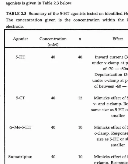
The action of 5-HT on identified neurones in the visceral ganglion was studied using the electrophysiological techniques of both voltage- and current-damp. Under voltage-dam p conditions the response of the identified neurones to iontophoretic application of 5-HT was seen to be an inward current of approximately 3-10 nA. Under current-damp conditions the response to 5-HT was an excitatory depolarization leading to the firing of action potentials of approximately 3-15 mV. Both responses showed rapid desensitization to repetitive applications of 5-HT and were blocked by tubocurarine. No specific 5-HT receptor antagonist to this 5-HT response in
Helix neurones was found. The action of 5-HT was mimicked by 5-CT and a-Me-5-HT both of which showed similar-sized responses to 5-HT, whereas sumatriptan gave smaller responses than those of 5-HT.
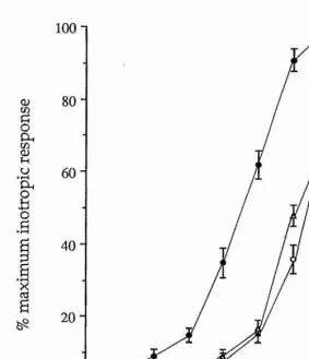
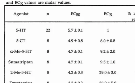
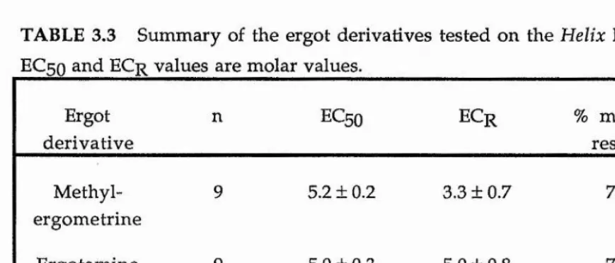
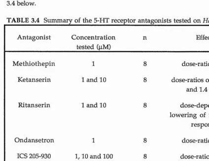
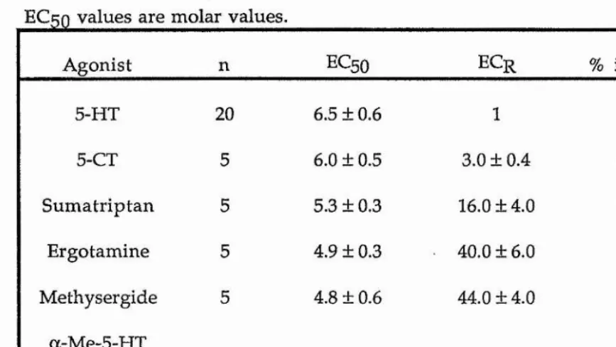
5-HT had a positive inotropic effect on the heart. The excitatory action of 5-HT on the heart was studied using an organ bath methodology with application of the 5-HT receptor agonists and antagonists at suitable concentrations. No specific 5-HT receptor antagonist was found for the cardioexcitatory effect of 5-HT. The full rank order of potency for the 5-HT receptor agonists tested was 5-HT > methylergometrine = ergotamine = 5-CT > a-Me-5-HT = sumatriptan > methysergide - 2-Me-5-HT = tryptamine > 8-OH-DPAT.
and antagonists at suitable concentrations. N o specific 5-HT receptor antagonist for the inhibition of ACh-induced contraction was found. The rank order of 5-HT receptor agonist potency was 5-HT > 5-CT > sumatriptan = ergotamine = methysergide » a-Me-5-HT = 2-Me-5-HT.
The effect of 5-HT on cyclic adenosine 3',5'-monophosphate (cAMP) levels within Helix heart and pharyngeal retractor muscle (PRM) tissue were monitored in this investigation. 5-HT caused a dose-dependent increase in cAMP both in Helix heart and PRM tissue.
ABBREVIATIONS
ABRM anterior byssus retractor muscle AC alternating current
ACh acetylcholine
ATP adenosine triphosphate A V a trio-ventricular
BSA bovine serum albumen
cAMP cyclic adenosine 3', 5'-monophosphate cGMP cyclic guanosine 3', 5'-monophosphate CNS central nervous system
D aspartic acid EXH direct current
DMSG dimethyl sulphoxide EC excitation-contraction
EDTA ethylene diamine tetraacetic acid F phenylalanine
g gram
Hepes N-2-Hydroxyethylpiperazine-N'-2-ethanesulfonic acid
Hz Hertz
IBMX 3-isobutyl-l -methylxanthine
ICS 205-930 (3a-tropanyl)-lH-indole-3-carboxylic acid ester
KQ kilohm
1 litre
L leucine
LSD lysergic acid diethylamide
M molar
M m ethionine
MAO monoamine oxidase
mCPP l-(3-chlorophenyl)piperazine mg milligram
MHz megahertz min m inute m l m illilitre mM m illim olar m m m illim etre
MQ m V |iF kig [iM [ im Hi nA nm nM P PAPP pm ole PQ PRM R RU 24969 TFMPP TRM UV 4-HT 5-CT 5-HT 5-HTP 5-MeOT 5-MeODMT 6-HT 7-HT 8-OH-DPAT m egohm m illivolt microfarad microgram micromolar micron microlitre nanoamp nanometre nanomolar proline p-amino-phenylTFMPP picomole pyroglutamate
pharyngeal retractor muscle arginine
5-methoxy-3(l,2,3,6-tetrahydropyridin-4-yl)lH indole
second
m-trifluoromethylphenyl pierazine tentacle retractor muscle
ultraviolet
- a
MOLECULAR STRUCTURES OF SOME OF THE RELEVANT 5-HT AGONISTS AND ANTAGONISTS
kïxi.V ,
# # # #
SAVX
"##
NMNM
0
####
'"'T'' SsS's^^ScO'-î-'4."s !>'
■Î* Vs\*>.
x^^yrÿsCsN
5 - H T
' v^- :■^V <5\-
I
TRYPTAMINE
* #
J f t ' "- - j v : ,# # N
5 - C T
# # # # # # # # ! # # # # #
m m m #
M h B N
0
#
NNT
M
-DC-M E-S-HT
#
N M M
mm
:!0!00!0i000^^^
########
liliiiiiiiiiiiiilliiiiiiP
Siiiiiiiiiiiiiiii
0##
#
i
*
3
a
z
CONTENTS
CHAPTER ONE GENERAL INTRODUCTION 1
CHAPTER TWO THE 5-FIT RECEPTOR IN HELIX NEURONES
Introduction 13
Methods and Materials 19
Results 25
Discussion 32
CHAPTER THREE THE 5-HT RECEPTOR IN HELIX HEART
Introduction 40
Methods and Materials 49
Results 57
Discussion 69
CHAPTER FOUR THE 5-HT RECEPTOR IN HELIX SMOOTH MUSCLE
Introduction 82
Methods and Materials 87
Results 92
Discussion 99
CHAPTER FIVE GENERAL DISCUSSION 107
CHAPTER ONE
GENERAL INTRODUCTION Historical perspective
5-hydroxytryptamine (5-HT) or serotonin, as it is also known, is one of the biogenic amines. However, unlike dopamine and noradrenaline which are catecholamines, 5-HT is derived from an indole nucleus and is therefore classified as an indoleamine.
Physiologists of the 19th century were aware of a substance in the blood serum that caused constriction of blood vessels and increased vascular tone. In the late 1940s, investigators were successful in isolating this serum-born substance, and named it serotonin (Rapport et aL, 1948). They later deduced that the active com ponent of their crystalline com plex was 5-hydroxytryptamine (Rapport, 1949). About the same time, a group of Italian workers had been investigating a substance found in high concentrations in enterochromaffin cells of the intestinal mucosa in the gut which they had called enteramine (Erspamer, 1940). Enteramine also occurred in the skin and salivary glands of lower animals such as toads, salamanders and octopuses. Erspamer and Asero (1952) went on to identify enteramine as being 5-hydroxytryptamine, identical to serotonin.
The distribution and possible roles of 5-HT
5-HT has a unusual distribution in nature and is not confined to vertebrates. Jacques and Schacter (1954) discovered 5-HT in the venom of the wasp, Vespa vulgaris, and it has been found in a number of fruits including bananas (Udenfriend et al, 1959) and in the stinging apparatus of the nettle
Urtica dioica (Brittain and Collier, 1957). It was found in the enterochromaffin cells of the intestine in all classes of vertebrates and in blood platelets and mast cells in mammals. Extensive listings of such occurrences have been given by Erspamer (1954).
an influential and important discovery in the history of 5-HT research. Using the Falck-Hillarp technique (Falck et a l, 1962) to visualise 5-HT within cells, with formaldehyde-induced fluorescence, they found that virtually all the 5-HT-containing neurones of the CNS of the rat were located in discrete clusters within the midbrain, pons and medulla (Dahlstrom and Fuxe, 1964). Over the years improvements in techniques have helped to define more clearly the locations of serotonergic cell bodies, fibres and terminals.
5-HT has been found to have multiple actions in the vertebrate nervous system. It affects sensory, motor, autonomic and enteric neuronal activity as well as influencing vascular and other smooth muscle responses. Within the CNS, 5-HT seemed to function in both an inhibitory and excitatory manner. In contrast, in the periphery, 5-HT has mainly excitatory postsynaptic actions on autonomic and enteric nervous system neurones, as well as producing contraction of the uterus, ileum and vasculature smooth muscle (see Vandermaelen, 1985).
The first systematic survey of the occurrence of 5-HT in invertebrates was undertaken by Welsh and Moorhead (1960). Using the fluorescence method to estimate the 5-HT content of nervous and non-nervous tissue from a variety of invertebrates, large amounts of 5-HT were detected both in annelids and molluscs. Although Welsh and Moorhead (1960) showed that 5-HT is widely distributed in both nervous and non-nervous tissues, they were not able to locate it in specific cell types. With the introduction of the Falck-Hillarp fluorescence microscopy technique (Falck et ah, 1962) for visualizing indoleamines and catecholamines in single cells and nerve tracts, it was possible to identify specific amine-containing cells. A further refinement was the introduction of biochemical techniques whereby the amine content of single neurones could be established quantitatively. Since then there have been many studies of the localization and possible function of 5-HT in a wide range of invertebrates, particularly in the molluscs.
5-HT has potent actions on many invertebrate tissues but it was the molluscan heart, particularly that of the quahog clam, Mercenaria mercenaria,
Endogenous 5-HT was shown to be released from the cerebro-buccal ganglionic ring of Aplysia (Gerschenfeld et ah, 1978). They demonstrated that the rate of spontaneous release of 5-HT varied between 0.4 and 1.2 pmol/hour. Direct stimulation of the gaint 5-HT-containing cerebral neurones resulted in an 80-100% increase in the release of 5-HT but only in the presence of chlorimipramine, a 5-HT uptake blocker.
The best evidence for 5-HT as a central transmitter comes from work on gastropods such as Helix (Cottrell and Macon, 1974) and Aplysia (Gerschenfeld and Paupardin-Tritsch, 1944a,b). It is the giant 5-HT-containing cerebral neurones in these gastropods which have been the subjects of extensive basically inhibitory, e.g. on the heart of Modiolus (Wilkens and Greenberg, 1973). At molluscan peripheral sites, 5-HT was not always found to be excitatory. Twarog and Cole (1972) found that 5-HT acted as a relaxing agent at the anterior byssus retractor muscle (ABRM) of Mytilus and was released at this site following nerve stimulation (Satchell and Twarog, 1978). 5-HT has also been shown to have a relaxing action of the penis retractor muscle (Wabnitz and von Watchendonk, 1976) and of the pharyngeal retractor muscle (PRM) (Lloyd, 1980a) of Helix.
Evidence for 5-HT as a transmitter in molluscan ganglia
The idea that 5-HT is a neurotransmitter in invertebrates had been proposed by Welsh (1954). His study with Moorhead (1960) had yielded important data on 5-HT levels in the nervous tissue of invertebrates with particularly high 5-HT levels in both annelids and molluscs. 5-HT has been demonstrated histochemically in the nervous systems of Helix, Anodonta,
j Buccinium and Limax (Dahl et ah, 1966, Sedden et ah, 1968, Osborne and 4 Cottrell, 1971). The levels of 5-HT in identified neurones of Tritonia and
Aplysia ganglia were found to be between 4 and 6 pm ol/ cell body (Weinreich
studies. These cells were first noted in Helix pomatia by Kunze (1921) and were then characterized electrophysiologically and according to their input properties by Kandel and Tauc (1966a,b). More general interest in these neurones was developed by Cottrell and Osborne (1970) who demonstrated that, in both Helix and Limax, these neurones contained significant amounts of 5-HT. Cottrell (1970) went on to show that these glo^nt 5-HT-containing neurones in Helix provided excitatory inputs to three identified neurones in the ipsilateral buccal ganglion, which were subsequently labelled anterior, middle and posterior. It was this pathway in Helix pomatia which was studied with electrophysiological methods by Cottrell and Macon (1974) to provide further evidence of a transmitter role for 5-HT. In their study, stimulation of the giant 5-HT-containing cerebral neurones evoked excitatory post synaptic potentials in the middle buccal cell. Repetitive stimulation of the cerebral neurones caused summation of these excitatory post synaptic potentials, giving rise to an action potential. Iontophoretic application of 5-HT onto the middle buccal neurone produced a depolarizing response which was shown to be sodium-dependent and could be antagonized by morphine. Reserpine which depletes 5-HT in the cerebral 5-HT-containing neurone, reduced but not completely abolished the efficacy of transmission between this neurone and the middle buccal neurone.
Evidence for 5-HT as a transmitter in molluscan hearts
That 5-HT served as a mediator for cardio-excitation in molluscs was probable. H ow ever confirmation of the release of 5-HT from the cardioregulatory nerves had to be achieved. A two Helix heart/Loew i-type experiment was set up by S-Rozsa and Perenyi (1966) in order that the release of 5-HT from the cardioregulatory nerves might be demonstrated. On stimulation of the nerve going to one heart, the perfusate from this first heart was found to contain material which activated the second heart. 5-HT was subsequently detected in the perfusate both by spectrophotometric and chromatographic techniques.
Evidence for 5-HT as a transmitter in molluscan visceral and smooth muscle. The other molluscan tissues where the action of 5-HT has been well investigated are the somatic and visceral muscles. These have included ABRM of M ytilus, the penis retractor muscle of Helix and Strophocheilos and the PRM of Helix. The visceral muscles which have been investigated include the rectum of Mercenaria and Tapes and the crop of Helix.
There is substantial evidence for 5-HT as a neurotransmitter mediating relaxation at the ABRM in Mytilus. 5-HT was found to mimic the effects of stimulating the relaxing nerves. When the muscle was exposed to 5-HT at concentrations >lnM , relaxation of catch contraction in the muscle was proportional to the log%g of 5-HT concentration (Twarog ,1954). Von Watchendonk and Kappler (1977), using liquid scintillation counting and microchromatography, found 5-HT levels up to 4.3|ig.g"^wet weight for the ABRM. 5-HT was also released from the ABRM during stimulation of the pedal ganglion (Satchell and Twarog, 1978). Relaxation from either ACh- or electrically-induced contraction was faster following prolonged stimulation of the muscle (York and Twarog ,1973); another indication of the release of 5-HT from the nerves supplying the muscle. The pharmacological evidence for 5-HT as a transmitter in the ABRM was less extensive. LSD and ergometrine
relaxed the ABRM while other ergot derivatives showed no relaxant action i (Twarog, 1959). Northrop (1964), reported that methysergide blocked neural
blocked both the response to 5-HT in the ABRM and the relaxant effect | induced by inhibiting nerve stimulation. The blocking action of mersalyl was j thought to be attributable to its combination with a sulphydryl group at or j near the receptor site to which the indole nitrogen of 5-HT attached (Twarog et j
a l, 1977).
j
Evidence was accumulating also for the involvem ent of 5-HT in j neuromuscular transmission in the Helix penis retractor muscle. The j presence of 5-HT was confirmed by von Watchendonk and Kappler (1977),
1
who found 5-HT levels of 3.2p,g.g"fwet weight. Wabnitz and von j 1 Watchendonk (1976) showed that 5-HT relaxed the muscle and reversed tonic | contractions induced by ACh. In contrast, a study of the penis retractor of Ij another snail, Strophocheilos oblongus, showed that the muscle was excited by | 5-HT. At lower concentrations 5-HT relaxed the muscle (Jaeger, 1963). LSD |! activated the muscle while 2-Bromo-LSD caused relaxation and antagonised | the action of 5-HT. The pharyngeal retractor muscle (PRM) of Helix was also | sensitive to 5-HT. Kerkut and Leake (1966) found that 5-HT increased the rate |I
of relaxation of the muscle following electrical stimulation of the brain. In an i]
■i earlier study, Kerkut and Cottrell (1962) had found that 5-HT increased the rate |
'i| of relaxation following contractions with ACh, |
Studies on the actions of 5-HT have not been limited solely to the ‘ molluscan somatic muscles but have also focused on the molluscan visceral
J
;j
muscles. Greenberg and Jegla (1963) investigated the actions of 5-HT on the |
I
rectum of Mercenaria. 5-HT excited the rectum, inducing rhythmical activity | at lower concentrations, while at high concentrations 5-HT increased the tone i|
I
of the rectum. !i:3
'I Inactivation of 5-HT within molluscs
5-HT uptake were carried out on the Helix brain by Stahl et al. (1977). They found that 5-HT was taken up by an active transport process which required extracellular sodium and was inhibited by 1-1 OmM ouabain. These studies indicated that several different mechanisms existed within the molluscs for the inactivation of 5-HT, the particular pathway depending upon the tissue in which the inactivation of 5-HT was taking place.
Multiple 5-HT receptors
The concept of multiple receptors for 5-HT was advanced first by Gaddum and Picarelli (1957) for the guinea pig ileum. They classified the receptors for 5-HT as M and D, based upon pharmacological studies in isolated segments of ileum. The D receptor, sited in the smooth muscle, was blocked by dibenzyline whereas the M receptor found in neuronal tissue was blocked by morphine. The existence also of multiple 5-HT receptors in the vertebrate brain had been suggested by electrophysiological studies in cat cerebral cortex, where the microiontophoresis of 5-HT produced either excitation or inhibition in the population of neurones tested (Roberts and Straughan, 1967). In their study, methysergide, LSD and cianserin blocked the excitatory but not the inhibitory effects. On the basis of these data, Roberts and Straughan (1967) proposed that there may be both excitatory and inhibitory types of 5-HT receptor in the brain. A similar multiple excitatory and inhibitory receptors concept was proposed for the invertebrate nervous system based on the diverse electrophysiological actions of 5-HT at several synaptic junctions in
Aplysia and Helix by Gerschenfeld and Paupardin-Tritsch (1974a).
Peroutka and Snyder (1979) presented evidence for two distinct 5-HT binding sites for which LSD had a high affinity. At one site, termed 5-HT%, 5-HT had a high affinity while at the other, termed 5-HT2, spiperone had a high affinity. Subsequent investigations have, however, revealed a far greater complexity, with at least five subtypes of 5-HT% recognition sites (termed 5-HT%^, 5-HT%g, 5-HT%(2, 5-HT|p> and 5-HT%p) being demonstrated to date (see Peroutka, 1988; Leonhardt et ah, 1989). Furthermore there was also a suggestion of possible heterogeneity, or multiple affinity states, within the
5-HT2 category (McKenna and Peroutka, 1989). In an attempt to reconcile
and nomenclature for functional 5-HT receptors, based upon data obtained from isolated peripheral tissues. A comparison of the pharmacological profiles of the 5-HT2 recognition site and the D receptor led to the conclusion that the two were essentially identical. The so-called D receptor therefore became redundant in favour of the more prevalent term 5-HT2. The category "5-HT%-like" was introduced to include those functional receptors which demonstrated pharmacological similarities to the heterogeneous 5-HT% binding sites described for the CNS, and the M receptor was incorporated into a newly defined 5-HT3 class. The central binding site equivalent of the 5-HTg receptor (Kilpatrick et a l, 1987) emerged subsequent to the recommendations of Bradley et ah (1986), along with the discovery of further subtypes of 5-HT receptor such as 5-HT4 (Clarke et al., 1989). The scheme developed by Bradley
et al. (1986) stressed the role of functional responses and the use of potent selective and competitively acting receptor antagonists, and, to a lesser extent, agonists in the classification of 5-HT receptors.
Specific agonists and antagonists for the different 5-HT receptor types
The 5-HT% like group of functional receptors is the least well-defined because there are, as yet, no selective antagonists available. Some 5-HT2 receptor antagonists, such as methysergide and methiothepin, have been found to have appreciable affinity for the 5-HT% binding sites and were found | not to discriminate between the various 5-HT% subtypes or 5-HT%-like 1
recep tor-m ed iated resp o n ses (Peroutka and Snyder, 1979). j 5-carboxamidotryptamine (5-CT) was found to be a selective agonist and, at | most 5-HT|-like receptors, 5-CT was either equipotent or more potent than j 5-HT (Bradley et ah, 1986). Selective agents have been defined for the four
j
5-HT% subtypes; 8-hydroxy-2-(di-n-propylamine) tetralin (8-OH-DPAT) | (5-HT%/^), propranolol (5-HT%g), cyanopinodolol (5-HT%/^ and 5-H Tig), j m esulergine (5-H T ic)/ metergoline (5-HT%(] and 5-HT%%)), rauwolscine ■ (5-HT|p)) and yohimbine (S-HT^a and 5-HT|p)) (see Peroutka, 1988). Another 1selective agonist at some 5-HT%-like receptors, particularly those in the J vasculature, is sumatriptan (Humphrey et al., 1988). j
Functional 5-HT2 receptors were identified by the potent blocking action of ketanserin (Bradley et al., 1986). Humphrey and Feniuk (1987), have
suggested that a-Methyl-5-HT was a useful selective agonist for identifying 5-HT2 receptors.
Until seven years ago no satisfactory antagonist of the 5-HTg receptors was available. Early investigations demonstrated blockade of 5-HT3 receptor-mediated responses by metoclopramide, quipazine and cocaine. MDL 72222 and ICS 205-930, the first potent and selective 5-HT3 receptor antagonists, were recognised in the mid-1980s (Fozard, 1984c; Richardson et al,, 1985). These were follow ed by other selective 5-HT3 antagonists, which included ondansetron (Butler et al, 1988), and 2-Methyl-5-HT proved to be a selective
5-HT3 receptor agonist (Richardson et al, 1985).
5-HT actions at the molecular level in vertebrates
patches of membrane excised from guinea-pig submucous plexus neurones (Derkach et al., 1989; Lambert et ah, 1989).
5-HT actions at the molecular level in molluscs
The second messenger concept of hormone or transmitter action, involving adenylate cyclase, which had been developed by Sutherland and Robison (1966), had been well established in the control of certain biochemical processes in vertebrates. Furthermore Mansour et al. (1960) had shown that adenylate cyclase of the liver fluke, Fasciola, could be activated by 5-HT. The existence of various transmitter-sensitive adenylate cyclases implied that transmitters might act through the alteration of cAMP, which was itself formed from adenosine triphosphate (ATP) by the membrane-bound adenylate cyclases. Cyclic AMP-dependent protein kinase activity had been shown in a wide range of invertebrates (Kuo and Greengard, 1969). The latter authors suggested that a variety of actions attributable to cAMP was mediated by the single reaction mechanism of phosphorylation of protein kinases, which itself increased their activity.
5-HT had been implicated in the activation of cyclic nucleotides in many invertebrate tissues: for example in neurones of Helix, 5-HT could induce an inward current which was associated with a decrease in potassium conductance (Deterre et a l, 1982). This response could also be induced by cAMP and dopamine. When the response to one compound was maximal, in terms of inward current, the response to the other two was blocked. This indicated that 5-HT and dopamine activated adenylate cyclase via two distinct receptors, although it was not possible to find specific antagonists for 5-HT and dopamine.
The distribution of receptors for 5-HT and dopamine in Aplysia and
Helix tissues was studied using [^H]LSD binding and adenylate cyclase stimulation techniques (Drummond et al., 1980a,b). Using [^HJLSD they identified dopamine and serotonin receptors in a particulate fraction derived from Helix CNS. These authors concluded that, in gastropod tissues, 5-HT-sensitive pH]LSD binding was related to a 5-HT receptor coupled to adenylate cyclase. In a subsequent paper, Drummond et al. (1980b) found that there was a high level of specific [^HjLSD binding in all the examined ganglia and
nerves. The ability of 5-HT and dopamine to inhibit [H^]LSD binding varied depending on the tissue; in muscle most of the binding was sensitive to 5-HT whereas in the nervous system the ganglia contained up to 50% dopamine sensitive binding. The IC50 values (concentration required to inhibit 50% of the binding) for A plysia CNS and gill muscle and Helix CNS have been compared with data from the rat CNS (Bennett and Snyder, 1976), and found to be remarkably similar.
Adenylate cyclase was found in Spisula solida heart and 5-HT stimulated this enzyme (Cottrell and Osborne, 1969). In heart muscle microsomes of a molluscan bivalve, Macrocallistra nimhosa, 5-HT and cAMP both stimulated phosphorylation of microsomal calcium uptake (Higgins and Greenberg, 1974). 5-HT and cAMP induced a similar increase directly. Higgins and Greenberg suggested that 5-HT increased intracellular cAMP and altered the amount of calcium available for contraction, whereas cAMP mediated the action of 5-HT, either by phosphorylation and subsequent activation of the protein kinase or by direct activation of calcium uptake.
cAMP was also implicated in having a second messenger function in molluscan smooth muscle. Cole and Twarog (1972) suggested that cAMP was involved in the 5-HT-induced catch relaxation of the ABRM. An investigation by Achazi et a l (1974), of the cAMP system in the molluscan ABRM, showed an increase of cAMP levels following application of 5-HT. The possible involvement of cAMP in the effect of 5-HT within the different molluscan tissues will be discussed at greater length in the relevant chapters. Aims of the present study
From the evidence presented here, 5-HT has potent actions on molluscan tissues and it is within this phylum that a transmitter role for 5-HT has been established most clearly. Very few studies of the action of 5-HT on nervous and non-nervous tissue have focused on the nature of the receptor or receptors responsible for the 5-HT effect seen in these tissues. The molluscan 5-HT receptor(s) has not been well characterized in contrast to those of vertebrates. This lack of characterization of molluscan receptors can, in part, be attributed to the fact that the demands of modern medicine have led to the development of specific 5-HT agonists and antagonists in vertebrates, with a
commercial view to drug development. As a consequence, investigations into the invertebrate, and in particular the m olluscan, 5-HT receptor characterization have been overlooked in favour of the more financially rewarding vertebrate 5-HT receptor studies. Invertebrate research by physiologists has, therefore, concentrated on the mechanism of action of 5-HT rather than the 5-HT receptor itself. In the present study an attempt was made to characterize the 5-HT receptor for three preparations from the common garden snail. Helix aspersa. This was achieved using previously developed specific 5-HT agonists and antagonists: these have helped, not only in the characterization of the different 5-HT receptors in vertebrates, but also in their classification. The three preparations that were utilised in this study were (i)identified cells in the visceral ganglion, which showed a fast depolarising response to 5-HT, (ii)the isolated heart, which showed a positive inotropic and chronotropic response to 5-HT and (iii)the isolated pharyngeal retractor muscle (PRM), which showed a relaxant response to 5-HT. The 5-HT receptor of Helix heart was studied using a standard organ bath technique with the specific 5-HT agonists and antagonists being bath-applied at suitable concentrations. The same method was used to study the 5-HT receptor mediating relaxation, or, in this case, inhibition of acetylcholine (ACh) induced contraction in Helix PRM. In the latter two cases the effect of 5-HT on levels of cAMP was also investigated in order to give a clearer insight into the mechanism of action of 5-HT at the receptor. The 5-HT receptor in identified cells of Helix visceral ganglion was studied using the electrophysiological techniques of current- and voltage-clamp. Each preparation is described separately with each chapter including a separate introduction, methods and materials, results and discussion sections. The final chapter includes a general discussion in order to compare and contrast the conclusions drawn from each previous chapter.
CHAPTER TWO
THE 5-HT RECEPTOR IN IDENTIFIED HELIX NEURONES INTRODUCTION
The 5-HT receptor in molluscan ganglia
Electrophysiological evidence: The molluscs, and in particular gastropods such as Aplysia and Helix, offer ideal preparations for neurobiological studies since they possess nervous systems with large neurones which can be easily identified from preparation to preparation. Many of these identified neurones have also been shown to contain 5-HT as discussed earlier. With the development of electrophysiological methods,, such as the single electrode circuit which allows for both current and voltage recording with a single electrode (Wilson and Goldner, 1975), 5-HT has been shown to have potent actions on many central gastropod neurones. Electrophysiological studies of neurones in the abdomino-visceral ganglionic mass of a gastropod.
Cryptophallus aspersa, showed that 5-HT depolarized the neurones through a selective increase of their membrane perm eability to sodium ions (Gerschenfeld and Stefani, 1966, 1968). They postulated that the 5-HT receptors were located at or near the axon hillock. These 5-HT receptors could be blocked by LSD and its derivative Bromo-LSD, dibenamine, morphine and tryptamine. In a later study of the central ganglia of Helix aspersa,
Gerschenfeld (1971) distinguished 5-HT inhibition mediated via two different mechanisms. 5-HT B receptors inhibited neurones by causing a selective increase in potassium permeability whereas 5-HT C receptors caused inhibition by selectively increasing the membrane permeability to chloride ions.
There is now evidence to suggest six different types of response, within molluscan neurones, to the iontophoretic application of 5-HT (Gerschenfeld and Paupardin-Tritsch, 1974a). These workers found that the iontophoretic application of 5-HT onto Helix and Aplysia neurones caused three different types of excitation. 5-HT could also evoke three kinds of inhibition. Table 2.1 overpage, taken from their paper, summarizes these 5-HT responses.
TABLE 2.1 Summary of different types of 5-HT response of ganglionic
Response Effect Reversal Potential(mV)
Ionic mechanism
A Fast
depolarisation
O Increase in sodium conductance
A' Slow
depolarisation
O Increase in sodium conductance B Slow hyper
polarisation
-75 Increase in potassium conductance C Fast hyper
polarisation
-56 Increase in chloride conductance a Depolarisation -75 Decrease in
potassium conductance
P Hyper
polarisation
-30 Decrease in sodium and potassium
conductance The reversal potential values for A and A' were estimated by extrapolation (Taken from Gerschenfeld and Paupardin-Tritsch, 1974a).
Four of the responses which they labelled A, A', B and C were consequent upon an increase in membrane conductance while the other two, labelled a and P, were caused by a decrease in membrane conductance. Four distinct receptors involved in the 5-HT responses associated with conductance increases were characterized by the action of specific antagonists. These data are summarized in Table 2.2 overpage.
TABLE 2.2 Pharmacological profile of four 5-HT responses of Helix and
Antagonist A A' B C
Tubocurarine block none none block
LSD block none block block
Tryptamine block none block block
7-Methyl-tryptamine
block none none none
Bufotenine block block block none
5-Methoxy-gramine
none none block none
Neostigm ine none none none block
The block was obtained with lOjiM antagonist (Taken from Gerschenfeld and Paupardin-Tritsch, 1974a).
7-Methyltryptamine blocked only the A-receptors, 5-methoxygramine only the B-receptors and neostigmine only the C-receptors. Curare was found to block the A- and C-receptors while bufotenine the A-, A'- abd B-receptors. No specific antagonists were found for the 5-HT responses caused by conductance decreases.
In a subsequent paper Gerschenfeld and Paupardin-Tritsch (1974b) studied the connexions made by a pair of gaint neurones, in Aplysia cerebral ganglia, to neurones situated in the ipsilateral buccal ganglion in order to investigate the possible physiological roles for the 5-HT responses they had previously described in the preceding paper. They found that of the six 5-HT responses, at least four intervened in the generation of synaptic potentials i.e. 5-HT released from a single neurone was then able to mediate opposite synaptic actions on different buccal cells : excitation associated with sodium conductance increases and inhibition resulting from an increase in potassium conductance.
An alternative approach to attempt to analyze the gastropod central 5-HT receptors in terms of the mammalian central classification into 5-HT%
and 5-HT2 as proposed by Peroutka and Snyder (1979) was undertaken by
Bokisch et a l (1983). The 5-HT excitatory type A response was used as the standard 5-HT response. RU 24969, a potent 5-HT% receptor agonist was tested together with MK 212, a preferential 5-HT2 agonist. MK 212 had clear agonist activity but it was 60-70 times less potent than 5-HT. In contrast RU 24969 had no clear agonist action. In this study MK 212 was also observed to be a specific antagonist of 5-HT. RU 24969 was also found to possess 5-HT antagonist activity though it was much less potent than MK 212. These data implied that the Helix excitatory type A receptor appeared to be similar to the mammalian 5-HT2 receptor.
The structural requirements for the molluscan 5-HT receptors has not been fully determined but there are indications that in binding the 5-HT molecule the 5-hydroxy group, the indole nucleus, the length of the side chain and its charged nitrogen group have vital importance (Walker and Woodruff, 1972, Walker, 1985).
Electrophysiology of 5-HTg receptors in neuronal cell lines
Cells of the murine neuroblastoma cell line, NIE-115, responded to locally applied 5-HT with a rapid membrane depolarisation associated with a conductance increase to monovalent cations (Peters and Usher wood, 1983). Both the 5-HT-induced depolarisation and the transient inwar(i current which underlay it, seen under voltage-clamp conditions, were blocked by selective
5-HT3 antagonists but were unaffected by antagonists acting at 5-HT%-like or
5-HT2 receptors. The responses to iontophoretically applied 5-HT were rapidly desensitized (Nejit et ah, 1988). The reversal potential obtained from the current-voltage curve of the peak amplitude of response in the NIE-115 cell line was 20mV (Nejit et a l , 1989), while the reversal potential of the neuroblastoma x Chinese hamster brain cell line, NCB-20, was -2mV (Lambert
et ah, 1989). This rapid depolarizing response to 5-HT, which was due to a transient inward current in these neuronal cell lines, was mediated by 5-HT3 receptors. However, the response to 5-HT observed in these cell lines was similar to the depolarizing response of 5-HT as noted, and labelled the A
response, by Gerschenfeld and Paupardin-Tritsch (1974a). The responses to 5-HT in both the neuronal cell lines and Helix neurones showed rapid desensitization. In the case of the A-depolarizing responses to 5-HT, which had previously been shown to be sensitive to changes in extracellular sodium concentration, voltage-clamp experiments have shown that they reverse near the zero level: this suggested that they involved an increase in both sodium and potassium conductances (Gerschenfeld et a l, 1981). More recently Bokisch and Walker (1986) have investigated the ionic mechanisms associated with the actions of putative transmitters on identified neurones of the snail. Helix aspersa. The ionic mechanism associated with 5-HT excitation was examined using two identified cells (El and E2) in the visceral ganglion. Iontophoretic application of 5-HT induced an inward current which had an extrapolated reversal potential of -3mV. In sodium-free saline the 5-HT-induced inward current was reduced by 30-50% of the control value. When sodium- and calcium-free solution was used the 5-HT-induced current was totally abolished. Bokisch and Walker concluded that this 5-HT excitatory response involved an increase both in sodium and calcium conductance. This was similar to the conductance increases in monovalent cations induced by 5-HT seen in the NIE-115 cell line by Peters and Usher wood (1983). Could it be a
5-HT3 receptor which mediates the A-depolarizing response in molluscan
neurones as described by Gerschenfeld and Paupardin-Tritsch (1974a)? Aim s
The aim of the present study was to investigate the 5-HT receptor present in Helix aspersa neurones, in order to attempt characterization of the 5-HT receptor into one of the vertebrate 5-HT receptor subtypes. This was to be achieved using the electrophysiological techniques of voltage-clamp and current-damp. The neurones in the suboesophageal ganglia of Helix aspersa
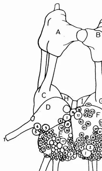
had been well mapped by Kerkut et al. (1975) who had also noted which cells were depolarized by the iontophoretic application of 5-HT onto the identified cell soma. A more detailed electrophysiological, pharmacological and fluorescent study on twelve neurones from the suboesophageal ganglia by Loker et al. (1975) identified five large 5-HT-containing neurones in the visceral ganglion. The six neurones under investigation in the visceral ganglion were El, E2, E3, E4, E5 and E6. Five of these neurones contained 5-HT, were depolarized by 5-HT and ACh and were hyperpolarised by dopamine.
In contrast, neurone E4 was found not to contain 5-HT and was hyperpolarized by both ACh and dopamine. It was five of these six large easily identifiable neurones that were utilised in this study. They were El, E2, E3, E4 and E5. Because of the similarity between the depolarizing response of 5-HT in Helix neurones to the 5-HT response observed in neuroblastoma cells which was mediated by a well-described 5-HTg receptor, 5-HTg receptor agonists and antagonists were tested first on the preparation. This was followed later by other specific 5-HT vertebrate receptor subtype agonists and antagonists.
METHODS AND MATERIALS Preparation of animals
Common garden snails. Helix aspersa, were collected locally and kept in a enclosed bucket at room temperature until they were required. Prior to dissection the snails were enticed out of their shells by being placed in warm water (20-3QOC). Once out of its shell, the head of the snail was rapidly cut off using a sharp pair of scissors and the detached head pinned through the mouth to a wax bottomed dissecting dish. The dorsal midline at the back of the head was cut from top to bottom and the skin pulled back exposing the buccal mass. The cerebral ganglia, which com prise part of the circumoesophageal ring, were visible as a collar over the oesophagus which is itself attached to the buccal mass. The oesophagus was pulled from beneath the cerebral ganglia and the buccal mass cut away. A pair of forceps was then placed underneath the cerebral ganglia and these lifted to expose the suboesophageal ganglia beneath. The nerve connections were cut away and the isolated circum oesophageal ring placed in a beaker containing physiological saline (80mM NaCl, 5mM KCl, 5mM MgCl2, 7mM CaCl2, 20mM Hepes pH 7.5) (Meech and Thomas, 1977). The ganglia were then transferred to the recording chamber for further dissection before the experiments were started. Once the suboesophageal ganglia had been pinned to a Sylgard base of the bath, the layers of connective tissue covering the ganglion cells could then be removed. The connective tissue had to be removed before the neurones could be impaled with microelectrodes. This careful microdissection was achieved with fine forceps and a pair of fine scissors. The outer connective sheath is spongy in texture and was relatively easy to remove. However, the inner sheath is thin and transparent, and had to be carefully torn to expose the cells without causing them further damage.
Experimental set up in the recording chamber
The recording chamber was made of a rectangular block of Perspex supported by four small Perspex legs. A 5mm identation in the perspex block formed the small bath. Sylgard was used to coat the bath and this made a suitable base into which dissecting pins could be inserted. The bath was illuminated by a conventional Watson light source. This was achieved by
19
using an tapered glass rod, the thick end of which was inserted by means of a rubber bung into the light source, while the thin tapering end was positioned in the bath below the level of the liquid. Adequate illumination was provided by the tip of the glass rod when it was positioned close to the preparation. The bath was observed through a Nikon stereoscan microscope.
The perfusion system
The preparation could be exposed to various solutions by means of a multi-way tap to which a maximum of six reservoirs could be connected. A diagram of the tap system is shown in Fig. 2.1. Large 100ml syringes were used as reservoirs and these were attached to smaller 5ml syringes. The syringes were held in place on the side of the Faraday cage surrounding the experimental apparatus by means of adjustable clips. Thin plastic tubing attached to the small syringes formed the inflow into the six-way tap. The single outflow of the tap led directly into the bath. The reservoirs were positioned higher than the recording chamber, thus providing the small hydraulic head necessary to create a flow of solution. The different solutions from the different reservoirs could be used to perfuse the bath by turning the tap outflow to the selected reservoir inflow. A clamp on the tubing from the outflow of the tap to the bath made it possible to control the flow of solution. The rate of flow per minute could then be measured by monitoring the amount of solution passing from the large to the small syringe. The flow rate was an important experimental parameter so it was measured periodically by the addition of a small amount of dye to the bath. The dye gave an indication of the movement of solution in the bath. For example, if the flow was too fast the dye would be swept around, but not over, the preparation whereas, if the flow was too slow small pockets of dye would remain in the bath. The flow had to be adjusted carefully in order to allow any drugs in the perfusate to have an effect, but also to allow them to be washed out of the bath once the effect had been observed.
A suction system disposed of the waste perfusate from the bath. The suction tube was positioned carefully so that the flow of solution was constant and the liquid in the bath was maintained at the desired level. The vacuum was produced by connecting the suction tube to an aspirator via a conical flask into which the waste collected.
20
».
FIG URE 2.1
.9 o .
o
>
<D
00
(D
O>
0) c/)
Q) E o $o
Intracellular recording
Intracellular recordings were made using the Dagan 8100 single electrode clamp system. This single electrode circuit had been developed by Wilson and Goldner (1975) to provide a means of voltage clamping with a single microelectrode. This technique is described in further detail in a following section. The electrical recordings were monitored with a Gould 20MHz oscilloscope, and permanent records were made on a Gould 200 brush chart recorder. Both current and voltage records were filtered before being displayed on the chart recorder, in order to eliminate high frequency oscillations and noise. This was accomplished by using a low pass filter consisting of a resistor and capacitor in series. A 160kQ resistor was used with a 0.1 pF capacitor. This low pass filter effectively removed noise above 9-lOHz. The experimental responses to 5-HT were sufficently slow to not be distorted by this filter. The bath was grounded with an Ag/AgCl wire connected to the virtual ground of the Dagan 8100 probe. The A g/A gC l wire was made by coating a piece of silver wire with molten AgCl. A Grass S44 stimulator was connected to the Dagan, via a Grass SIU5 stimulus isolation unit so that pulses could be given to impaled cells. The stimulation rate, the delay between stimuli and the duration of stimuli could all be modified with this system.
Intracellular electrodes
Microelectrodes were made using filamented glass capillary tubes (Clark Electromedical) of 1.5mm diameter. They were pulled on a Narishige PP83 electrode puller and kept on plasticine in petri dishes until required. Recording electrodes were filled with 200mM KCl solution using a syringe and fine needle. The solution was filtered with a Millipore Swinnex 25 filter unit containing 0.22pm filter paper before use. The microelectrodes had a resistance of between 1 and 5MQ .
Voltage- and current-clamp techniques
The single electrode circuit in the Dagan 8100 requires that a single electrode both records potential and passes current. This is accomplished through a switching circuit that alternates between a current-injecting mode and a voltage recording mode. The percentage of time spent injecting current and recording voltage is called the duty cycle. Current is injected for 50% of the time while membrane potential is recorded only during intervals between applications of current. The rate at which the clamp alternates between the two modes is called the switching frequency. The clamp relies on the time constant of the cell (membrane resistance x membrane capacitance) to smooth out the injected square waves of current and to provide an essentially steady DC current across the cell membrane. It was therefore important that the switching frequency be less than the time constant so that the membrane potential did not have time to wane appreciably. In all experiments the switching frequency was set at 3Hz. The voltage-clamp system used a negative feedback circuit to compare the actual potential of the cell to a commanded holding potential. Current is injected to compensate for any offset, including spontaneous events, that would otherwise shift the membrane potential. The injected current, equal and opposite to that flowing across the cell membrane, is monitored. From the holding potential, voltage commands to other membrane potentials can be applied. The currents required to produce and sustain the voltage step are recorded; they reflect the various currents present in the cell.
The Dagan could also be used in switched current-clamp mode. So that a neurone could be voltage-clamped adequately the electrode had to be able to pass current reliably during the course of the experiment. Electrodes with a resistance of 1-5MQ were found to be reliable. Before impaling a neurone and with the electrode in solution, the resistance of the recording electrode was tested using a bridge current circuit with the z test facility of the Dagan. Electrodes with resistances higher than 5MQ were discarded. The recording electrode was held in a micromanipulator. The gain of the negative feedback amplifier was set close to the maximum value because the higher the gain of the clamp, the more accurately the membrane voltage will follow the voltage command. The negative capacity compensation was set to a minimum.
Electrodes were then inserted into the soma of identified neurones once the fine microdissection had exposed the neurones.
Application of agonists by iontophoresis
Drugs such as 5-HT and the other 5-HT agonists were applied onto the soma of the impaled neurones by iontophoresis. Iontophoresis was performed using a microiontophoresis W Pl M odel 160 programmer. Iontophoretic electrodes were made from filamented glass capillary tubes (Clark Electromedical) of 1.5mm diameter, pulled on a Narishige PN3 8903 electrode puller. They were kept in petri dishes, held in place with plasticine until required. They were filled with appropriate drug solutions which had been filtered using Costar centrifuge filter units with 0.22pm cellulose acetate filters. The drugs were made up in distilled water at a concentration of 40mM. A small retain current was applied to the iontophoretic electrode to prevent leakage of the drugs into the bath. The retain current of 20nA was used routinely. Ejection currents were in the range of 200nA with an ejection time of two seconds. This could all be set up by connecting the iontophoresis programmer to a custom -m ade small pulse generator. With this arrangement, the time between pulses could also be set to whatever was required. Blockage of electrodes could be detected easily by either a decrease in retain current or by the appearance of a large artefact during the eject current. Blocked electrodes were replaced immediately.
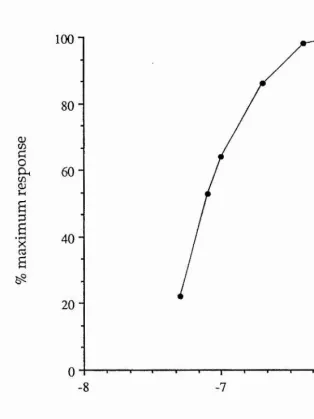
Before each experiment the eject current was increased to ensure selection of a submaximal ejection pulse. A sigmoid curve was obtained if eject current was plotted against the size of response. It was important that the eject current was on the rising phase and not on the plateau phase of the curve. The iontophoretic electrodes, once in the bath solution, were positioned with their tips close to the impaled neurones. However, care had to be taken not to place the tip too close because the eject current might cause the iontophoretic electrode to impale the cell. With continuous flow through the bath, if the iontophoretic electrode was too far away from the impaled neurone, the drug was automatically washed away without effect. The agonists applied in this way were 5-CT and sumatriptan (both 5-HT% receptor agonists), 2-Me-5-HT (a 5-HTg receptor agonist) and a-Me-5-HT (a 5-HT2 receptor agonist). The full names of the drugs and from where they were
obtained are as follows: 5-HT creatinine sulphate (Sigma), 5-CT maleate, a-Me-5-HT maleate, 2-Me-a-Me-5-HT hydrochloride and sumatriptan (all synthesised by the Chemistry Research Department at Glaxo Group Research).
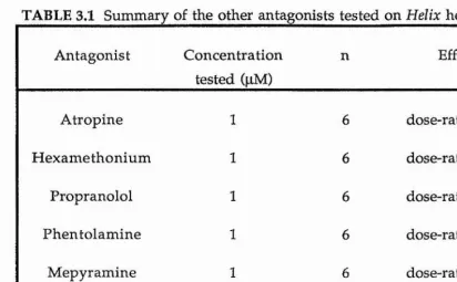
Antagonists by bath application
Application of antagonists required that they be perfused through the bath at the relevant concentrations. The antagonists were made up in physiological saline at appropriate concentrations with the exception of ketanserin and ondansetron which were dissolved in O.IM tartaric acid to give a ImM stock solution. The subsequent dilutions were then made in physiological saline. All antagonists were made up fresh on the day of use and kept in the fridge (4 ^ 0 until required. The antagonists tested were methiothepin (a 5-HT% receptor antagonist), ketanserin (a 5-HT2 receptor antagonist), ondansetron, MDL 72222 and ICS 205-930 (all 5-HTg receptor antagonists). Methysergide was tested as an antagonist. The full names of the drugs tested and from where they were obtained are listed: methiothepin maleate (Hoffman La Roche), ketanserin tartrate (Janssen), MDL 72222 (Merrel Dow), ICS 205-930 (Sandoz), ondansetron (Glaxo) and methysergide maleate (Sandoz).
Layout of equipment
The recording chamber, m icrom anipulators, lighting system , microscope and perfusion system were all placed on a steel baseplate which was supported by four specially reinforced legs. This reduced vibration and aided stable recordings. A diagram of the layout is shown in Fig. 2.2. The baseplate with all the equipment on it was enclosed in a Faraday cage which was connected to earth to reduce electrical interference. The microscope baseplate and micromanipulators were also earthed. All the electronic apparatus was stacked in a freely moveable rack to the left of the cage.
FIG URE 2.2
Q . CL
CL
CL
O)
JZ O)
*0
O)