Submitted2 March 2018 Accepted 16 June 2018 Published27 July 2018
Corresponding author Christopher R. Madan,
Academic editor Silvia Comani
Additional Information and Declarations can be found on page 11
DOI10.7717/peerj.5176
Copyright 2018 Madan
Distributed under
Creative Commons CC-BY 4.0
OPEN ACCESS
Age differences in head motion and
estimates of cortical morphology
Christopher R. Madan
School of Psychology, University of Nottingham, Nottingham, United Kingdom
ABSTRACT
Cortical morphology is known to differ with age, as measured by cortical thickness, fractal dimensionality, and gyrification. However, head motion during MRI scanning has been shown to influence estimates of cortical thickness as well as increase with age. Studies have also found task-related differences in head motion and relationships between body–mass index (BMI) and head motion. Here I replicated these prior findings, as well as several others, within a large, open-access dataset (Centre for Ageing and Neuroscience, CamCAN). This is a larger dataset than these results have been demonstrated previously, within a sample size of more than 600 adults across the adult lifespan. While replicating prior findings is important, demonstrating these key findings concurrently also provides an opportunity for additional related analyses: critically, I test for the influence of head motion on cortical fractal dimensionality and gyrification; effects were statistically significant in some cases, but small in magnitude.
SubjectsNeuroscience, Radiology and Medical Imaging, Computational Science
Keywords Head motion, Cortical structure, Fractal dimensionality, Age, Cortical thickness, Gyrification, Cortical morphology, Movie watching, BMI
INTRODUCTION
Head motion during the acquisition of magnetic resonance imaging (MRI) can lead to artifacts when estimating brain activity and structure. With functional MRI (fMRI), volumes are acquired relatively quickly–often every 1–3 s–allowing for the estimation and correction of head motion artifacts. Using innovative techniques such as prospective motion correction (Dosenbach et al., 2017;Federau & Gallichan, 2016;Maclaren et al., 2013;
Stucht et al., 2015;Tisdall et al., 2016) and custom-designed, individualized head-cases (https://caseforge.co), effects of head motion can be attenuated. However, these solutions are not suitable for large studies of inter-individual differences in brain morphology where changes to the MRI scan sequence or custom-built equipment for each participant are often not practical. In the current study, I assessed relationships between age and body–mass index (BMI) on head motion, task-related differences in head motion, and the influence of head motion on estimates of cortical morphology. In light of these findings, many of which are replications, I propose a potential method for attenuating head motion during structural MRIs, as well as discuss limitations of this method.
head motion can lead to lower cortical thickness estimates (Alexander-Bloch et al., 2016;
Pardoe, Hiess & Kuzniecky, 2016; Reuter et al., 2015;Savalia et al., 2017), as such, age-related differences in cortical thickness (e.g., Fjell et al., 2009;McKay et al., 2014;Salat et al., 2004) may be exaggerated by age-related differences in head motion. In addition to age, obesity has also been associated with head motion (Beyer et al., 2017;Hodgson et al., 2017). In particular, these associations have been shown with respect to body–mass index (BMI; kg/m2), which is measured as body weight (in kg) divided by body height (in m) squared–despite the relatively coarse nature of BMI (e.g., does not differentiate between muscle vs. fat mass) (Diverse Populations Collaborative Group, 2005; Romero-Corral et al., 2008). Findings of relationships between obesity and cortical thickness have been mixed (Shaw et al., 2017;Shaw et al., 2018;Veit et al., 2014). More generally, head motion has been suggested to be a neurobiological trait–being both stable over time and heritable (Engelhardt et al., 2017;Hodgson et al., 2017;Zeng et al., 2014).
There is also evidence that fMRI tasks can differ in the degree of associated head motion (Alexander et al., 2017;Huijbers et al., 2017;Greene et al., 2018;Vanderwal et al., 2015;Wylie et al., 2014;Yuan et al., 2009). With this in mind, it may be beneficial to present participants with a task to attend toduring structural scans, with the objective of decreasing head motion; typically structural scans are accompanied by the presentation of a blank screen or otherwise lack of instruction of attending to a visual stimulus.
Madan & Kensinger (2016) showed that a structural metric, fractal dimensionality (FD), may be more sensitive to age-related differences in cortical structure than cortical thickness (also see Madan & Kensinger, 2018). In a preliminary analysis to examine the influence of head motion on age-related differences in cortical fractal dimensionality,Madan & Kensinger (2016) showed qualitative evidence of age-related differences in fractal dimensionality in a small sample (N=7) of post-mortem MRIs.
However, as this sample was small and also less indicative of potential head motion effects inin vivoMR imaging, further work is necessary. To more directly test for the additive influence of head motion on estimates of cortical morphology, beyond aging, here I also tested for a relationship of fMRI-estimated head motion on cortical fractal dimensionality, as well as on mean cortical thickness. Additionally, as recent studies have found that gyrification also decreases with age (Cao et al., 2017;Hogstrom et al., 2013;
Madan & Kensinger, 2016;Madan & Kensinger, 2018), it was also included in the analysis presented here. Test-retest reliability of estimates for these structural measures has recently been compared (Madan & Kensinger, 2017b), but robustness to head motion has yet to be assessed.
METHODS
DatasetData used in the preparation of this work were obtained from the Cambridge Centre for Ageing and Neuroscience (CamCAN) repository, available at http://www.mrc-cbu.cam.ac.uk/datasets/camcan/(Shafto et al., 2014;Taylor et al., 2017). The CamCAN dataset includes structural and functional MRI data for a sample of 648 adults across the adult lifespan (aged 18–88; Mean (SD)= 54.2 (18.5)). All participants were cognitively
healthy (MMSE>24) and were free of any neurological or serious psychiatric conditions. SeeShafto et al. (2014)for additional details about the sample inclusion and exclusion criteria.
A total of eight participants were excluded from further analyses due to problems with cortical reconstruction or gyrification estimation, yielding a final sample size of 640 adults (326 female, 314 male). Height and weight measurements were available for 559 of the 648 participants (280 female, 279 male), additionally allowing for the calculation of body–mass index (BMI) for this subset of participants (also seeRonan et al., 2016).
Structural measures are derived from a T1-weighted volume acquired using a 3 T Siemens Trio MRI scanner with an MPRAGE sequence. Scan parameters were as follows: TR =2,250 ms, TE =2.99 ms, flip angle =9◦, voxel size =1×1×1 mm, GRAPPA =2,
TI =900 ms. Head motion was primarily estimated from two fMRI scans, during rest
and a movie-watching task. Both scans lasted for 8 min and 40 s (i.e., 520 s total). For the rest scan, participants were instructed to rest with their eyes closed. For the movie scan, participants watched and listened to condensed version of Alfred Hitchcock’s (1961) ‘‘Bang! You’re Dead’’ (Campbell et al., 2015;Hasson et al., 2008). Note that different scan sequences were used for both of these scans, with volumes collected every 1.970 s or 2.470 s for the rest and movie scans, respectively (seeTaylor et al., 2017for more details); both rest and movie scans had the same voxel size, 3×3×4.44 mm (32 axial slices, 3.7 mm thick,
0.74 mm gap).
Preprocessing of the structural MRI data
The T1-weighted structural MRIs were processed using FreeSurfer v6.0 (https://surfer. nmr.mgh.harvard.edu/) (Dale, Fischl & Sereno, 1999; Fischl, 2012; Fischl & Dale, 2000). Surface meshes and cortical thickness was estimated using the standard processing pipeline, i.e.,recon-all, and no manual edits were made to the surfaces. Gyrification was calculated using FreeSurfer, as described inSchaer et al. (2012).
Fractal dimensionality (FD) is a measure of the complexity of a structure and has previously been shown to decrease in relation to aging for cortical (Madan & Kensinger, 2016; Madan & Kensinger, 2018) and subcortical (Madan & Kensinger, 2017a;Madan, 2018) structures and has been shown to have high test-retest reliability (Madan & Kensinger, 2017b). FD was calculated using the calcFD toolbox (http://cmadan.github. io/calcFD/) (Madan & Kensinger, 2016) using the dilation method and filled structures (denoted asFDf in prior studies). Briefly, FD measures the effective dimensionality of a
scale-invariant measure of the complexity of a structure. This is mathematically calculated as
FD= −1log
2(
Count
)/1log2(Size
), whereSize
was set to{1,2,4,8,16}(i.e., powersof 2, ranging from 0 to 4). To correct for the variability in FD estimates associated with the alignment of the box-grid with the structure, a dilation algorithm was used which instead relies on a 3D-convolution operation (convnin MATLAB) as this approach yields more reliable estimates of FD. This computational issue is described mathematical and demonstrated in simulations inMadan & Kensinger (2016), and empirically shown in Madan & Kensinger (2017b). SeeMadan & Kensinger (2016)andMadan & Kensinger (2018)for additional background on fractal dimensionality and its application to brain imaging data.
Estimates of head motion
Head motion was estimated using two approaches:
(1) Measured as the frame-wise displacement using the three translational and three rotational realignment parameters. Realignment parameters were included as part of the preprocessed fMRI data (Taylor et al., 2017), in the form of therp_*.txtoutput generated by the SPM realignment procedure. Rotational displacements were converted from degrees to millimeters by calculating the displacement on the surface of a sphere with a radius of 50 mm (as inPower et al., 2012). Frame-wise displacement was substantially higher between volumes at the beginning of each scan run, so the first five volumes were excluded. This is the same approach to estimating head motion that is commonly used (e.g.,Alexander-Bloch et al., 2016;Engelhardt et al., 2017;Power et al., 2012;Savalia et al., 2017).
(2) Estimated directly from the T1-weighted volume as ‘average edge strength’ (AES) (Aksoy et al., 2012;Zacà et al., in press). This approach measures the intensity of contrast at edges within an image. Higher AES values correspond to less motion, with image blurring yielding decreased tissue contrast. AES was calculated using the toolbox provided byZacà et al. (in press), on the skull-stripped volumes generated as an intermediate stage of the FreeSurfer processing pipeline. AES is calculated on two-dimensional image planes and was performed on each plane orientation (axial, sagittal, and coronal).
Model comparison approach
Effects of head motion on estimates of cortical morphology (thickness, fractal dimensionality, and gyrification) were assessed using a hierarchical regression procedure using MATLAB. Age was first input, followed by BMI (both with and without age), followed by estimates of head motion from each fMRI scan and the related interaction term with age. In total, eight models were examined, as listed inTable 1. Model fitness was assessed using bothR2and1BIC.
Bayesian Information Criterion, BIC, is a model fitness index that includes a penalty based on the number of free parameters (Schwarz, 1978). SmallerBIC values correspond to better model fits. By convention, two models are considered equivalent if
1BIC<2 (Burnham & Anderson, 2004). AsBICvalues are based on the relevant dependent variable,1BIC values are reported relative to the best-performing model (i.e., 1BIC=0
Table 1 Variance explained and model fits of cortical measures by age, BMI, and head motion estimates. Note thatR2decreases after the
inclu-sion of BMI as models 2 and 3 can only be calculated on a subset of participants (559 out of 640 participants) since height and weight informa-tion was not available for all participants.
Thickness FD Gyrification
Model Predictors R2 1BIC R2 1BIC R2 1BIC
1 Age .425 6.98 .497 3.15 .192 3.65
2 BMI .029 455.85 .028 805.25 .007 243.17
3 Age+BMI .425 168.82 .487 454.69 .183 140.23
4 Age+Movement(Rest) .429 10.01 .500 5.65 .192 10.07
5 Age+Movement(Movie) .437 0.00 .504 0.00 .194 8.44
6 Age+Movement(Movie)+Age×Movement (Movie) .427 11.64 .499 6.58 .205 0.00
7 Age+AES(axial) .443 0.23 .507 3.18 .194 14.83
8 Age+AES(axial)+Age×AES(axial) .428 17.58 .500 12.42 .208 3.76
RESULTS
fMRI-estimated head motion
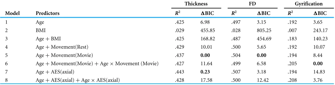
As shown in Fig. 1, older adults head increased head motion relative to younger adults in both the rest and movie scans (rest: r(638)=.351, p< .001; movie:r(638)=.430, p< .001). Head motion was also greater in the rest scan than during the movie watching (t(639)=23.35,p< .001, Cohen’sd=0.99,Mdiff =1.528 mm/min). Nonetheless, head
motion was correlated between the fMRI scans [r(638)=.484,p< .001]. While this
correlation between scans is expected, particularly since both were collected in the same MRI session, studies have provided evidence that head motion during scanning may be a trait (Engelhardt et al., 2017;Hodgson et al., 2017;Zeng et al., 2014). Moreover, this correlation provides additional evidence that motion during the fMRI scans is consistently larger in some individuals than others, suggesting it similarly affected the structural scans more for some individuals than others and appropriate to include as a predictor for the cortical morphology estimates.
As expected based on prior literature (Beyer et al., 2017;Hodgson et al., 2017), head motion was also correlated with body–mass index (BMI) (rest:r(557)=.456,p< .001;
movie: r(557)=.335, p< .001) (Fig. 1). While BMI was also correlated with age
(r(557)=.274,p< .001), BMI-effects on head motion persisted after accounting for
age differences (rest:rp(555)=.340,p< .001; movie:rp(555)=.249,p< .001).
20 30 40 50 60 70 80 90 Age (years) .1 .5 2 5 10 20
Head Motion (mm/min)
Rest
20 30 40 50 60 70 80 90
Age (years) .1 .5 2 5 10 20
Head Motion (mm/min)
Movie
.1 .5 2 5 10 20
Rest (mm/min) .1 .5 2 5 10 20 Movie (mm/min)
15 20 25 30 35 40 45 50
BMI (kg/m2)
.1 .5 2 5 10 20
Head Motion (mm/min)
15 20 25 30 35 40 45 50
BMI (kg/m2)
.1 .5 2 5 10 20
Head Motion (mm/min)
20 30 40 50 60 70 80 90
Age (years) 15 20 25 30 35 40 45 50 BMI (kg/m 2 )
A
B
C
D
E
F
[image:6.612.188.576.84.415.2]Rest Movie
Figure 1 Age-related differences in head motion.Correlations between average head motion (mm/min) with age for the (A) rest and (B) movie fMRI scans, with (D–E) body–mass index (BMI), (C) between fMRI scans, and (F) between age and BMI. Head motion axes are log-10 scaled to better show inter-individual variability.
Full-size DOI: 10.7717/peerj.5176/fig-1
T1-estimated head motion
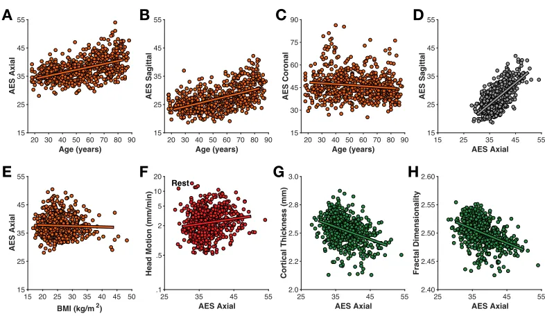
Head motion was also estimated directly from the T1-weighted volume as the average edge strength (AES), following fromZacà et al. (in press); higher AES values correspond to less motion. Here I calculated AES for each plane orientation. AES in the axial and sagittal planes was moderately related to age (axial:r(639)=.493,p< .001; sagittal:r(639)=.525, p< .001) (Fig. 3); AES in the coronal was only weakly correlated with age (r(639)= −.131, p< .001). AES in the axial and sagittal planes were strongly correlated with each other (r(639)=.702,p< .001).
Interestingly, AES was relatively not related to BMI (all |r|’s< .2). AES was also
relatively unrelated to fMRI-estimated head motion (rest:r(639)=.112,p=.005; movie: r(639)=.148,p< .001). Thus, while AES is sensitive to an MR image property related to
0 60 120 180 240 300 360 420 480 520 0 1 2 3 4 5 6
Head Motion (mm/min)
Young Adults
(Age<35; N=117)0 60 120 180 240 300 360 420 480 520
Time (s) 0 1 2 3 4 5
6
Older Adults
(Age>75; N=110)A
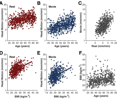
[image:7.612.185.576.87.357.2]B
Figure 2 Averaged time-course of head motion for rest (red) and movie (blue) fMRI scans for young (A) and older adults (B).Bands represent 95% confidence intervals.
Full-size DOI: 10.7717/peerj.5176/fig-2
20 30 40 50 60 70 80 90
Age (years) 15 25 35 45 55 AES Axial
20 30 40 50 60 70 80 90
Age (years) 15 25 35 45 55 AES Sagittal
20 30 40 50 60 70 80 90
Age (years) 15 30 45 60 75 90 AES Coronal
15 25 35 45 55
AES Axial 15 25 35 45 55 AES Sagittal
15 20 25 30 35 40 45 50
BMI (kg/m2)
15 25 35 45 55 AES Axial
25 35 45 55
AES Axial .1 .5 2 5 10 20
Head Motion (mm/min)
Rest
25 35 45 55
AES Axial 2.0 2.2 2.5 2.8 3.0
Cortical Thickness (mm)
25 35 45 55
AES Axial 2.40 2.45 2.50 2.55 2.60 Fractal Dimensionality
A B C D
E F G H
Figure 3 Relationships between motion estimated from the structural volume using average edge strength (AES) in (A–C) different planes with age, (D) between planes, (E) BMI, (F) rest-fMRI estimated motion, (G) cortical thickness, and (H) fractal dimensionality.
[image:7.612.184.577.410.646.2]20 30 40 50 60 70 80 90
Age (years)
2.0 2.2 2.5 2.8 3.0
Cortical Thickness (mm)
20 30 40 50 60 70 80 90
Age (years)
2.40 2.45 2.50 2.55 2.60
Fractal Dimensionality
20 30 40 50 60 70 80 90
Age (years)
2.5 2.7 2.9 3.1 3.3 3.5
Gyrification Index
15 20 25 30 35 40 45 50
BMI (kg/m2)
2.0 2.2 2.5 2.8 3.0
Cortical Thickness (mm)
15 20 25 30 35 40 45 50
BMI (kg/m2)
2.40 2.45 2.50 2.55 2.60
Fractal Dimensionality
15 20 25 30 35 40 45 50
BMI (kg/m2)
2.5 2.7 2.9 3.1 3.3 3.5
Gyrification Index
A
B
C
[image:8.612.189.578.83.418.2]D
E
F
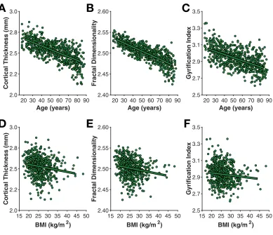
Figure 4 Age- and BMI-related differences in the three cortical morphology measures examined here: (A, D) thickness, (B, E) fractal dimensionality, and (C, F) gyrification.
Full-size DOI: 10.7717/peerj.5176/fig-4
head motion is likely apparent–rather than real–head motion caused by respiratory chest motion producing susceptibility variations in the B0 field (Raj, Anderson & Gore, 2001;
Van de Moortele et al., 2002;Van Gelderen et al., 2007).
Cortical morphology
As shown inFig. 4, mean cortical thickness significantly decreased with age (r(638)= −.652, p< .001,−0.0432 mm/decade), as did fractal dimensionality (r(638)= −.705,p< .001, −0.0097FDf/decade) and gyrification (r(638)= −.427,p< .001, −0.0372GI/decade).
All three slopes (change in metric per decade) are nearly identical to those first calculated byMadan & Kensinger (2016), as is the finding of higher age-related differences in fractal dimensionality and weaker differences in gyrification (also seeMadan & Kensinger, 2018). However, it is also worth acknowledging that AES in the axial and sagittal planes were comparably correlated with age as gyrification. Effects of BMI on all three measures of cortical morphology were relatively weak (thickness: r(557)= −.169, p< .001; fractal
Of particular interest, I examined the influence of head motion on the cortical morphology estimates. For all three measures, head motion explained only a small amount of additional variance beyond age, as shown inTable 1. Nonetheless, head motion from the movie scan did explain significant additional variance, as measured by1BIC, however, this only accounted for an additional 1% variance in the cortical morphology measures. In the model of cortical thickness including head motion from the movie scan (but not the interaction), age related changes corresponded to−0.0398 mm/decade, while head motion
contributed−0.0135 mm/(mm/min).
DISCUSSION
In the current study, I replicated several prior findings as well as tested for a few novel effects of head motion. First I outline the key findings of prior studies that were replicated here:
(1) Increased head motion in older adults (replicatingSavalia et al., 2017;Pardoe, Hiess & Kuzniecky, 2016).
(2) BMI is correlated with fMRI-estimated head motion (replicating Beyer et al., 2017;
Hodgson et al., 2017).
(3) Less head motion occurs when watching a movie than during rest (replicating
Vanderwal et al., 2015;Huijbers et al., 2017).
(4) Head motion in different scans from the same individuals is correlated and indexes reliable inter-individual differences (replicatingZeng et al., 2014;Engelhardt et al., 2017;Hodgson et al., 2017).
(5) Cortical thickness decreases with age (replicatingFjell et al., 2009;Salat et al., 2004). (6) Fractal dimensionality and gyrification also decrease with age (replicatingMadan &
Kensinger, 2016;Madan & Kensinger, 2018;Hogstrom et al., 2013).
(7) More head motion leads to lower estimates of cortical thickness (replicatingReuter et al., 2015;Savalia et al., 2017).
In addition to these replications, the new findings were:
(8) Head motion leads to nominally lower estimates of fractal dimensionality and gyrification.
(9) Head motion estimated from the structural volume itself (i.e., average edge strength [AES]) correlated with age, but not BMI.
(10) AES may be sensitive to gray/white matter contrast ratio (GWR). (11) AES was only weakly related to fMRI-estimated head motion. (12) Global cortical morphology is weakly related to BMI.
it has been used in some recent large-scale studies, such as the Human Connectome Project (HCP) (Marcus et al., 2013) and Adolescent Brain Cognitive Development (ABCD) study (Casey et al., in press), and has also been suggested and used elsewhere, particularly in MRI studies with children (Greene, Black & Schlaggar, 2016;De Bellis et al., 2001;Howell et al., in press;Overmeyer, 1996;Pliszka et al., 2006;Raschle et al., 2009;
Theys, Wouters & Ghesquière, 2014; Von Rhein et al., 2015; Wu Nordahl et al., 2008). However, it is also important to consider the context that this movie watching would occur in. For instance, if the structural scan is followed by a resting-state fMRI scan, cognitive processes related to the movie watching will ‘spill over’ and influence patterns of brain activity in a subsequent rest period (e.g.,Tambini & Davachi, 2013;Van Kesteren et al., 2010;Eryilmaz et al., 2011).
Estimates of cortical thickness were significantly influenced by head motion (replicating
Savalia et al., 2017;Reuter et al., 2015), though the influence of this appeared to be relatively small. Effects of head motion on fractal dimensionality were also significant, but even smaller in magnitude, while head motion did not significantly influence estimates of gyrification. The results here also served as a replication age-related differences in fractal dimensionality and gyrification (Madan & Kensinger, 2016;Madan & Kensinger, 2018).
Interestingly, average edge strength (AES) did not correlate well with fMRI-estimated motion, but did correlate with age. This may be related to age-related differences in gray/white matter contrast ratio (GWR), as AES corresponds to the degree of tissue intensity contrast. This finding may be important when examining differences in AES between different samples (e.g., patients vs. controls).
While the results here are predominately replications of prior work, they nonetheless integrate the key findings of several papers through a single, open-access dataset, that also has a larger sample size than these previous studies. Moreover, these results serve as an example to highlight the benefits of open data sharing on improving our understanding of brain morphology (seeMadan, 2017for a detailed discussion).
CONCLUSION
Head motion influences estimates of cortical morphology, but can be attenuated by using an engaging task, such as movie watching, rather than merely instructing participants to rest. Decreasing head motion is particularly important when studying aging populations, where head motion is greater than for young adults, but considerations are necessary to see how this may ‘carry over’ and influence a subsequent scan, such as resting-state fMRI.
ACKNOWLEDGEMENTS
ADDITIONAL INFORMATION AND DECLARATIONS
Funding
CamCAN funding was provided by the UK Biotechnology and Biological Sciences Research Council (BBSRC) (BB/H008217/1), together with support from the UK Medical Research Council (MRC) and the University of Cambridge. The funders had no role in study design, data collection and analysis, decision to publish, or preparation of the manuscript.
Grant Disclosures
The following grant information was disclosed by the author:
UK Biotechnology and Biological Sciences Research Council (BBSRC): BB/H008217/1. UK Medical Research Council (MRC).
University of Cambridge.
Competing Interests
The authors declare there are no competing interests.
Author Contributions
• Christopher R. Madan conceived and designed the experiments, analyzed the data,
contributed reagents/materials/analysis tools, prepared figures and/or tables, authored or reviewed drafts of the paper, approved the final draft.
Data Availability
The following information was supplied regarding data availability:
The unprocessed T1 structural data, head-motion regressors from the processed resting-state and movie-watching functional MRI data, along with demographic (age, sex) and physical (height, weight) data are available at Cam-CAN Data Portal
https://camcan-archive.mrc-cbu.cam.ac.uk/dataaccess/.
The measures derived in this article’s analysis are available here: Christopher R. Madan. (2018). Derived brain morphology measures from CamCAN data [Data set]. Zenodo.
http://doi.org/10.5281/zenodo.1258016.
Supplemental Information
Supplemental information for this article can be found online athttp://dx.doi.org/10.7717/ peerj.5176#supplemental-information.
REFERENCES
Aksoy M, Forman C, Straka M, Çukur T, Hornegger J, Bammer R. 2012.Hybrid prospective and retrospective head motion correction to mitigate cross-calibration errors.Magnetic Resonance in Medicine67:1237–1251DOI 10.1002/mrm.23101.
J, Cohen S, Dufek S, Eaves M, Fradera B, Gardner J, Grant-Villegas N, Green G, Gregory C, Hart E, Harris S, Horton M, Kahn D, Kabotyanski K, Karmel B, Kelly SP, Kleinman K, Koo B, Kramer E, Lennon E, Lord C, Mantello G, Margolis A, Merikangas KR, Milham J, Minniti G, Neuhaus R, Levine A, Osman Y, Parra LC, Pugh KR, Racanello A, Restrepo A, Saltzman T, Septimus B, Tobe R, Waltz R, Williams A, Yeo A, Castellanos FX, Klein A, Paus T, Leventhal BL, Craddock RC, Koplewicz HS, Milham MP. 2017.An open resource for transdiagnostic research in pediatric mental health and learning disorders.Scientific Data4:170181
DOI 10.1038/sdata.2017.181.
Alexander-Bloch A, Clasen L, Stockman M, Ronan L, Lalonde F, Giedd J, Raznahan A. 2016.Subtle in-scanner motion biases automated measurement of brain anatomy from in vivo MRI.Human Brain Mapping 37:2385–2397DOI 10.1002/hbm.23180.
Andrews-Hanna JR, Snyder AZ, Vincent JL, Lustig C, Head D, Raichle ME, Buckner RL. 2007.Disruption of large-scale brain systems in advanced aging.Neuron 56:924–935DOI 10.1016/j.neuron.2007.10.038.
Beyer F, Masouleh SK, Huntenburg JM, Lampe L, Luck T, Riedel-Heller SG, Loeffler M, Schroeter ML, Stumvoll M, Villringer A, Witte AV. 2017.Higher body mass index is associated with reduced posterior default mode connectivity in older adults.Human Brain Mapping 38:3502–3515DOI 10.1002/hbm.23605.
Burnham KP, Anderson DR. 2004.Multimodel inference.Sociological Methods & Research33:261–304 DOI 10.1177/0049124104268644.
Campbell KL, Shafto MA, Wright P, Tsvetanov KA, Geerligs L, Cusack R, Tyler LK, Tyler LK, Brayne C, Bullmore E, Calder A, Cusack R, Dalgleish T, Duncan J, Henson R, Matthews F, Marslen-Wilson W, Rowe J, Shafto M, Campbell K, Cheung T, Davis S, Geerligs L, Kievit R, McCarrey A, Price D, Taylor J, Tsvetanov K, Williams N, Bates L, Emery T, Erzin¸clioglu S, Gadie A, Gerbase S, Georgieva S, Hanley C, Parkin B, Troy D, Allen J, Amery G, Amunts L, Barcroft A, Castle A, Dias C, Dowrick J, Fair M, Fisher H, Goulding A, Grewal A, Hale G, Hilton A, Johnson F, Johnston P, Kavanagh-Williamson T, Kwasniewska M, McMinn A, Norman K, Penrose J, Roby F, Rowland D, Sargeant J, Squire M, Stevens B, Stoddart A, Stone C, Thompson T, Yazlik O, Dixon M, Barnes D, Hillman J, Mitchell J, Villis L. 2015.Idiosyncratic responding during movie-watching predicted by age differences in attentional control.Neurobiology of Aging 36:3045–3055
DOI 10.1016/j.neurobiolaging.2015.07.028.
Cao B, Mwangi B, Passos IC, Wu M-J, Keser Z, Zunta-Soares GB, Xu D, Hasan KM, Soares JC. 2017.Lifespan gyrification trajectories of human brain in healthy individuals and patients with major psychiatric disorders.Scientific Reports7:511
DOI 10.1038/s41598-017-00582-1.
DA, Dale AM.The Adolescent Brain Cognitive Development (ABCD) study: imaging acquisition across 21 sites.Developmental Cognitive NeuroscienceIn Press
DOI 10.1016/j.dcn.2018.03.001.
Chan MY, Park DC, Savalia NK, Petersen SE, Wig GS. 2014.Decreased segrega-tion of brain systems across the healthy adult lifespan.Proceedings of the Na-tional Academy of Sciences of the United States of America111:E4997–E5006
DOI 10.1073/pnas.1415122111.
Dale AM, Fischl B, Sereno MI. 1999.Cortical surface-based analysis: I. Segmentation and surface reconstruction.NeuroImage9:179–194DOI 10.1006/nimg.1998.0395.
De Bellis MD, Keshavan MS, Beers SR, Hall J, Frustaci K, Masalehdan A, Noll J, Boring AM. 2001.Sex differences in brain maturation during childhood and adolescence.
Cerebral Cortex 11:552–557DOI 10.1093/cercor/11.6.552.
Diverse Populations Collaborative Group. 2005.Weight-height relationships and body mass index: some observations from the diverse populations collaboration.American Journal of Physical Anthropology128:220–229DOI 10.1002/ajpa.20107.
Dosenbach NU, Koller JM, Earl EA, Miranda-Dominguez O, Klein RL, Van AN, Snyder AZ, Nagel BJ, Nigg JT, Nguyen AL, Wesevich V, Greene DJ, Fair DA. 2017. Real-time motion analytics during brain MRI improve data quality and reduce costs.
NeuroImage161:80–93DOI 10.1016/j.neuroimage.2017.08.025.
Engelhardt LE, Roe MA, Juranek J, DeMaster D, Harden KP, Tucker-Drob EM, Church JA. 2017.Children’s head motion during fMRI tasks is heritable and stable over time.
Developmental Cognitive Neuroscience25:58–68DOI 10.1016/j.dcn.2017.01.011.
Eryilmaz H, Ville DVD, Schwartz S, Vuilleumier P. 2011.Impact of transient emotions on functional connectivity during subsequent resting state: a wavelet correlation approach.NeuroImage54:2481–2491DOI 10.1016/j.neuroimage.2010.10.021.
Federau C, Gallichan D. 2016.Motion-correction enabled ultra-high resolution in-vivo 7 T-MRI of the brain.PLOS ONE11:e0154974DOI 10.1371/journal.pone.0154974.
Fischl B. 2012.FreeSurfer.NeuroImage62:774–781
DOI 10.1016/j.neuroimage.2012.01.021.
Fischl B, Dale AM. 2000.Measuring the thickness of the human cerebral cortex from magnetic resonance images.Proceedings of the National Academy of Sciences of the United States of America97:11050–11055DOI 10.1073/pnas.200033797.
Fjell AM, Westlye LT, Amlien I, Espeseth T, Reinvang I, Raz N, Agartz I, Salat DH, Greve DN, Fischl B, Dale AM, Walhovd KB. 2009.High consistency of regional cortical thinning in aging across multiple samples.Cerebral Cortex19:2001–2012
DOI 10.1093/cercor/bhn232.
Greene DJ, Black KJ, Schlaggar BL. 2016.Considerations for MRI study design and implementation in pediatric and clinical populations.Developmental Cognitive Neuroscience18:101–112DOI 10.1016/j.dcn.2015.12.005.
Hasson U, Landesman O, Knappmeyer B, Vallines I, Rubin N, Heeger DJ. 2008.
Neurocinematics: the neuroscience of film.Projections2:1–26
DOI 10.3167/proj.2008.020102.
Hitchcock A. 1961.Bang! You’re Dead [Motion Picture]. Hollywood: Shamley Produc-tions.
Hodgson K, Poldrack RA, Curran JE, Knowles EE, Mathias S, Gring HH, Yao N, Olvera RL, Fox PT, Almasy L, Duggirala R, Barch DM, Blangero J, Glahn DC. 2017.Shared genetic factors influence head motion during MRI and body mass index.Cerebral Cortex 27:5539–5546DOI 10.1093/cercor/bhw321.
Hogstrom LJ, Westlye LT, Walhovd KB, Fjell AM. 2013.The structure of the cerebral cortex across adult life: age-related patterns of surface area, thickness, and gyrifica-tion.Cerebral Cortex23:2521–2530DOI 10.1093/cercor/bhs231.
Howell BR, Styner MA, Gao W, Yap P-T, Wang L, Baluyot K, Yacoub E, Chen G, Potts T, Salzwedel A, Li G, Gilmore JH, Piven J, Smith JK, Shen D, Ugurbil K, Zhu H, Lin W, Elison JT.The UNC/UMN baby connectome project (BCP): an overview of the study design and protocol development.NeuroImageIn Press
DOI 10.1016/j.neuroimage.2018.03.049.
Huijbers W, Van Dijk KRA, Boenniger MM, Stirnberg R, Breteler MMB. 2017.Less head motion during MRI under task than resting-state conditions.NeuroImage 147:111–120DOI 10.1016/j.neuroimage.2016.12.002.
Knight MJ, McCann B, Tsivos D, Couthard E, Kauppinen RA. 2016.Quantitative T1 and T2 MRI signal characteristics in the human brain: different patterns of MR contrasts in normal ageing.Magnetic Resonance Materials in Physics, Biology and Medicine29:833–842 DOI 10.1007/s10334-016-0573-0.
Maclaren J, Herbst M, Speck O, Zaitsev M. 2013.Prospective motion correc-tion in brain imaging: a review.Magnetic Resonance in Medicine69:621–636
DOI 10.1002/mrm.24314.
Madan CR. 2017.Advances in studying brain morphology: the benefits of open-access data.Frontiers in Human Neuroscience11:405 DOI 10.3389/fnhum.2017.00405.
Madan CR. 2018.Shape-related characteristics of age-related differences in subcortical structures.Aging & Mental HealthIn PressDOI 10.1080/13607863.2017.1421613.
Madan CR, Kensinger EA. 2016.Cortical complexity as a measure of age-related brain atrophy.NeuroImage134:617–629DOI 10.1016/j.neuroimage.2016.04.029.
Madan CR, Kensinger EA. 2017a.Age-related differences in the structural complex-ity of subcortical and ventricular structures.Neurobiology of Aging 50:87–95
DOI 10.1016/j.neurobiolaging.2016.10.023.
Madan CR, Kensinger EA. 2017b.Test–retest reliability of brain morphology estimates.
Brain Informatics4:107–121DOI 10.1007/s40708-016-0060-4.
Madan CR, Kensinger EA. 2018.Predicting age from cortical structure across the lifespan.European Journal of Neuroscience47:399–416DOI 10.1111/ejn.13835.
Magnaldi S, Ukmar M, Vasciaveo A, Longo R, Pozzi-Mucelli R. 1993.Contrast between white and grey matter: MRI appearance with ageing.European Radiology3:513–519
Marcus DS, Harms MP, Snyder AZ, Jenkinson M, Wilson JA, Glasser MF, Barch DM, Archie KA, Burgess GC, Ramaratnam M, Hodge M, Horton W, Herrick R, Olsen T, McKay M, House M, Hileman M, Reid E, Harwell J, Coalson T, Schindler J, Elam JS, Curtiss SW, Essen D. CV. 2013.Human Connectome Project informatics: quality control, database services, and data visualization.NeuroImage80:202–219
DOI 10.1016/j.neuroimage.2013.05.077.
McKay DR, Knowles E. EM, Winkler A. AM, Sprooten E, Kochunov P, Olvera RL, Cur-ran JE, Kent JW, Carless MA, Göring HHH, Dyer TD, Duggirala R, Almasy L, Fox PT, Blangero J, Glahn DC. 2014.Influence of age, sex and genetic factors on the hu-man brain.Brain Imaging and Behavior8:143–152DOI 10.1007/s11682-013-9277-5.
Overmeyer S. 1996.Angstverarbeitung von psychisch aufflligen Kindern im Kernspinto-mogramm.Monatsschrift Kinderheilkunde 144:1337–1341
DOI 10.1007/s001120050091.
Pardoe HR, Hiess RK, Kuzniecky R. 2016.Motion and morphometry in clinical and nonclinical populations.NeuroImage135:177–185
DOI 10.1016/j.neuroimage.2016.05.005.
Pliszka SR, Lancaster J, Liotti M, Semrud-Clikeman M. 2006.Volumetric MRI dif-ferences in treatment-naive vs chronically treated children with ADHD.Neurology 67:1023–1027DOI 10.1212/01.wnl.0000237385.84037.3c.
Power JD, Barnes KA, Snyder AZ, Schlaggar BL, Petersen SE. 2012.Spurious but systematic correlations in functional connectivity MRI networks arise from subject motion.NeuroImage59:2142–2154DOI 10.1016/j.neuroimage.2011.10.018.
Raj D, Anderson AW, Gore JC. 2001.Respiratory effects in human functional magnetic resonance imaging due to bulk susceptibility changes.Physics in Medicine and Biology 46:3331–3340DOI 10.1088/0031-9155/46/12/318.
Raschle NM, Lee M, Buechler R, Christodoulou JA, Chang M, Vakil M, Stering PL, Gaab N. 2009.Making MR imaging child’s play—pediatric neuroimaging protocol, guidelines and procedure.Journal of Visualized Experiments29:e1309
DOI 10.3791/1309.
Reuter M, Tisdall MD, Qureshi A, Buckner RL, Van der Kouwe AJ, Fischl B. 2015.Head motion during MRI acquisition reduces gray matter volume and thickness estimates.
NeuroImage107:107–115DOI 10.1016/j.neuroimage.2014.12.006.
Romero-Corral A, Somers VK, Sierra-Johnson J, Thomas RJ, Collazo-Clavell ML, Korinek J, Allison TG, Batsis JA, Sert-Kuniyoshi FH, Lopez-Jimenez F. 2008.
Accuracy of body mass index in diagnosing obesity in the adult general population.
International Journal of Obesity32:959–966 DOI 10.1038/ijo.2008.11.
Ronan L, Alexander-Bloch AF, Wagstyl K, Farooqi S, Brayne C, Tyler LK, Fletcher PC. 2016.Obesity associated with increased brain age from midlife.Neurobiology of Aging 47:63–70DOI 10.1016/j.neurobiolaging.2016.07.010.
Salat DH, Lee SY, Van der Kouwe AJ, Greve DN, Fischl B, Rosas HD. 2009. Age-associated alterations in cortical gray and white matter signal intensity and gray to white matter contrast.NeuroImage48:21–28DOI 10.1016/j.neuroimage.2009.06.074.
Savalia NK, Agres PF, Chan MY, Feczko EJ, Kennedy KM, Wig GS. 2017. Motion-related artifacts in structural brain images revealed with independent estimates of in-scanner head motion.Human Brain Mapping 38:472–492DOI 10.1002/hbm.23397.
Schaer M, Cuadra MB, Schmansky N, Fischl B, Thiran J-P, Eliez S. 2012.How to measure cortical folding from MR images: a step-by-step tutorial to compute local gyrification index.Journal of Visualized Experiments59:e3417DOI 10.3791/3417.
Schwarz G. 1978.Estimating the dimension of a model.Annals of Statistics6:461–464
DOI 10.1214/aos/1176344136.
Shafto MA, Tyler LK, Dixon M, Taylor JR, Rowe JB, Cusack R, Calder AJ, Marslen-Wilson WD, Duncan J, Dalgleish T, Henson RN, Brayne C, Matthews FE. 2014.
The Cambridge Centre for Ageing and Neuroscience (Cam-CAN) study protocol: a cross-sectional, lifespan, multidisciplinary examination of healthy cognitive ageing.
BMC Neurology 14:204DOI 10.1186/s12883-014-0204-1.
Shaw ME, Abhayaratna WP, Anstey KJ, Cherbuin N. 2017.Increasing body mass index at midlife is associated with increased cortical thinning in Alzheimer’s disease-vulnerable regions.Journal of Alzheimer’s Disease59:113–120
DOI 10.3233/JAD-170055.
Shaw ME, Sachdev PS, Abhayaratna W, Anstey KJ, Cherbuin N. 2018.Body mass index is associated with cortical thinning with different patterns in mid- and late-life.
International Journal of Obesity42:455–461 DOI 10.1038/ijo.2017.254.
Stucht D, Danishad KA, Schulze P, Godenschweger F, Zaitsev M, Speck O. 2015.
Highest resolution in vivo human brain MRI using prospective motion correction.
PLOS ONE10:e0133921DOI 10.1371/journal.pone.0133921.
Tambini A, Davachi L. 2013.Persistence of hippocampal multivoxel patterns into pos-tencoding rest is related to memory.Proceedings of the National Academy of Sciences of the United States of America110:19591–19596DOI 10.1073/pnas.1308499110.
Taylor JR, Williams N, Cusack R, Auer T, Shafto MA, Dixon M, Tyler LK, Cam-CAN, Henson RN. 2017.The Cambridge Centre for Ageing and Neuroscience (Cam-CAN) data repository: structural and functional MRI, MEG, and cognitive data from a cross-sectional adult lifespan sample.NeuroImage144:262–269
DOI 10.1016/j.neuroimage.2015.09.018.
Theys C, Wouters J, Ghesquière P. 2014.Diffusion tensor imaging and resting-state functional MRI-scanning in 5- and 6-year-old children: training protocol and motion assessment.PLOS ONE9:e94019DOI 10.1371/journal.pone.0094019.
Tisdall MD, Reuter M, Qureshi A, Buckner RL, Fischl B, Van der Kouwe AJ. 2016.
Prospective motion correction with volumetric navigators (vNavs) reduces the bias and variance in brain morphometry induced by subject motion.NeuroImage 127:11–22DOI 10.1016/j.neuroimage.2015.11.054.
postencoding rest in humans.Proceedings of the National Academy of Sciences of the United States of America107:7550–7555DOI 10.1073/pnas.0914892107.
Vanderwal T, Kelly C, Eilbott J, Mayes LC, Castellanos FX. 2015.Inscapes: a movie paradigm to improve compliance in functional magnetic resonance imaging.
NeuroImage122:222–232DOI 10.1016/j.neuroimage.2015.07.069.
Van de Moortele P, Pfueffer J, Glover GH, Ugurbil K, Hu X. 2002.Respiration-induced B0 fluctuations and their spatial distribution in the human brain at 7 Tesla.Magnetic Resonance in Medicine47:888–895DOI 10.1002/mrm.10145.
Van Gelderen P, De Zwart JA, Starewicz P, Hinks RS, Duyn JH. 2007.Real-time shim-ming to compensate for respiration-induced B0 fluctuations.Magnetic Resonance in Medicine57:362–368 DOI 10.1002/mrm.21136.
Veit R, Kullmann S, Heni M, Machann J, Häring H-U, Fritsche A, Preissl H. 2014.
Reduced cortical thickness associated with visceral fat and BMI.NeuroImage: Clinical 6:307–311DOI 10.1016/j.nicl.2014.09.013.
Von Rhein D, Mennes M, Van Ewijk H, Groenman AP, Zwiers MP, Oosterlaan J, Heslenfeld D, Franke B, Hoekstra PJ, Faraone SV, Hartman C, Buitelaar J. 2015.
The NeuroIMAGE study: a prospective phenotypic, cognitive, genetic and MRI study in children with attention-deficit/hyperactivity disorder. Design and descriptives. Eu-ropean Child & Adolescent Psychiatry24:265–281DOI 10.1007/s00787-014-0573-4.
Wu Nordahl C, Simon TJ, Zierhut C, Solomon M, Rogers SJ, Amaral DG. 2008. Meth-ods for acquiring structural MRI data in very young children with autism without the use of sedation.Journal of Autism and Developmental Disorders38:1581–1590
DOI 10.1007/s10803-007-0514-x.
Wylie GR, Genova H, DeLuca J, Chiaravalloti N, Sumowski JF. 2014.Functional magnetic resonance imaging movers and shakers: does subject-movement cause sampling bias?Human Brain Mapping 35:1–13DOI 10.1002/hbm.22150.
Yuan W, Altaye M, Ret J, Schmithorst V, Byars AW, Plante E, Holland SK. 2009.
Quantification of head motion in children during various fMRI language tasks.
Human Brain Mapping 30:1481–1489DOI 10.1002/hbm.20616.
Zacà D, Hasson U, Minati L, Jovicich J.Method for retrospective estimation of natural head movement during structural MRI.Journal of Magnetic Resonance ImagingIn PressDOI 10.1002/jmri.25959.
Zeng L-L, Wang D, Fox MD, Sabuncu M, Hu D, Ge M, Buckner RL, Liu H. 2014.
Neurobiological basis of head motion in brain imaging.Proceedings of the National Academy of Sciences of the United States of America111:6058–6062