Neural Plasticity in Sensory
Processing Investigated at the
Levels of Single Neurons,
Networks, and Perception
Sebastian Thomas Philipp
Neural Plasticity in Sensory
Processing Investigated at the
Levels of Single Neurons,
Networks, and Perception
Dissertation
an der Graduate School of Systemic Neurosciences
der Ludwig-Maximilians-Universit¨
at
M¨
unchen
vorgelegt von
Sebastian Thomas Philipp
geboren am 13.10.1982 in Eschwege
...
Erstgutachter: PD Dr. Thomas Wachtler Zweitgutachter: Prof. Dr. Andreas Herz
...
”agitate; annotate; arbitrate; artistry; back and forth; brevity; ca d’etait; can-didate; can’t you see; can’t you stay; cape cod you say; card estate; cardio tape; car district; catch a tape; cavitate; cha cha che; cogitate; computate; conjugate; conscious state; counter tape; count to ten; count to three; count yer tape; cut the steak; entity; fantasy; God to take; God you say; got a date; got your pay; got your tape; gratitude; gravity; guard the tit; gurgitate; had to take; kinds of tape; majesty; marmalade.”
Contents
1 Summary 5
2 Main Introduction 7
2.1 Main Questions . . . 8
2.2 Outline . . . 9
2.3 Sensory Organs and Neurons . . . 10
2.3.1 Sensory Organs . . . 10
2.3.2 Neurons, Synapses & Action Potentials . . . 11
2.4 Neural Representations . . . 13
2.4.1 Receptive Fields . . . 13
2.4.2 Topographic Maps . . . 14
2.5 Contextual or Multimodal Integration . . . 15
2.6 Learning and Early Perceptual Development . . . 16
2.6.1 The Brain as a Self-Organizing System . . . 16
2.6.2 Sensory Experience and Development . . . 16
2.6.3 Synaptic Plasticity . . . 17
2.6.4 Spike-Timing-Dependent Plasticity . . . 18
2.6.5 Self-Organization of Topographic Maps . . . 18
2.6.6 Temporal Invariance Learning . . . 19
2.6.7 Invariance Learning in Self-Organizing Networks . . . . 20
2.6.8 Self-Organization of Contextual Interactions . . . 21
2.7 Neural Plasticity in Adults . . . 21
2.7.1 Adult Sensory Representations Adapt to Significant Changes in the Environment . . . 22
2.7.2 Perceptual Learning . . . 22
2.8 Attention . . . 23
2.8.1 Input Attention and Controlled Attention . . . 24
2.8.2 Neural Correlates of Attention . . . 25
2.8.3 Attention and Perceptual Learning . . . 25
3.2 Introduction . . . 27
3.2.1 Permissive Gating . . . 30
3.2.2 Permissive Gating and Multiplicative Interactions . . . 31
3.2.3 Scope of this study . . . 32
3.3 Methods . . . 32
3.4 Results . . . 36
3.4.1 Generation of Single Spikes - Complete Model Neuron . 36 3.4.2 Influence of the Gating Mechanism on Output Firing Rates - Simplified Model Neuron . . . 37
3.4.3 Gating Mechanism in the Complete Model Neuron . . 44
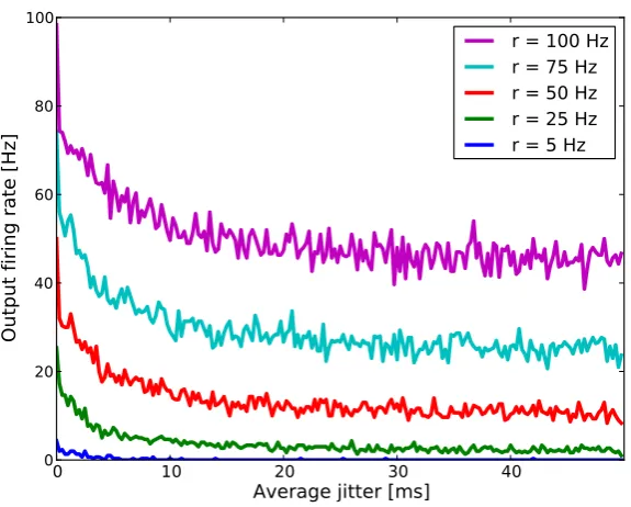
3.4.4 Effects of Input Synchrony . . . 47
3.5 Discussion . . . 49
3.5.1 Comparison to a Detailled Biophysical Model . . . 49
3.5.2 Multiplicative Interactions with Small Gating Weights 49 3.5.3 Exact and Nearly Multiplicative Interactions . . . 50
3.5.4 Robustness of Models that Explain Multiplicative In-teractions via a Network Approach . . . 51
3.6 Outlook to Chapter 3 . . . 52
4 Unsupervised Learning of Gaze-Invariance 53 4.1 Abstract . . . 53
4.2 Introduction . . . 53
4.2.1 Coordinate Transformations and Basis Function Networks 55 4.2.2 Development of Gaze-Invariant Representations . . . . 57
4.3 Methods . . . 58
4.3.1 Network Architecture . . . 58
4.3.2 Neuron Model & Synapse Types . . . 60
4.3.3 Learning Rule . . . 62
4.3.4 Stimuli, Training & Testing Procedure . . . 63
4.4 Results . . . 65
4.4.2 Receptive Fields and Coding Properties of Output Layer
Neurons . . . 67
4.4.3 Reduction of the Dimension of the Map Formation Layer 73 4.4.4 Stable Head-Centered Representations and Choice of Model Neurons . . . 74
4.5 Discussion . . . 76
4.5.1 Gain Fields and Structure of Topographic Maps . . . . 77
4.5.2 Coordinate Transformations and Eye Velocity Gain Fields 78 4.5.3 A General Mechanism for Information Integration? . . 79
4.6 Outlook to Chapter 4 . . . 80
5 Improvement of Tactile Perception by Meditation 81 5.1 Abstract . . . 81
5.2 Introduction . . . 81
5.2.1 Neural Plasticity Without External Stimulation . . . . 82
5.3 Methods . . . 83
5.3.1 Measures of Tactile Abilities . . . 83
5.3.2 2-Point Discrimination Thresholds . . . 84
5.3.3 Localization Performance . . . 85
5.3.4 Subjects, Measurement Protocol & Meditative Inter-vention . . . 86
5.3.5 Data Aquisition, Software, and Investigator . . . 88
5.4 Results . . . 88
5.4.1 2pd Thresholds . . . 89
5.4.2 Localization Performance . . . 92
5.5 Discussion . . . 93
5.5.1 Learning Mechanisms that Could Underlie the Observed Effects . . . 95
5.5.2 Joint Changes in Perceptual Measures and Immobilization 96 5.5.3 Possible Experimenter Effects . . . 96
5.5.4 Relation to Other Studies . . . 97
6 Main Discussion 100
6.1 Multiplicative Interactions in Single Neurons . . . 100
6.2 Unsupervised Learning of Gaze-Invariance . . . 101
6.3 Improvement of Tactile Perception by Meditation . . . 103
6.4 Neural Plasticity and Mental States . . . 105
6.5 Activity-Dependent Plasticity, the Inside, and the Outside . . 107
7 Acknowledgements 109 8 Supplementary Material 110 8.1 Multiplication in Neurons via Permissive Gating . . . 110
8.1.1 Models of Multiplicative Interactions - Network Level . 110 8.1.2 Models of Multiplicative Interactions - Single Neuron Level . . . 111
8.2 Improvement of Tactile Perception by Meditation . . . 114
8.2.1 Original Data - Sensory Focussing Group . . . 114
8.2.2 Original Data - Control Group . . . 115
8.2.3 Evaluated Two Point Discrimination Thresholds - Right Index Finger (r2) . . . 116
8.2.4 Evaluated Two Point Discrimination Thresholds - Right Middle Finger (r3) . . . 116
8.2.5 Evaluated Two Point Discrimination Thresholds - Left Index Finger (l2) . . . 117
8.2.6 Evaluated Localization Performance - Right Index Fin-ger (r2) . . . 117
8.2.7 Evaluated Localization Performance - Right Middle Finger (r3) . . . 118
8.2.8 Evaluated Localization Performance - Left Index Finger (l2) . . . 118
1
Summary
In this doctoral thesis, several aspects of information integration and learning in neural systems are investigated at the levels of single neurons, networks, and perception.
In the first study presented here, we asked the question of how contextual, multiplicative interactions can be mediated in single neurons by the physiolog-ical mechanisms available in the brain (chapter 3). Multiplicative interactions are omnipresent in the nervous system [Salinas and Sejnowski (2001)] and although a wealth of possible mechanisms were proposed over the last decades, the physiological origin of multiplicative interactions in the brain remains an open question [Koch (1999); Nezis and van Rossum (2011)]. We investi-gated permissive gating [Katz (2003); Gisiger and Boukadoum (2011)] as a possible multiplication mechanism. We proposed an integrate-and-fire model neuron that incorporates a permissive gating mechanism and investigated the model analytically and numerically due to its abilities to realize multipli-cation between two input streams. The applied gating mechanism realizes multiplicative interactions of firing rates on a wide range of parameters and thus provides a feasible model for the realization of multiplicative interactions on the single neuron level.
stimulation and eye position signals under natural viewing conditions. In the third study we investigated psychophysically the effect of a three day meditative Zen [Kapleau (2000)] retreat on tactile abilities of the finger tips. Here, meditators strongly altered the statistics of their attentional focus by focussing sustained attention on their right index finger for hours. Our data shows that sustained sensory focussing on a particular body part, here the right index finger, significantly affects tactile acuity indicating that merely changing the statistics of the attentional focus without external stimulation or training can improve tactile acuity.
2
Main Introduction
As humans, we are in constant perceptual contact with our environment via our sensory organs that translate physical states into chemical and electrical signals which are meant to be the language of our nervous systems. Our nervous systems integrate these different signals coming from our multiple sensory organs and creates our percept of the environment. In the best case, the internally perceived environment and the external physical environment fit in a way that the organisms re-actions to mental phenomena in the internally perceived environment lead to results in the physical environment that enable the organism to survive. In the worst case they do not fit. If the canvas, the brushes, and the colors that create my internal representation of the environment are not adequate, my actions will lead to tremendous problems for me - and maybe also for the environment.
2.1
Main Questions
Let us assume that our environments constantly change1. In a constantly
changing environment it is not beneficial for organisms to genetically pass on a fixed set of perceptual abilities from one generation to the next. This is because these fixed sets would quickly become obsolete when faced with the changing environment and next generations would no longer be able to interact with their environment in an adequate way.2
In contrast to genetically passing on a fixed set of perceptual abilities from one generation to the next is the idea of passing on the ability of the nervous system todevelop and toadapt its perceptual and acting abilities according to that actual world that calls the organism to respond to it. The central idea of this developmental, adaptational view of the nervous system is a steady state of use dependent self-organization that serves to constantly match perceptual and acting abilities to the actual environment the organism is confronted with [Dinse and Merzenich (2002)]. In this view, perception is a circular process in which the world we perceive and in which we act, forms the way we perceive the world.
Viewing brains as self-organizing systems, the main driving force for devel-opment and adaptation of neural representations is neural activity [Singer (1986)]. Patterns of neural activity shape our brains during development and significant changes in the patterns of neural activity inevitably change mature neural representations. At the same time, the patterns of neural activity are formed by environmental sensory inputs as well as by contextual, multiplicative inputs like gaze-direction or by internally generated signals like
1Two hundred years ago, there were no cars. Five years ago, cars looked different than
today. My girlfriend left last year, a new one came, my neighbour has got new glasses, in summer, leaves are green, in winter there are no leaves. I do not own a cat nor a car, but I think about getting both, ten years ago I had five friends in my home town, today, I am supposed to have fivethousand on Facebook. I lived in Munich last year, now I live in Freiburg. It seems reasonable to assume that our environments constantly change.
2If you are interested in a funny but nevertheless serious statement of what may happen,
the focus of controlled attention. It all collapses into two questions:
• How does our environment shape our neural representations and the way we perceive the environment and which role do contextual influences play in this process?
• How do factors in our inner world, for example our interests, the way we pay attention to things and the way we are aware of phenomena, shape our neural representations and the way we perceive the environment?
This doctoral thesis will try to contribute scientific knowledge to these ques-tions. Moreover, the results will support the adaptational, developmental view of the nervous system. But before I begin presenting the conducted studies, let me introduce you to a few fundamentals of neuroscience that are important in order to understand the presented studies and to classify them into the contemporary view of the nervous system.3
2.2
Outline
In the next chapters I will go deeper into the above mentioned topics and questions concerning learning and perception. I will briefly review the neuro-scientific view of how physical stimuli in the environment are transformed into the language of the nervous system: chemical signals, electrical potentials, and action potentials. I will review the concept of receptive fields and topographic maps which lie at the core of the question of how the brain encodes and represents its environment. I will look at the ability of the nervous system to integrate signals originating from different sensory modalities into one neural response, a topic known as multimodal integration. Here, the interaction between primary sensory information and contextual information is especially relevant. The study ”Multiplication in Neurons via Permissive Gating”, which will be presented in chapter 3, is thematically rooted here, because contextual interactions are thought to be mediated by multiplicative interactions in
3Fundamentals of neuroscience will be presented densely and briefly in this introductory
neurons. Furthermore, I will discuss how neural representations are formed by stepping into the developmental, adaptational view of the nervous system, reviewing literature on learning and self-organization. This will form the basis for the study ”Unsupervised Learning of Gaze-Invariance” presented in chapter 4 in which it is shown that gaze-invariant representations of visual space can be learned by a self-organizing network. However, once established, these neural representations are not static but are in a constant state of use dependent self-organization that keeps brain organization and functionality plastic for the whole lifetime of an organism. Reviewing the concepts of neural plasticity, perceptual learning and attention, I will form the basis for the study ”Improvement of Tactile Perception by Meditation” which will be presented in chapter 5. In this study, I will present an experiment investigating the effects of sustained attention in adults on the perceptual abilities in the somatosensory area.
Regarding the above mentioned main questions of this thesis, the study ”Multiplication in Neurons via Permissive Gating” can be seen as a prepara-tory investigation for the study ”Unsupervised Learning of Gaze-Invariance”, where the two studies ”Unsupervised Learning of Gaze-Invariance” and ”Im-provement of Tactile Perception by Meditation” directly relate to the main questions.
After the introduction I will go on presenting the three studies separately and I will discuss the results of the three studies in chapter 6.
2.3
Sensory Organs and Neurons
2.3.1 Sensory Organs
our ear is specialized in detecting sounds in a frequency range between around 16Hz and 20000Hz. The huge part of the physical environment beyond those tiny ranges is not considered.
Sensory receptors in the eye are selective for special frequency bands of light and translate the frequency-dependent light intensity into chemical and electri-cal signals. Sensory receptors in the inner ear are mechanielectri-cal receptors: Sound reaching the eardrum undergoes a frequency analysis in the inner ear resulting in deviations of the basial membrane where different sound frequencies lead to deviations at different positions of the basilar membrane. Frequency-selective sensory receptors detect those deviations and translate them into chemical and electrical signals. Sensory receptors in the skin detect different aspects of touch and also translate them into chemical and electrical signals. Our smell and taste receptors detect that specific aspects of the environment on which they are specialized on - odorants. All sensory receptors share one principle: physical states in the environment are transformed into chemical and electrical signals.4
Our sensory organs, confronted with an unthinkable wealth of physical states, measure those tiny aspects of these states that proved to be functionally mean-ingful in evolution. To me it feels like a miracle that our organisms are able to, first, effectively detect the specific information important for our survival and that, second, it creates from these different information sources a single, continuous and integrated internal representation of the environment on the canvas of our consciousness that feels so unbeliveably real and overarching. The basis for this miracle is that the results of the measurements made by the sensory receptors can be transmitted into stages that are able to construct use-full content from these measurements. This will be elaborated in the following.
2.3.2 Neurons, Synapses & Action Potentials
In the common neuroscientific view, the basic mediators of information con-tent in the brain are neurons. The human brain contains about 1012 neurons,
4Statements in this paragraph are based on scientific views presented in textbooks such
each forming several thousand synapses, the functional contacts between neurons. Physiologically, neurons are cellular components of the brain [Cowan and Sharp (1988); Ramon y Cajal (1911, 1984)] able to conduct electrical pulses rapidly over large distances.
Neurons. Typically, neurons are viewed as consisting of dendrites, a soma, an axon, and synapses. A dendrite is a short, branching process of cellular extensions specialized to act as the receptive network of the neuron receiving electrical and chemical stimulation from sensory receptors or other neurons and transmiting it to the soma. Each neuron has numerous dendrites with profuse dendritic branches. The soma is the cell body of a neuron and contains the nucleus and most of the metabolic machinery of the cell. The axon is the electrically active process of a neuron being able to transmit action potentials, the main mediator of the neural code [Hodgkin (1951); Hodgkin and Huxley (1952); Rieke et al. (1999)]. Neurons mostly have only one axon, but this undergoes extensive branching enabling communication with many target cells.
Synapses. The functional contacts between neurons are synapses. Synapses consist of a presynaptic terminal bouton separated by a narrow gap, called the synaptic cleft, from an area of postsynaptic membrane containing receptors. At a chemical synapse, a release of neurotransmitters from the presynaptic terminal triggered by changes in the membrane potential of the presynaptic neuron carries a signal to the receptors on the postsynaptic membrane. In this way, the electrical excitability in the postsynaptic neuron is influenced. Chemical synapses can mediate either excitatory or inhibitory effects. Here, excitation is the depolarisation of the membrane potential of a postsynaptic neuron, the electrical potential difference across a neuron’s membrane. Excita-tion increases the likelihood of an acExcita-tion potential in the postsynaptic neuron to occurr. Inhibition is the hyperpolarization of the membrane potential of a postsynaptic neuron, reducing the likelihood of an action potential in the postsynaptic neuron.
and short (≈ 1ms) electrical signal that is executed when the membrane potential of a neuron is strongly depolarized such that it exceeds a spiking threshold [Lapicque (1907); Brunel and van Rossum (2007); Adrian (1914); Hodgkin (1951); Hodgkin and Huxley (1952)].5 Action potentials propagate
without failure, in an all-or-none fashion, along the neurons axon to its presy-naptic terminal and evoke a response in a postsypresy-naptic neuron [Eccles (1964)] in the form of a postsynaptic potential. When the membrane potential of the postsynaptic neuron exceeds its threshold due to input by synaptic potentials, another action potential is generated in the postsynaptic neuron.
The specificity of synaptic connections between neurons forms the basis for selectivities of neurons and for the development of neural representations as discussed in the next chapter.
2.4
Neural Representations
2.4.1 Receptive Fields
The receptive field of a neuron is defined as the region of sensory state space the stimulation of which leads to excitation or inhibition of the neuron. The receptive field in this way describes the selectivity of a neuron to specific stimuli [Sherrington (1909)]. In the special case of visual receptive fields in early stages of computation, the term refers to the spatial position in a respective coordinate frame (e.g. retinal position) and to the quality (e.g. color, spatial shape) a visual stimulus must have to cause a response in the neuron. The response of a neuron can be characterized by determining its average firing rate as a function of the stimulus parameter under consideration. The firing rate of a neuron is defined as the number of action potentials per second produced by a neuron. Other neural codes such as timing, latency or synchrony codes are possible [Theunissen and Miller (1995); Rieke et al. (1999); Vanrullen et al. (2005); Gollisch (2008); Brostek (2012)]. However, in
this study I will focus on the firing rate code. Determining the firing rate for different stimuli allows to determine a response tuning curve [Dayan and Abbott (2002)], which specifies the selectivity of the neuron with respect to
the parameters under consideration. For example, neurons in primary visual cortex (V1) of cats are tuned to bright or dark bars of certain orientations that are presented at specific retina-centered positions [Hubel and Wiesel (2005)]. Here, the retina-centered position of an object specifies the position of an objects image on the retina with respect to the center of the retina. The retina-centered position of objects changes when the direction of gaze changes. Objects positions can also be represented in other frames of reference: a head-centered and body-centered frame of reference is specified by an objects position with respect to the center of the head or the body, respectively (read more in chapter 4). Most visual neurons at early visual stages have receptive fields that are retina-centered. Selectivities for stimuli are mediated by specific patterns of connectivity in neural networks combining excitatory and inhibitory connections as well as other forms of synaptic and dendritic integration [Dayan and Abbott (2002)].
A feature of neural representations is the organization of selectivities in topographic feature maps.
2.4.2 Topographic Maps
Topographic maps are found in many areas of sensory and motor pathways [Kaas (1997); Swindale (2000); Brown et al. (2000); Chklovskii and Koulakov (2004); Thivierge and Marcus (2007)]. In a topographic map, neurons that are spatially close together encode similar stimulus properties [Thivierge and Marcus (2007)]. An example of topographic maps is found in visual area V1 [Hubel and Wiesel (1974); Hirsch and Gilbert (1991); McLaughlin and O’Leary (2005)]. Neurons in area V1 are retinotopically organized. This retinotopic organization is superimposed by an orientation topography, where neighbouring populations of neurons respond to edges of similar orientation [Hubel and Wiesel (1974)]. Topographic maps are not limited to the case of ordered mapping of visual content. For example, in somatosensory cortex, selectivities for skin regions are arranged in a topographic map that reflects the positions of the skin regions in the body.6
6In the cochlea, the organ of hearing in the inner ear, there is a tonotopic map ordered
Before I will go on reviewing theories of how receptive fields and topographic maps develop, let me review the concept of multimodal integration.
2.5
Contextual or Multimodal Integration
Contextual or multimodal integration is the ability of a neuronal system to integrate information from different sensory sources into one neural response. Multimodal interaction is widespread in the brain [Hassenstein and Reichardt (1956); Reichardt (1961); Andersen and Mountcastle (1983); Andersen et al. (1985); Brotchie et al. (1995); Treue and Martinez-Trujillo (1999); McAdams
and Maunsell (2000); Gabbiani et al. (2002); Freeman (2004); Womelsdorf et al. (2006)]. The benefit for an organism having the capability for this multimodal integration is that it must not only rely on one specific type of sensory modality in order to orient itself in the world but that it can combine information from different sensory modalities and form a more reliable percept of the envoironment.
A physiological mechanism that could underlie multimodal interactions is gain-modulation [Salinas and Sejnowski (2001)]. Here, primary sensory inputs and contextual inputs are combined in the output firing rate of one neuron. A primary sensory input elicits an output firing rate in a neuron due to its receptive field [Hubel and Wiesel (1962)]. The gain of this tuning function is modulated by a contextual input source in a nearly multiplicative way while the tuning concerning the primary sensory input is unaffected. Parallel to the concept of the receptive field, the selectivity of the gain-modulatory effect with regard to its sensory source can be called gain field. A prominent example of gain-modulation is the nearly multiplicative modulation of retina-centered tuning curves in parietal cortex of macaque monkeys by gaze direction [Andersen and Mountcastle (1983)] which will be a central aspect in the
study ”Unsupervised Learning of Gaze-Invariance” presented in chapter 4. In the study ”Multiplication in Neurons via Permissive Gating” that is presented in chapter 3 we investigate permissive gating [Katz (2003); Kepecs and Raghavachari (2007); Gisiger and Boukadoum (2011)] as a possible physiological mechanism that could underlie gain modulation and contextual interactions.
Let us now focus on theories of how receptive fields and topographic maps develop.
2.6
Learning and Early Perceptual Development
One problem in neurophysiology has been to understand the origin of feature-sensitive neurons and their spatial order with regard to topographic represen-tations: How are receptive fields, selectivities and topographic maps formed in sensory areas? How do they develop?
2.6.1 The Brain as a Self-Organizing System
Genetic information is insufficient to explain the huge amount of specific connectivity between the approximately 1012neurons in a human brain [Singer
(1986)]. In contrast to this is the view of the brain as a self-organizing system. Here, during the early phase of brain development, the detailed form and function of cells as well as the coarse connectivity between cells are shaped by biochemical signals that are communicated between nearby neurons and glial cells [Singer (1986)]. However, when neurons become electrically active, connections between nerve cells undergo a process of activity-dependent self-organization. In embryogenesis, basic connections in the nervous system are shaped by self-generated spontaneous activity [Singer (1986); Firth et al. (2004); Feller (2009)]. After this process, self-organization as a function of
sensory experience becomes more and more important.
2.6.2 Sensory Experience and Development
self-organization. In visual cortex I of grown up cats, monkeys, and other species, neurons being selective for bars of different orientations can be found [Hubel and Wiesel (1962, 1968, 2005); Kuffler et al. (1984); Bosking et al. (1997)]. Orientation selectivities of different cells are distributed around the clock with each orientation appearing with equal probability [Hubel and Wiesel (1962); Blakemore and Cooper (1972)]. Restricting the early visual environment of cats to stripe patterns of one orientation biased this distribution of orientation selectivities towards the stripe patterns a cat was exposed to [Hirsch and Spinelli (1971); Blakemore and Cooper (1972); Pettigrew et al. (1973)]. If a cat was exposed to an environment with horizontal or vertical stripes, mostly cells could be found that were selective for horizontal or vertical stripes, respectively. The selectivity and organizational structure of neurons in early visual cortex seems to depend strongly on the environment an organism is exposed to during its early development. These representations seem to be shaped by experience.
2.6.3 Synaptic Plasticity
2.6.4 Spike-Timing-Dependent Plasticity
In 1949, Donald Hebb supposed that if a presynaptic neuron A often con-tributed to the firing of a postsynaptic neuron B, the synapse from neuron A
to neuron B would be strengthened [Hebb (1949)]. Indeed, experiments have shown that the temporal order of pre- and postsynaptic spiking determines the sign and amplitude of changes in the synaptic efficacy [Zhang et al. (1998)]. Within a window on the order of tenths of milliseconds between pre- and postsynaptic spike, the sign of long-lasting synaptic modification depends on the order of spiking: A presynaptic spike that precedes a postsynaptic spike produces long-term potentiation (LTP), the persistent strengthening of synapses lasting hours to days [Bliss and Lomo (1973)]. A presynaptic spike that follows a postsynaptic spike results in long-term depression (LTD) [Murkey and Malenka (1992); Kirkwood and Bear (1994)] of synaptic efficacies, a persistent weakening of synapses lasting hours to days. This mechanism is referred to as spike-timing-dependent plasticity (STDP) [Markram et al. (1997); Magee and Johnston (1997); Bell et al. (1997); Bi and Poo (1998); Dan
and Poo (2004); Song et al. (2000)]. These changes in synaptic efficiacy are able to shape receptive fields and the neurons selectivities. Hebb’s postulate is in line with these findings as a synapse is strengthened only when a presy-naptic spike precedes a postsypresy-naptic spike and therefore can be interpreted as having contributed to it.
2.6.5 Self-Organization of Topographic Maps
weights due to the activity in the network and the input layers, the feature maps form according to the interaction of stimuli, recurrent connections in the network and learning parameters.
Self-organizing networks can learn orientation maps similar to the ones found in visual cortex when they are trained with a stimulus set of oriented bars [Kohonen (1982, 1984); Choe and Miikkulainen (1998)]. In these models, spatial correlations (i.e. the spatial similarity between stimuli) are the crucial feature which enables the models to form topographic maps: Orientations that are highly spatially correlated are represented in nearby regions of the topographic map. The mapping of spatial statistics in the environment onto spatial relations in neural representations is a substantial feature of self-organization of topographic maps in the brain.
2.6.6 Temporal Invariance Learning
Not only spatial statistics, but also temporal statistics play a major role in the self-organizing process of the brain. These temporal statistics are especially important in the development of invariant object recognition: the ability to recognise familiar objects independent of the viewing conditions. Here, the problem is that a small change in viewing angle, viewing distance, illumination, and gaze direction can cause a dramatic change in the retinal image of an object. In order to achieve invariant object recognition, our visual system has to develop a representation which is insensitive to the dramatic changes in the retinal image; but which is sensitive to object-specific qualities of the sensory input. These relevant qualities provide a cue for object identity. In a natural scene object identities are largely temporally invariant on the time scale of changes in gaze direction, viewing angle or other parameters. Therefore a cue for learning object identities is temporal invariance.
on the postsynaptic spiking history, which is preserved in a memory trace and can be described as a temporal low pass filtering of the postsynaptic activity. This has the effect that activity at one moment influences learning at a later moment and allows for learning features in the environment that are invariant over time - e.g. object identities. Several other mechanisms were proposed for learning invariant representations through temporal correlations which explain the development of essential neural properties such as the phenomenon of invariant object recognition [Wallis (1996); Wallis and Rolls (1997); Einh¨auser et al. (2005); Wallis et al. (2009); Stringer et al. (2006)], place cells [O’Keefe (1976); Wyss et al. (2006); Franzius et al. (2007)] or complex cells in visual cortex [Wiskott and Sejnowski (2002); Einh¨auser et al. (2002); Berkes and Wiskott (2005)]. A crucial feature in these models is the
extraction of temporal statistics under natural viewing conditions: the models learn invariances for slowly changing features in input streams which relate to slowly changing features under natural viewing conditions. Those systems adapt to the spatial and temporal correlations in the input streams they are presented to.
2.6.7 Invariance Learning in Self-Organizing Networks
A biologically plausible mechanism that combines self-organization via spatial and temporal statistics in input streams was proposed by Michler et al. (2009). They showed that it is possible to map spatio-temporal correlations
topography in the inferotemporal cortex [Gross et al. (1985); Rolls et al. (1985); Rolls and Baylis (1986); Rolls et al. (1987); Tovee et al. (1994); Ito
et al. (1995); Tanaka (1996); Wang et al. (1996); Tanaka (2003)].
2.6.8 Self-Organization of Contextual Interactions
In this section the view of how sensory representations and receptive fields develop in resonance with sensory experience in young organisms was reviewed. Spatio-temporal statistics, synaptic plasticity and self-organizing networks are the main key-words to keep in mind here. By this it can be explained, how receptive fields and topographic maps are formed. However, it is still an open question how contextual interactions like gain fields develop. Attempts have been made to answer this question [Zipser and Andersen (1988); Mazzoni et al. (1991); Salinas and Abbott (1997); White and Snyder (2004); Davison and
Fregnac (2006)]. However, these models are quite unphysiological. In chapter 4 we will present the study ”Unsupervised Learning of Gaze-Invariance” in which a physiologically plausible model for the development of contextual gain fields in visual cortex is investigated which is based on the network presented by Michler et al. (2009).
In the last chapter we focussed on reviewing the neuroscientific view of neural development concerning the question of how neural representations are formed in young organisms. Let us now consider the question of how plastic cortical representations in adult brains are.
2.7
Neural Plasticity in Adults
columns was strongly altered. Transferring the patch during a critical phase of the first few months of the kittens life to the open eye inverted the effect. After that critical phase, an inversion of the effects could not be observed any more. These experiments established a view of early sensory areas as being plastic during early development but being hardwired and static after a certain critical age in adult organisms [Fahle and Poggio (2002)].
2.7.1 Adult Sensory Representations Adapt to Significant Changes in the Environment
However, adult early sensory areas show a high degree of plasticity: Once developed, the neural representations in early sensory areas are not static but are in a constant process of use dependent adaptation [Dinse and Merzenich (2002)]. In their pioneering work, Jenkins et al. (1990) showed in a
neuro-physiological experiment with adult owl monkeys, that finger stimulation over about ten days altered the neural representation of the stimulated fingers in primary somatosensory cortex. Characteristics of receptive fields as well as topographic representations of fingers differed greately from that recorded in control experiments. The results of Jenkins et al. (1990) showed a clear effect of training on early sensory representations in adult owl monkeys and were a milestone - if not the foundation stone - in the field of Neuroplasticity. Use dependent plasticity of somatsensory areas was also shown in adult humans. Comparing the cortical representation of fingers in professional string players and controls showed that the cortical representation of the left hand of string players was larger than that in controls [Elbert et al. (1995)]. A wealth of studies report similar effects of training on cortical reorganization [Godde et al. (1996); J¨urgens and Dinse (1997); Dinse and Merzenich (2002); Dinse et al. (2003); Lissek et al. (2009)].
2.7.2 Perceptual Learning
of a horizontally aligned visual stimulus leads to improvements in perceptual performance for horizontal stimuli but not for vertically aligned stimuli [Fiorentini and Berardi (1980)]. In the same way improvements of vernier discrimination are specific for stimulus orientation [Fahle (1997)]. Moreover, perceptual improvements of texture discrimination are highly eye-specific [Karni and Sagi (1995); Fahle and Morgan (1996)]. This has led to the assumption that the neural correlates of these types of perceptual learning are native to a relatively early stage of processing where cells are both monocular (unlike in areas beyond primary visual cortex) and orientation-specific (unlike in the retina) [Fahle and Poggio (2002)]. Physiological experiments in primary visual areas [Gilbert and Wiesel (1992); Fahle and Skrandies (1994)], primary auditory areas [Recanzone et al. (1993)], and primary somatosensory areas [Diamond et al. (1993); Wang et al. (1995); Godde et al. (2000)] inspired by Jenkins et al. (1990) support this view of the plasticity of adult primary sensory areas [Fahle and Poggio (2002)].
A crucial factor in perceptual learning is the phenomenon of attention [Ahissar and Hochstein (2002)] which will be introduced in the next section.
2.8
Attention
As organisms, equipped with multiple sensory organs, we are confronted with a tremendous wealth of messages from the environment that our brain is asked to process in a meaningfull way in order to yield adequate information about the environment whereas most of the messages from the environment are behaviorally irrelevant at the moment [Treue (2001)]. Having finite processing resources, our cognitive system needs to select some behaviorally relevant messages or tasks and withdraw others [Broadbent (1956); Deutsch and Deutsch (1963); Strayer and Johnston (2001); Spence and Read (2003)]. By this top-down influence the brain optimizes its processing resources by concentrating processing on a very small portion of incoming messages [Treue (2001)]. William James, one of the founders of psychology, stated in his book
the principles of psychology [James (1890)]:
the mind in clear and vivid form, of one out of what seem several simultaneously possible objects or trains of thought. Focalization, concentration of conciousness are of its essence. It implies withdrawal from some things in order to deal effectively with others.”
The view of attention as a selecting mechanism is still up-to-date in psychology [Ahissar and Hochstein (2002); Ashcraft (2005)].
2.8.1 Input Attention and Controlled Attention
In cognitive psychology, mainly two forms of attention are distinguished: input attention and controlled attention [Ashcraft (2005)]. Input attention denotes a mostly input-driven, reactive, involuntary process. The orienting reflex, the immediate response of an organism to significant changes in the environment by directing attention to the novel phenomenon, is one example that can be captioned by the term input attention [Cowan (1995); Abrams and Christ (2003)]. In contrast to input attention, controlled attention denotes the act of voluntarily directing attention to phenomena that are of interest for an organism without the necessary condition of significant changes in the environment. ”I” control or select what I want to pay attention to and what I want to ignore instead. This is why the ability to attend to one phenomenon while ignoring another is also called selective attention. Here, phenomena can be physical objects in the environment as well as thought or ideas [Ashcraft (2005)].
2.8.2 Neural Correlates of Attention
The process of paying attention changes the neural responses of single neurons in the abscence of changes in sensory inputs as reviewed in [Treue (2001)]. For example, when a monkey pays attention to a given stimulus, the neurons that encode the stimulus show enhanced responsiveness and selectivity as well as altered tuning curves [Spitzer et al. (1988); Mountcastle et al. (1987); Richmond and Sato (1987)]. The amount of arousal also has an effect on the responsiveness of single neurons in visual cortex of cats in a way that neurons of sleeping cats show a reduced signal-to-noise ratio compared to awake cats [Livingstone and Hubel (1981)]. Top-down attentional modulations of neural responses and neurons tuning curves go all the way back to the very primary sensory cortices [McAdams and Maunsell (1998); Roelfsema et al. (1998); Ito and Gilbert (1999); McAdams and Maunsell (1999); Lee et al. (1999); Gilbert et al. (2000); McAdams and Maunsell (2000); Corbetta et al. (1990); Brefczynski and De Yoe (1999); Gandhi et al. (1999); Martinez et al. (1999); Somers et al. (1999); Treue (2001)]. Here, directing attention on a stimulus seems to induce a gain-modulating effect in those neurons in primary sensory cortices that encode the specific stimulus [Treue (2001)].
2.8.3 Attention and Perceptual Learning
Psychophysical experiments indicate that attention is essential for the learning of simple tasks in perceptual learning [Ahissar and Hochstein (1993, 2002)]. Experimenters found a near absence of learning when subjects did not attend to the stimulus aspects that were relevant for the task. This indicates that the attentional focus may prevent the organism from learning irrelevant stimulus aspects [Ahissar and Hochstein (2002)]. Furthermore, the attentional focus increases the effectiveness of perceptual training in the auditory, visual, and somatosensory domain [Seitz and Dinse (2007)].
3
Multiplication in Neurons via Permissive
Gating
3.1
Abstract
Multiplicative interactions in neurons are omnipresent in the nervous system [Salinas and Sejnowski (2001)]. However, the mechanisms that underlie these multiplicative interactions are unclear. In this study, we investigated permissive gating [Katz (2003); Kepecs and Raghavachari (2007); Gisiger and Boukadoum (2011)] as a possible multiplication mechanism. Permissive gating was modelled via two functionally different input pathways which interact in a specific manner: input from the feeding pathway F contributes to the somatic membrane potential of the neuron only if input from thegating pathway G exceeds a specific gating threshold γ.
The applied gating-mechanism realized multiplicative interactions of firing rates on a wide range of parameters and thus provides a feasible model for the realization of multiplicative interactions on the single neuron level.
3.2
Introduction
neuron which describes the selectivity of the neuron for a given stimulus space. The canonical computations in those kinds of models are summation, thresholding, and averaging. Studies published over the last decades suggest, that this view of a neuron as a simple summation and thresholding stage that provides an output firing rate should be revised.
In 1983 Anderson and Mountcastle found evidence for a multiplicative effect of gaze direction on the output firing rate of neurons in area 7a of parietal cortex of macaque monkeys that are selective for visual stimuli [Andersen and Mountcastle (1983)]. Additional to the well known properties of neurons showing selectivities for sensory stimuli, these neurons were found to include contextual information about gaze direction in a non canonical, multiplicative fashion. Such multiplicative interactions have turned out to be omnipresent in the nervous system. The most prominent examples are source-position estimations in the barn owl auditory system [Pena and Konishi (2001); Fisher et al. (2007)], looming stimulus detection [Gabbiani et al. (2002)], binocular interaction [Freeman (2004)], motion detection in the visual system [Hassen-stein and Reichardt (1956); Reichardt (1961); Borst (2011)], gaze direction gain fields and coordinate transforms in the visual system [Andersen and Mountcastle (1983); Andersen et al. (1985); Brotchie et al. (1995); Ono et al. (2010)], and modulation of neurons output firing rates by attentional context
[Treue and Martinez-Trujillo (1999); McAdams and Maunsell (2000); Wom-elsdorf et al. (2006)].
as the neural correlate of the Stone-Weiserstrass theorem as they allow to compute a wealth of nonlinear functions of their inputs [Dayan and Abbott (2002)].7
Subsuming, multiplication is omnipresent in the nervous system and the ability to multiply inputs leads to a wealth of computational abilities and perceptual benefits. Multiplicative interactions can be modelled phenomeno-logically in black-box models of neurons without caring about the biophysical mechanisms that realize multiplication in the brain. This has led to many insights concerning the possibilities that the application of multiplicative mechanisms in neural networks allows for [Zipser and Andersen (1988); Sali-nas and Abbott (1995); Pouget and Snyder (2000)]. However, the biophysical basis of multiplicative interactions remains unclear.
A wealth of possible biophysical mechanisms has been proposed over the last decades in order to answer the question of how multiplication could in principle be realized by the physiological mechanisms available in the brain [Koch and Poggio (1992); Salinas and Sejnowski (2001)]. They differ with respect to two main approaches. One set of approaches focusses on the explanation of multiplicative mechanisms as an emergent property of neural networks composed of ordinary single neurons that themselves are not capable of mediating multiplicative interactions. Here, ordinary means that neurons do not need more functional properties than the traditional averaging, thresholding, summation, and subtraction to realize multiplication in the networks. Here, multiplication is mediated by specific feedforward networks [Nezis and van Rossum (2011)] as well as by recurrent network connectivities [Salinas and Abbott (1996); Salinas and Sejnowski (2000)]. The second approach focuses on the explanation of multiplicative interactions via biophysical mechanisms in single cells. Here, dendritic interactions [Tal and Schwartz (1997); Larkum et al. (2004)], coincidence detection [Srinivasan and Bernard (1976)], background synaptic activity [Chance et al. (2002); Brostek
7Chapter 4 deals with the computational power of basis function networks: the described
(2012)], or nonlinear properties of ion channels [Mel (1992, 1999); Schiller et al. (2000)] are taken into account in order to explain multiplicative interactions.
The above mentioned mechanisms that propose answers to the question of how multiplication could in principle be mediated by the physiological mechanisms available in the brain are presented in detail in the supplementary materials (chapter 8.1.1 and 8.1.2). In the following I will focus on a mechanism called permissive gating which is another such candidate mechanism and serves as the basis for the multiplicative interactions in the model neuron presented in this study.
3.2.1 Permissive Gating
In permissive gating, the presence of the gating-input A opens a gate for input B and thus allows input B to pass and contribute to the membrane potential of the neuron. By this, input B can only contribute to the membrane potential of the neuron, if the gating-input A is present. However, the gating-input A does not directly contribute to the membrane potential but only input B does. This permissive form of gating can be thought of as being a basis for gain-modulation and multiplicative interactions as it implements a sort of AND gate.
amount of neurotransmitters released from sensory neurons [Byrne and Kan-del (1996)] leading to permissive gating. Other canditate mechanisms that could realize permissive gating involve ionotropic receptors [MacDermott et al. (1999); Vitten and Isaacson (2001)] or intrinstic neuromodulation [Katz and
Frost (1996)]. Other studies focus on explaining permissive gating via the interaction between resting membrane potentials and integrative electrophys-iological properties of neurons [Ivanov and Calabrese (2003); Evans et al. (2003); Herberholz et al. (2002); Katz (2003)]. Last but not least, bistable or
up/down neurons that oscillate between silentdownstates and firingup states are thought to be a basis for gating information in the cortex [Gisiger and Boukadoum (2011)] and are meant to be common in the brain [MacLean et al. (2005)]. Here, the down state names a situation with hyperpolarized mem-brane potentials, theup state names a situation where the neurons membrane potential is just below the neurons firing threshold. Furthermore, the down state is accompanied with the complete abscence of action potential genera-tion by the neuron, whereas in theup state action potentials can be observed.8
3.2.2 Permissive Gating and Multiplicative Interactions
In 2007 Kepecs and Raghavachari examined the roles of NMDA-receptors in postsynaptic integration and found that NMDA-receptors may serve to gate the flow of information as well as control the gain of information transfer [Kepecs and Raghavachari (2007)]. The authors proposed a biophysical model that realizes the phenomena of an up/down neuron via two input pathways with different AMPA/NMDA receptor content. The proposed model is a detailled two-compartmental Hodgkin-Huxley like model [Hodgkin and Huxley (1952)] composed of a spike-initiation zone and an active dendrite.
8For example, neurons in the nucleus accumbens (NAC) show these transitionalup/down
properties [O’Donnell and Grace (1995)]. Here, stimulation of the prefrontal cortex triggers almost no action potentials in NAC neurons when they are in a down state, where stimulation of hippocampal neurons leads to a shift of NAC neurons from adown to an
up state - without invoking spike initiation in NAC neurons. However, stimulation of prefrontal cortex reliably leads to spikes in NAC neurons when those neurons are in an up
Here, the NMDA-rich pathway gates the spike generation of the AMPA-rich pathway: the AMPA-rich pathway can not trigger an up-state and alone can not generate enough current to generate an action potential. However, sufficiently strong input to the NMDA-rich pathway can trigger an up-state and allow input to the AMPA-rich pathway to elicit a spike. Once in the up-state, a further increase in input to the NMDA-rich pathway changes the gain of the neuron by realizing a multiplicative increase in the output firing rate of the neuron without changing its selectivity resulting from inputs to the AMPA-rich pathway. These gain-modulatory properties correspond to physiological findings [Andersen and Mountcastle (1983)]. The results of Kepecs and Raghavachari (2007) suggest that permissive gating mechanisms in principle are capable of realizing multiplicative interactions between two input streams.
3.2.3 Scope of this study
Multiplicative mechanisms as well as mechanisms of permissive gating are common in the brain [Katz (2003)]. The study by Kepecs and Raghavachari (2007) showed that permissive gating may lead to multiplicative interactions in a detailled biophysical model neuron. However, in simulations of learning mechanisms in large networks, computational time has to be kept as small as possible from a practical point of view. Here, simple integrate-and-fire models are more feasible than detailled biophysical models, as they skip the time-consuming process of calculating the detailed generation of action potentials. In this study we investigate a simple implementation of a permissive gating mechanism due to its ability to realize multiplicative interactions between two input streams in an integrate-and-fire model neuron [Eckhorn et al. (1990)].
3.3
Methods
dendritic regions. The time course of the feeding (F) and gating (G) EPSPs (measured in mV) is modeled by an instantaneous jump in the postsynaptic
potential followed by an exponential decay
F(t) = wf ·e−dt·t/τf G(t) =wg·e−dt·t/τg (3.1)
where t is an integer time-step with duration dt= 0.25ms. The amplitude of the impulse response is specified by the synaptic weights wf and wg (in mV). The permissive gating mechanism is implemented by a specific interaction between the two dendritic regions: EPSPs from the feeding stream F only contribute to the somatic membrane potential of the neuron if EPSPs from the gating stream G exceed a specific gating threshold γ. By this, the gating stream permissively gates the feeding input stream. Successive EPSPs elicited in the same dendritic region superimpose linearly. The feeding input stream represents junctions in the main stimulus driven pathway of a neuron where the gating stream represents junctions receiving contextual signals that are ment to modulate the feeding inputs influence on the output firing rate. The somatic membrane potential of a neuron at time step t thus computes as
U(t) = F(t)·Θ(G(t)−γ) (3.2)
where Θ(x) is the Heaviside step function and γ (in mV) is the gating-threshold. The resting membrane potential is 0mV.
To allow for a preliminary investigation of the gating mechanism, mechanisms of spike thresholding in the soma will be skipped in a simplified neuron model. In this situation the output spike train is a simple copy of the feeding input stream if the gate is opened all the time. The output firing rate of the model neuron decreases when the opening probability of the gate is lowered. This skipping of thresholding mechanisms in the soma allows us to investigate the influence of the gating mechanism with regard to the opening probability of the gate independent of nonlinear interactions in the somatic region. This neuron model will be called simplified neuron model in the following.
dynamical integrate-and-fire nature of action potential generation. Thus, we set up a second model neuron which will be called complete model neuron in the following. We extended the simplified model neuron with skipped somatic mechanisms by a somatic thresholding mechanism similar to the one used in the Marburg Model Neuron proposed by Eckhorn et al. in 1990 [Crair and Malenka (1995); Eckhorn et al. (1990); Pauly (2000)]. Synaptic decay time constants are chosen to be τf = τg = 7ms initially. An action potential is generated when the somatic membrane potential U(t) exceeds a dynamical threshold Γ(t). This dynamical threshold consists of a baseline threshold Γbase = 1mV and a threshold potential Γξ(t).9 When a spike is generated at
time step ts, the threshold potential is increased by a fixed value ξ= 1mV and then exponentially decays with time constant τξ= 10ms. The dynamical threshold at time step t thus computes as
Γ(t) = Γbase+ Γξ(t) (3.3)
Γξ(t) = ξ·X
ts
exp(−dt·(t−ts)/τξ). (3.4)
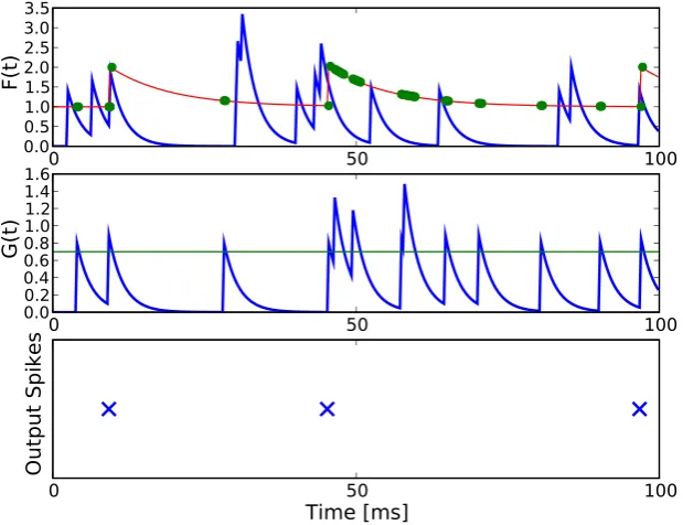
This kind of thresholding serves as a simulation of the absolute and relative refractory period in real neurons [Eckhorn et al. (1990)]. The model neuron is illustrated in figure 1.
Input spike trains to the model neuron are modelled as a homogeneous Poisson process which is often used as an approximation of stochastic neural firing [Dayan and Abbott (2002)]. Evaluation methods will be described in the re-spective passage in the results section. Simulations were executed on aLenovo x61s laptop running Ubuntu 10.04 LTS. Simulation software was written in the programming language Python (version 2.6.5) using standard scientific libraries (Scipy, Numpy and Pylab). Each parameter set was simulated for 10s with a sampling rate of dt= 0.25ms.
9All voltage-related units in the theoretical studies presented in this thesis (chapters
3.4
Results
In this section the model neurons will be investigated focussing on the effect of the gating mechanism on the neurons output. We will first consider the generation of single spikes in the complete model neuron that incorporates the interaction between the gating and the feeding input stream as well as somatic spike thresholding. After that we consider the influence of the gating mechanism on the average output firing rate of the simplified model neuron with skipped somatic thresholding mechanisms. We will analytically derive a relationship between parameters of the gating input stream and the opening probability of the gate and relate it to the output firing rate of the simplified model neuron. Simulations with the simplified and complete model neuron show that the gating mechanism realizes multiplicative interactions on a wide range of input parameters.
3.4.1 Generation of Single Spikes - Complete Model Neuron
Typically, integrate-and-fire neurons elicit a spike when the sufficient condition that the sum over synaptic input currents exceeds a spiking threshold is met [Lapicque (1907); Eckhorn et al. (1990)]. Only one condition has to be met here to allow the model neuron to elicit a spike. In the model neuron presented here, two conditions have to be met to allow the input streams to elicit a spike. First, the gating potential G(t) has to be larger than the gating thresholdγ
in order to let the Heaviside function Θ(G(t)−γ) become 1 and thus to let feeding inputs pass on to the somatic thresholding mechanism (equation 3.2). Second, the feeding potential F(t), that determines the membrane potential
U(t) when the gate is open, has to be larger than the dynamical spiking threshold Γ(t). Only when these two conditions
(G(t)> γ) AND (F(t)>Γ(t))
simplified model neuron, an output spike is elicited in the model neuron if (1) a spike in the feeding input stream and (2) an open gate (G(t)> γ) occurr simultaneously.
3.4.2 Influence of the Gating Mechanism on Output Firing Rates - Simplified Model Neuron
The concept of gain modulation assumes a modulatory input stream to multiplicatively modulate the output firing rate of a neuron with respect
0
50
100
0.0
0.51.0
1.5 2.0 2.5 3.0 3.5
F(t)
0
50
100
0.0 0.2 0.4 0.6
0.81.0
1.2 1.4 1.6
G(t)
0
50
100
Time [ms]
Output Spikes
to a sensory input stream [Andersen and Mountcastle (1983); Salinas and Sejnowski (2001)]. To investigate if the gating mechanism presented here is a feasible model for mediating gain modulation we test how inputs to the gating input stream influence output firing rates of the model neuron with respect to the feeding input stream. We first skip the spike thresholding mechanism in the soma of the model neuron setting Γ(t) = 0mV and τf = 0ms as described in the methods section for the simplified model neuron. Thus, each spike in the feeding input stream elicits one output spike if the gate is open (G(t)> γ). When the gate is open all the time, the output stream is a copy of the feeding input stream and the output firing rate ro equals the feeding inputs firing rate rf. If the gate is closed all the time, no output spike is elicited at all (ro = 0Hz). In between these two extreme situations, we expect the gating mechanism to modulate the output firing rate ro between 0Hz (gate closed all the time) and the firing rate of the feeding input stream ro = rf (gate open all the time). We express the output firing rate ro as the product of the opening probability of the gate popen and the feeding input frequencyrf:
ro =rf ·popen (3.5)
We then analytically describe the opening probability of the gate popen as a function of the gating input firing rate rg, the gating decay time constant τg and the gating input weight wg. When the input weightwg is larger than the gating threshold γ, one spike in the gating input stream opens the gate and it takes a time of tc milliseconds until the gate closes again. The closing time
tc can be calculated via the time it takes until the exponentially decaying gating EPSP with amplitude wg relaxes to the gating threshold value of γ:
wg·e−tc/τg =γ (3.6)
⇔ tc =τg·ln(
wg
γ ) (3.7)
product of tc and the input firing rate rg:
popen(rg, τg, wg, γ) = rg ·tc (3.8)
This suggests that the opening probability popen is proportional to the gating input firing rate with proportionality factor tc given that isig >> tc and
wg > γ. Under these conditions the combination of equation 3.5 and equation 3.8 suggests that the output firing rate ro with respect to rf is modulated multiplicatively by the gating firing rate rg with proportionality factor tc:
ro =rf ·popen =rf ·rg·tc when..isig >> tc..and..wg > γ. (3.9)
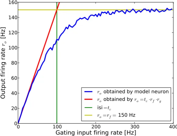
Figure 3 illustrates this for a feeding firing rate of rf = 150Hz and tc =
0
100
200
300
400
Gating input firing rate [Hz]
0
20
40
60
80
100
120
140
160
Ou
tp
ut
fi
rin
g
ra
te
r
o[H
z]
ro
obtained by model neuron
ro
obtained by
ro=tc·rf·rgisi
=tcro=rf=
150 Hz
Figure 3: Output firing rate as a function of gating input firing rate in the simplified model neuron. Output firing rates ro of the simplified model neuron are shown in the blue trace as a function of gating input firing rates. The red trace shows output firing rates as predicted by equation 3.9. The predicted output firing rate deviates from the models output firing rate when the claim
isig >> tcis violated. Inter spike intervals equal the closing time tc whenrg= 100 Hz (green trace isi= tc). The yellow trace illustrates the saturation frequency
10ms (wg = 3.0mV, τg = 7ms). Output firing rates of the model neuron are well described by equation 3.9 when isig >> tc (which corresponds to
rg << 100Hz) but not when interspike intervals are in the range oftc. When the gating firing rate is rg = 100Hz average interspike intervals equal the closing time (isig =tc). Here, equation 3.9 predicts an output firing rate of 150Hz that should equal the feeding input firing rate because the gate should be opened all the time. Nevertheless, the saturation frequency of 150Hz was foremost reached at much higher gating input firing rates due to overlapping gating EPSPs which decrease the opening probability. This is not considered in the analytical description of the model. Output firing rates deviated from predicted firing rates at a gating input frequency larger than approximately 50Hz. Thus, in the following we simulated the model neuron in a range where
isig >2tc.
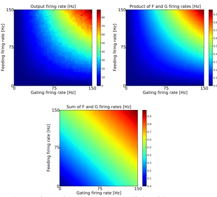
We tested the model for different feeding and gating input firing rates rf = [0, ...,150]Hz and rg = [0, ....,150]Hz with a closing time of tc= 2.5ms (wg = 1.0mV, τg = 7ms). As the smallest average inter spike intervals in the gating input stream were larger than the closing time isimin = 1501Hz = 6.7ms >2tc we expected the output firing rate to be the weighted product of the feeding and the gating input firing rates under the assumption that the analytical equation 3.9 describes the model neuron sufficiently. Figure 4 shows the 2-d-matrix of output firing rates as a function of feeding and gating input firing rates. Here, it can be seen that gating inputs modulated the output firing rate for a wide range of feeding input firing rates. To quantify the type of interaction between the feeding and the gating inputs, the 2d-array of output firing rates was fit based on a multiplicative and an additive model as done in previous studies [Pena and Konishi (2001); Fisher et al. (2007); Kepecs and Raghavachari (2007)]:
rout
mult = am·rf ·rg (3.10)
raddout = as·rf +bs·rg. (3.11)
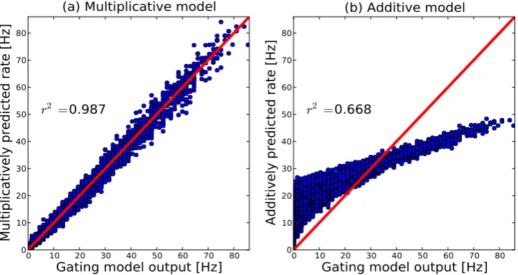
equation 3.9 with the fitting parameter am representing the closing time..tc. Correlations between the multiplicative analytical model and the output of the gating neuron model were high r2 = 0.987 (figure 5). The additive
0
75
150
Gating firing rate [Hz]
0
75
150
Feeding firing rate [Hz]
Output firing rate [Hz]
0 10 20 30 40 50 60 70 80
0
75
150
Gating firing rate [Hz]
0
75
150
Feeding firing rate [Hz]
Product of F and G firing rates [Hz]
0.0 0.1 0.2 0.3 0.4 0.5 0.6 0.7 0.8 0.9
0
75
150
Gating firing rate [Hz]
0
75
150
Feeding firing rate [Hz]
Sum of F and G firing rates [Hz]
0.0 0.1 0.2 0.3 0.4 0.5 0.6 0.7 0.8 0.9
model showed a lower correlation value r2 = 0.668. Moreover, the fitting
parameter am of the multiplicative analytical model reflected the constant closing time factor tcin equation 3.9: tc= 2.5ms and am = 2.3ms. Repeating the simulations for parameters that violate the condition isig > 2tc with
rg = [0, ....,400]Hz with a minimal inter spike interval ofisimin = 4001 = 2.5ms and a closing time of tc= 10ms (wg = 3.0mV, τg = 7ms) yielded relatively small correlation values of r2 = 0.8 for the multiplicative and r2 = 0.6 for the
additive model. This is as overlapping gating EPSPs and saturation effects that appear when isig ≯tc are not considered in the analytical models. To test equation 3.9 for different parameters, we did two experiments. First, we varied the gating input weights between wg = [1.0, ....,10.0]mV while mutually varying τg to keep tc constant. Here, we also kept the gating threshold γ = 0.7mV constant; second, we varied gating input weights
be-0 10 20 30 40 50 60 70 80
Gating model output [Hz]
0 10 20 30 40 50 60 70 80Multiplicatively predicted rate [Hz]
r
2 =0.987
(a) Multiplicative model
0 10 20 30 40 50 60 70 80
Gating model output [Hz]
0 10 20 30 40 50 60 70 80Additively predicted rate [Hz]
r
2 =0.668
(b) Additive model
Figure 5: Correlations between the responses predicted by analyti-cal models and the data obtained with the gating threshold model. Firing rates obtained by fitting the multiplicative (eq. 3.10) and the additive ana-lytical model (eq. 3.11) are plotted against the output of the gating model neuron. The red line denotes the diagonal with correlation value 1. Analytical models are explained in the main text. (a) Correlation between data and multiplicative model
routmult with r2 = 0.99. (b) Correlation between data and additive model routadd with
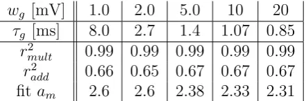
wg [mV] 1.0 2.0 5.0 10 20
τg [ms] 8.0 2.7 1.4 1.07 0.85
r2
mult 0.99 0.99 0.99 0.99 0.99
r2
add 0.66 0.65 0.67 0.67 0.67 fitam 2.6 2.6 2.38 2.33 2.31
Table 1: Correlation valuesbetween the multiplicative analytical model and the output of the simplified gating neuron model r2mult and correlation values between the additive analytical model and the output of the simplified gating neuron model
r2add. Gating weightswg and gating time constantsτg were varied mutually in order to keep tc≈2.8 ms constant (eq. 3.7 with constant gating thresholdγ = 0.7mV). Correlation values were high for the multiplicative analytical model and low for the additive analytical model. The multiplicative fitting parameter am was in the range of the closing time tc.
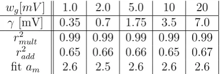
tween wg = [1.0, ....,20.0]mV while mutually varying the gating threshold
wg[mV] 1.0 2.0 5.0 10 20
γ [mV] 0.35 0.7 1.75 3.5 7.0
r2
mult 0.99 0.99 0.99 0.99 0.99
r2
add 0.65 0.66 0.66 0.65 0.67 fitam 2.6 2.5 2.6 2.6 2.6
Table 2: Correlation valuesbetween the multiplicative analytical model and the output of the simplified gating neuron model r2mult and correlation values between the additive analytical model and the output of the simplified gating neuron model
r2add. Gating weights wg and gating thresholds γ were varied mutually in order to keep tc≈2.8 ms constant (eq. 3.7 with constant gating time constantτg= 5ms). Correlation values were high for the multiplicative analytical model and low for the additive analytical model. The fitting parameteram was in the range of the closing timetc.
3.4.3 Gating Mechanism in the Complete Model Neuron
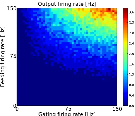
We now transfer the findings obtained with the simplified model neuron to the model neuron that incorporates dynamical thresholding of feeding EPSPs. The influence of the gating mechanism on the output firing rate of the simplified model neuron was described by the opening probability. Analytical equations concerning the opening probability of the gate are still valid here. Equation 3.9 describes the conditions that have to be met for the gating stream to have a multiplicative influence on the output firing rate of the model neuron. In the last chapter it was shown that this equation describes the gating mechanism invariant of mutual changes in gating parameters. Thus, in the following gating parameters are fixed to one set of parameters which fullfils equation 3.9 for gating firing rates between 0 and 150 Hz (wg = 1.0mV,
τg = 8.0ms, γ = 0.7mV). We hypothesize that with these parameters a multiplicative interaction between the feeding and the gating input stream is obtained.