A Survey on Detecting Brain Tumorinmri
Images Using Image Processing Techniques
A.Sindhu1,S.Meera2
M.Phil. Research Scholar, PSGR Krishnammal College for Women, Peelamedu, Coimbatore, India1
Assistant Professor, Dept. of I.T, PSGR Krishnammal College for Women, Peelamedu, Coimbatore, India 2
ABSTRACT: Medical Image Processing is the fast growing and challenging field now a days. Medical Image techniques are used for Medical diagnosis. Brain tumor is a serious life threatening disease. Detecting Brain tumor using Image Processing techniques involves four stages namely Image Pre-Processing, Image segmentation, Feature Extraction, and Classification. Image processing and neural network techniques are used to improve the performance of detecting and classifying brain tumor in MRI images. In this survey various Image processing techniques are reviewed particularly for Brain tumor detection in magnetic resonance imaging. More than twenty five research papers of image processing techniques are clearly reviewed.
KEYWORDS:
Pre
-processing; Image segmentation; Feature extraction; Classification; Braintumour; MRIimagesI. INTRODUCTION
A brain tumor occurs when abnormal cells form within the brain. There are two main types of tumors: malignant(fast growing) and benign (slowgrowing) tumors. Primary brain tumors also be malignant and affect surrounding tissues and it’s contain cancerous cells. The secondary brain tumors are spread to the brain from another place in the body. Imaging plays an important role in the diagnosis of brain tumors. Scientist have classified brain tumor according to their location and type of tissue involved to detect whether it is cancerous and non-cancerous. World Health Organization classified 120 types of tumor and it is done based on the behaviour of the cell from less aggressive to more aggressive. It involves high –resolution techniques especially MRI (magnetic resonance imaging) and CT (Computed tomography).
Fig.1: Brain MRI containing tumor
II. MAGNETIC RESONANCE IMAGING
Magnetic resonance imaging [2] is a powerful tool for investing the body’s internal structure. MRI provides better quality images for the brain, the muscles, the heart and cancerous tissues compared with other medical imaging techniques such as computed tomography (CT) or X-rays. In tumorous brain. MR images intensity level of tumorous tissues exhibit different intensity level on T1-w and T2-w images based on the type of tumor. On T1-w most tumors have low or intermediate signal intensity. On T2-w most tumors have bright intensity. The anatomy of the Brain can normally be viewed by the MRI scan. So this technique is a special one for the brain tumor detection and cancer imaging.
III. LITERATURE SURVEY
A.Image pre- processing
Pre-processing of MRI images is the primary step in image Analysis which perform image enhancement and noise reduction techniques which are used to enhance the image quality. Image is enhanced in the way that finer details are improved and noise is removed from the image. Enhancement and noise reduction techniques are implemented in brain tumor detection that can give best possible results. Enhancement will result in more prominent edges and a sharpened image like tumor is obtained noise will be reduced thus reducing the blurring effect from the image.
Priyanka. Balwinder Singh proposed Median Filter technique for de-noising the salt and pepper noise and Poisson
noise from the images. In a median filter, a window slides along the image and the median intensity value of the pixels within the window becomes the output intensity value of the pixel being processed. Median filter preserves edges in an image while reducing random noise. Each pixel is set to median of the pixel values in the neighborhood of the corresponding input pixels. This filter is used to remove these noises and bounding box method is implemented to identify the location of the tumor [4].
Order statistics filters present a simple and efficient technique to remove noise from the medical images which combines both median filtering and mean filtering to determine the pixel value in the noise less image. This method is used to remove the Rician noise which affects the MRI images [5].This method performs much better than the other filtering methods which was developed by M. N. Nobi and M. A. Yousuf.
C.Ramalakshmi and A.Jaya Chandrandeveloped anisotropic filter to remove the background noise and thus preserving
the edge points in the image. This technique applies a concurrent filtering and contrast stitching. A diffusion constant related to the noise gradient and smoothing the background noise by filtering a proper threshold value is chosen.[6].
De-noising using weighted median filter is applied to remove high frequency components and it can remove salt and
pepper noise from MRI without disturbing of the edges. It is applied for each pixel of a 3*3,5*5,7*7,9*9.11*11 window of neighborhood pixels are extracted and analyzed the mean gray value of foreground mean value of background and contrast value has been employed by J.Jaya. K.Thanushkodi, M.Karnan in 2009[7].
B.Image segmentation
The purpose of image segmentation is to partition an image into regions (spatially connected groups of pixels called classes. or subsets) and objects with respect to one or more characteristics or features.Image segmentation plays a significant role in image processing as it helps in the extraction of suspicious regions from the medical images. The idea behind segmentation is to segment an image into several clusters. The results will be such that, it is possible to identify regions of interest and objects in the original image.
R. Rajeswari. G. Gunasekaran proposed Watershed segmentation algorithm[8] for brain tumor segmentation and this
Color based segmentation using k-means clustering identifies the tumor region significantly form the pre-processed MR image as a clustering feature. Here the pre-processed gray-level brain MR image is converted into RGB color image. Histogram equalization technique is performed and it takes advantages of the neglected pixel values. The RGB color image is then been coarsely represented using 25 bins. Coarse representation uses the spatial information from a histogram based windowing process. K-means is been used to cluster the coarse image data. This shows better result when compared with other edge detection algorithm and enhance the tumor detection accuracy in less time. This was developed by Sarbani Datta. Dr. Monisha Chakraborty [9].
Easha Noureen. Dr. Md. Kamrul Hassan proposed Histogram Thresholding segmentation method for detecting the
brain tumor in MRI image. This is based on thresholding of histogram features and gray level thresholding .It is suitable for an image with region or object of uniform brightness placed against a background of different gray level. A threshold must be applied to segment the object and background. Histogram presents the intensity values of an image and the thresholding is a technique for converting the grayscale or color image in to a binary image based on threshold values. MRI image of the brain is divided and the histogram of each part is drawn. Threshold point of the histogram is calculated and the segmentation is done using the threshold point for both the halves. Plot the histogram and it is between number of pixel and pixel intensity .Bar graph can be used to plot the histogram. Difference of the two histogram is calculated and the resultant difference is plotted using bar graph to select the threshold point. This results gives the great importance in detecting the brain tumor in MRI image [10].
Seeded Region Growing method is an approach to segmentation where it examines neighboring pixels of initial ‘seed points’ and determines pixel neighbors should be added to the region. It is a technique for determining the regions directly. Formulation of the region –based segmentation is, it must be complete and every pixel in the region must be disjoint so that clear separation from each other can be identified. It satisfies the condition that the gray level of pixel is in the range of region. This segmentation is used to find the abnormality is present in the image or not. Fast and fully automatic algorithm, both the homogenous texture features and spatial features of the MRI are used to find the seed points and segmentation results obtained are accurate. This was developed by Mukesh Kumar, Kamal Mehta in 2011[11].
C. Feature extraction
Feature extraction is the technique of extracting specific features from the pre-processed images of different abnormal categories. This technique extracts high-level features needed in order to perform classification of targets. Features are those items which uniquely describe such as size, shape, composition, location etc.Feature Extraction is an important step in the construction of any pattern classification and aims at the extraction of the relevant information that characterizes each class. Gaurav Kumar and Pradeep Kumar Bhatia reviewed various types of features, feature extraction techniques and importance of Using this techniques in image processing systems [12].
SivaSankari.S. Sindhu proposed an image processing technique to extract the optimal features of brain tumor in MRI
by utilizing GLCM [13] (Gray Level Co-occurrence Matrix) and Gabor feature extraction algorithm with the help of
k-means clustering segmentation. Some features are extracted using GLCM techniques and the Gabor features extractions are Contrast. Correlation. Homogeneity. Entropy. Energy. Shape. Color. Texture and Intensity. Thus the feature was extracted and compared with other metric and gives efficient result. [14]
Pratik P. Singhai, Siddharth A. Ladhake developed CCA (Connected Component Analysis) technique in Digital MRI
images to extracts the region which are not supported by boundary after region boundaries have been detected. CCA is to detect the large sized connected foreground region or object. In image analysis, the object is extracted using the connected component labeling operation which consist of assigning a unique label to each maximally connected foreground region of pixel. Any set of pixels which is not separated by the boundary is called connected component. The set of connected components partition an image into segments and thus the area of detected tumor is calculated in pixels using connected component analysis. [15]
Neelam Marshkole, Bikesh Kumar Singh. A.S Thoke used feature extraction based on texture and shape features and
present in the image itself. Set of features which used to describe a medical image is texture feature. Before Feature Extraction region of interest (ROI) consisting of tumor region was extracted for further analysis .Features are extracted and represented in a single array called as feature vector. Haralick‟s texture features were experimented for further
classfication.Feature vector is a row consisting of shape features such as Fourier Descriptor coefficients and seven moment invariants along with 13 texture features[16]. Texture and shape features can give satisfactory result in analysis and classification of brain tumors.
Dina Aboul Dahab. Samy S. A. Ghoniemy proposed the method based on automatic utilization of Specified regions of interest (ROIs) within the tumor area in the MRI images. Form each ROI, set of extracted features include tumor shape and intensity characteristics are extracted and normalized. Each ROI is then given a weight to estimate the PDF of each brain tumor in the MR image. These weights are used as a modeling process to modify the conventional PNN. This
method is based on learning vector quantization (LVQ) which is a supervised competitive learning technique. This
model is successfully tested by using a set of infected brain MRI-scan images to classify brain tumor [17]
D.Image classification
Classification is the labelling of a pixel or a group of pixels. Multiple features are used for a set of pixels i.e. many images of a particular object are needed. Image classification refers to the labelling of images into one of a number of predefined categories.Image classification is more important as it is a critical step for high-level processing such as brain tumor classification. Classification is the last step in the process of brain tumor detection used to classify the image into normal or abnormal and classify the abnormality type whether it is benign or malignant. This study evaluates various techniques which are used in tumor detection from brain MRI.
Kailash D.Kharat & Pradyumna Kulkarni [18] proposed two approaches for Brain Tumor classification based on artificial neural networks. The networks were categorized into feed-forward neural networks and Back propagation
neural Network. First classifier based on feed forward artificial neural network (FF-ANN) and second classifier based
on Back propagation Neural Network (BP-ANN).FF-ANN classifier was created with 500 nodes in the first (input)
layer. 1 to 50 nodes in the hidden layer and 1 node as the output layer and varied the nodes in order to determine the optimal number of hidden nodes. This was to avoid the fitting or under fitting the data. The most widely used neural-network learning method is the BP algorithm. Learning in a neural neural-network involves modifying the weights and biases of the network in order to minimize a cost function. The classifiers have been used to classify subjects as normal or abnormal MRI brain images.
The MR images are classified by wrapper approach with Multi class Support Vector Machine classifier (MC-SVM)
using color, texture and shape features. To reduce the large numbers of features to a smaller set of features wrapper algorithm with multi-class SVM is used. Performance of the MC-SVM classifier is compared with different kernel functions. From the analysis and performance measures like classification accuracy, it is inferred that the brain MRI classification is best done using MC- SVM with Gaussian RBF kernel function than linear and polynomial kernel Functions, the wrapper approach MC-SVM with Gaussian RBF kernel function enhance the classification of MR brain image with normal and benign or malignant classes This approach is efficient for classification of the human brain normal or abnormal (benign or malignant tumor) with high sensitivity, specificity and accuracy rates. This was developed by N.Rajalakshmi and Lakshmi Prabha. [19].
Neelam Marshkole, Bikesh Kumar Singh. A.S Thoke presents a hybrid approach to classify malignant and benign tumors using fusion of texture and shape futures. Both tumor and non-tumor regions appear with little distinction on an MR, image processing toolbox is used for feature extraction and ANN toolbox has been used for classification. Before feature extraction, region of interest (ROI) consisting of tumor region was extracted using MATLAB imtool function for further analysis. Textures features of both benign and malignant tumors are very close to each other and hence
texture feature alone may not give desired classification efficiency. Linear vector quantization (LVQ) is finally used
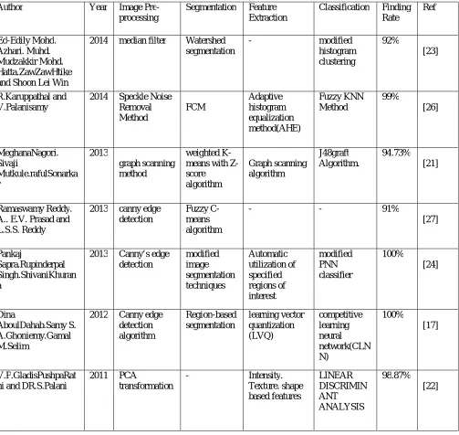
Table 1. Observation and Analysis of Existing System
In this table Existing system shows the various methods which has been used in the Image processing techniques and also displayed the finding rate of the methods varying from 91% to 100%.
IV. CONCLUSION AND FUTURE WORK
This paper describes different image processing techniques for detecting Brain tumor in MRI image. Four components were discussed in MRI images to improve the performance, classification and accuracy of detecting the brain tumor. They are Pre-processing, segmentation, feature extraction and classification. Table I presents the overview of various
Author Year Image
Pre-processing
Segmentation Feature
Extraction
Classification Finding
Rate Ref Ed-Edily Mohd. Azhari. Muhd. Mudzakkir Mohd. Hatta.ZawZawHtike and Shoon Lei Win
2014 median filter Watershed
segmentation
- modified
histogram clustering 92% [23] R.Karuppathal and V.Palanisamy
2014 Speckle Noise
Removal Method FCM Adaptive histogram equalization method(AHE) Fuzzy KNN Method 99% [26] MeghanaNagori. Sivaji Mutkule.rafulSonarka r 2013 graph scanning method weighted K-means with Z-score algorithm Graph scanning algorithm J48graft Algorithm. 94.73% [21] Ramaswamy Reddy. A.. E.V. Prasad and L.S.S. Reddy
2013 canny edge
detection
Fuzzy C-means algorithm
- - 91%
[27]
Pankaj
Sapra.Rupinderpal Singh.ShivaniKhuran a
2013 Canny’s edge
detection modified image segmentation techniques Automatic utilization of specified regions of interest modified PNN classifier 100% [24] Dina AboulDahab.Samy S. A.Ghoniemy.Gamal M.Selim
2012 Canny edge
detection algorithm Region-based segmentation learning vector quantization (LVQ) competitive learning neural network(CLN N) 100% [17] V.P.GladisPushpaRat hi and DR.S.Palani
2011 PCA
transformation
- Intensity.
image processing techniques among the existing systems and also displays the finding rate of the methods and shown the different accuracy rate. Future research lead towards improving the accuracy and also it can be done more advanced in detecting the tumor and growth can be analyzed. Fig 1 shows the MRI image containing tumor which can also define the tumor type.This work will be extendedfor Median Filter and Region Based algorithms to detect the types of tumor in MRI which will provide more efficient results.
REFERENCES
1. Damadian, R., Goldsmith, M.,and Minkoff, L., NMR in Cancer: XVI, FONAR Image of the Live Human Body, Physiological Chemistry and Physics, Vol. 9, pp. 97-100, 1977.
2. ‘National Brain Tumor Society’, [Online] [Available] http:/www,braintumor,org/patients-family-friends/about-brainTumors/brain-tumor-faq,html.
3. Logeswari, T., and Karnan, M.,“An Enhanced Implementation of Brain Tumor Detection Using Segmentation Based onSoft Computing”, IEEE International Conference on Signal Acquisition and Processing, pp. 243-247, 2010.
4. Priyanka, and Balwinder Singh, “AN IMPROVEMENT IN BRAIN TUMOR DETECTION USING SEGMENTATION AND
BOUNDING BOX” IJCSMC, Vol. 2, pp.239 – 246, 2013.
5. Nobi, M. N., and Yousuf, M. A., “A New Method to Remove Noise in Magnetic Resonance and Ultrasound Images”,JOURNAL OF SCIENTIFIC RESEARCH, Vol. 3, pp. 81-89, 2011.
6. Ramalakshmi, C., andJaya Chandran, A.,“Automatic Brain Tumor Detection in MR Images Using Neural Network Based Classification”,International Journal of Computer Science and Network security,Vol. 14, pp.5,2014.
7. Jaya, J., Thanushkodi, K., and Karnan, M., “Tracking Algorithm for De-Noising of MR Brain Images”, IJCSNS International Journal of Computer Science and Network Security, Vol. 9, pp. 11, 2009.
8. Rajeswari, R., and Gunasekaran, G.,“Tumor Detection and Segmentation Using Watershed and Hierarchical Clustering Algorithms”, IJIRCCE,Vol. 2, Special Issue 5, 2014.
9. Sarbani Datta, and Dr. Monisha Chakraborty,“Brain Tumor Detection from Pre-Processed MR Images using Segmentation Techniques”,IJCA, 2011.
10. Easha Noureen,and Dr. Kamrul Hassan, Md.,“ Brain Tumor Detection Using Histogram Thresholding to Get the Threshold point” IOSR Journal of Electrical and Electronics Engineering, Vol. 9,pp.14-19, 2014.
11. Mukesh Kumar, and Kamal K, Mehta, “A Texture based Tumor detection and automatic Segmentation using Seeded Region Growing Method” International Journal of ComputerTechnology, Vol.2, pp. 855-859, 2011.
12. Gaurav Kumar, and Pradeep Kumar Bhatia,“A Detailed Review of Feature Extraction in Image Processing System” International conference an advanced computing and communication Technologies, 2014.
13. Jainy Sachdeva, Vinod Kumar, Indra Gupta, Niranjan Khandelwal,and Chirag Kamal Ahuj ,“Multiclass Brain Tumor Classification using GA-SVM” , 2011.
14. SivaSankari, and Sindhu, S., “Feature Extraction of Brain Tumor Using MRI”, International Journal of Innovative Research in ScienceEngineering and Technology, Vol. 3, 2014.
15. Pratik, P., Singhai , Siddharth, A., and Ladhake, “Brain Tumor Detection Using Marker Based Watershed Segmentation from Digital MR Images” , IJITEE, Vol. 2, pp. 2278-3075, 2013.
16. Neelam Marshkole, and Bikesh Kumar Singh, “Texture and Shape based Classification of Brain Tumors using Linear Vector Quantization”, International Journal of Computer Applications, Vol. 30, pp.0975 – 8887, 2011.
17. Dina Aboul Dahab,Samy, S. A., Ghoniemy, Gamal, M. Selim, “Automated Brain Tumor Detection and Identification Using Image Processing and Probabilistic Neural Network Techniques”, International Journal of Image Processing and Visual Communication, Vol.1, 2012.
18. Kailash, D.Kharat,and PradyumnaKulkarni,“Brain Tumor Classification Using Neural Network Based Methods”, International Journal of Computer Science and Informatics, Vol.1, pp. 2231 –5292, 2012.
19. Rajalakshmi, N.,and Lakshmi Prabha,“Automated classification of brain MRI using color converted k-means clustering segmentation and application of different kernel functions with multi-class svm”, International Interdisciplinary Conference, pp.24-26.
20. Neelam Marshkole, Bikesh Kumar Singh,and Thoke, A.S., “Texture and Shape based Classification of Brain Tumors using Linear Vector Quantization” International Journal of Computer Applications,Vol.30, pp.0975 – 8887, 2011.
21. Meghana Nagori,SivajiMutkule,and Praful Sonarkar, “Detection of Brain Tumor by Mining fMRI Images”, International Journal of Advanced Research in Computer and Communication Engineering, Vol. 2, 2013.
22. Gladis, V. P., PushpaRathi, and Palani, S.,”Brain tumor mri image classification with feature selection and extraction using linear discriminant analysis”.
23. Ed-Edily Mohd, Azhari, Muhd, Mudzakkir Mohd, Hatta,” Brain Tumor Detection and Localization InMagnetic Resonance Imaging” ,International Journal of Information Technology Convergence and Services , Vol. 4, 2014.
24. Pankaj Sapra, Rupinderpal Singh,and Shivani Khurana, “Brain Tumor Detection Using Neural Network”, International Journal of Science and Modern Engineering, Vol.1,2013.
25. Janki Naik,and Prof, Sagar Patel,“Tumor Detection and Classification using Decision Tree in Brain MRI”.
26. Karuppathal, R., and Palanisamy, P., “Fuzzy based automatic detection and classification approach for mri-brain tumor”,ARPN Journal of Engineering and Applied Sciences,Vol. 9,2014.
BIOGRAPHY
A.Sindhu is a Research Scholar in the Computer Science Department, PSGR Krishnammal College for women, Coimbatore. She received Master of Computer Application (MCA) degree in 2005 from Bharathiar University, Coimbatore, India. Her research interests are Digital Image Processing, Neural Network Algorithms etc.