1
Increased zinc and albumin but lowered copper in children with transfusion-dependent
thalassemia.
Zainab Hussein Alhillawi a, Hussein Kadhem Al-Hakeim b, Shatha Rouf Moustafa c, Michael
Maesd,e,f .
a Department of Chemistry, College of Science, University of Kufa, Iraq. E-mail :
b Department of Chemistry, College of Science, University of Kufa, Iraq. E-mail :
c Clinical Analysis Department, College of Pharmacy, Hawler Medical University, Havalan City,
Erbil, Iraq. E-mail: [email protected].
d *Department of Psychiatry, Faculty of Medicine, Chulalongkorn University, Bangkok, Thailand;
e Department of Psychiatry, Medical University of Plovdiv, Plovdiv, Bulgaria;
f IMPACT Strategic Research Centre, Deakin University, PO Box 281, Geelong, VIC, 3220,
Australia .
Corresponding author
Prof. Dr. Michael Maes, M.D., Ph.D.
Department of Psychiatry, Faculty of Medicine, Chulalongkorn University, Bangkok, Thailand
https://scholar.google.co.th/citations?user=1wzMZ7UAAAAJ&hl=th&oi=ao
2 Abstract
Background: Measurements of copper and zinc in transfusion-dependent thalassemia (TDT) show
contradictory results.
Aim of the study: To examine serum levels of these minerals in TDT in relation to iron overload
indices and erythron variables.
Methods: This study recruited 60 children with TDT and 30 healthy children aged 3-12 years old.
Results: Zinc was significantly higher in TDT children than in control children, whilst copper and
the copper to zinc ratio were significantly lowered in TDT. Serum zinc was significantly associated
with the number of blood transfusions and iron overload variables (including serum iron and TS%)
and negatively with erythron variables (including hemoglobin, mean corpuscular volume, mean
corpuscular hemoglobin). Serum copper was significantly and negatively associated with the same
iron overload and erythron variables. The copper to zinc ratio was significantly correlated with
iron, TS%, ferritin, hemoglobin, mean corpuscular volume, and mean corpuscular hemoglobin.
Albumin levels were significantly higher in TDT children than in control children.
Conclusion: Our results suggest that the increase in zinc in children with TDT may be explained
by iron loading anemia and hemolysis and the consequent shedding of high amounts of
intracellular zinc into the plasma. Increased albumin levels and treatment with Desferral may
further contribute towards higher zinc levels in TDT. We suggest that the elevations in zinc in
TDT are a compensatory mechanism protecting against infection, inflammation, and oxidative
stress. Previous proposals for prophylactic use of zinc supplements in TDT may not be warranted.
3
Keywords: Copper, transfusion-dependent thalassemia, zinc, oxidative stress, antioxidants,
4 Introduction
Beta-thalassemia major (β-TM) is a genetic disorder caused by mutations in the β-globin
gene resulting in a lack or reduction of the β-globin chain synthesis leading to the globin chain
imbalances (1). This imbalance is characterized by an excess of free α-globin chains in maturing
red blood cells (RBCs) that lead to hemolysis with consequent anemia (2). Beta-TM patients
require frequent blood transfusions to maintain normal hemoglobin levels and to suppress
ineffective erythropoiesis (3). Frequent RBC transfusions may cause a state of iron overload,
which may cause precipitation of iron in most vital organs (4-6). As a consequence, patients with
transfusion-dependent thalassemia (TDT) frequently develop immune disorders including
decreased synthesis or increased consumption of complement factors (C3 and C4) (7), aberrations
in cell-mediated immunity (8), lymphocyte proliferative responses to mitogens and antigens (9),
and antibody production and responses, decreased activity of T- and B-lymphocytes, alterations in
cytokine responses (10), changes in natural killer cell activity, neutrophil chemotaxis, phagocytic
and chemotaxis activity of macrophages, and increased susceptibility to infections (11-14).
Moreover, the accumulation of iron in various tissues is associated with induction of macrophage
polarization (15) and increased gene expression of inflammatory molecules (16) frequently
causing harmful inflammatory responses (17).
Zinc is a metal that is required by the immune system to function normally (18) and
additionally zinc is involved in the regulation of hepcidin production, which is important in the
iron absorption process (19). Zinc plays a vital role in immune system homeostasis affecting both
innate and adaptive immunity (20, 21) and modulates the number and function of immune cells,
including T and B cells, macrophages, dendritic cells, mast cells, and neutrophils (20-22). This
5
zinc concentrations suppress T-cells (23). Furthermore, maintaining normal zinc levels seems to
be essential to avoid negative concentration-dependent effects of zinc on T-cell activation (22).
The decrease in zinc availability affects the survival, proliferation, and maturation of cells involved
in innate and adaptive immunity (monocytes, polymorphonuclear cells, natural killer cells) in
addition to the balance between the different T helper cell subsets (24).
Another trace element that plays a role in iron metabolism is copper. Copper and zinc, are
found in enzymes that act on iron metabolism (25) and intracellular copper could prompt RBCs
hemolysis (26). Moreover, copper can act as a pro-oxidant in addition to its antioxidant activity.
When copper acts as a pro-oxidant, it promotes harmful free radical effects (27). In the body, free
radicals occur naturally and they can damage cell membranes and genetic material thereby
inducing disease (28). When copper acts as an antioxidant, it scavenges the free radicals and
neutralizes its potentially harmful effects (29). In patients with thalassemia major, serum
concentrations of copper depends on several factors including the amount of copper intake in the
daily diet, intestinal uptake of copper, iron accumulation, kidney function, copper to zinc ratio,
and administration of Desferal (30).
The published data on serum levels of copper and zinc in major thalassemia show some
discrepant results with some papers showing decreased or increased levels or no significant
changes (31-37). Importantly, the levels of these minerals may be affected by iron overload-related
oxidative stress and the use of chelators, which may cause mineral deficiencies, which, in turn,
may lead to or aggravate the complications of thalassemia (38). As a consequence, some authors
advocate the use of zinc supplements in order to treat the oxidative stress which accompanies
thalassemia (39, 40). However, there is no evidence from randomized controlled trials that zinc
6
Hence, the present study aims to examine the zinc and copper levels in TDT in association
with iron load and erythron indices.
Subjects and methods
Participants
This study recruited 90 participants, namely 30 normal controls and 60 TDT children, aged
3-12 years old and of both sexes. The TDT patients were 31 females and 29 males recruited at the
Thalassemia Unit at Al-Zahra'a Teaching Hospital, Najaf, Iraq. The diagnosis of β-thalassemia
major was made by pediatricians and hematologists according to the 2019 ICD-10-CM Diagnosis
Code D56.1. The diagnosis was based on the typical clinical symptoms (e.g. severe anemia,
hepatosplenomegaly, and abnormal bone growth), hematological parameters including
hemoglobin <7g/dl and hypochromic microcytic RBCs with anisopoikilocytosis and increase
reticulocyte percentage, and by elevated HbA2 levels as assayed using HPLC (VARIANT TM
β-Thalassemia Short Program). The healthy controls were 30 apparently healthy children (13 male
and 17 female). None of the controls was anemic or had an obvious inflammatory or systemic
disease. We excluded patients or controls with splenectomy, systemic diseases such as diabetes
mellitus, renal failure, or patients with overt inflammation defined as serum C-reactive protein
(CRP) levels > 6mg/l. The later exclusion criterion is needed to confirm that the increase of ferritin
is due to iron overload rather than to an acute phase response (42).
All patients had regular blood transfusions of packed RBCs at two to four-week intervals
depending on the transfusions needed to maintain the pre-transfusion Hb level above 9 g/dL. The
patients were on iron-chelating therapy with deferoxamine mesylate USP (Desferal®) infusion at
7
3-5 times/week. TDT patients were also given vitamin C to facilitate the binding of iron to deferral
through increased iron release from the reticuloendothelial system. One-alpha® capsules were
administered to TDT patients if they had calcium imbalance due to reduced 1-α hydroxylation.
Folic acid was also given to most patients to reduce ineffective erythropoiesis. Written informed
consent was obtained from the patient’s first-degree relatives (mother or father) after appropriate
oral explanation according to the Declaration of Helsinki. The study was approved by the IRB of
the University of Kufa number 397/2018.
Measurements
Five milliliters of venous blood samples were drawn from all participants after an
overnight fast. The patients' samples were collected just before their blood aspiration session.
Blood was left at room temperature for 10 minutes for clotting, centrifuged 3000 rpm for 5 minutes,
and then serum was separated and transported into new Eppendorf tubes. Serum Cu and Zn were
measured by flame atomic absorption spectrophotometry AA990 (PG Instruments Ltd.). Samples
were diluted 1:10 with 6% n-butanol as diluent before measurement. This method achieved a 30%
increase in sensitivity compared to the use of deionized water only due to decrease viscosity and
the difference in droplet formation that produces more accurate results (43, 44).
Serum iron was measured spectrophotometrically by using the kit from LINEAR
CHEMICALS Co. (Barcelona, Spain) based on the ferrozine method. TIBC was measured by
saturation of serum transferrin with iron and the unbound iron portion
is precipitatedwith magnesium carbonate and the total amount of iron is then determined by the
ferrozine method. Serum ferritin levels were measured by using ELISA kit supplied by
8
saturation percentage (TS%) was calculated from the following equation: TS% = Iron * 100/TIBC
and transferrin concentrations were estimated from the percentage of transferrin saturation and
serum iron using the formula: transferrin (g/L)= serum iron (µM) /(3.98 * TS%) (42). The formula
is based on the maximal binding of 2 moles ofFe3+ to each mole of mole of transferrin and a
molecular weight of 79,570 kD fortransferrin (42). For samples with highly concentrated analytes,
we employed sample dilutions. CRP was measured using a kit supplied by Spinreact®, Spain,
which is based on latex agglutination. Serum albumin was measured spectrophotometrically using
the bromocresol green method with a kit supplied by LINEAR CHEMICALS (Barcelona, Spain).
Hematological parameters were measured by a five-part differential Mindray
BC-5000 hematology analyzer (Mindray Medical Electronics Co., Shenzhen, China) and comprised
hemoglobin (Hb) measured with a cyanide-free spectophotometric method, and an
impedance-based method used for RBCs count, white blood cells count (WBC), packed cell volume (PCV),
and platelet (PLT) count in subject’s blood. Mean corpuscular hemoglobin (MCH), mean
corpuscular volume (MCV), and mean corpuscular hemoglobin concentration (MCHC) were
calculated automatically from the results. All these results were generated automatically by the
Mindray hematology analyzer.
Statistical analysis
Analysis of contingency tables (χ2 test) was used to check associations between nominal
variables and analysis of variances (ANOVAs) to check differences in continuous variables among
diagnostic groups. Associations between scale variables were computed using Pearson’s
product-moment correlation coefficients. Multivariate general linear model (GLM) analysis was employed
9
variables). We computed a z unit-weighted composite score reflecting the copper to zinc ratio as
z score of copper (zCu) – z zinc (zCu-zZn).
Results
Demographic data and Clinical data
The socio-demographic and clinical data in the TDT and healthy control children are
presented in Table 1. There were no significant differences in age, sex ratio, and rural/urban ratio
between the study groups. Our patients are dependent on transfusion with a mean blood transfusion
number of 97 (SD=59.2). Our data showed results which are typical of the iron overload status in
TDT, namely a significant increase in serum iron, TS%, and ferritin besides a decrease in
transferrin, Hb, PCV, RBCs count, MCV, MCH and transferrin concentration. Serum albumin was
significantly increased in TDT patients when compared with the control group.
Differences in biomarkers between TDT and healthy controls
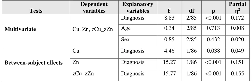
Table 2 shows the results of multivariate GLM analysis examining the association between
serum copper, zinc, and zCu_zZn and diagnosis (TDT versus controls) while adjusting for age and
sex. We found that there was a significant association between diagnosis and the biomarkers with
an effect size of 0.172 while there were no significant effects of age and sex. Tests for
between-subject effects and Table 3 show that copper was significantly lower in TDT than in controls, zinc
was higher in TDT than in controls, while the zCu_zZn ratio was lower in TDT than in controls.
10
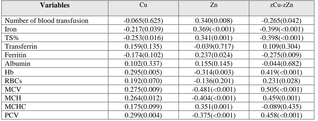
In the total study group, there was a weak inverse correlation between zinc and copper
levels (r=-0.212, p=0.045, n=90). The intercorrelation matrix between copper, zinc, and their ratio
and iron/erythron parameters is shown in Table 4. The results revealed a significant negative
correlation between copper and both iron and TS% but positive correlations with Hb, MCV, MCH,
and PCV. Serum zinc was negatively correlated with MCV, MCH, Hb, and PCV and significantly
positively correlated with the number of blood transfusions, iron, TS%, ferritin, and MCHC.
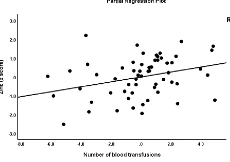
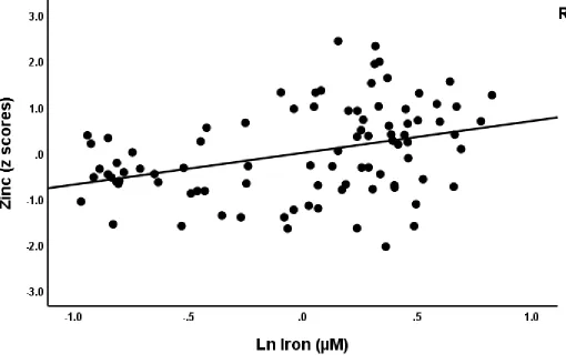
Figure 1 shows the partial association between serum zinc and the number of transfusions
(adjusted for age and sex). Figure 2 shows the partial correlation between serum zinc and serum
iron (adjusted for age and sex). The composite zCu_zZn score was significantly and negatively
correlated with number of transfusions, iron, and TS%, and positively with Hb, RBCs, MCV,
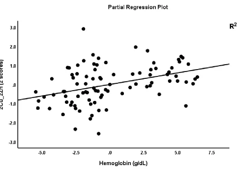
MCH, and PCV. Figure 3 shows the partial regression of zCu_zZn on hemoglobin levels.
Discussion
Increased zinc levels in TDT patients
The first finding of our study is that serum zinc was significantly higher in TDT patients as
compared with controls. Some previous studies reported lowered zinc levels in TDT patients (37,
45, 46). In another study, the prevalence of zinc deficiency (zinc levels>50 µg/dl) was 22.2% and
the deficiency rate was higher in males with a duration of illness > 10 years (34). In contrast,
Kosarian et al. reported that serum zinc levels in major thalassemia patients and controls were
within normal limits (47). In another study, no significant signs of zinc deficiency were found and
no differences between patients and siblings of patients (48). Another study showed that 77% of
11
levels (49). In accordance with our study, a statistically significant increase in zinc in TDT patients
was reported in Iran and these authors noted that zinc deficiency is rare in thalassemia (31).
A first plausible explanation of the increase in zinc in TDT established in our study is that
severe hemolysis leads to the shedding of high amounts of intracellular zinc into the plasma (35).
This theory is supported by the strong intercorrelations between serum zinc and indices of RBC
variables (e.g. negative correlations with Hb, MCV, and MCH) and iron overload (positive
correlations with the number of transfusions, iron, and TS%) established in our study. Zinc
concentrations are up to ten times higher in RBCs than in plasma (50, 51). A second explanation
is that we omitted participants with increased CRP values thereby excluding those with overt
inflammation (52, 53). Other papers, on the other hand, did not always mention whether they
excluded TDT patients with inflammation, which is a prevalent condition in TDT due to the iron
overload (54) and RBC hemolysis, which releases large quantities of damage-associated molecular
patterns into the circulation (55). Moreover, zinc concentrations are about 7 times higher in
leukocytes than in erythrocytes (56) and, therefore, during inflammation, an increased number of
leukocytes may concentrate a significant amount of zinc and remove it from plasma leading to a
reduction in plasma zinc levels. Furthermore, in patients with overt inflammation there is a
significant inverse correlation between serum zinc and inflammatory biomarkers CRP, IL-6, and
TNFα (57) indicating that - for proper interpretation - zinc levels should be adjusted for the
inflammatory state in children (58) and elderly people (59). Lowered zinc levels in inflammatory
conditions may also be explained by lowered albumin levels because zinc is bound to albumin in
the circulation (60). Hence, the presence of inflammation in thalassemia patients may explain zinc
12
patients with overt inflammation should be excluded to evaluate the independent associations
between zinc and diagnoses (62).
Because zinc acts as a signaling agent to modulate (immune) cell activity (63), both zinc
toxicosis and zinc sequestration are used as strategies by the human body compartments during
bacterial and fungal infections (64). Therefore, we suggest that the elevations in zinc in TDT may
be a compensatory mechanism protecting against infection an providing anti-inflammatory and
antioxidant effects (65-67). Likewise, the increased albumin levels established in our TDT patients
may function as a compensatory mechanism because albumin is an antioxidant and is protective
against oxidative stress toxicity in thalassemia patients (68). However, serum albumin was not
always significantly different between thalassemia patients and normal subjects (69, 70). Albumin
binds around 80% of all plasma zinc and is thought to act as a major zinc transporter, while
zinc-albumin complexes have rapid exchange kinetics and contribute to the modulation of free zinc in
the plasma (71). As such, the increased albumin levels established in our study could be another
factor explaining increased zinc levels.
The increased serum zinc levels in TDT patients established in our study and another study
(31) indicate that findings on zinc deficiency in thalassemia may not be generalized. Therefore,
the proposals for prophylactic zinc supplements as routine management of TDT may not be
warranted (34). Even moderate zinc supplementation may interfere with copper metabolism and
may adversely affect the concentrations of high-density lipoprotein cholesterol (72).
Lowered copper levels and copper to zinc ratio in TDT patients
In our study, there is a significant reduction in serum copper in TDT patients as compared
13
sometimes found increased copper levels (77) or no significant changes in TDT (32). As reviewed
in the Introduction, serum concentrations of copper in patients with thalassemia major depend on
several factors including iron accumulation, effects of increased zinc, and administration of
desferrioxamine (Desferal) (78). The inverse correlations between copper levels and indices of
iron overload established in the present study suggest that lowered copper is indeed associated
with iron accumulation or with Desferal treatment (79). Our findings that lowered copper is
significantly associated with lowered Hb, MCV, and MCH may be explained by the knowledge
that low serum copper aggravates the anemia in TDT patients. Copper deficiency may lead to the
development of anemia by the reduction of erythropoietin (80, 81). Furthermore, the decrease in
erythrocyte Cu-Zn superoxide dismutase activity, a key RBCs antioxidant enzyme, may shorten
the life span of erythrocytes in response to oxidative stress (36, 82).
The lowered zCu_zZn ratio in TDT and the strong associations with iron overload indices
(inverse associations) and erythron variables (positive correlations) are a consequence of lowered
copper and increased zinc levels and the effects of different TDT-related factors on both minerals.
It was suggested that the copper to zinc ratio may be used as an inflammatory marker in patients
with sickle cell disease (83). In hemolytic anemia, the copper to zinc correlated positively with
CRP levels (83). Nevertheless, in our study, we excluded patients with overt inflammation,
suggesting that the low copper to zinc ratio in TDT is independent from inflammatory processes.
Also, patients with hemoglobinopathies with complications showed a higher plasma copper to zinc
ratio than those with normal development (84).
14
Children with TDT how a significant increase in zinc and decrease in copper levels as compared
with healthy controls. We suggest that the elevations in zinc in TDT may be a compensatory
mechanism protecting against infection, inflammation, and oxidative stress. Previous proposals
for prophylactic use of zinc supplements in TDT may not be warranted.
Acknowledgment
We acknowledge the staff of the Thalassemia Unit at Al-Zahra'a Teaching Hospital, Najaf,
Iraq for their help in the collection of samples. We also acknowledge the work of the high-skilled
staff of Asia Clinical Laboratory in Najaf city for their help in the ELISA measurements.
Funding
There was no specific funding for this specific study.
Conflict of interest
The authors have no conflict of interest with any commercial or other association in
connection with the submitted article.
Author’s contributions
All the contributing authors have participated in the preparation of the manuscript .
References
1. Thein SL. The molecular basis of β-thalassemia. Cold Spring Harb Perspect Med. 2013;3(5):a011700-a.
15
3. de Dreuzy E, Bhukhai K, Leboulch P, Payen E. Current and future alternative therapies for beta-thalassemia major. Biomedical journal. 2016;39(1):24-38.
4. Al-Hakeim HK, Al-Khakani MM, Al-Kindi MA. Correlation of hepcidin level with insulin resistance and endocrine glands function in major thalassemia. Advances in clinical and experimental medicine : official organ Wroclaw Medical University. 2015;24(1):69-78.
5. Uysal A, Alkan G, Kurtoğlu A, Erol O, Kurtoğlu E. Diminished ovarian reserve in women with transfusion-dependent beta-thalassemia major: Is iron gonadotoxic? European journal of obstetrics, gynecology, and reproductive biology. 2017;216:69-73.
6. Koohi F, Kazemi T, Miri-Moghaddam E. Cardiac complications and iron overload in beta thalassemia major patients-a systematic review and meta-analysis. Annals of hematology. 2019;98(6):1323-31. 7. Ghafourian M, Esmaeili M, Dashti-Gerdabi N, Sadeghi A, Malekei Naseri A, Kazemi A. Evaluation of serum levels of C3 and C4 complement factors in patients with beta thalassemia major in Khuzestan Province, Southwest Iran. Journal of immunoassay & immunochemistry. 2017;38(6):629-38. 8. Hagag AA, Elgamasy MA, Abd Elbar ES. Study of Serum Immunoglobulin Levels and T lymphocyte
Subsets in Children with Beta Thalassemia with Iron Overload in Egypt. The Egyptian journal of immunology. 2016;23(1):97-105.
9. Gharagozloo M, Karimi M, Amirghofran Z. Double-faced cell-mediated immunity in beta-thalassemia major: stimulated phenotype versus suppressed activity. Annals of hematology. 2009;88(1):21-7.
10. Wang MQ, Gao JJ, Xie RW, Wang QH. [Analysis of Irregular Antibody Production in Severe β -Thalassemia Children with long-term Blood Transtusion and Its Correlation with RH Gene and Anemia Gene Mutation Site]. Zhongguo shi yan xue ye xue za zhi. 2017;25(6):1756-60.
11. Ricerca BM, Di Girolamo A, Rund D. Infections in thalassemia and hemoglobinopathies: focus on therapy-related complications. Mediterranean journal of hematology and infectious diseases. 2009;1(1):e2009028.
12. Javad G, Saeid A, Mohammadmehdi N. Thalassemia and immune system dysfunction-review article. Int J Curr Res. 2011;3:105-8.
13. Bao W, Zhong H, Li X, Lee MT, Schwartz J, Sheth S, et al. Immune regulation in chronically transfused allo-antibody responder and nonresponder patients with sickle cell disease and β -thalassemia major. American journal of hematology. 2011;86(12):1001-6.
14. Vento S, Cainelli F, Cesario F. Infections and thalassaemia. The Lancet Infectious diseases. 2006;6(4):226-33.
15. Zhou Y, Que KT, Zhang Z, Yi ZJ, Zhao PX, You Y, et al. Iron overloaded polarizes macrophage to proinflammation phenotype through ROS/acetyl‐p53 pathway. Cancer medicine. 2018;7(8):4012 -22.
16. Maras JS, Das S, Sharma S, Sukriti S, Vyas AK, Kumar D, et al. Iron-overload triggers ADAM-17 mediated inflammation in severe alcoholic hepatitis. Scientific reports. 2018;8(1):1-14.
17. Wessling-Resnick M. Iron homeostasis and the inflammatory response. Annual review of nutrition. 2010;30:105-22.
18. Maares M, Haase H. Zinc and immunity: An essential interrelation. Archives of biochemistry and biophysics. 2016;611:58-65.
19. Bjørklund G, Aaseth J, Skalny AV, Suliburska J, Skalnaya MG, Nikonorov AA, et al. Interactions of iron with manganese, zinc, chromium, and selenium as related to prophylaxis and treatment of iron deficiency. Journal of Trace Elements in Medicine and Biology. 2017;41:41-53.
20. Maywald M, Wessels I, Rink L. Zinc signals and immunity. International journal of molecular sciences. 2017;18(10):2222.
16
22. Daaboul D, Rosenkranz E, Uciechowski P, Rink L. Repletion of zinc in zinc-deficient cells strongly up-regulates IL-1β-induced IL-2 production in T-cells. Metallomics. 2012;4(10):1088-97.
23. Rink L, Gabriel P. Extracellular and immunological actions of zinc. Zinc Biochemistry, Physiology, and Homeostasis: Springer; 2001. p. 181-97.
24. Bonaventura P, Benedetti G, Albarède F, Miossec P. Zinc and its role in immunity and inflammation. Autoimmunity reviews. 2015;14(4):277-85.
25. Hacibekiroglu T, Basturk A, Akinci S, Bakanay S, Ulas T, Guney T, et al. Evaluation of serum levels of zinc, copper, and Helicobacter pylori IgG and IgA in iron deficiency anemia cases. European review for medical and pharmacological sciences. 2015;19(24):4835-40.
26. Tasneem S, Sultana N, Snover A, Alam K. Clinical utility of red blood cell count, red cell distribution width: will it provide more accurate differentiation of Beta thalassemia trait and iron deficiency anemia in pregnancy. Rawal Med J. 2016;41(4):424-7.
27. Rehmani N, Zafar A, Arif H, Hadi SM, Wani AA. Copper-mediated DNA damage by the neurotransmitter dopamine and L-DOPA: a pro-oxidant mechanism. Toxicology in Vitro. 2017;40:336-46.
28. Nimse SB, Pal D. Free radicals, natural antioxidants, and their reaction mechanisms. Rsc Advances. 2015;5(35):27986-8006.
29. Cruces‐Sande A, Méndez‐Álvarez E, Soto‐Otero R. Copper increases the ability of 6‐ hydroxydopamine to generate oxidative stress and the ability of ascorbate and glutathione to potentiate this effect: potential implications in Parkinson's disease. Journal of neurochemistry. 2017;141(5):738-49.
30. Rakhra G, Masih D, Vats A, Verma SK, Singh VK, Rana RT, et al. Effect of physical activity and age on plasma copper, zinc, iron, and magnesium concentration in physically active healthy males. Nutrition. 2017;43:75-82.
31. Mehdizadeh M, Zamani G, Tabatabaee S. Zinc status in patients with major beta-thalassemia. Pediatric hematology and oncology. 2008;25(1):49-54.
32. Mahyar A, Ayazi P, Pahlevan A-A, Mojabi H, Sehhat M-R, Javadi A. Zinc and copper status in children with Beta-thalassemia major. Iran J Pediatr. 2010;20(3):297-302.
33. Banihashem A, Ghahramanlu E, Tavallaie S, Mirhosseini N, Taherpour M, Saber H, et al. Serum zinc and copper concentrations in patients with Beta-thalassemia major. Trace Elem Electrolytes. 2013;30:108-13.
34. Sultan S, Irfan SM, Kakar J, Zeeshan R. Effect of iron chelator desferrioxamine on serum zinc levels in patients with beta thalassemia major. The Malaysian journal of pathology. 2015;37(1):35-8. 35. Wieringa FT, Dijkhuizen MA, Fiorentino M, Laillou A, Berger J. Determination of zinc status in
humans: which indicator should we use? Nutrients. 2015;7(5):3252-63.
36. Walter PB, Minkley M, Curtis C, Maeve H, Morty R, Ewing S, et al. Implications of Low Zinc and Copper Levels As Well As Altered Iron Trafficking Proteins on Oxidant Stress in Patients with Transfusion Dependant Thalassemia. American Society of Hematology Washington, DC; 2016. 37. Zekavat OR, Bahmanjahromi A, Haghpanah S, Ebrahimi S, Cohan N. The Zinc and Copper Levels in
Thalassemia Major Patients, Receiving Iron Chelation Therapy. Journal of pediatric hematology/oncology. 2018;40(3):178-81.
38. Ozturk Z, Genc GE, Gumuslu S. Minerals in thalassaemia major patients: An overview. Journal of Trace Elements in Medicine and Biology. 2017;41:1-9.
17
40. Ghahramanlu E, Banihashem A, Mirhossini N-Z, Hosseini G, Mostafavi-Toroghi H, Tavallaie S, et al. Effect of zinc supplementation on serum antibody titers to heat shock protein 27 in patients with thalassemia major. Hematology. 2014;19(2):113-9.
41. Swe KMM, Abas AB, Bhardwaj A, Barua A, Nair N. Zinc supplements for treating thalassaemia and sickle cell disease. Cochrane Database of Systematic Reviews. 2013(6).
42. Kennedy A, Kohn M, Lammi A, Clarke S. Iron status and haematological changes in adolescent female inpatients with anorexia nervosa. Journal of paediatrics and child health. 2004;40(8):430-2. 43. Meret S, Henkin R. Simultaneous direct estimation by atomic absorption spectrophotometry of
copper and zinc in serum, urine, and cerebrospinal fluid. Clinical Chemistry. 1971;17(5):369-73. 44. Twayej AJ, Al-Hakeim HK, Al-Dujaili AH, Maes M. Lowered zinc and copper levels in drug-naïve
patients with major depression: Effects of antidepressants, ketoprofen and immune activation. The World Journal of Biological Psychiatry. 2019:1-12.
45. Mashhadi MA, Sepehri Z, Heidari Z, Shirzaee E, Kiani Z. The prevalence of zinc deficiency in patients with thalassemia in South East of iran, sistan and baluchistan province. Iranian Red Crescent Medical Journal. 2014;16(8).
46. Karunaratna AMDS, Ranasingha JGS, Mudiyanse RM. Zinc status in beta thalassemia major patients. Biological trace element research. 2018;184(1):1-6.
47. Kosarian M, Valaee N, Mahdyanee A. Do the Desferal thalassemic patients have zinc deficiency receiver. Journal of Mazandaran University of Medical Sciences. 2000;10(26):1-8.
48. El Missiry M, Hamed Hussein M, Khalid S, Yaqub N, Khan S, Itrat F, et al. Assessment of serum zinc levels of patients with thalassemia compared to their siblings. Anemia. 2014;2014:125452.
49. Reshadat S, Kiani A, Iranfar S. Zinc level of major thalassemic patients in Kermanshah. Behbood. 2006;2(10):157-67.
50. Bogden JD, Klevay LM. Clinical nutrition of the essential trace elements and minerals: the guide for health professionals: Springer Science & Business Media; 2000.
51. Akanli L, Lowenthal DB, Gjonaj S, Dozor AJ. Plasma and red blood cell zinc in cystic fibrosis. Pediatric Pulmonology. 2003;35(1):2-7.
52. Mburu AS, Thurnham DI, Mwaniki DL, Muniu EM, Alumasa FM. The influence of inflammation on plasma zinc concentration in apparently healthy, HIV+ Kenyan adults and zinc responses after a multi-micronutrient supplement. European journal of clinical nutrition. 2010;64(5):510-7.
53. Abdulzahra MS, Al-Hakeim HK, Ridha MM. Study of the effect of iron overload on the function of endocrine glands in male thalassemia patients. Asian J Transfus Sci. 2011;5(2):127-31.
54. Ganz T. Iron and infection. International Journal of Hematology. 2018;107(1):7-15.
55. Mendonça R, Silveira AA, Conran N. Red cell DAMPs and inflammation. Inflammation research : official journal of the European Histamine Research Society [et al]. 2016;65(9):665-78.
56. Hinks LJ, Clayton BE, Lloyd RS. Zinc and copper concentrations in leucocytes and erythrocytes in healthy adults and the effect of oral contraceptives. Journal of clinical pathology. 1983;36(9):1016-21.
57. Jung S, Kim MK, Choi BY. The Relationship between Zinc Status and Inflammatory Marker Levels in Rural Korean Adults Aged 40 and Older. PloS one. 2015;10(6):e0130016.
58. Karakochuk CD, Barr SI, Boy E, Bahizire E, Tugirimana PL, Akilimali PZ, et al. The effect of inflammation on serum zinc concentrations and the prevalence estimates of population-level zinc status among Congolese children aged 6-59 months. European journal of clinical nutrition. 2017. 59. MacDonell SO, Miller JC, Harper MJ, Reid MR, Haszard JJ, Gibson RS, et al. A comparison of
18
60. Maes M, De Vos N, Demedts P, Wauters A, Neels H. Lower serum zinc in major depression in relation to changes in serum acute phase proteins. Journal of affective disorders. 1999;56(2-3):189-94.
61. Prasad AS. Zinc: role in immunity, oxidative stress and chronic inflammation. Current Opinion in Clinical Nutrition & Metabolic Care. 2009;12(6):646-52.
62. Gammoh NZ, Rink L. Zinc in Infection and Inflammation. Nutrients. 2017;9(6):624.
63. Wessels I, Maywald M, Rink L. Zinc as a gatekeeper of immune function. Nutrients. 2017;9(12):1286.
64. Vignesh KS, Deepe Jr GS. Immunological orchestration of zinc homeostasis: the battle between host mechanisms and pathogen defenses. Archives of biochemistry and biophysics. 2016;611:66-78.
65. Prasad AS. Zinc is an Antioxidant and Anti-Inflammatory Agent: Its Role in Human Health. Frontiers in Nutrition. 2014;1(14).
66. Prasad AS. Zinc in human health: effect of zinc on immune cells. Mol Med. 2008;14(5-6):353-7. 67. Olechnowicz J, Tinkov A, Skalny A, Suliburska J. Zinc status is associated with inflammation,
oxidative stress, lipid, and glucose metabolism. The Journal of Physiological Sciences. 2018;68(1):19-31.
68. Bazvand F, Shams S, Borji Esfahani M, Koochakzadeh L, Monajemzadeh M, Ashtiani M-TH, et al. Total Antioxidant Status in Patients with Major β-Thalassemia. Iran J Pediatr. 2011;21(2):159-65. 69. Omar Mousa S, Afifi M, Saedii A, El-Setohy A. Ischemia modified albumin in children with
transfusion-dependent β-thalassemia: a new marker for an old problem. The Egyptian Journal of Haematology. 2016;41(2):45-9.
70. Sultan S, Irfan SM, Ahmed SI. Biochemical markers of bone turnover in patients with β-thalassemia major: a single center study from southern Pakistan. Advances in hematology. 2016;2016.
71. Lu J, Stewart AJ, Sadler PJ, Pinheiro TJ, Blindauer CA. Albumin as a zinc carrier: properties of its high-affinity zinc-binding site. Biochemical Society transactions. 2008;36(Pt 6):1317-21.
72. Fosmire GJ. Zinc toxicity. The American journal of clinical nutrition. 1990;51(2):225-7.
73. Shamshirsaz AA, Bekheirnia MR, Kamgar M, Pourzahedgilani N, Bouzari N, Habibzadeh M, et al. Metabolic and endocrinologic complications in beta-thalassemia major: a multicenter study in Tehran. BMC endocrine disorders. 2003;3(1):4.
74. Eshghi P, Alavi S, Ghavami S, Rashidi A. Growth impairment in β-thalassemia major: the role of trace element deficiency and other potential factors. Journal of pediatric hematology/oncology. 2007;29(1):5-8.
75. De Franceschi L, Brugnara C, Beuzard Y. Dietary magnesium supplementation ameliorates anemia in a mouse model of β-thalassemia. Blood, The Journal of the American Society of Hematology. 1997;90(3):1283-90.
76. Ghone RA, Kumbar KM, Suryakar AN, Katkam RV, Joshi NG. Oxidative stress and disturbance in antioxidant balance in beta thalassemia major. Indian J Clin Biochem. 2008;23(4):337-40.
77. Claster S, Wood JC, Noetzli L, Carson SM, Hofstra TC, Khanna R, et al. Nutritional deficiencies in iron overloaded patients with hemoglobinopathies. American journal of hematology. 2009;84(6):344-8.
78. Faranoush M, RAHIMINEZHAD M, Karamizadeh Z, Ghorbani R, OUJI S. Zinc supplementation effect on linear growth in transfusion dependent β Thalassemia. 2008.
79. Genc GE, Ozturk Z, Gumuslu S, Kupesiz A. Mineral Levels in Thalassaemia Major Patients Using Different Iron Chelators. Biological Trace Element Research. 2016;170(1):9-16.
19
81. Kraemer K, Zimmermann MB. Nutritional anemia: Sight and Life press Basel; 2007.
82. Iuchi Y, Okada F, Onuma K, Onoda T, Asao H, Kobayashi M, et al. Elevated oxidative stress in erythrocytes due to a SOD1 deficiency causes anaemia and triggers autoantibody production. Biochemical Journal. 2007;402(2):219-27.
83. Emokpae MA, Fatimehin EB. Copper-to-Zinc Ratio Correlates with an Inflammatory Marker in Patients with Sickle Cell Disease. Sci. 2019;1(2):34.
20
Table 1. Sociodemographic and biomarkers data in children with TDT (transfusion-dependent thalassemia) and healthy children (HC).
Variables HC TDT F/χ2 df p
Age years 7.1(2.5) 8.0(3.1) 1.54 1/88 0.217
Sex Female/Male 13/17 31/29 0.56 1 0.456
Residency Rural / Urban 4/26 16/44 2.06 1 0.151
#Blood transfusion - 97.6(59.2) - - -
Iron µM 15.2(3.4) 43.3(8.0) 581.77 1/88 <0.001
Transferrin g/l 0.154(0.043) 0.131(0.015) 13.06 1/88 0.001
TS% % 26.7(2.6) 82.9(11.7) 544.08 1/88 <0.001
Ferritin ng/ml 153.4(44.2) 3298.1(1841.0) 899.55 1/88 <0.001
Albumin g/l 39.2(3.7) 43.9(5.9) 16.56 1/88 <0.001
RBCs *106 Cell/µl 4.49(0.62) 3.38(0.57) 29.79 1/88 <0.001
Hb g/dl 14.11(1.41) 7.92(1.38) 397.19 1/88 <0.001 MCV fl 96.06(8.14) 64.87(7.82) 309.28 1/88 <0.001
MCH pg 31.67(2.68) 21.06(0.32) 348.02 1/88 <0.001
MCHC % 32.97(0.21) 32.48(0.93) 8.06 1/88 <0.001
PCV % 42.80(4.23) 24.39(4.15) 389.75 1/88 <0.001
21
Table 2: Results of multivariate GLM analysis examining the differences in zinc (Zn), copper (Cu) and their ratio between children with TDT (transfusion-dependent thalassemia) and heathy children.
Tests
Dependent variables
Explanatory
variables F df p
Partial η2
Multivariate Cu, Zn, zCu_zZn
Diagnosis 8.83 2/85 <0.001 0.172
Age 0.34 2/85 0.713 0.008
Sex 0.85 2/85 0.432 0.020
Between-subject effects
Cu Diagnosis 4.46 1/86 0.038 0.049
Zn Diagnosis 15.27 1/86 <0.001 0.151
zCu_zZn Diagnosis 15.77 1/86 <0.001 0.155
22
Table 3: Model-generated estimated marginal mean (SE) values in children with transfusion-dependent thalassemia (TDT) and healthy children (HC).
Dependent Variables HC TDT
Copper mg/l 0.808(0.049) 0.693(0.035)
z Copper (z score) 0.312(0.180) -0.156(0.127)
Zinc mg/l 0.573(0.042) 0.782(0.030)
z Zinc (z score) -0.550(0.172) 0.275(0.121)
zCu-zZn (z score) 0.554(0.170) -0.277(0.120)
23
Table 4. Intercorrelation matrix of iron and erythron status parameters with Zn, Cu, and their ratio.
Variables Cu Zn zCu-zZn
Number of blood transfusion -0.065(0.625) 0.340(0.008) -0.265(0.042)
Iron -0.217(0.039) 0.369(<0.001) -0.399(<0.001)
TS% -0.253(0.016) 0.341(0.001) -0.398(<0.001)
Transferrin 0.159(0.135) -0.039(0.717) 0.109(0.304)
Ferritin -0.174(0.102) 0.237(0.024) -0.275(0.009)
Albumin 0.102(0.337) 0.155(0.145) -0.044(0.682)
Hb 0.295(0.005) -0.314(0.003) 0.419(<0.001)
RBCs 0.192(0.070) -0.136(0.201) 0.231(0.028)
MCV 0.275(0.009) -0.481(<0.001) 0.505(<0.001)
MCH 0.264(0.012) -0.404(<0.001) 0.459(0.001)
MCHC 0.175(0.099) 0.351(0.001) -0.089(0.435)
PCV 0.299(0.004) -0.375(<0.001) 0.458(<0.001)
Shown are the correlation coefficients with exact p-values.
24
Figure 1. Partial regression (adjusted for age and sex) of serum zinc levels (in z scores) on the number of blood transfusions in children
25
26