Drug Design, Development and Therapy
Dove
press
R e v i e w open access to scientific and medical research
Open Access Full Text Article
Developing therapeutic approaches
for metachromatic leukodystrophy
Shilpa A Patil1
Gustavo HB Maegawa1,2
1McKusick-Nathans institute
of Genetic Medicine, 2Department
of Pediatrics, The Johns Hopkins School of Medicine, Baltimore, MD, USA
Correspondence: Gustavo HB Maegawa McKusick-Nathans institute of Genetic Medicine, Department of Pediatrics, The Johns Hopkins School of Medicine, Baltimore, MD 21205, USA Tel +1 443 287 3505
Fax +1 410 502 5677 email [email protected]
Abstract: Metachromatic leukodystrophy (MLD) is an autosomal recessive lysosomal dis-order caused by the deficiency of arylsulfatase A (ASA), resulting in impaired degradation of sulfatide, an essential sphingolipid of myelin. The clinical manifestations of MLD are character-ized by progressive demyelination and subsequent neurological symptoms resulting in severe debilitation. The availability of therapeutic options for treating MLD is limited but expanding with a number of early stage clinical trials already in progress. In the development of therapeutic approaches for MLD, scientists have been facing a number of challenges including blood–brain barrier (BBB) penetration, safety issues concerning therapies targeting the central nervous system, uncertainty regarding the ideal timing for intervention in the disease course, and the lack of more in-depth understanding of the molecular pathogenesis of MLD. Here, we discuss the current status of the different approaches to developing therapies for MLD. Hematopoietic stem cell transplantation has been used to treat MLD patients, utilizing both umbilical cord blood and bone marrow sources. Intrathecal enzyme replacement therapy and gene therapies, administered locally into the brain or by generating genetically modified hematopoietic stem cells, are emerging as novel strategies. In pre-clinical studies, different cell delivery systems including microencapsulated cells or selectively neural cells have shown encouraging results. Small molecules that are more likely to cross the BBB can be used as enzyme enhancers of diverse ASA mutants, either as pharmacological chaperones, or proteostasis regulators. Specific small molecules may also be used to reduce the biosynthesis of sulfatides, or target different affected downstream pathways secondary to the primary ASA deficiency. Given the progressive neurodegenerative aspects of MLD, also seen in other lysosomal diseases, current and future therapeutic strategies will be complementary, whether used in combination or separately at specific stages of the disease course, to produce better outcomes for patients afflicted with this devastating inherited disorder.
Keywords: metachromatic leukodystrophy, arylsulfatase A, enzyme replacement therapy, gene therapy, enzyme enhancement therapy, small molecules
Introduction
Metachromatic leukodystrophy (MLD) is an autosomal recessive lysosomal stor-age disorder caused by the deficiency of arylsulfatase A (ASA; EC 3.1.6.8), a lysosomal sulfatase that hydrolyzes the 3-O ester bond from sulfatides including 3-O-sulfogalactosylceramide and 3-O-sulfolactosylceramide.1,2 These sulfatides are
expressed in specific tissues and are mostly abundant in the central (CNS) and periph-eral nervous systems (PNS). Sulfatides are important components of myelin, which is a crucial insulator sheath that surrounds axons as a bilayer membrane.3 An intact
myelin sheath is important for the proper conduction of nerve impulse in both CNS
Drug Design, Development and Therapy downloaded from https://www.dovepress.com/ by 118.70.13.36 on 22-Aug-2020
For personal use only.
Number of times this article has been viewed
This article was published in the following Dove Press journal: Drug Design, Development and Therapy
Dovepress
Patil and Maegawa
and PNS.3 The inappropriate recycling of sulfatides results in
alterations of the myelin structure, affecting the physiological transmission of electrical impulse between nerve cells.4 This
process of degeneration of the myelin sheath surrounding axons, mostly known as demyelination, is the hallmark of MLD. Patients afflicted with this condition present with a broad spectrum of neurological manifestations centered on the impairment of CNS and PNS, including progressive coordination and speech problems, seizures, and behavioral disturbances.5 In the design and development of
disease-specific therapies for MLD, the biochemical pathogenesis and secondary molecular events to the ASA deficiency are important to be considered.
Epidemiology and pseudodeficiency
ARSA
alleles
Inherited disorders affecting the lysosomes have a collective incidence of 1 in 2,300–7,000 live births, making them a prevalent class of inborn organelle disorders.6,7 Among
dif-ferent lysosomal disorders, the general prevalence of MLD varies from 1:40,000 to 1:100,000.8 In the Polish population,
its incidence was reported as 4:100,000.9 MLD is a pan-ethnic
lysosomal storage disease with affected patients described in several populations including European, Japanese, Jew-ish, Lebanese, Muslim Arab, South African, Iranian, Indian, Polynesian, Algerian, Habbanite Jew, Navajo Indian, Alaskan Eskimo, and Christian Arab, with ranging from mild to severe forms of MLD.8 The deficiency of ASA is caused by
muta-tions in the ARSA gene in homo- or heterozygosity. Over 150 mutations have been reported in MLD patients.10 Some
specific alterations in the ARSA gene sequence result in an enzymatic activity of 10%–15% of the normal (wild-type) ASA, which is sufficient to physiologically hydrolyze sulfati-des, maintaining their recycling process and avoiding their accumulation in the lysosomal compartment. These non-del-eterious ARSA gene alterations resulting in reduction of ASA enzymatic activity are known as pseudodeficiency alleles. These ARSA alleles are considered polymorphisms, with no disease-associated symptoms either in hetero or homozygous states. Two most common ARSA pseudodeficiency alleles are c.1049A.G/p.N350S and c.*96A.G. As they occur in cis-trans, they are described as c.[1049A.G; c.*96A.G].11,12
The c.1049A.G/p.N350S results in modification of one of the N-glycosylation sites of ASA, affecting the structural stability of ASA and its targeting to the lysosomes.12 The
c.*96A.G is found in the 3′ non-translated region that alters the signaling of the polyadenylation of mRNA, substantially reducing the amount of ASA produced.13 The frequency
of these ARSA pseudodeficiency alleles is up to 5% in the European population.13–17 The carrier frequency of the ARSA
pseudodeficiency alleles in Australia is estimated to be 20%.16
Its correlation with occurrence of other neurodegenerative disorders that occur later in life remains debatable.14 The
existence of the ARSA pseudodeficiency alleles demonstrates that the sulfatide degradation can occur normally in the presence of ASA variants functioning at 10–15% of the wild type ASA enzymatic activity. This biochemical observation has crucial implications in the development of therapeutic strategies for MLD.
Biochemistry and molecular
genetics of MLD
As with other lysosomal hydrolases, ASA is synthesized in the rough endoplasmic reticulum (ER) and co-translationally transported to the lumen of ER.18–20 Once properly folded, ASA
is targeted to the trans-Golgi network where it becomes a sub-strate of uridine diphosphate (UDP)-N-Acetylglucosamine-1 phosphotransferase (EC 2.7.8.17) and N-acetylglucosamine-1-phosphodiester alpha-N-acetylglucosaminidase (EC 3.1.4.45). These two enzymes catalyze the binding and formation of mannose-6-phosphate residue, the recognition marker of lysosomal hydrolases. This modification is impor-tant to allow ASA to interact with the mannose-6-phosphate receptor, and be targeted to the lysosomal compartment.21
In terms of its molecular structure, 2.1-Å resolution crystal X-ray structure showed that ASA is a homo-octamer com-posed of a tetramer of dimers (α2)4.22 In fact, the formation
of ASA homodimers (α2) occurs in the ER. Subsequently, ASA only assumes the homo-octomeric conformation at the acidic pH of the lysosomal compartment.22 The identification
of the c.1277T.G/p.P426L mutation showed it preventing the formation of homo-octomers, as the P426 residue locates proximal to the E424 residue, whose protonation is necessary for the octomerization process.23 The mutant ASA-P426L
is degraded by cathepsin-L, a lysosomal protease, which recognizes a cleavage site in the ASA homo-dimer interface that is normally protected by the octomerization conforma-tion seen in wild-type ASA.23,24 Functionally, ASA catalyzes
the desulfation of 3-O-sulfogalactosyl-bound sphingolipids. Sulfatides, or 3-O-galactosylceramides, are the main sphin-golipids containing sulfate residues. Sulfatides are abundant in both the CNS and PNS. These glycosphingolipids are the main components of the myelin sheath. In the CNS, oligo-dendrocytes synthesize sulfatides, which are located in the myelin surrounding neural axons. In the PNS, the Schwann cells are the myelin-producing cells. Interestingly, in the gall
Drug Design, Development and Therapy downloaded from https://www.dovepress.com/ by 118.70.13.36 on 22-Aug-2020
Dovepress Development of therapies for MLD
bladder of MLD-affected patients, sulfatides accumulate in the epithelium cells, resulting in sludge, gallstones, hemobilia, cholecystitis, polyposis, papillomatosis, and small or enlarged gallbladder.25–29 Sulfatides are also present, but
at low levels, in the respiratory tract,30 gastric mucosa,31 and
uterine endometrium.32 The 3-O-sulfatelactosyl ceramide,
which is another sulfatide substrate of ASA, is found in the renal tubules and liver.33 Another deacylated sulfatide,
known as lysosulfatide or psychosine sulfatide, is present in the CNS and renal tubules.34,35 The presence of sulfatides in
the kidney is the cornerstone of the main diagnostic tool for MLD, which is based on measuring urinary sulfatides.36 ASA
is also responsible for catalyzing the desulfation of glycerol-containing sulfated sphingolipids, which have a sulfate bond to the galactose C-3 hydroxyl residue. These 3-O-sulfaga-lactosyl sphingolipids are present in the mammalian testes and sperm, which are mostly known as seminolipids.37 These
sphingolipids are produced by spermatocytes, and their roles in different stages of the spermatogenesis are still to be determined.37
In MLD, the deficiency of ASA results in sulfatide accu-mulation in the myelin-producing cells: oligodendrocytes in CNS, and Schwann cells in PNS. With the progressive accumulation of sulfatides, the lysosomal-endosomal system becomes dysfunctional, and other secondary pathogenic cas-cades occur, ultimately resulting in apoptosis.38 This results
in progressive demyelination in both CNS and PNS, which correlates with the major clinical manifestations of MLD.39
However, cellular inclusions called membranous cytoplasmic bodies (MCBs), which can correspond to accumulation of sulfatides, are observed in pyramidal neurons and Purkinje cells in the brain of MLD patients.40
The deficiency of ASA is caused by mutations in the
ARSA gene. Amongst the over 150 mutations reported in the ARSA gene, more than 70% are missense mutations.10
The most common mutation is the splice donor site muta-tion of the exon2/intron2 border, IVS459+1G.A, which occurs in 15%–43% of MLD alleles, and is associated with the late infantile clinical form of MLD.41 The second most
common ARSA mutation is c.1277C.T/p.P426L, which, when in homozygosity or heterozygosity with an infantile mutation (eg, IVS459+1G.A), is associated with juvenile onset of MLD.41 Each of these two common mutant alleles
represents approximately 25% of all ARSA mutant alleles in MLD patients with European ancestry.42 The third most
prevalent ARSA mutation is c.536T.G/p.I179S, which, when in heterozygosity with the c.1277C.T/p.P426L mutation, is associated with adult onset of the disease.41,42 Interestingly,
ASA-p.I179S presents the higher residual ASA enzymatic activity, and is correlated with later onset and more protracted MLD course.41 The ARSA c.536T.G/p.I179S allele is
pres-ent in 12%–13% of the total European population of mutant alleles.41,42 Though several missense mutations have been
identified, few of them have been biochemically character-ized.10,43 Most missense mutations result in misfolded mutant
ASA that is early selected for the ER-associated degradation (ERAD) pathway and targeted to the ubiquitin-proteasome system.44 Therefore, small amounts of partially folded and/
or misfolded mutant ASA pass the ER post-translational quality control and ultimately reach the lysosomal compart-ment, resulting in the residual ASA enzymatic activity.45 Such
molecular characterizations, and their correlation with the MLD pathogenesis, may assist in delineating the molecular mechanism to correct the lysosomal enzymatic deficiencies at genetic and biochemical levels.
Clinical presentations
MLD is a neurological lysosomal storage disorder with a broad and heterogeneous clinical spectrum characterized by CNS and PNS demyelination resulting in a progressive demy-elination and encephalopathy. The clinical presentation and disease course of MLD can be classified into three clinical forms. The three clinical forms are determined on the basis of the age of onset of first symptoms, the rate of progression of the neurological symptoms, and their severity. The late infantile form is the most severe form, in which patients develop first symptoms within a few months to 2 years of age.46 The juvenile MLD clinical form includes patients who
have the onset of symptoms occur between 3 and 16 years of age. Patients with the adult MLD clinical form have the onset of disease when older than 16 years. The two late-onset forms of MLD, juvenile and adult, sometimes overlap, and they usually present with a more insidious manifestation of a wide range of neurological symptoms, offering ample opportunity for possible therapeutic interventions.
Patients with the late infantile form of MLD present with delays in psychomotor development characterized by impairment of speech, gross, and fine motor development. Additionally, patients can present with muscular hypotonia, hyporreflexia, lately followed by pyramidal signs character-ized by spasticity, and hypereflexia. Ataxia, spastic paresis, and optic dystrophy were also seen. The juvenile form of MLD is characterized by later onset of the above symptoms, which are usually accompanied by poor school performance, and some behavioral disturbance and peripheral neuropathies. Later on, MLD patients become debilitated, losing gait
Drug Design, Development and Therapy downloaded from https://www.dovepress.com/ by 118.70.13.36 on 22-Aug-2020
Dovepress
Patil and Maegawa
independence and speech. As disease progresses, seizures and recurrent pulmonary infections occur frequently.8,43 The
adult form of MLD is characterized by gradual impairment of intellectual function, emotional instability, and behavioral/ psychiatric disturbances. Polyneuropathy occurs later and does not usually present symptoms in this clinical form of MLD. In general, the course of disease is slower than the juvenile and late infantile clinical forms.8
Pathogenesis
Sulfatide formation and its role in myelin
Sulfatides are membrane sphingolipids that show a cell type-specific expression.3 They are expressed in the kidney
and bile duct, in addition to brain tissue. Sulfatides are the most abundant sphingolipid in myelin, accounting for 4% of its composition. The stability of the myelin structure is one of its distinct features, as demonstrated in the intact myelin found in the 5,000-year old Tyrolean Iceman.47 Two
main factors confer stability of the myelin structure. First, the hydrophobic components of myelin work to repel the
hydrophilic environment around its structure, represented by cytosol and extracellular fluids. Second, the same hydrophobic structures have strong attractive forces among them, making myelin a compact structure.3 The sulfatide and
galactosylceramide structures are major contributors to these attractive forces through formation of hydrogen bonds.48 In
MLD, the excess of sulfatides and deficiency of cerebrosides can lead to abnormal myelin composition and possibly affect the formation of a stable lipid bilayer.49 Further, the sulfatide
metabolite galactosylsphingosine can also cause cytotoxicity and lead to death and dysfunction of neuronal cells.50
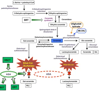
Sulfatides are formed by the action of a series of enzymes, as shown involved in the biosynthesis and degradation of sulfatides in Figure 1. The importance of different myelin components has been investigated in specific mutant murine models with deficiencies in genes encoding the enzymes in the sulfatide biosynthetic pathway. UDP-galactose:ceramide galactosyltransferase (CGT) knockout mice (cgt−/−) were
unable to synthesize the galactosylceramide or sulfatide structures.51 Though the myelin structure seems normal,
SGal-Glc-cer
SGal-Cer (Sulfatide)
HSCT
SRT
ASA ASA
NB-DNJ eliglustat
tartrate
EET ERT
Gal-ceramide Lactosylceramide
Lactosylceramide N-acylsphingosine
galactosyltransferase
Galactosylceramidase glucosyltransferaseCeramide Sphingolipid
delta-4-desaturase 3-Dehydrosphinganine
reductase Serine
palmitoyltransferase
Ceramide synthase 1
Ceramide Ceramide
Gal-ceramide 3-Dehydro-sphinganine L-Serine + palmitoyl-CoA
Glc-ceramide Dihydro-sphingosine
Dihydro-ceramide Ceramidase
Glc
Galactosyl sulfotransferase
Glucosyl ceramidase Beta-1,4-galactosyl
transferase 6 Galactosyl
sulfotransferase
SO42− SO42−
Figure 1 Sulfatide metabolic pathway.
Notes: The figure shows the biosynthetic pathways of sulfatide, the sphingolipid that accumulates during metachromatic leukodystrophy MLD. The specific enzymes described in each step of the pathway are in italics. The rounded boxes depict the products or substrates of the reactions. The green shaded square shapes show different therapeutic approaches for MLD treatment. Two current substrate reduction therapies for another lysosomal disease (Gaucher disease) are also shown: N-Butyldeoxynojirimycin (NB-DNJ) miglustat (Zavesca®, Actelion Pharmaceuticals Ltd, Allschwil, Switzerland), and eliglustat tartrate. Both of these small molecules competitively inhibit ceramide glucosyltransferase. Abbreviations: ASA, arylsulfatase A; EET, enzyme enhancement therapy; ERT, enzyme replacement therapy; Gal, galactose; HSCT, hematopoietic stem cell transplantation; Glc, glucose; SGal-Glc-cer, sulfo-galactose-glucosylceramide; MLD, metachromatic leukodystrophy; SRT, substrate reduction therapy.
Drug Design, Development and Therapy downloaded from https://www.dovepress.com/ by 118.70.13.36 on 22-Aug-2020
Dovepress Development of therapies for MLD
the cgt−/− mice develop progressive hind limb paralysis and
extensive vacuolation of the ventral region of the spinal cord as they age.2,51,52 Thus, these results indicate that the
sulfati-des are important in myelin stability and its physiological function. In the absence of CGT, the cgt−/− mice synthesize
2-OH glucocerebroside as the main component of myelin, a glycosphingolipid that is absent in the normal myelin composition. The 2-OH glucocerebroside gives a seemingly normal structure to myelin, and was hypothesized to prevent the severe effects of the loss of hydroxylated fatty acids such as galactosylceramide.52,53 To validate this hypothesis,
a CGT/FA2H double knockout mouse (cgt−/−/fa2h−/−) was
generated with an additional deletion of the FA2H gene encoding the fatty acid 2-hydroxylase.54 These mice showed
myelin comprised of non-hydroxylated sphingomyelin.54 No
phenotypic differences were observed between cgt−/−/fa2h−/−
and cgt−/− knockout mice.54 Thus, these results suggest that
the presence of glucosylceramide and hydroxylated sphin-gomyelin in cgt−/− mice does not functionally compensate
for the loss of hydroxylated fatty acid galactosylceramide.54
On the other hand, the fa2h−/− mice with intact CGT showed
absence of 2-OH galactosylceramide and sulfatide in normal myelin.48 However, myelin sheath degeneration was observed
with aging.48,55 These studies of different murine knockout
models deprived of specific myelin sphingolipids demonstrate that myelin biogenesis in the nervous system remains intact, likely due to the redundancies of these myelin components. The murine model with ceramide synthase 2 (CerS2) also showed interesting observations with regard to the impor-tance of galactosylceramide and sulfatides in myelin.56 The
CerS2 knockout mice showed decreased levels of galactosyl-ceramide, sulfatide, and very long-chain fatty acids.56 From
early adulthood, the CerS2 null mice showed progressive myelin instability, with 50% loss of the compacted myelin pattern and ∼80% loss of myelin-basic protein.56 In a
differ-ent study of the hepatic biochemical aspects of CerS2-null mice, the biophysical properties of lipid extracts isolated from liver microsomes were shown to be altered, indicating higher membrane fluidity and morphological changes.57
Taken as a whole, these observations on Cer2S knockout mice reiterate the role of the sulfatides in the maintenance of myelin stability, which seems to be of crucial importance during the aging process.
Mechanism of leukodystrophy
In MLD, ASA deficiency causes impairment of sulfatide degradation, and its accumulation mostly in nervous sys-tem (Figure 1). Sulfatide accumulation does not impair
the function of the renal tubular, respiratory epithelium, or endometrium as it does in the CNS and PNS.58 Of note is that
some degree of accumulation of sulfatides in the mucosa of the biliary tract is observed in MLD, resulting in mild to mod-erate clinical manifestations.26–29 This observation is explained
because, physiologically, the levels of sulfatides present in the nervous system are much higher, reflecting its crucial func-tion in the composifunc-tion of myelin. In the arsa−/− MLD mouse
model generated by Hess et al, mild neurological symptoms are only observed by the end of a normal life span.59 As the
arsa−/− mice fail to show sulfatide accumulation, no signs
of demyelination are observed.59 However, the arsa−/− mice
showed neuromotor incoordination, suggesting that such neurological signs might occur before demyelination.59 The
lack of a complete recapitulation of the clinical and pathologi-cal MLD phenotype led to the generation of the arsa−/− mice
with neural cells overexpressing the sulfatide synthesizing enzymes, including UDP-galactose:ceramide galactosyl-transferase (CGT) and cerebroside sulfogalactosyl-transferase (CST).60
These CGT/arsa−/− and CST/arsa−/− mice showed increased
sulfatide storage in myelin-forming cells, resulting in axonal degeneration and development of neurological symptoms similar to MLD.60 The hallmark of the disease is progressive
demyelination with resulting neurological manifestations. The precise pathogenic mechanism of the demyelination is still not clearly understood.43
Pathogenic cascades
In lysosome storage disorders (LSDs), several pathogenic cascades have been described secondary to the substrate accumulation in the lysosomal-endosomal system.38,61,62
These cascades include calcium signaling, altered lipid trafficking, oxidative stress, ER stress/unfolded protein response, autophagy, and inflammation.38 The role of these
different pathogenic cascades in MLD is largely unchar-acterized, but the cascades in some lysosomal disorders presenting with neurodegenerative processes are well studied. Calcium signaling is essential for cell motility and myelination of neurons. Therefore, perturbations of cal-cium signaling are likely to contribute to the demyelination observed in MLD.63,64 Studies in brain tissue from Gaucher
disease patients indicate that defective calcium homeostasis may be a mechanism that leads to neuropathophysiology in the acute and chronic neuronopathic forms of Gaucher disease.65 Studies in mice models of
mucopolysacchari-dosis type III showed that neurodegeneration proceeds independently of the signaling cascade associated with heparin sulfate.66 Signaling cascades may direct cells towards
Drug Design, Development and Therapy downloaded from https://www.dovepress.com/ by 118.70.13.36 on 22-Aug-2020
Dovepress
Patil and Maegawa
autophagy, as observed in a lysosomal disease characterized by neuromuscular presentation known as Pompe disease. This inborn organelle disease is caused by deficiency of lysosomal acid α-glucosidase (GAA). In the fibroblasts from Pompe disease patients carrying c.546G.T mutation in the
GAA gene, the misfolded mutant GAA was shown to induce ER stress, and subsequently elicit autophagy through activa-tion of the p38 mitogen-activated protein kinase pathway.67
Autophagy is the pathway through which a cell can recycle defective proteins in the system. Thus, autophagy is a key cleansing and regenerating pathway, in which products of protein breakdown can make amino acids available to the cell again. Also, autophagy and apoptosis have been shown previously to be inversely related in hematopoietic stem cells.68 In another study in cultured Pompe patient
fibro-blasts, the inactivation of Akt can lead to autophagy, in a ER stress-independent manner, via the suppression of mTOR signaling.69 In the absence of ER stress in Pompe disease
fibroblasts, the inactivated Akt signaling cascade drives the cells to autophagy, which can be reversed with insulin, which activates Akt signaling.69 In Niemann-Pick disease type C
(NPC), sphingosine storage inhibits calcium uptake into the lysosomal acidic compartment, leading to insufficient calcium release for function of the endocytic pathway.70
In this lysosomal disease, the altered lipid trafficking was demonstrated using fluorescent analogues of different lipids. Fluorescent dye-labeled sulfatides were shown to accumulate in lysosomes from fibroblasts obtained from MLD patients.71
Studies have shown evidence that altered chain length of the lipids can affect its trafficking along the endocytic pathway.72
Further, the storage of such metabolites in lysosomes is linked with defective autophagosome-lysosome fusion,73
ultimately resulting in impaired autophagy. The processes of autophagy and apoptosis have been shown to be comple-mentary in some cell types.68 Thus, the impaired autophagy
pathway in lysosomal disorders can lead to apoptosis. Inflam-mation is associated with the development of neurodegen-erative diseases like multiple sulfatase deficiency (MSD).74
MSD is pathogenically similar to MLD, since in both disor-ders sulfatide accumulation is noted. In MSD, inflammation was observed in the mouse model with null mutation in the
SUMF1 gene,75 which encodes the formylglycine-generating
enzyme that is responsible for the oxidation of cysteine and formation of formylglycine (alanine-semialdehyde), the critical catalytic residue present in the active site of eukary-otic sulfatases.76 The sumf1−/− mice demonstrated complete
loss of sulfatase activities and, consequently, early death, growth retardation, and neurological defects.75 These mice
showed inflammatory response characterized by the presence of vacuolated macrophages and activation of microglia in different regions of the brain.75 In sumf1−/− mice between 4
and 6 months of age, elevated expression of inflammatory cytokines and apoptotic markers were observed in the CNS and liver, indicating that inflammation and apoptosis occur later in the disease and play an important role in the neuro-logical phenotype.75 Thus, arresting the disease after these
processes occur becomes very difficult as, by this point in time, permanent and irreversible neural damage is already established in the CNS.77
Challenges in development
of treatment options
Blood-brain barrier
The blood-brain barrier (BBB) controls the migration of components from circulating blood to brain tissue. The BBB is formed by several types of cells, including endothelial cells with tight junctions, pericytes, astrocytes with foot processes, and perivascular neurons. Thus, the arterial capillaries in the CNS form a permeability barrier distinct from any other tissue, which harbors endothelial fenestra-tions between cells. The tight juncfenestra-tions found in the BBB generate high transendothelial electrical resistances of 1,500–2,000 Ω cm2. As a comparison, in tissues other than
brain, the transendothelial electrical resistance ranges from 3–33 Ω cm2.78 Thus, the BBB contributes with the essential
protection of neural structures preventing cellular damage from extra-cellular insults.
However, from the standpoint of the therapeutic devel-opment for inherited neurological disorders such as MLD, the BBB represents “a problem standing before the major problem”. In other words, BBB constitutes the major obstacle to novel therapeutic agents that require reaching the affected neural tissue to treat the neurological manifestations of the disease. Amongst the drugs in the current medicinal chemis-try database, only a few – approximately 5%–6% – cross the BBB and are able to produce CNS effects.79 These are small
molecules with high Log P coefficient of lipophilicity, and molecular weight ranging from 400 to 450 D.79 These drugs
include antipsychotics, antidepressants, and hypnotics.79 The
BBB imposes the main obstacle to delivery of several of the novel therapies developed for MLD, including the recombi-nant enzyme, which is a large molecular weight therapeutic agent (∼52 kD). Additionally, certain cells of hematopoietic origin, including macrophages, activated T-cells, and micro-glial cells, are capable of crossing the BBB and migration to the CNS.80 Thus, both specific strategies, utilizing small
Drug Design, Development and Therapy downloaded from https://www.dovepress.com/ by 118.70.13.36 on 22-Aug-2020
Dovepress Development of therapies for MLD
molecules and specific cells, constitute potential novel avenues to deliver CNS disease-modifying agents for MLD patients.
incomplete understanding
of MLD pathogenesis
Sulfatides account for 4%–6% of the lipids forming the myelin and, as above, are essential sphingolipids in the myelin sheath surrounding the axons. They modulate the differentiation of myelinating cells, and are required for axon-glial interactions. Sulfatides are extracted from the myelin by saposin B (SapB), a heat-stable sphingolipid activator protein that is necessary to solubilize sulfatides and make them accessible to ASA (Figure 1). Consequently, the molecular and clinical manifestations of MLD can also be resultant from defects in SapB, which was the first saposin described.81 The arsa−/− mouse model showed a similar
pattern of accumulation of sulfatides in the CNS and other organs.59 However, the levels of sulfatide accumulation were
lower, which resulted in mild phenotype with no myelin loss until the second year of mouse life, when progressive astro-gliosis and microastro-gliosis in the CNS and peripheral nerves were noticeable.59 In the arsa−/− mouse, reduction of the
levels of membrane raft-associated sulfatide-binding myelin and myelin and lymphocyte (MAL) protein were observed in the oligodendrocyte-generated myelin. This phenomenon indicates the presence of a compensatory mechanism to limit MAL-mediated delivery of sulfatide to the myelin membrane.82 The sulfatide in the mice CNS has a long
half-life (.6 months). Therefore, a short lifespan of the MLD mice model can limit the extent of sulfatide accumulation. How the accumulation of sulfatides causes the disruption of myelin sheath resulting in apoptosis of the myelin-forming cells, as observed in older arsa−/− mice, is still unclear.59 In
ultrastructural transmission electron microscopy studies of neurons from the arsa−/− mice, several MCBs were
notice-able, indicating that sulfatides are likely accumulated in these cells.83 The accumulation of sulfatides is possible
in oligodendrocytes and Schwann cells of affected MLD patients, resulting in failure to produce and maintain myelin in axons located in the CNS and PNS, respectively. Thus, the complete inhibition of sulfatide biosynthesis is not an option in treatment, considering its importance as a myelin component. In addition, the biological effects of sulfatide accumulation in neural cells other than oligodendrocytes and Schwann cells are still unknown.83 The arsa−/− mouse
model of MLD showed incoordination signs in the absence of pathological signs of demyelination59, alluding that other
pathogenic process may occur that can result in neurological symptoms, beyond demyelination.83 Therefore, a better
understanding of the molecular pathogenesis of MLD is a substantial challenge in developing therapies for MLD.
Time of intervention in the course
of disease
In case of MLD, as in others lysosomal disorders, the classifi-cation of patients in clinical forms such as late infantile, juve-nile, and adult is based on the age of onset of first symptoms.58
In general, the earlier the onset of the disease, the more rapid is the disease progression. The different treatment options, though currently limited, can produce a better outcome when started in the earlier stages of the disease.84 Generally, the
ear-lier the intervention, the better the responses to that specific therapy observed. The importance of earlier intervention can be noted in MLD patients who underwent hematopoietic stem cell transplantation (HSCT).85 MLD patients who received
HSCT earlier usually had a better outcome than those trans-planted at later stages of the disease. This can considerably increase an already high risk of mortality strictly related to the procedure.85–89 Some studies have shown that lysosomal
storage in neuronal cells can be reduced to some extent in different LSDs. However, the cellular changes occurring downstream of storage are usually not reversible.90 Thus, it
is necessary to arrest such a primary cellular event at earlier stages of the natural history of the disease. Hence, the time of intervention becomes an important parameter in determining the prognosis and outcome.
Current treatments for MLD
Since MLD is caused by defective ASA, most therapeutic approaches have tried to correct the biochemical defect by providing wild-type ASA (Figure 1). The different methods and sources of wild-type ASA constitute distinct therapeutic approaches. Enzyme replacement therapy (ERT) and HSCT rely on providing normal ASA to the deficient cells, while gene therapy approaches are based on the overexpression of wild-type ASA in different cell types (Figure 1).
Enzyme replacement therapy
ERT has been a therapeutic modality used with remarkable success in treating some LSDs. Its applicability to MLD is challenged by the BBB. Lysosomal ASA is a high molecu-lar weight therapeutic agent, and consequently is unable to penetrate the BBB to reach the affected brain tissue. In mice models, some studies have shown positive results,91 leading
to early phase clinical trials in MLD-affected patients. The
Drug Design, Development and Therapy downloaded from https://www.dovepress.com/ by 118.70.13.36 on 22-Aug-2020
Dovepress
Patil and Maegawa
efficacy of this treatment modality is inversely related to the age of mice.92 The ERT approach also faces the difficulty
of producing the recombinant enzyme at a large scale and in high purity, which has been overcome already by several pharmaceuticals.93 In this setting, the BBB limitation has
been addressed with different routes of administration, including intracerebroventricular or intrathecal ERT agent delivery. These approaches have been carefully evaluated as modes of delivery for ERT. The current ongoing clinical trials using Metazym, (Shire Human Genetics Therapeutics, Inc., Lexington, MA, USA) administered intrathecally as an ERT agent, are listed in Table 1. The final results of these clinical studies will be important to determine the efficacy of this mode of ERT delivery for MLD and other lysosomal diseases.
Hematopoietic stem cell therapy
As described earlier, the BBB prevents the delivery of ASA enzyme to the affected neural cells. This sets the scene for the development of various strategies that focus on overcoming the BBB and making the enzyme available to CNS cells. HSCT is one such approach, where hematopoi-etic stem cells (HSCs) cross the BBB and differentiate to microglial cells.94 In principle, the donor microglial cells
are able to secrete the wild-type ASA enzyme, which is uptaken by the recipient neural cells that are ASA-deficient. Uptake of the secreted wild-type ASA occurs through the mannose-6-phosphate receptor system, which brings the enzyme directly to the lysosomes, where sulfatide is accumulated in the recipient ASA-deficient neural cells.95
One of the limitations of HSCT is the rate of repopulation of microglia into the recipient CNS, which should be a sufficient cell number to provide an adequate amount of wild-type ASA for the affected neural cells of the recipient. HSCT was shown to be efficacious in the treatment of MLD as early as 1990.84 However, HSCT failed to show efficacy
in the late infantile clinical form of MLD.96 Though HSCT
is a well-established therapeutic approach for a few LSDs, including mucopolysaccharidosis type I and globoid-cell leukodystrophy (Krabbe disease), this treatment modality is limited only to MLD patients who are diagnosed at earlier stages of the disease course (Table 1).97–99 HSCT brings
other potential factors to be considered, such as identifi-cation of matched related donors, the rate of repopulation of microglial cells, along with specific transplant-related complications including acute and chronic graft-versus host diseases, and the level of engraftment.100,101 An alternative
approach to HSCT is using umbilical cord blood as a source of hematopoietic and mesenchymal stem cells, as reviewed elsewhere.102 Recently, the outcomes of 27 patients with
different clinical forms of MLD were reported.85 In this
study, an inverse relationship between time of intervention and outcome was observed. In addition, children with the juvenile form of MLD showed better outcomes than those with late infantile onset.85
Gene therapy
Types of gene therapies have been studied for the treatment of MLD. Autologous HSCs obtained from the affected MLD patient can be genetically modified to overexpress the
ARSA gene using retrovirus gene transfer.103 This approach
has been shown to be highly successful in the arsa−/− mice
model, as it prevents the development of motor conduc-tion impairment, learning and coordinaconduc-tion deficits, and neuropathological abnormalities typical of the disease.104
Thus, genetically modified HSCs can help prevent neurode-generation or slow its rate of progression. The synthesized wild-type ASA secreted by these genetically modified HSCs is uptaken by the adjacent ASA-deficient cells via the mannose-6-phosphate receptor system.95 The overexpression
of ARSA gene by the autologous HSCs before transplant is performed using retro- or lentivirus vectors.105,106 Different
studies using this strategy have been discussed elsewhere.103
The advantage of this therapeutic approach is the reduction in risk of graft-versus host disease. The major limitation of this approach is safety consideration, because of unknown risk of mutagenesis of cancer-related genes or oncogenes.107
Currently, a phase I/II clinical study is ongoing with MLD
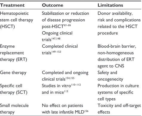
Table 1 Current therapeutic modalities in metachromatic leukodystrophy (MLD)
Treatment Outcome Limitations
Hematopoietic stem cell therapy (HSCT)
Stabilization or reduction
of disease progression post-HSCT97–99
Ongoing clinical trials147,148
Donor availability, risk and complications related to the HSCT procedure
Enzyme
replacement therapy (eRT)
Completed clinical trials149–153
Blood-brain barrier, non-homogeneous distribution of eRT agent to CNS Gene therapy Completed and ongoing
clinical trials154,155
Safety and oncogenecity
Specific cell
therapy (SCT)
Studies in vitro110–112
and in mice113
Production in culture
systems of specific
cell types Small molecule
therapy
No effect on patients with late infantile MLD156
Toxicity and off-target effects
Abbreviation: CNS, central nervous system.
Drug Design, Development and Therapy downloaded from https://www.dovepress.com/ by 118.70.13.36 on 22-Aug-2020
Dovepress Development of therapies for MLD
patients receiving autologous HSCT with genetically modified HSCs (Table 1).
Specific cell therapy
In addition to HSCT, various other cell-based approaches can be used to deliver ASA to affected cells.102 Briefly,
microencapsulated recombinant cells that overexpress the
ARSA gene can be directly delivered. These cells offer substantial flexibility in terms of the dosage and control of delivery, since they can be easily retracted.108,109 Similarly,
oligodendrocyte progenitor cells110 and neural progenitor
cells111,112 are other cell types that can be used for
cell-based therapy. Embryonic stem cells constitute another cell source for ARSA overexpression and delivery. Studies in the
arsa−/− mice, using embryonic stem cells, have demonstrated
significant decreases in sulfatide accumulation.113 These
specific cell therapies are attractive options for the treatment of neurological manifestations of MLD. All these studies have demonstrated considerable clearance of the storage of sulfatides in animal models.111–113 However, their efficacy
and (of utmost importance) safety and tolerability are still to be demonstrated in humans.
Potential novel therapies
Small molecule-based therapies
In MLD, no specific treatment has been shown to completely arrest disease progression. Therapies using small molecules (in general, ,500 D) have hardly been explored in MLD. Small molecule-based therapies have shown encouraging results in the case of some lysosomal disorders like Gaucher disease and NPC. N-butyldeoxynojirimycin (NB-DNJ) miglustat (Zavesca®, Actelion Pharmaceuticals Ltd, Allschwil,
Switzerland) was showed to reduce the biosynthesis of glu-cosylceramide by competitively inhibiting the UDP-glucose-N-acysphingosine D-glucosyltransferase (EC 2.4.1.80) or ceramide-specific glucosyltransferase.114 The mechanism of
action of miglustat was shown using an in cellulo model of Gaucher disease.115 In studies using cultured human
mac-rophages pre-exposed with conduritol-β-epoxide (CBE), miglustat was shown to reduce glucosylceramide storage, which is the main substrate accumulated in Gaucher disease.115
CBE is an irreversible inhibitor of glucocerobrosidase, leading to accumulation of glucosylceramide in cultured cells.116,117
Ceramide-specific glucosyltransferase is the key enzyme on the biosynthetic pathway of several glycosphingolipids, including the ganglio- series (acidic glycosphingolipids), lacto(neo)- series (neutral glycosphingolipids), and globo- series (Figure 1).118 Based on this principle, miglustat was
tested in the murine model for Tay-Sachs disease (hexa−/−),
and was showed to reduce the accumulation of GM2 ganglio-side.119 However, the hexa−/− mouse lacks an overt Tay-Sachs
disease phenotype, as mice can partially bypass the blocked catabolic pathway and escape disease.119 In an early clinical
trial with patients with the juvenile clinical form of GM2 gan-gliosidosis, miglustat showed adequate pharmacokinetics and safety profiles,120 but failed to arrest some aspects of
disease progression.121 However, patients affected by NPC,
a lysosomal disorder resulting in intra-cellular lipid traffick-ing defect and secondary glycosphtraffick-ingolipid storage, showed improvement and stabilization of a number of clinical end-points, including swallowing capacity and ambulatory index, when receiving miglustat in a clinical trial.122
Small molecule-based therapy can potentially overcome limitations of the current therapies for MLD and may also tackle, at multiple levels, different pathogenic and molecular mechanisms involved in MLD. Small molecules can eas-ily cross the BBB, which is a major hindrance in the ERT strategy. Warfarin (coumadin) is a classical anti-coagulant that functions by competitively inhibiting the multi-unit vitamin K epoxide reductase complex, resulting in impair-ment of the activation of vitamin K-dependent coagulation factors II, VII, IX, and X. Based on microorganism studies in vitamin K, warfarin administration in mice showed to reduce the levels of sulfatides by 33%, along with cerebro-sides and sphingomyelin in the brain.123 Warfarin was then
hypothesized as a potential substrate-reducing agent to be explored therapeutically for MLD. Recently, a clinical trial in a small cohort of MLD patients tested whether warfarin would control the progression of MLD.124 During a 45-day
treatment with warfarin at doses of 0.2 mg/kg/day, adjusted to maintain INR within the range 2–2.5, no beneficial effect of this small molecule was observed in four enrolled patients with the late infantile form of MLD (Table 1).124 The urinary
sulfatide levels of these patients remained elevated. Brain magnetic resonance spectroscopy showed that the levels of N-acetyl-aspartate and myo-Inositol remained unchanged after the treatment period.124
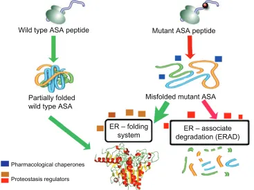
Small molecules as enzyme
enhancement agents
Lysosomal storage disorders can be caused by missense mutations in specific genes that encode misfolded mutant lysosomal enzymes (Figure 2). Most of these misfolded mutant lysosomal enzymes are earlier degraded in the ERAD pathways125 and, consequently, a very small amount of the
mutant enzyme reaches the lysosomal compartment and is
Drug Design, Development and Therapy downloaded from https://www.dovepress.com/ by 118.70.13.36 on 22-Aug-2020
Dovepress
Patil and Maegawa
functionally active. The resultant residual enzymatic activ-ity, in the case of ASA mutants, ranges between 0%–10% of the normal activity detected from the wild-type ASA.126
This ASA enzymatic activity, above which the activity is sufficient to degrade the sulfatides and prevent their accu-mulation, is known as the critical threshold – which is ∼10% of that measured from the wild-type ASA.126 Interestingly,
the levels of ASA enzymatic activity of the pseudodeficiency
ARSA alleles are just above the critical threshold. Therefore, in ASA pseudodeficiency, the levels of enzyme activity between 10%–15% of wild-type ASA levels result in no sulfatide accumulation, and consequently no disease-related symptoms of MLD.126
Specific small molecules are able to rescue misfolded proteins. These molecules are able to physically interact with a specific misfolded protein, assist its folding process, and prevent it from being targeted to the ERAD pathway and ultimately degraded by the ubiquitin-proteasomal system
(Figure 2).127 Thus, by evading the ERAD pathway, and
sub-sequently enhancing the level of the available mutant ASA to the lysosomal compartment, the residual ASA enzymatic activity will be increased and potentially overcome the 10% critical threshold.126 In this regard, the ER folding
system, with its numerous ER-resident chaperones, plays an important physiological role in promoting adequate and functional folding of several lysosomal enzymes, including ASA. The ER-resident chaperones are proteins that assist the folding or unfolding of nascent proteins synthesized in the ER that are not able to do so spontaneously.127 Similarly,
pharmacological chaperones (PCs) are small molecules that function like chaperones, enhancing the levels of the misfolded-prone mutant enzymes by assisting their ER folding and preventing their interaction with the ERAD pathway and subsequent degradation.128–130 Small molecules
functioning as PCs were identified for misfolded mutant hexosaminidase A, the lysosomal enzyme deficient in
Wild type ASA peptide
Partially folded wild type ASA
ER – folding
system degradation (ERAD)ER – associate
Pharmacological chaperones
Proteostasis regulators
Mutant ASA peptide
Misfolded mutant ASA
Figure 2 Schematic representation of the mechanism of action of enzyme enhancement agents including pharmacological chaperones (PCs) and proteostasis regulators (PRs).
Notes: ASA is synthesized in the endoplasmic reticulum (ER) as several other proteins target to different intracellular organelles, plasma membrane, and those destined to be secreted to the extracellular environment. Concomitant to the translation process, nascent ASA peptide interacts with several components of the eR folding system. Once folded properly, ASA is targeted to the eR exit pathway and ultimately reaches the lysosomal compartment, where it exerts its physiological function. Missense mutations in the ARSA gene can affect physicochemical interactions between amino acids, resulting in impairment of the folding process of the mutant ASA. PCs (blue boxes) are small molecules that physically interact with the target protein, in this case ASA, and assist its folding, preventing it from being targeted to the eRAD pathway and ultimately degraded by the ubiquitin-proteasomal system. Thus, increased levels of mutant ASA reach the lysosomes to degrade the natural substrate (sulfatides). eventually, other small molecules function as PRs (beige/brown boxes), which can improve the cellular folding capacity by interacting with eR folding components. These molecules can also promote physiological cellular responses, such as unfolded protein response, which can be advantageous to enhance folding of specific misfolded ASA mutants. Other PRs (red boxes) can also potentially inhibit components of eRAD and favor the interactions of misfolded ASA mutants with the eR folding elements, increasing the probability of reaching a folding-like conformation. Abbreviations: ASA, arylsulfatase A; eR, endoplasmic reticulum; eRAD, endoplasmic reticulum associate degradation; PC, pharmacological chaperones; PR, proteostasis regulators.
Drug Design, Development and Therapy downloaded from https://www.dovepress.com/ by 118.70.13.36 on 22-Aug-2020
Dovepress Development of therapies for MLD
GM2 gangliosidosis,128 and for misfolded mutant
gluco-cerebrosidase (Glc-ase), the lysosomal enzyme deficient in Gaucher disease.129,131 In addition to PCs, proteostasis
regulators (PRs) are small molecules that improve the protein folding capacity of cells, resulting in the enhance-ment of folded mutant proteins.132 PRs work by interacting
with the ER folding components and eventually activating physiological cellular response mechanisms, including unfolded protein response and heat shock proteins (Fig-ure 2). PRs can also function by inhibiting the degradation of the misfolded mutant enzyme, by interacting with the ERAD components or HSC molecules (Figure 2).133–135 The
effect of PRs can be enhanced by simultaneously using PCs for assisting the folding of misfolded-prone mutant enzymes.127 Interestingly, these two classes of small
mol-ecules can work together synergistically to guide the mis-folded mutant enzyme to a mis-folded state (assisted by PRs) and maintain its structural stability (assisted by PCs).136 Some
PRs have been identified for specific lysosomal mutant enzymes.136,137 Inhibitors of histone deacetylase (HDAC)
are showed to reduce the acetylation of heat shock protein 90 (Hsp90)138. Since Hsp90 plays an important role in the
degradation pathway of the misfolded N370S and L444P Glc-ase mutants, HCAD inhibitors resulted in increased levels of mutant Glc-ase enzymatic activity.138 Thus, these
inhibitors act as PRs by preventing the degradation of mutant enzymes while interacting with components of the degradation pathway. MG-132 and celastrol are other examples of small molecules that were shown to function as PRs for some mutant Glc-ase and Hex A enzymes.136 PRs
can be potentially applied to a range of LSDs, since these small molecules target the cellular enzyme folding pathway and their effect can be enhanced using PCs. These specific small molecule-based therapies can work in conjunction with ERT by interacting with and, consequently, stabilizing the administered recombinant ASA, extending its half-life in either extra or intracellular compartments.
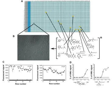
How can these therapeutic small molecules be identi-fied? Small molecules that work as PCs or PRs for specific lysosomal mutant enzymes can be identified using high throughput screening (HTS) assays. Recently, the use of cell-based assays in the HTS of chemical compound collections has become increasingly common, as targets are presented in a more biologically relevant context than classical in vitro biochemical-based HTS assays.139 In cell-based HTS assays,
the identification of small molecules that penetrate cellular membranes and reach specific cellular organelles, along with their cytotoxicity information, can already be obtained at the
primary screening stage.140 However, in most cellular HTS
assays, the cells utilized are usually not disease relevant. Thus, using patient-derived cell lines for HTS campaigns brings considerable advantages, compared to the traditional, single-target biochemical assays (Figure 3).141,142 Cellular
assays utilizing cells from affected patients provide a unique opportunity to assess a target protein and/or pathway in the context of potentially disrupted biochemical and/or signal-ing pathways secondary to the disease process. In addition, this pathogenic in cellulo setting permits the evaluation of multiple intervention points, which are potentially altered in the disease, as opposed to the commonly-used cell expres-sion systems of a specific protein target, or a predefined step of a purified or recombinant protein-based assay (Figure 3). Therefore, cell-based HTS can identify both PCs and PRs.141,142 A cell-based HTS assay was developed to identify
small molecules for mutant ASAs.141 Using a transformed
fibroblast cell line from a MLD patient with measurable residual ASA activity, a fluorescence quench absorbance ASA assay was developed. This HTS assay showed robust signals in diverse multi-well plates, including 1,536-well plates.141
The implementation of the cell-based HTS assay against larger and diverse small molecule libraries should allow the identification of novel potential drugs for MLD. This and other cell-based HTS assays open a novel opportunity to identify numerous small molecules, as several potential therapeutic targets are accessible in this disease-cellular environment.141,142
Small molecules as substrate
reduction agents
Some small molecules can also have their point of action upstream to the biochemical defect: ie, sulfatide accumu-lation in MLD (Figure 1). Miglustat is an example of a substrate reduction therapy (SRT) agent that functions as a competitive inhibitor of ceramide-specific transferase.118
Similarly, the sulfatide synthesis pathway can be potentially targeted for substrate (sulfatide) reduction at multiple points in the pathway, as seen in Figure 1. Specifically, ceramide synthase can be targeted using small molecules, which would lead to reduction of the biosynthesis of sulfatides (Figure 1). Reducing ceramide synthase catalytic activity can result in the reduction of further accumulation of sulfatides. However, complete arrest of sulfatide biosynthesis may cause dis-ruption of myelin stability, as shown in different murine models described above.48,51,54 Thus, physiological levels of
sulfatides are crucial, as these are main glycosphingolipids of the myelin sheath. Hence, the optimality of therapeutic
Drug Design, Development and Therapy downloaded from https://www.dovepress.com/ by 118.70.13.36 on 22-Aug-2020
Dovepress
Patil and Maegawa
dose of potential small molecules functioning as SRT agents is a prerequisite for the success of this therapeutic strategy. Therefore, SRT agent doses should be determined by the molecule concentrations capable of partially inhibiting the catalytic activity of a target enzyme in the biosynthetic pathway of sulfatides at intracellular levels (ie, in oligo-dendrocytes and Schwann cells) (Figure 1). This will be crucial to avoid potential adverse events of SRT agents in the treatment of MLD.
Small molecules to control
pathogenic cascades
Small molecules can also be used to manipulate downstream pathways that are initiated or disturbed secondary to the primary lysosomal enzymatic defect. Diverse pathways have been described to be impaired in lysosomal storage diseases. These are known as pathogenic cascades, which
are characterized by disturbances in molecular signaling pathways occurring secondary to sulfatide accumulation, ultimately leading to apoptosis.
In a few lysosomal disorders, some small molecules have been identified to target specific pathogenic cascades. In stud-ies of NPC patient fibroblasts and Npc1−/− mice, sphingosine
storage was shown to precede low calcium levels in the lyso-somal compartment, which explains the profound block in late endosome to lysosome transport, resulting in the storage of cholesterol and glycosphingolipids – the cellular hallmark of this lysosomal disorder.70 In the presence of curcumin, a
small molecule that is a weak antagonist of the sarco (endo) plasmic reticulum ATPase (SERCA), the levels of calcium in the acidic compartments were normalized and clearance of lipid storage was noted, resulting in increased Npc1−/− life
expectancy.70 SERCA is a pump that transports calcium ions
from the cytoplasm into the sarco(endo)plasmic reticulum.143
6000 5000 4000 3000 2000 1000 0
0 10 20 30 4.6 µM Row number Fluorescent signal 6000 5000 4000 3000 2000 1000 0
0 10 20 30
23 µM
Row number
Fluorescent signal −5.0 −4.5 −4.0 −3.5
Log [ −5.5 −10 0 10 20 30 40 50 60 70 80
% of HMU signal
−3.5 −4.0 −4.5 −5.0 −5.5 −10 0 10 20 30 40 50 60 70 80
% of HMU signal
] M Log [ ] M
O N H N H H N O O O OH CI NO2 CH3 CH3 CH3 HCI • H3C
H3C HO
HO OHHN
O O H N N H NH
NH O O HO OHOH OH OH OH N N N H N H N H H N H N N HCI CH3 CH3 CI CI N H N NH NH2 NH2 NO2 OH O NH2 OCH3 N N N N H
HO CH CHCH2CH3
H2N
CI O
H3C CH3 O O
CH3 CH3
HCI H3CN
H CH3SO3H
NHCH(CH3)2
O O O HCI CI • • • HCI • 1 1 2 2 3 3 4 4 5 5 6 6 7 7 8 8 9 9 10 10 11 11
12 13 14 15 16 17 18 19 20 21 22
12 13 14 15 16 17 18 19 20 21 22
23 24 25 26 27 28 29 30 31 32 33
23 24 25 26 27 28 29 30 31 32
34 35 36 37 38 39 40 41 42 43 44 45 46 47 48 A
B
C
n
D
Figure 3 Cell-based high-throughput screening (HTS) assay.
Notes: Here, a cell-based HTS assay for galactocerebrosidase (GALC), which is the lysosomal enzyme deficient in Krabbe disease or globoid cell leukodystrophy (GLD), is depicted for (A and B). The 1,536-well plate contains GLD patient cells that are cultured and treated with a small molecule collection for a specific period of time. After the treatment period, the specific enzyme GALC can be measured using a fluorescent substrate, 6-hexadecanoylamino-4-methylumbelliferyl-β-D-galactoside (HMU) for the enzyme. In each 1,536-well plate, control cells (columns 2 and 3) are seeded and later assayed. These control cells have normal Gal-ase enzymatic activity and are not treated with the library. The columns 1 and 2 contain only culture medium and no cells, and are used as backgrounds for the assays. Columns 5–48 contain cultured patients cells (B) – in this case, fibroblasts. Each well (yellow square) corresponds to a specific small molecule from the library, which is tested in different concentrations. (C) Two-plate view diagrams are shown with the fluorescent signals generated from the HMU against the different rows of a 1,536-well plate. Substantial assay window is observed between GALC enzymatic activity of controls (triangles) and GLD patient cells (circles). The small molecule candidates are selected based on the enhancement of the residual GALC enzyme activity. (D) The quantitative nature of the HTS assay, where seven different concentrations of the target library were tested, allows the generation of concentration-response curves (CRCs) given by the HMU fluorescence signal increase. Based on the characteristics of the CRCs,146 prioritization of “hits” can be done during the validation process with secondary and tertiary assays.
Drug Design, Development and Therapy downloaded from https://www.dovepress.com/ by 118.70.13.36 on 22-Aug-2020
Dovepress Development of therapies for MLD
The inflammatory cascades can be targeted using non-steroidal and anti-inflammatory drugs, which have resulted in clini-cal benefits in other lysosomal diseases, including Sandhoff disease144 and NPC145. Most of these small molecules have
already been characterized to interact with specific molecular targets. To discover further small molecules, cell-based HTS assays can be developed using as the readout signal a specific relevant target of a pathogenic cascade related to ASA defi-ciency. These HTS assays will identify potential therapeutic small molecules to tackle signaling pathways that are disturbed secondary to the ASA deficiency. These small molecules may produce synergistic effects along with the therapeutic modali-ties that tackle primarily the ASA deficiency, such as ERT, gene therapy, and cell-based therapies. By using combinations of therapies to specific targets, it is more likely that the arrest or even reversal of disease progression can be achieved.
Conclusion
Metachromatic leukodystrophy is a lysosomal disorder causing progressive neurological disability secondary to the accumulation of sulfatides and the resultant demyelination process that affects both CNS and PNS. The range of age of onset of symptoms and rate of disease progression are broad. The available therapeutic options are limited. In developing novel therapeutic approaches for MLD, BBB penetration, lack of a better understanding of the disease pathogenesis, uncertainty regarding the ideal time for intervention in the disease course, and safety issues related to specific thera-pies are the main challenges. Diverse therapeutic strategies have been developed and are now moving to clinical trials including cell-based therapies, gene therapy, ERT, and small molecule-based therapies. Most of these therapies tackle specific pathogenic aspects of the disease. Some of these strategies may result in better outcomes at specific stages of the natural disease course. These diverse strategies comple-ment each other, either when applied in combination or in a stepwise manner. The determination of the more appropriate combinations and times of administration of these therapies in the disease course will certainly produce the best out-comes for patients afflicted with this devastating inherited neurological disorder.
Acknowledgments
We are grateful for our collaborators including Marc Ferrer PhD and his group at the NIH-Chemical Genomic Center (NCGC) for the instrumental assistance in the development of high-throughput screening cell-based assays targeting different lysosomal enzymes. We are also thankful for our
collaborators who donated cell lines including Dr Hirokazu Furuya MD, PhD, neurologist from NHO Omuta Hospital, Omuta, Fukuoka, Japan.
Disclosure
Shilpa Patil is partially supported by National MPS Society. Gustavo Maegawa is currently supported by grants NIH-NIMH 1R03MH098689 and NIH-NINDS 1R01NS079655. Dr Maegawa also receives research grants from the National MPS Society and received research grants from the National Tay-Sachs Disease and Allied Diseases Association. Dr Maegawa receives research funds for clini-cal and educational studies from Genzyme Therapeutics Ltd, Biomarin Pharmaceutical Inc, Protalix Biotherapeutics Inc, and Shire HGT. The authors declare no other conflicts of interest in this work.
References
1. Austin J, McAfee D, Armstrong D, O’Rourke M, Shearer L, Bachhawat B. Low sulfatase activities in metachromatic leukodystro-phy (MLD). A controlled study of enzymes in 9 living and 4 autopsied patients with MLD. Trans Am Neurol Assoc. 1964;89:147–150. 2. Bosio A, Binczek E, Stoffel W. Functional breakdown of the lipid bilayer
of the myelin membrane in central and peripheral nervous system by dis-rupted galactocerebroside synthesis. Proc Natl Acad Sci U S A. 1996;12; 93(23):13280–13285.
3. Aggarwal S, Yurlova L, Simons M. Central nervous system myelin: struc-ture, synthesis and assembly. Trends Cell Biol. 2011;21(10): 585–593. 4. Faust PL, Kaye EM, Powers JM. Myelin lesions associated with lyso-somal and peroxilyso-somal disorders. Expert Rev Neurother. 2010;10(9): 1449–1466.
5. Gieselmann V. Metachromatic leukodystrophy: genetics, patho-genesis and therapeutic options. Acta Paediatr Suppl. 2008; 97(457):15–21.
6. Mechtler TP, Stary S, Metz TF, et al. Neonatal screening for lysosomal storage disorders: feasibility and incidence from a nationwide study in Austria. Lancet. 2012;379(9813):335–341.
7. Meikle PJ, Hopwood JJ, Clague AE, Carey WF. Prevalence of lysosomal storage disorders. JAMA. Jan 1999;281(3):249–254.
8. Von Figura K. Metachromatic Leukodystrophy. In: Scriver C, Beaudet A, Arthur L, Sly WS, Valle D, editors. Metabolic and Molecular Basis of
Inherited Disease. New York: McGraw-Hill; 2001;3:3696–3724.
9. Lugowska A, Poninska J, Krajewski P, Broda G, Ploski R. Population carrier rates of pathogenic ARSA gene mutations: is metachromatic leukodystrophy underdiagnosed? PLoS One. 2011;6(6):e20218. 10. Stenson PD, Ball EV, Howells K, Phillips AD, Mort M, Cooper DN.
The Human Gene Mutation Database: providing a comprehensive central mutation database for molecular diagnostics and personalized genomics. Hum Genomics. 2009;4(2):69–72.
11. Gieselmann V. An assay for the rapid detection of the arylsul-fatase A pseudodeficiency allele facilitates diagnosis and genetic counseling for metachromatic leukodystrophy. Hum Genet. 1991;86(3): 251–255.
12. Gieselmann V, Polten A, Kreysing J, von Figura K. Molecular genetics of metachromatic leukodystrophy. J Inherit Metab Dis. 1994;17(4): 500–509.
13. Gort L, Coll MJ, Chabas A. Identification of 12 novel mutations and two new polymorphisms in the arylsulfatase A gene: haplotype and genotype-phenotype correlation studies in Spanish metachromatic leukodystrophy patients. Hum Mutat. 1999;14(3):240–248.
Drug Design, Development and Therapy downloaded from https://www.dovepress.com/ by 118.70.13.36 on 22-Aug-2020