Clinical and Experimental Gastroenterology
Dove
press
O r i G i n a l r E s E a r C h
open access to scientific and medical research
Open access Full Text article
application of colon capsule endoscopy (CCE)
to evaluate the whole gastrointestinal tract:
a comparative study of single-camera
and dual-camera analysis
José María remes-Troche1
Victoria alejandra Jiménez-García2
Josefa María García-Montes2
Pedro hergueta-Delgado2
Federico roesch-Dietlen1
Juan Manuel herrerías-Gutiérrez2
1Digestive Physiology and Motility lab, Medical Biological research institute, Universidad Veracruzana, Veracruz, México; 2Gastroenterology service, Virgen Macarena University hospital, seville, spain
Correspondence: Juan Manuel herrerías-Gutierrez
Gastroenterology service, Virgen Macarena University hospital, avda Dr. Fedriani, s/n41071 sevilla, spain Tel +34 95 500 88 01
Fax +34 95 500 88 05 Email [email protected]
Background and study aims: Colon capsule endoscopy (CCE) was developed for the evaluation of colorectal pathology. In this study, our aim was to assess if a dual-camera analy-sis using CCE allows better evaluation of the whole gastrointestinal (GI) tract compared to a single-camera analysis.
Patients and methods: We included 21 patients (12 males, mean age 56.20 years) submitted for a CCE examination. After standard colon preparation, the colon capsule endoscope (PillCam Colon™) was swallowed after reinitiation from its “sleep” mode. Four physicians performed the analysis: two reviewed both video streams at the same time (dual-camera analysis); one analyzed images from one side of the device (“camera 1”); and the other reviewed the opposite side (“camera 2”). We compared numbers of findings from different parts of the entire GI tract and level of agreement among reviewers.
Results: A complete evaluation of the GI tract was possible in all patients. Dual-camera analysis provided 16% and 5% more findings compared to camera 1 and camera 2 analysis, respectively. Overall agreement was 62.7% (kappa = 0.44, 95% CI: 0.373–0.510). Esophageal (kappa = 0.611) and colorectal (kappa = 0.595) findings had a good level of agreement, while small bowel (kappa = 0.405) showed moderate agreement.
Conclusion: The use of dual-camera analysis with CCE for the evaluation of the GI tract is feasible and detects more abnormalities when compared with single-camera analysis. Keywords: capsule endoscopy, colon, gastrointestinal tract, small bowel
Introduction
Capsule endoscopy (CE) has been established as an extremely useful diagnostic tool for the evaluation of obscure gastrointestinal bleeding (OGIB) and other small-bowel conditions.1,2 Currently, CE is approved in adult and pediatric patients older than 10 years for evaluation of OGIB,3,4 celiac disease,5 polyposis syndromes,6,7 and small-bowel abnormalities detected in imaging studies.8–10
This technology has evolved over the last decade, and new and better devices are available, such as capsules with two lenses and cameras and those capable of capturing more frames per second.2,11 The PillCam ESO™ esophageal capsule (Given Imaging, Yoqneam, Israel), for example, has two cameras, each with the capability of taking images at seven frames per second.12,13 This device has been approved for screening and follow-up of esophageal varices and screening for Barrett’s esophagus.12,14,15 More recently, a colon capsule endoscope (PillCam Colon™; Given Imaging) has been
Clinical and Experimental Gastroenterology downloaded from https://www.dovepress.com/ by 118.70.13.36 on 20-Aug-2020
For personal use only.
Number of times this article has been viewed
This article was published in the following Dove Press journal: Clinical and Experimental Gastroenterology
Dovepress
remes-Troche et al
developed.16 The colon capsule is similar to the conventional capsule, but has two cameras that are able to record video images from both ends.17 The device measures 31 × 11 mm and acquires images at a rate of four frames per second. The preprogrammed “sleep” mode allows recording of images from the esophagus and stomach for 3 minutes, after which the capsule switches to sleep mode for 1 hour 45 minutes. During this period, the capsule is likely to transit most of the small bowel; thus, by the time the capsule “wakes up” from its sleep period, it will have reached approximately the terminal ileum. The preprogrammed sleep mode saves battery, thereby allowing an almost complete evaluation of the colon.
Several studies have pointed out the effectiveness of the PillCam Colon™ capsule endoscope compared to colonoscopy for the study of colon diseases.18–20 The high-priority objective of colon capsule endoscopy (CCE) is screening for colorectal cancer in at-risk populations.21 CCE can be performed when there is a contraindication for colonoscopy, and it is suitable for patients who are unwilling to undergo colonoscopy or to complete a failed colonoscopy. It can also be used for follow-up of condi-tions different to adenomas or polyps, such as ulcerative colitis (UC).22
Although CCE was developed for evaluation of the colon, this dual device can also be used to assess the entire GI tract if the patient takes the capsule after early activation and as soon as the capsule recovers from its sleep mode. In a previous study, Almeida et al23 performed a study in which the colon capsule was given after the device woke up from sleep mode to ten patients for small-bowel evaluation. In this study, the authors found that enteroscopy with a double-camera device allowed the detection of more findings and increased diagnostic accuracy when compared to a single-camera analysis.23
In our study, our aim was to determine if the use of CCE and a dual-camera analysis allows a better evaluation of the entire gastrointestinal (GI) tract when compared to a single-camera analysis. In addition, we also analyzed the level of agreement between double-camera and single-camera analysis.
Patients and methods
Patients
This prospective, exploratory, and pilot study was conducted between September 2007 and October 2009 at the Hospital Virgen de la Macarena, Seville, Spain. Twenty-one (12 male, mean age 56.20 years, range 23–97 years) consecutive
Hispanic patients submitted for a PillCam Colon™ examina-tion were included. These patients were submitted for CCE for: screening to rule out organic diseases (n = 10); follow-up of inflammatory bowel disease ([IBD] eg, Crohn’s or UC history [n = 5]); chronic anemia (n = 3); chronic abdominal pain (n = 3); and chronic diarrhea (n = 1). Subjects were invited to participate, and those who agreed gave their written informed consent for the procedure and the protocol analysis. This protocol was approved by the internal ethics committee of the Hospital Virgen de la Macarena.
study protocol
small-bowel and colon preparation
All patients underwent a 48-hour standard colon prepara-tion for the colon capsule procedure previously described by Herrerías-Gutiérrez et al.17 Forty-eight hours before the study, the patients were put on a low-fiber diet and were instructed to drink ten glasses of water during the day. Four pills of Pursennid (12 mg sennosides A and B) were prescribed to be taken at night. The day before the test, a clear liquid diet was prescribed (water, apple juice, tea or black coffee, transparent carbonated fluids, ice cream or jelly without red or violet color, chewing gum), then the patients received 1 L polyethylene glycol solution between 7 pm and 9 pm and another liter of this solution between 7 am and 8.30 am on the day of the examination. Before (at 9.15 am) and 15 minutes after swallowing the capsule, the patients received 20 mg of domperidone. Two hours later, after confirming the capsule’s exit from the stomach with the real-time viewing system (Rapid Access Real Time; Given Imaging); the patient received 30 mL sodium phosphate solu-tion (Fleet Phospho Soda; Casen-Fleet Laboratories, Utebo, Spain) as a booster dose. A second booster dose of 30 mL sodium phosphate solution was administered 3 hours after the first one if the capsule was not excreted in this period. If necessary, a bisacodyl suppository (Dulcolax; Boehringer Ingelheim, Ingelheim am Rhein, Germany) was used. Small-bowel and colon cleanliness were assessed using a grading scale, previously reported by Herrerías-Gutiérrez et al, with four grades: very good, good, fair, and poor.17
PillCam Colon™ CCE device
The PillCam Colon™ capsule used in this study is the con-ventional capsule (31 × 11 mm) with two cameras that enable the device to acquire images from opposite directions with a wide coverage area, acquiring pictures at a rate of four frames per second. This device has a 10-hour battery life. After 3 minutes of running, the capsule turns off for a period of 1
Clinical and Experimental Gastroenterology downloaded from https://www.dovepress.com/ by 118.70.13.36 on 20-Aug-2020
Dovepress single- and dual-camera CCE analysis of the gastrointestinal tract
hour 45 minutes and then “wakes up” and restarts the trans-mission of images. For this protocol, we activated the PillCam prior to the procedure; the patient swallowed the capsule after the sleep period in order that images could be obtained from the whole GI tract.
Data interpretation and analysis
Four different physicians with experience in PillCam exami-nations interpreted the recorded data. After ingestion of the capsule, the images were read at a maximum speed of eight frames per second. Since we wanted to analyze the entire GI tract, all pathological findings, from the mouth to the rectum, were considered. When CCE is performed, the Rapid Viewer Software™ (v 6; Given Imaging) allows simultaneous analy-sis of both cameras (dual-camera analyanaly-sis) or single-camera analysis by deactivation of one of the two streams. In order to achieve a complete and more accurate evaluation and analysis, two physicians (PHD and JMHG) observed both video streams at the same time (dual-camera analysis). One physician (VAJG) saw only the images taken from one side of the device (conventionally named “camera 1”), and the other (JMRT) saw only the images from the opposite side (“camera 2”). The physicians were blinded to each other’s results to avoid bias.
statistical analysis
The physicians counted the total number of findings and described these for each case. The findings were catego-rized according to the area of the GI tract in which they were located: esophagus, stomach, small bowel (duodenum, jejunum, ileum); or colon, rectum, and other (eg, mouth). We compared the number of individual findings detected by camera 1 versus by dual-camera analysis (ie, VAJG vs PHD and JMHG) and then by camera 2 (JMRT vs PHD and JMHG) to determine the level of agreement between the involved physicians and to exclude interobserver dis-agreement bias. We also compared the findings between camera 1 and camera 2 to determine the levels of agreement and reproducibility. An overall agreement level for all three analyses (dual-camera analysis, camera 1, and camera 2) was also calculated. Strength of agreement was classified as fol-lows: ,0.20 = poor; 0.21–0.40 = fair; 0.41–0.60 = moderate; 0.61–0.80 = good; 0.81–1.00 = very good.
A descriptive analysis was performed using absolute val-ues and percentages, with mean and range when appropriate. To evaluate the strength of agreement between dual-camera and single-camera observers, chance-adjusted kappa coef-ficients with 95% confidence intervals (CIs) were calculated
for overall analysis as well as for individual GI tract areas of evaluation (esophagus, stomach, small bowel, and colon and rectum), and for specific diagnoses (eg, hiatal hernia, erosive esophagitis, diverticular disease, vascular lesions, etc).
Results
A total evaluation of the entire GI tract was possible in all patients. The quality of small-bowel and colon prepara-tion was very good in all cases. The mean total endoscope transit time was 325 minutes (range 150–691 minutes). Mean esophageal transit time was 7.78 seconds (range 1–42 seconds), mean gastric transit time was 65.28 minutes (range 11–343 minutes), mean small bowel transit time was 131.33 minutes (range 48–254 minutes), and mean colonic transit time was 123.80 minutes (range 14–406 minutes). No difficulties in swallowing the capsule nor adverse or side effects related to the procedure were observed.
Overall, dual-camera analysis detected a total number of findings of 148, single-camera analysis with camera 1 detected 128 findings, and single-camera analysis with camera 2 detected 135 findings. Thus, dual-camera analysis provided 16% and 5% more findings compared to camera 1 and camera 2, respectively. The types of findings and their locations are summarized in Table 1.
Agreement between dual-camera analysis and camera 1 was 66% (kappa = 0.322, 95% CI: 0.211–0.434), correspond-ing to a fair agreement. The agreement between dual-camera analysis and camera 2 was 76% (kappa = 0.513, 95% CI: 0.414–0.613), corresponding to a moderate agreement. The agreement between camera 1 and camera 2 was 64% (kappa = 0.278, 95% CI: 0.16–0.394), corresponding to a fair agreement. Overall agreement level for all three analyses (dual-camera analysis, camera 1, and camera 2), concerning all findings, was 62.7% (kappa = 0.442, 95% CI: 0.373– 0.510). Overall agreement levels (three-level agreement) according to the site of the GI tract where the findings were detected are shown in Table 2. Esophageal (kappa = 0.611), and colorectal (kappa = 0.595) findings had the best overall level of agreement, while small bowel (kappa = 0.405) was the site with the least agreement (Table 2).
A total of 31 small-bowel angiodysplasias were detected (eleven by dual-camera analysis, nine by camera 1, and eleven by camera 2). Forty-seven colorectal polyps were detected (17 by dual-camera analysis, 15 by camera 1, and 14 by camera 2). Most of the polyps were less than 6 mm. Table 3 shows the overall levels of agreement for specific diseases. Perfect agreement was observed in UC and Crohn’s disease (kappa = 1), where all observers reported
Clinical and Experimental Gastroenterology downloaded from https://www.dovepress.com/ by 118.70.13.36 on 20-Aug-2020
Dovepress
remes-Troche et al
Table 1 Whole gastrointestinal tract findings detected by each camera separately and by both cameras simultaneously
Findings Dual analysis (both cameras) (n)
Single analysis (camera 1) (n)
Single analysis (camera 2) (n)
Total number of findings 148 128 135
Esophageal Esophageal diverticuli schatzki ring hiatal hernia Erosive esophagitis Barrett esophagus Venous ectasia Esophageal varices Glycogenic acanthosis 1 1 7 5 2 1 1 4 0 0 6 5 1 0 2 3 0 1 7 6 2 0 1 3 stomach Congestive gastropathy Gastric polyps Gastric angiodysplasia Bile reflux Gastric hyperplasia 9 4 1 0 0 9 4 3 2 3 10 3 2 2 1 small bowel
Brunner’s gland hyperplasia Duodenitis
Duodenum polyps lymphangiectasia Celiac disease (atrophy) Duodenal varices Duodenal angiodysplasia Duodenum diverticuli Jejunal angiodysplasia Jejunal polyps Jejunal venous ectasia isolated jejunal ulcers Jejunal xanthoma ileal angiodysplasia
nodular lymphoid hyperplasia ileal Crohn’s disease nsaiD enteropathy ileal cyst
ileal diverticuli ileal polyp ileal venous ectasia
1 3 2 16 5 1 1 0 2 2 0 4 0 1 5 2 1 2 2 0 0 1 4 2 15 1 0 2 1 4 2 4 3 1 0 2 2 0 1 4 2 4 1 3 1 13 4 0 1 1 3 1 1 3 0 0 6 2 2 0 2 1 0 Colon and rectum
Diverticular disease Colon ulcers Ulcerative colitis Cecum angiodysplasia sigmoid angiodysplasia Transverse angiodysplasia rectal polyps sigmoid polyps right colon polyps Transverse colon polyps sigmoid malignancies rectal ulcer Proctitis hemorrhoids 17 4 2 4 3 0 7 5 2 1 2 1 0 13 18 2 2 2 1 1 4 8 3 2 1 1 1 5 16 2 2 5 1 1 5 6 2 2 1 1 0 8 Other Geographic tongue soft palate erythema
1 0 0 2 0 0
Abbreviation: NSAID, nonsteroidal anti-inflammatory drug.
Clinical and Experimental Gastroenterology downloaded from https://www.dovepress.com/ by 118.70.13.36 on 20-Aug-2020
Dovepress single- and dual-camera CCE analysis of the gastrointestinal tract
Table 2 Overall agreement (three levels) according to different sections of the gastrointestinal tract
Site of findings Dual analysis (both cameras) (n)
Single analysis (camera 1) (n)
Single analysis (camera 2) (n)
Level of agreement
Kappa (95% CI)
Esophagus 22 17 20 0.741 0.611
(0.43–0.792)
stomach 14 21 18 0.673 0.510
(0.319–0.701)
small bowel 50 55 45 0.605 0.405
(0.291–0.520)
Colon and rectum 61 51 52 0.730 0.595
(0.48–0.709)
Table 3 Overall agreement (three levels) according to specific diagnoses among observers
Disease Level of agreement
Kappa 95% CI
Crohn’s disease 1.0 1.0 –
Ulcerative colitis hiatal hernia Erosive esophagitis
Diverticular disease of the colon lymphangiectasia
hemorrhoids Gastropathy Celiac disease Colorectal polyps nodular lymphoid hyperplasia nsaiD enteropathy
1.0 0.90 0.875 0.875 0.864 0.786 0.714 0.667 0.660
0.615 0.600
1.0 0.851 0.812 0.812 0.795 0.643 0.571 0.500 0.491
0.393 0.375
– 0.54–1.156 0.465–1.159 0.612–1.012 0.585–1.004 0.369–0.916 0.309–0.832 0.175–0.825 0.290–0.691
0.001–0.785
-2.64–1.014 small-bowel
angiodysplasias
0.58 0.372 0.126–0.618
Abbreviation: NSAID, nonsteroidal anti-inflammatory drug.
similar findings in all cases (Figure 1). A good agreement was observed for hiatal hernia (kappa = 0.851), erosive esophagitis (kappa = 0.812), and diverticular disease of the colon (kappa = 0.812 ). The lowest levels of agreement were observed in small-bowel diseases such as angiodysplasias (kappa = 0.375), and nonsteroidal anti-inflammatory drug (NSAID) enteropathy (kappa = 0.372) (Figure 1).
Discussion
According to the European Society of Gastrointestinal Endoscopy (ESGE), CCE is considered a feasible and safe technique that seems to be accurate when used in average-risk individuals, may be cost-effective in screening for colorectal cancer compared with colonoscopy, and represents a useful tool for visualization of the colonic mucosa in patients with incomplete colonoscopy.21
Information regarding the use of small-bowel cap-sule endoscopy and CCE beyond its indications is scarce.
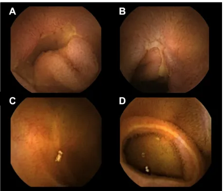
Figure 1 small-bowel images obtained with capsule endoscopy of the colon.
Notes: images of a patient with ileal Crohn’s disease obtained with camera 1 (A) and camera 2 (B). in this case, the two cameras showed the presence of the disease. Images of a patient with nonsteroidal anti-inflammatory drug enteropathy obtained with camera 1 (C) and camera 2 (D). in this case, only camera 2 revealed the presence of a circumferential ulceration.
Kobayashi et al,24 in a study of 55 patients with OGIB, evalu-ated the diagnostic yield of small-bowel capsule endoscopy for gastric diseases, and found that this technique has, for diffuse lesions, sensitivity and specificity of 70% and 82%, respectively. However, for localized lesions, sensitivity and specificity were 28% and 63%, respectively. In another study, Rana et al25 found that up to 10% of 87 subjects evaluated with the small-bowel capsule had colonic abnormalities (including angiodysplasias, neoplasias, polyps, and Crohn’s disease), suggesting that, along with appropriate preparation, the colon should also be carefully evaluated in patients undergoing small-bowel capsule endoscopy. Almeida et al,23 with the opposite approach to Rana et al’s study, found that double-camera ent-eroscopy using CCE increases the rate of detection for small-bowel abnormalities. Although Almeida et al23 first described the use of CCE to perform a double-camera enteroscopy, this
Clinical and Experimental Gastroenterology downloaded from https://www.dovepress.com/ by 118.70.13.36 on 20-Aug-2020
Dovepress
remes-Troche et al
study included only ten patients and the aim was solely to evaluate the small bowel.
Following a protocol similar to that used by Almeida et al,23 we evaluated the whole GI tract; to our knowledge, this was the first study to do so. The entire GI tract evaluation was feasible because the double-camera capsule was swallowed after it restarted from the sleep mode. Thus, CCE may be used as a noninvasive, all-in-one technique to visualize the entire GI tract from the esophagus to the colon and rectum.
A major benefit of the double camera is that it significantly increases the area able to be evaluated, but also, as shown in our study, both cameras capture different sets of images that are not mutually reproducible (kappa between camera 1 and camera 2 was 0.278). Thus, a dual and simultaneous analysis of both cameras allows a more precise assessment of the GI tract. Although the overall agreement between observers was between fair and moderate for the entire GI evaluation, the dual-camera analysis detected between 5% and 16% more abnormalities compared to single-camera analysis. Previous studies have estimated that the miss rate for single-capsule endoscopy ranges between 11% and 19%.2,26 Problems such as the potential to miss a single-mass lesion and poor visual-ization of some segments of the small bowel are less likely to occur if a CCE is used.
Interestingly, we found that, for some areas of the GI tract and for some diseases, the overall concordance was moderate to good. For example, kappa for overall esophageal findings, erosive esophagitis, and hiatal hernia were 0.611, 0.51, and 0.812, respectively. Similar results have been previously reported with kappa values ranging from 0.42 to 0.67 for esophagitis.12,27 In contrast, the use of esophageal capsule endoscopy demonstrates moderate sensitivity and specificity for the diagnosis of Barrett’s esophagus. In our study, the number of patients with Barrett’s esophagus was small (n = 2); thus, we cannot assess the agreement for this condition.
Small-bowel capsule endoscopy provides a high diagnostic yield for small-bowel mucosal lesions, and its use is recom-mended in specific scenarios of IBD, such as in the pediatric population, where it may help lead to a definitive diagnosis and sometimes can result in reclassification of UC/indetermi-nate colitis to Crohn’s disease.28–30 To date, the role of CCE in IBD has been evaluated in only one case series.22 Specifically, CCE has been compared with colonoscopy with the aim of evaluating its accuracy in monitoring colonic inflammation in patients with suspected or known UC. In this preliminary experience, CCE yielded encouraging results for detecting active ulcerative colitis (ie, sensitivity 77%, specificity 78%)
and good agreement with colonoscopy. Although we had a small sample size, we found that, for detecting IBD, CCE is a good method with a high level of agreement.
Regarding other colorectal findings, a good agreement level was found for diverticular disease of the colon. To our knowledge, ours is the first study to report on this disease. It is essential to remark that, in all cases, diverticular dis-ease was considered non-complicated. Further studies are required to analyze diagnostic accuracy and clinical use of CCE for diverticular disease of the colon. We found 47 polyps, most of which were small. As reported by others, most colonic polyps discovered at screening are diminutive, with negligible risk of harboring advanced features (high-grade dysplasia, villous component, or malignancy).20,31 Moreover, 40% of diminutive colonic polyps are hyperplastic rather than adenomatous.32 Diminutive lesions identified by a noninvasive test may also be missed by colonoscopy, because of the imperfect sensitivity of the latter for diminu-tive lesions.33
It is also important to remark that, in up to 62% (13/21) of our subjects, internal hemorrhoids were detected by CCE. This finding makes obvious that a bowel preparation using booster doses of 30 mL sodium phosphate solution can guarantee that the capsule reaches the rectum, allowing a complete GI evaluation.
Comparing single-camera analysis and dual-camera analysis, the lower level of agreement was observed in small-bowel diseases such as angiodysplasias (kappa = 0.375), and NSAID enteropathy (kappa = 0.372 ). This finding could be explained because single-camera analysis has limited vision and each camera may capture different, non-reproducible findings. Thus, according to our results, as in the Almeida study,23 a double-camera enteroscopy using CCE is a better option for complete evaluation of small-bowel diseases.
There are some limitations in our study that should be acknowledged. First of all, the study had a small sample size and the CCE was used in different diseases. Also, we did not evaluate the upper part of the GI tract, the small bowel, or the colon with other invasive techniques (such as endoscopy, enteroscopy, and colonoscopy), but we must say that this was beyond our aim. Further studies are required to compare findings detected by CCE in the whole GI tract versus those detected with standard techniques. The statistical agreement analysis was calculated for overall findings and for some lesions because there were not enough subjects to do a per-patient analysis. Also, because each per-patient had more than one abnormality detected by CCE, an analysis per patient was extremely difficult to perform.
Clinical and Experimental Gastroenterology downloaded from https://www.dovepress.com/ by 118.70.13.36 on 20-Aug-2020
Dovepress single- and dual-camera CCE analysis of the gastrointestinal tract
Although we have shown, using CCE, that more find-ings could be detected in the colon and other parts of the GI tract, this technique does not exclude false negative results at all. For example, a CCE examination may be incomplete, mainly because of a slow GI transit that may mean that the capsule stays longer periods in some seg-ments of the GI tract. Therefore, some segseg-ments may not be visualized by CCE. In addition, although we describe that an entire GI tract evaluation was possible in all cases, we must acknowledge that some parts of the stomach, specifically the fundus, are not able to be assessed using capsule endoscopy.
Conclusion
The use of a dual-camera analysis using CCE for the evalua-tion of the GI tract is feasible and detects more abnormalities when compared with a single-camera analysis. However, further studies are required that include larger sample sizes, specific populations (eg, obscure bleeding), and a cost-effectiveness analysis.
Acknowledgments
The authors wish to thank Mr Eli de la Cruz Patiño and Mr Job Ulises Reyes Huerta for their technical support.
Disclosure
The authors report no conflicts of interest in this work.
References
1. Delvaux M, Gay G. International Conference on Capsule and Double-Balloon Endoscopy (ICCD). Paris, August 27–28, 2010. Endoscopy. 2011;43(6):533–539.
2. Leighton JA. The role of endoscopic imaging of the small bowel in clinical practice. Am J Gastroenterol. 2011;106(1):27–36.
3. Triester SL, Leighton JA, Leontiadis GI, et al. A meta-analysis of the yield of capsule endoscopy compared to other diagnostic modalities in patients with obscure gastrointestinal bleeding. Am J Gastroenterol. 2005;100:2407–2418.
4. Liu K, Kaffes AJ. Review article: the diagnosis and investiga-tion of obscure gastrointestinal bleeding. Aliment Pharmacol Ther. 2011;34(4):416–423.
5. Rokkas T, Niv Y. The role of video capsule endoscopy in the diagnosis of celiac disease: a meta-analysis. Eur J Gastroenterol Hepatol. 2012;24(3): 303–308.
6. Günther U, Bojarski C, Buhr HJ, Zeitz M, Heller F. Capsule endoscopy in small-bowel surveillance of patients with hereditary polyposis syndromes. Int J Colorectal Dis. 2010;25(11):1377–1382.
7. Postgate AJ, Will OC, Fraser CH, Fitzpatrick A, Phillips RK, Clark SK. Capsule endoscopy for the small bowel in juvenile polyposis syndrome: a case series. Endoscopy. 2009;41(11):1001–1004.
8. Bailey AA, Debinski HS, Appleyard MN, et al. Diagnosis and out-come of small bowel tumors found by capsule endoscopy: a three-center Australian experience. Am J Gastroenterol. 2006;101(10): 2237–2243.
9. Schwartz GD, Barkin JS. Small-bowel tumors detected by wireless capsule endoscopy. Dig Dis Sci. 2007;52:1026–1030.
10. Caunedo A, Rodríguez-Téllez M, García-Montes JM, et al. Usefulness of capsule endoscopy in patients with suspected small bowel disease.
Rev Esp Enferm Dig. 2004;6(1):10–21. English, Spanish.
11. Niv Y. Capsule endoscopy: no longer limited to the small bowel. Isr
Med Assoc J. 2010;12(3):178–180.
12. Galmiche JP, Sacher-Huvelin S, Coron E, et al. Screening for esophagi-tis and Barrett’s esophagus with wireless esophageal capsule endoscopy: a multicenter prospective trial in patients with reflux symptoms. Am J
Gastroenterol. 2008;103:538–545.
13. Eliakim R, Yassin K, Shlomi I, Suissa A, Eisen GM. A novel diagnostic tool for detecting oesophageal pathology: the PillCam oesophageal video capsule. Aliment Pharmacol Ther. 2004;20(10):1083–1089. 14. Fernandez-Urien I, Carretero C, Armendariz R, Muñoz-Navas M.
Esophageal capsule endoscopy. World J Gastroenterol. 2008;14(34): 5254–5260.
15. Guturu P, Sagi SV, Ahn D, Jaganmohan S, Kuo YF, Sood GK. Capsule endoscopy with PILLCAM ESO for detecting esophageal varices: a meta-analysis. Minerva Gastroenterol Dietol. 2011;57(1):1–11. 16. Eliakim R, Fireman Z, Gralnek IM, et al. Evaluation of the PillCam
Colon capsule in the detection of colonic pathology: results of the first multicenter, prospective, comparative study. Endoscopy. 2006;38(10): 963–970.
17. Herrerías-Gutiérrez JM, Argüelles-Arias F, Caunedo-Álvarez A, et al. PillCamColon Capsule for the study of colonic pathology in clinical practice. Study of agreement with colonoscopy. Rev Esp Enferm Dig. 2011;103(2):69–75.
18. Schoofs N, Devière J, Van Gossum A. PillCam colon capsule endoscopy compared with colonoscopy for colorectal tumor diagnosis: a prospec-tive pilot study. Endoscopy. 2006;38:971–977.
19. Van Gossum A, Munoz-Navas M, Fernandez-Urien I, et al. Capsule endoscopy versus colonoscopy for the detection of polyps and cancer.
N Engl J Med. 2009;361:264–270.
20. Hassan C, Zullo A, Winn S, Morini S. Cost-effectiveness of capsule endoscopy in screening for colorectal cancer. Endoscopy. 2008;40: 414–421.
21. Spada C, Hassan C, Galmiche JP, et al; European Society of Gastro-intestinal Endoscopy. Colon capsule endoscopy: European Society of Gastrointestinal Endoscopy (ESGE) Guideline. Endoscopy. 2012;44(5): 527–536.
22. Sung JJY, Ching JY, Leung WK, et al. Assessment of colonic inflam-matory lesions and ulcerative colitis with PillCam Colon capsule endoscopy compared to colonoscopy. Endoscopy. 2008;40:A199. 23. Almeida N, Figuereido P, Lopes S, et al. Is it the end of single camera
small bowel capsule as we know it? Presented at the 16th European Gastroenterology Week “UEGW;” October 18–22;2008; Vienna, Austria. OP 144.
24. Kobayashi Y, Watabe H, Yamada A, et al. Diagnostic yield of capsule endoscopy for gastric diseases. Abdom Imaging. 2012;37(1):29–34. 25. Rana SS, Bhasin DK, Singh K. Colonic lesions in patients
undergo-ing small bowel capsule endoscopy. Int J Colorectal Dis. 2011;26(6): 699–702.
26. Rondonotti E, Villa F, Mulder CJ, Jacobs MA, de Franchis R. Small bowel capsule endoscopy in 2007: indications, risks and limitations.
World J Gastroenterol. 2007;13:6140–6149.
27. Delvaux M, Papanikolaou IS, Fassler I, et al. Esophageal capsule endoscopy in patients with suspected esophageal disease: double blinded comparison with esophagogastroduodenoscopy and assessment of interobserver variability. Endoscopy. 2008;40(1):16–22.
28. Herrerías JM, Caunedo A, Rodríguez-Téllez M, Pellicer F, Herrerías JM Jr. Capsule endoscopy in patients with suspected Crohn’s disease and negative endoscopy. Endoscopy. 2003;35:564–568. 29. Marmo R, Rotondano G, Piscopo R, et al. Capsule endoscopy versus
enteroclysis in the detection of small-bowel involvement in Crohn’s dis-ease: a prospective trial. Clin Gastroenterol Hepatol. 2005;3: 772–776. 30. Gralnek IM, Cohen SA, Ephrath H, et al. Small bowel capsule endoscopy
impacts diagnosis and management of pediatric inflammatory bowel disease: a prospective study. Dig Dis Sci. 2012;57(2):465–471.
Clinical and Experimental Gastroenterology downloaded from https://www.dovepress.com/ by 118.70.13.36 on 20-Aug-2020
Clinical and Experimental Gastroenterology
Publish your work in this journal
Submit your manuscript here: http://www.dovepress.com/clinical-and-experimental-gastroenterology-journal
Clinical and Experimental Gastroenterology is an international, peer-reviewed, open access journal, publishing all aspects of gastroenterology in the clinic and laboratory, including: Pathology, pathophysiology of gastrointestinal disease; Investigation and treatment of gastointes-tinal disease; Pharmacology of drugs used in the alimentary tract;
Immunology/genetics/genomics related to gastrointestinal disease. This journal is indexed on CAS. The manuscript management system is completely online and includes a very quick and fair peer-review system. Visit http://www.dovepress.com/testimonials.php to read real quotes from published authors.
Dovepress
Dove
press
remes-Troche et al
31. Lieberman D, Moravec M, Holub J, Michaels L, Eisen G. Polyp size and advanced histology in patients undergoing colonoscopy screening: impli-cations for CT colonography. Gastroenterology. 2008;135:1100–1105. 32. Weston AP, Campbell DR. Diminutive colonic polyps: histopathology,
spatial distribution, concomitant significant lesions, and treatment complications. Am J Gastroenterol. 1995;90:24–28.
33. van Rijn JC, Reitsma JB, Stoker J, Bossuyt PM, van Deventer SJ, Dekker E. Polyp miss rate determined by tandem colonoscopy: a sys-tematic review. Am J Gastroenterol. 2006;10:343–350.
Clinical and Experimental Gastroenterology downloaded from https://www.dovepress.com/ by 118.70.13.36 on 20-Aug-2020