CLUSTER HEADACHE
DR ANISH BAHRA
The Headache Group Institute of Neurology University College London
London, UK
Thesis submitted for the examination for
DOCTOR OF MEDICINE (MD)
UNIVERSITY OF LONDON
ProQuest Number: 10015022
All rights reserved
INFORMATION TO ALL USERS
The quality of this reproduction is dependent upon the quality of the copy submitted.
In the unlikely event that the author did not send a complete manuscript and there are missing pages, these will be noted. Also, if material had to be removed,
a note will indicate the deletion.
uest.
ProQuest 10015022
Published by ProQuest LLC(2016). Copyright of the Dissertation is held by the Author.
All rights reserved.
This work is protected against unauthorized copying under Title 17, United States Code. Microform Edition © ProQuest LLC.
ProQuest LLC
789 East Eisenhower Parkway P.O. Box 1346
Abstract
Cluster headache is a relatively uncommon primary head pain syndrome. In this thesis a total of 230 individuals with cluster headache were interviewed, of whom 19 underwent functional imaging with positron emission tomography (PET) to address pathophysiological mechanisms of the syndrome, and to better understand the clinical problem. Seventy-six percent were recruited through national support groups. The male to female ratio was 2.5:1. Although there were some shared clinical features with migraine the syndrome of cluster headache was clinically distinct: a strictly unilateral first division trigeminal head pain, with ipsilateral autonomic features and a characteristic circadian periodicity to short-lived attacks and bouts of attacks. With presumed better access to neurologists the time to diagnosis has improved over the decades, however despite this, the management of such patients remains suboptimal.
Description of thesis
This thesis is divided into four parts.
Part 1 provides an introduction to cluster headache. The clinical characteristics and epidemiology of the syndrome are followed by a discussion o f current hypotheses pertaining to pathophysiology, and a review of therapeutic management relevant to both the described PET studies and clinical data obtained. The final chapter of this section addresses the principles of PET and summarises functional imaging studies in the investigation o f pathophysiological mechanisms in primary head pain.
Part 2 comprises of three PET studies. The first is a study of patients with chronic cluster headache imaged during an acute attack of cluster headache. Using the same study design the second is a study of individuals with episodic cluster headache imaged out of the active bout. The third is a single PET study of a patient with migraine and cluster headache imaged during the active bout of cluster headache but during an acute typical migraine headache attack.
Part 3 comprises a clinical and epidemiological study of the 230 patients who were interviewed during recruitment of suitable individuals for the three PET studies.
The studies are presented in the order in which the data was obtained and analysed during the three year term of my research.
Abbreviations ACC BA CGRP CBF CSF CCH CH CHA CHI CT DHE ENT EEC ECAT ECH F FSH FWHM hrs HBO IHS LH LHRH MRA
Anterior cingulate cortex Brodmann area
Calcitonin gene related peptide Cerebral blood flow
Cerebrospinal fluid Chronic cluster headache Cluster headache
Cluster headache active Cluster headache inactive Computerised tomography Dihydroergotamine
Ear, nose and throat Electroencephalogram
Emission Computerised Axial Tomography Episodic cluster headache
Female
Follicle stimulating hormone Full width half maximum Hours
Hyperbaric oxygen
International Headache Society Luteinising hormone
M Male
HA* Mild generalised featureless headache post-nitroglycerine MMPI Minnesota multiphasic personality inventory
mins Minutes
GTN Nitroglycerine
PET Positron emission tomography
REM Rapid eye movement
rCBF Regional cerebral blood flow
SPECT Single photon emission computerised tomography SPM Statistical parametric mapping
TSH Thyroid stimulating hormone
TRH Thyrotropin releasing hormone VIP Vasoactive intestinal peptide
VAS Visual analogue scale
Tables Table 2.1 Table 2.2 Table 2.3 Table 2.4 Table 7.1 Table 7.2 Table 8.1 Table 8.2 Table 9.1 Table 9.2 Table 10.1 Table 10.2
Table 10.3
Table 10.4
Table 10.5
Associated features in cluster headache Associated features in cluster headache Attack duration and frequency
Bout duration and frequency
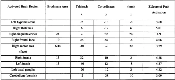
Number of scans for each condition o f interest
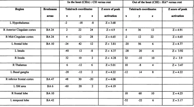
Autonomic features Additional features 34 34 43 44 124 Increases in blood flow during an induced attack o f 125 acute cluster headache compared to the pain-free state Conditions of interest following GTN in patients with 131 cluster headache in (CHA) and out (CHI) of the active bout
Significant increases in blood flow in patients in 137 (CHA) / out (CHI) of the bout during the scans defined for CH / HA* compared with rest
Regional increases in blood flow during migraine 148 headache compared to rest
Regional increases in blood flow post-sumatriptan 149 compared to rest
Site of pain 170
Side of attacks 171
Table 10.6
Table 10.7
Table 10.8
Table 10.9
Table 10.10
MaleiFemale (M:F) ratio, time to diagnosis and practitioners seen relative to year of onset of cluster headache
Specialists seen prior to diagnosis Acute therapy
Preventative therapy Alternative therapy
Figures
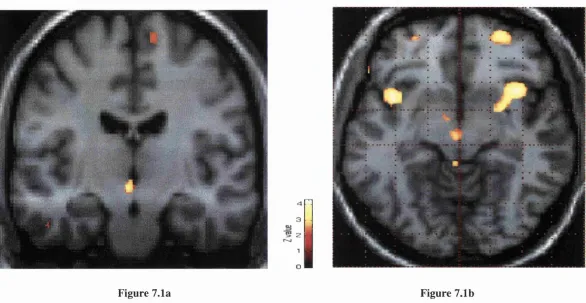
Figure 7.1a Comparison of the nitroglycerine induced acute cluster 126
and 7.1b headache attack and rest (no pain) conditions in nine patients with chronic cluster headache
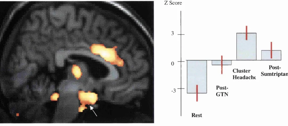
Figure 7.2 Activation of structures involved in pain processing and of 127 the intracranial vessels
Figure 7.3 Comparison of GTN-induced acute cluster headache attack 128 versus rest in nine patients with chronic cluster headache - Activation of the intracranial vessels
Figure 7.4 Activation of the intracranial vessels (arrowed) in a single 129 case of spontaneous cluster headache
Figure 9.1 Dorsorostral brainstem activation during migraine 150 headache compared to rest
Figure 9.2 Cortical and subcortical activations during migraine 151 headache compared to rest
Figure 9.3 Activation of the intracranial vessels during migraine 152 headache compared to rest
Figure 9.4 Activation pattern of the dorsorostral brainstem and 153 intracranial vessels in the three conditions o f interest
Figure 10.1 Distribution of bout frequency amongst the cohort of 180 patients studied
Figure 10.2 The distribution of the diagnosis of cluster headache made 181 by neurologists by year in which the diagnosis was made
Publications arising from this thesis
Bahra, A., May, A. & Goadsby, P J. (2002). Cluster Headache : A prospective study with diagnostic implications. Neurology, 58 (3), 354-361
Bahra, A., Matharu, M., Buechel, C., Frackowiak, R.S. & Goadsby, P.J. (2001). Brainstem activation specific to migraine headache. The Lancet, 31, 1016-1017
May, A., Bahra, A., Buechel, C., Frackowiak, R.J.S. & Goadsby, P.J. (2000). PET and MRA findings in cluster headache and MRA in experimental pain. Neurology, 55,
1328-1335
Bahra, A, May, A and Goadsby, PJ (1999). Diagnostic patterns in cluster headache. Cluster Headache and Related Conditions. J. Olesen and P. J. Goadsby. Oxford, Oxford University Press .
Goadsby, P.J., Bahra, A. & May, A. (1999). Mechanisms of cluster headache. Cephalalgia, 19, 19-23
Publications during research period not reported in this thesis
Bahra, A. & Goadsby, P J. (2000). Chronic tension-type headache. Clinical Evidence, December, 710-715
Bahra, A., Gawel, M.J., Hardebo, J.-E., Millson, D., Breen, S. & Goadsby, P.J. (2000). Oral Zolmitriptan is effective in the acute treatment of cluster headache. Neurology, 54,
1832-1839
Bahra, A. & Goadsby, P.J. (1999). Chronic overuse of acute anti-migraine preparations. Prescriber, 5th September, 109-115
Bahra, A., Evers, S. & Goadsby, P.J. (1999). Komorbiditat von depression and migraine - eine ubersicht. Nervenheilkunde, 18, 267-271
May, A., Buchel, C., Bahra, A., Goadsby, P.J. & Frackowiak, R.S.J. (1999). Intra cranial vessels in trigeminal transmitted pain: a PET study. Neuroimage, 9, 453-460
Evers, S., Bahra, A. & Goadsby, P.J. (1999). Coincidence of familial hemiplegic migraine and hemicrania continua? A case report. Cephalalgia, 19, 533-535
Bahra, A. & Goadsby, P.J. (1998). Cough headache responsive to methysergide. Cephalalgia, 18, 495-496
Bahra, A. & Goadsby, P J. (1998). The comorbidity o f migraine and depression: a review. Neurology and Psychiatry, 2, 23-27
Acknowledgements
I would like to acknowledge all the patients who participated in the work detailed in this thesis, the Migraine Action Association and the Migraine Trust, the support of the radiographers at the Wellcome Department of Cognitive Neurology and Zeneca Pharmaceuticals for their sponsorship.
My thanks to the following individuals - Lindsay Haas, Paul Hammond, Manjit Matharu, Arne May, Mary Reilly, Jo Simmons and Sophie Ryan. In particular for his support throughout the last five years, my thanks to Professor Peter Goadsby.
Contents
Abstract
Description of thesis Abbreviations Tables
Figures
Publications arising from this thesis
Publications during research period not reported in this thesis Acknowledgements Contents 2 4 5 7 9 10 11 13 14
P A R T I C hapter 1
1.1 1.2 1.3 1.4 1.5 1.6 1.7 L 7A 7.7.2 1.7.3 1.8
INTRODUCTION AND BACKGROUND 21
Epidemiology 22
Prevalence 22
Male:Female (M:F) ratio 22
Age of onset 24
Ethnic origin 25
Family history of cluster headache 25 History and family history of migraine 26
Patient characteristics 27
Smoking 27
Alcohol and Triggering o f attacks 28
Personality traits 28
1.8.1 Peptic ulcer disease Cardiac disease 29 30 Chapter 2 2.1 2.2 2.3 2.3.1 2.4 2.4.1 2.5 2.6 2.7 2.8 Clinical Characteristics
Localisation of pain Associated features
Symptomatic Cluster Headaehe Post-traumatic cluster headache
32 32 32 Duration and frequency of attacks 35
Periodicity o f attacks 35
Episodic Cluster Headache - Duration and 36 frequency of bouts of cluster headache
Periodicity o f bouts 37
Chronic Cluster Headache 38
Differences Between Episodic and Chronic 38 Cluster Headaehe 40 41 Chapter 3 3.1 3.2 3.3 3.3.1 3.3.2
The Pathophysiology of Cluster Headache 45
The pain phenomenon 45
The autonomic phenomena 47
Pathophysiological hypotheses 50 A pathophysiological focus in the region o f 50 the pericarotid cavernous sinus
Central pathophysiological mechanisms 53 involving the hypothalamus
3.3.3 The role o f the carotid body in cluster 61 headache
C hapter 4 Treatm ent of Cluster H eadache 64
4.1 Introduction 64
4.2 Acute attack treatment 64
4.27 Subcutaneous sumatriptan 64
4.22 Intranasal sumatriptan 66
4.22 Oral Zolmitriptan 67
4.24 Oxygen Therapy 68
4 .2 J Ergotamine 71
4 .2 6 Intranasal lignocaine and cocaine 72
4.3 Preventative treatment 74
4.3.1 Calcium channel blockers 74
4.2.2 Lithium 76
4.2.2 Corticosteroids 78
4.2.4 Methysergide 79
4.2.2 Ergotamine 80
4.2.6 Pizotifen 81
4.2.7 Sodium Valproate 81
4.2.^ Other prophylactic agents 82
4.4 Surgical Treatment 82
4.4.1 Introduction 82
4.4.2 Trigeminal ganglion 83
4.4.2 Trigeminal nerve 84
4.4.5 Nervus Intermedins 86
4.4.6 Greater superficial petrosal nerve 87
4.5 Summary 88
Chapter 5 Functional Imaging in Head and Facial 89
Pain
5.1 Functional imaging studies in migraine 89 headache
5.1.1 Introduction 89
5.1.2 Xenon-133 with the use o f stationary 90 detectors and single photon poisitron
emission tomography (SPECT) in migraine
with aura
5.1.3 Perfusion weighted magnetic resonance 93
imaging in migraine with aura
5.1.4 Xenon-133 and the use o f stationary 94 detectors and SPECT studies in migraine
without aura
5.1.5 Positron emission tomography (PET) 95
5.1.6 PET in migraine with and without aura 99
5.2 Functional imaging studies in cluster 101 headache
5.2.1 Xenon-133 with the use o f stationary 101 detectors and SPECT studies in cluster
headache
5.2.2 PET studies in cluster headache 104
5.3 Summary 104 PART 2 Chapter 6 6.1 6.2 6.3 6.4
PET STUDIES OF CLUSTER HEADACHE 106
Oxygen-15 Tracer Positron Emission 107
Tomography Introduction Method Statistical Analysis Ethics Approval 107 108 111 115 Chapter 7 7.1 7.2 7.3 7.4 7.5
PET Study 1 - Acute Cluster Headache 116
Attacks Introduction Methods Results Discussion Summary 116 117 117 119 123 Chapter 8 8.1 8.2 8.3 8.4 8.5
PET Study 2 - Cluster Headache : A 130
Comparison of Patients in and out of the
Chapter 9 PET Study 3 - Co-existent Migraine and
Cluster Headache
138
9.1 Introduction 138
9.2 Methods 139
9.3 Results 140
9.4 Discussion 142
9.5 Summary 146
PART 3 A CLINICAL STUDY OF CLUSTER
HEADACHE
154
Chapter 10 Cluster Headache : A Prospective Clinical
Study of 230 Patients
155
10.1 Introduction 155
10.2 Methods 155
10.3 Results 156
10.3.1 Sample Group 156
70.3.2 Clinical Characteristics- attacks 156
10.3.3 Clinical characteristics - patients 158
10.3.4 Cluster headache in Women 159
10.3.5 Accuracy and Rapidity o f Diagnosis 160
10.3.6 Treatment Regimens 161
10.4 Discussion 161
10.4.1 Clinical characteristics 162
10.4.2 Cluster headache and women 163
10.4.3 Genetics, Cluster Headache and Migraine 165
10.4.4 Natural History o f Cluster Headache
19
10.4.5
10.4.6
10.5
Diagnostic (in)accuracy 166
Management Strategies in Cluster 167 Headache
Summary 168
PART 4
Chapter 11
CONCLUSION
Conclusion
183 184
PART 1
INTRODUCTION AND BACKGROUND
CHAPTER 1
Epidem iology
1.1 Prevalence
Cluster headache is a relatively uncommon primary pain disorder. Current prevalence data are based mainly on estimates. The prevalence from a study of 18 year old Swedish army recruits was 0.09% (Ekbom et aL, 1978). Extrapolation of these results yielded prevalence rates in the United States of 0.4% for men and 0.08% for women (Kudrow, 1980). The most comprehensive prevalence data is from the population of the Republic of San Marino. This was based upon review of 15 year medical records and writing to each of almost 22,000 inhabitants with a resultant prevalence rate of 0.07% (D' Alessandro et aL, 1986).
1.2 MaleiFemaie (M:F) ratio
to control groups (Facchinetti et aL, 1986; Kudrow, 1976b; Kudrow, 1980; Murialdo et a l, 1989; Romiti et a l, 1983). There is little known about the contribution of female hormones in cluster headache. The onset of cluster headache in women is later than migraine (Kudrow, 1980) and unrelated to menarche (Manzoni et aL, 1988) (see 1.3). Menstruation does not seem to influence the symptoms (Ekbom & Waldenlind, 1981; Manzoni et aL, 1988). It has been reported that the majority of females notice remission during pregnancy although Manzoni et aL found no differences between cluster headache sufferers and controls (Manzoni et aL, 1988). The use of the oral contraceptive pill is not different to the general population but only anecdotal reports exist about any influences of the pill on the attacks of cluster headache. Women who develop cluster headache before having children tend to have fewer children than age- matched migraineurs, whilst in those with onset after their pregnancies the mean number of childbirths are similar to the general population. Infertility associated with the onset of the syndrome, or psychological factors have been put forward to explain this observation (Ekbom & Waldenlind, 1981; Manzoni et aL, 1988). Finally cluster headache can continue into the menopause or may start de novo (Ekbom & Waldenlind, 1981; Peatfield et aL, 1982); infact the age of onset in women shows a second peak in the 6‘*’ decade (see 1.3).
An increased incidence of past head injury has been reported in cluster headache sufferers compared to age- and sex-matched controls, and individuals with migraine, and tension-type headache (Friedman & Mikropoulos, 1958, Manzoni, 1983; Lance & Goadsby, 1998; Manzoni, 1999). It has been postulated that the male predominance may be due to a greater tendency of males to accidents (Manzoni, 1999; Manzoni et aL,
1983b). The most recent reported series of cluster headache sufferers (Manzoni, 1998) addressed the M:F ratio by decade of onset. A gradual reduction in the M:F ratio was
seen over time from 6.2:1 before 1960 to 2.1:1 for those individuals with onset between 1990 and 1995. This was attributed to significant changes in lifestyle, such as smoking and employment rate, which have shown a similar fall in M:F ratio for the corresponding decades.
An atypical pattern of cluster headache in women has been noted, although not validated (Lovshin, 1961; Peatfield et aL, 1982). This observation may be accounted for by the more recently described syndrome of paroxysmal hemicrania (Antonaci & Sjaastad, 1989). Paroxysmal hemicrania is a syndrome clinically related to cluster headache and differentiated by a female preponderance, shorter attack duration, increased daily attack frequency and an absolute response to indomethacin (Headache Classification Committee of the International Headache Society, 1988).
1.3 Age of onset
primary and secondary cluster headache. The mean age of onset of primary compared to secondary cluster headache appears to be later (Manzoni, 1999) and one study has shown the onset of primary cluster headache does not seem to diminish until the 6* decade while that of secondary cluster headache shows a bimodal pattern with an increased frequency in the 5^^ and 6* decades (Kudrow, 1980).
1.4 Ethnic origin
Few studies have addressed race. Cluster headache occurs in all races (Kudrow, 1980). Two studies reported cluster headache to be relatively more common in the Afro- Carribean population particularly in the women, with a M:F ratio of 3-3.5:1 (Kudrow,
1980; Lovshin, 1961).
1.5 Family history of cluster headache
Cluster headache has not generally been thought to have a genetic prediposition. However a positive family history had been reported in 1.9-10 % of patients with cluster headache (Klapper et al., 2000; Kudrow & Kudrow, 1994) and there have been occasional reports in monozygotic twins (Couturier et al., 1991; Roberge et a l, 1992; Sjaastad et a l, 1993). In a study of 200 women with cluster headache, 24 (12%) were found to have at least one first-degree relative with the disorder. Three generations of cluster headache were found in 7 of 24 kindreds (29%). Parenteral cluster headache was found in 19/24 of the probands (79%); in 14/24 (74%) the transmission was from father to proband. Of the 1652 first-degree relatives of 300 male and female patients, 3.45% had cluster headache. This is 13 times the expected frequency of cluster headache in the general population (taken as 0.26%) (Kudrow & Kudrow, 1994). Notably not all relatives were interviewed and descriptions from some probands was taken as adequate. Russell and colleagues found a positive family history of cluster headache (in first- and
second-degree relatives) in 25/366 (7%) o f families of 370 probands (21% women) with cluster headache (seven patients belonged to 3 families) (Russell et a l, 1995a). The risk of cluster headache in first- and second-degree relatives was 14 and 2 times that of the general population. This was based upon a population prevalence of 69 per 100,000 adjusted for age and sex (D' Alessandro et al., 1986). Montagna and colleagues found a postive family history o f cluster headache in 2.3% of first- and second-degree relatives of 222 probands (Montagna et a l, 1998). Leone and colleagues studied the occurrence of cluster headache in the families of 220 patients with cluster headache. Compared with the general population, first degree relatives had a 39-fold increased risk of cluster headache and second degree relatives an 8-fold increased risk (Leone et al., 2001). The population prevalence was again taken as 69 per 100,000. In summary, these studies have confirmed a higher familial occurrence of cluster headache. Some families have shown a pattern compatible with autosomal dominant inheritance (D'Amico et al., 1996; Montagna et al., 1998; Russell et al., 1995b; Spierings & Vincent, 1992). Methodological differences may explain the differences in familial risk observed in these studies.
1.6 History and family history of migraine
cluster headache (Andersson, 1985; Kudrow & Kudrow, 1994; Solomon & Cappa, 1986). However the co-existence of migraine and cluster headache does occur. One report of 10 patients commented that a ‘migraine crisis’ did not occur during the cluster period in any patient and moreover the attacks of migraine and cluster headache did not present in close proximity to one another (D'Amico et a l, 1997). However other authors have witnessed occasional migraine attacks during, and often limited to, the cluster period, with migraine attacks witnessed in some male and female patients with no clear history of migraine (Kudrow & Kudrow, 1994).
Similar discrepancies exist with regard to the familial incidence of migraine in individuals with cluster headache. However most of the larger published series have reported the familial incidence of migraine in first degree relatives of individual with cluster headache to be the same as the incidence in the general population (Ekbom,
1974; Ekbom & Waldenlind, 1981; Kudrow, 1980; Manzoni et a i, 1983b).
1.7 Patient characteristics
i. 7.1 Smoking
The majority o f individuals with cluster headache have a longstanding history of smoking, current or past. Significantly more patients with cluster headache smoked cigarettes in comparison to a control group; the number of cigarettes smoked per day was also significantly greater in cluster headache sufferers than controls (Kudrow, 1980). O f a series of 370 cluster headache patients, 330 were smokers or former smokers (Manzoni, 1999). Seventy-eight percent had started smoking before the onset of cluster headache, 11% after the onset, and 11% had never smoked. Those who had stopped smoking following the onset of cluster headache continued to experience
attacks. However since the series was clinic-based this group of individual may have represented a biased sample.
1.7.2 Alcohol and Triggering o f attacks
During the active bout cluster headache attacks can be precipitated by alcohol (Friedman & Mikropoulos, 1958; Krabbe et al., 1984; Kudrow, 1980; Symonds, 1956) and nitroglycerine (Krabbe et al., 1984). Ekbom showed a 100% success rate at triggering acute attacks during the middle of a bout with Img sublingual nitroglycerine (Ekbom, 1968). The attack was preceded by a relatively consistent latency period between 30-50 minutes and followed by a refractory period of a few hours. A higher frequency of severe attacks could be provoked during the middle of the bout in comparison to the end of the bout. Attacks could not be triggered when patients were out of the active bout. Triggered attacks were clinically identical to spontaneous attacks. Triggered attacks have also been shown experimentally to be identical to spontaneous attacks (Fanciullacci et al., 1995; Goadsby & Edvinsson, 1994a). Although sufferers tend to avoid alcohol consumption during the active bout, a higher consumption of alcohol in cluster headache patients compared to controls has been reported (Kudrow,
1980; Manzoni, 1999).
1.7.3 Personality traits
with cluster headache using the Minnesota Multiphasic Personality Inventory (MMPI) in comparison with 217 individual with other types of headache and a group of 30 controls. There was no difference between migraine and cluster headache sufferers. Although cluster headache sufferers did score higher than controls on some scales, the mean of the five MMPI scales was not different to controls. This was confirmed in a subsequent study comparing 40 cluster headache sufferers with 49 migraineurs (Cuypers et a l, 1981). However cluster headache sufferers did show elevated ‘anxiety’ scores and slightly diminshed scores for ‘masculinity’. It has been proposed that cluster headache sufferers are depressed. Marchesi et a l studied the prevalence of different types of headache in 160 depressed patients. Cluster headache was diagnosed in 1.2%, and migraine and tension-type headache in 22.5 and 24.4% respectively (Marchesi et a l, 1989).
1.8 Comorbid disorders
1.8.1 Peptic ulcer disease
Several investigators have reported an association between cluster headache and peptic ulcer disease, the prevalence ranging between 13 and 22% (Kudrow, 1980). Kudrow reported the prevalence to be 21.1% in a group of 119 male cluster headache sufferers compared to 10.7% in a group of age-sex matched migraineurs. A subsequent survey of 355 male cluster headache sufferers showed similar results. The prevalence in a group of 21 female sufferers was reported to be significantly different from the migraine ‘controls’, but twice the estimated incidence (2.5%) in the general population. The type of ulcer was noted to be predominantly duodenal (Kudrow, 1976c; Kudrow, 1980). Manzoni also reported a higher prevalence of 14.4% in his series (n=180) compared to 7.2 and 2.8% in the migraine and tension-type headache controls groups (each n=180). Peptic ulcer disease was more common in chronic than episodic cluster headache but
the patient numbers were markedly disproportionate with only 19 chronic sufferers (Manzoni et al., 1983b). It has been unclear from the aforementioned reports whether the history of peptic ulceration relates to all-time prevalence, or in relation to the patients’ cluster headache. With regard to the latter concomitant drug consumption was also not formally addressed. High gastric acid secretion after histamine stimulation was observed by Graham in 50% of patients (n=16), in some cases in the Zollinger-Ellison range (Sjaastad, 1992). However no study has addressed formally acid secretion during the acute attack, in the bout and out of the bout compared to a control group. If gastric acid secretion is truly elevated in cluster headache sufferers this may be a systemic manifestation of the associated parasympathetic discharge.
1.8.2 Cardiac disease
Ekbom, 1970b; Jacobsen, 1969; Russell & Storstein, 1983; Russell & von der Lippe, 1982). Moreover, Jacobson found that neither symptoms nor the observed cardiovascular phenomena were altered by administration of intravenous atropine diphenhydramine or saline (Jacobsen, 1969); occlusion of the ipsilateral carotid artery resulted in marked sinus bradycardia and arrhythmia, supraventicular ectopics and sinus arrest without affecting the pain. Russell and Storstein formally assessed and heart rate with 24 hour Holter ECG monitoring and recorded paroxysmal atrial fibrillation, atrioventricular block and sinoatrial block in three patients during the acute attack (Russell & Storstein, 1983). Cardiovascular reflexes in cluster headache have been shown to be significantly different to controls. It has been suggested that the observed changes may be a response to pain. Some studies have suggested that heart rate and blood pressure changes are correlated with the degree of pain (Ekbom, 1970b) but other studies have not (Russell & Storstein, 1983). A more plausible and the most theorised concept is of central autonomic dysfunction. Cardiovascular disturbances in association with conditions affecting the central nervous system are well known (Vaisrub, 1975) and a central autonomic dysfunction is in keeping with the syndrome of cluster headache (Jacobsen, 1969; Russell & Storstein, 1983).
CHAPTER 2
Clinical Characteristics
2.1 Localisation of pain
The site of pain in cluster headache most commonly involves the ocular (retro-orbital and supra-orbital) and temporal regions, although the pain can be experienced over a wide area which includes the cheek, jaw, upper and lower teeth, nose, ear, occiput, neck, shoulder or whole hemicranium (Friedman & Mikropoulos, 1958; Kudrow, 1980; Manzoni et al., 1983b; Sutherland & Eadie, 1972). The pain is usually strictly unilateral. More patients experience right than left sided attacks and 9-16% have experienced attacks on both the right and left side during different attacks. This is more common in different bouts, but can occur during the same bout (Friedman & Mikropoulos, 1958; Kudrow, 1980; Lance & Anthony, 1971; Manzoni et a l, 1983b; Sutherland & Eadie, 1972). Pain occurring simultaneously on both sides during an attack has been reported but is rare (Kudrow, 1980; Sjaastad et al., 1985; Young & Rozen, 1999). A change of side within the same attack is even more rare (Sutherland & Eadie, 1972). The nature of the pain at worst is excruciating (Symonds, 1956) and has been described as ‘often one of the most severe forms of human suffering’ (Sutherland & Eadie, 1972). Very characteristically during the pain patients prefer to be active (Ekbom, 1970a; Kudrow, 1980; Manzoni et al., 1983b; Russell, 1981).
2.2 Associated features
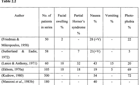
(Table 2.2). Facial swelling is usually localised orbitally/perio-orbitally. ‘Lumps in the mouth’ (Lance & Anthony, 1971) and focal palatal swelling (Kudrow, 1980) have been reported and may be a manifestation of mucosal oedema. The partial Homer’s syndrome occurs during acute attacks but may persist between attacks (Dmmmond, 1988; Nieman & Hurwitz, 1961; Sutherland & Eadie, 1972). Patients have also described visual blurring on the side ipsilateral to the pain which may be due to the concomitant autonomic abnormalities. Some patients may experience generalised sweating during the attacks (Friedman & Mikropoulos, 1958; Lance & Anthony, 1971; Manzoni et al., 1983b). Although earlier reports have suggested that nausea and vomiting do not occur (Horton, 1941), such symptoms have since been consistently reported with varying frequency, as has photophobia (Table 2.2).
There are also reports of other accompanying features during the attack such as anorexia and diarrhoea, facial hyperalgesia (Lance & Anthony, 1971), contralateral involuntary twitching of the foot (Sutherland & Eadie, 1972) and focal neurological symptoms. The described focal neurological symptoms have included visual disturbances such as flashing lights (Lance & Anthony, 1971), and scintillating scotomatous defict, contralateral facial and limb paraesthesia (Sutherland & Eadie, 1972) and vertigo and mild ataxia (Lance & Anthony, 1971). Aura symptoms in patients with cluster headache has been reported in 6 o f 101 patients (4M, 2F) (Silberstein et al., 2000). Five of the patients described visual and one described olfactory symptoms of 5-120 minutes duration associated with the cluster headache attack. One female patient had a history of migraine without aura, and two patients were noted to be related.
Associated Features in cluster headache
Table 2.1
Author No. of
patients in series Lacrimation % Conjunctival injection % Nasal congestion (NC) and rhinorrhoea (R)
% (Friedman & Mikropoulos,
1958)
50 80 50 NC + R = 88
(Sutherland & Eadie, 1972) 58 62 45 NC = 35;R = 7 (Lance & Anthony, 1971) 60 82 45 NC = 47;R= 15
(Ekbom, 1970a) 105 82 84 NC + R = 68
(Kudrow, 1980) 500 84 78 NC + R = 72
(Manzoni et al., 1983b) 180 84 45 NC = 48;R = 43
Table 2.2
Author No. of
patients in series Facial swelling % Partial Homer’s syndrome % Nausea % Vomiting % Photo phobia % (Friedman & Mikropoulos, 1958)
50 2 28 (+V) 22
(Sutherland & Eadie, 1972)
58 7 21(+V) 5
(Lance & Anthony, 1971) 60 10 32 43 15 20
(Ekbom, 1970a) 105 10 18 19 5 69
(Kudrow, 1980) 500 - - 54 - 72
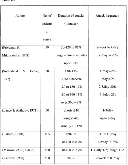
-2.3 Duration and frequency of attacks
The reported duration of a single attack of cluster headache has varied from a few minutes up to a couple of days (Friedman & Mikropoulos, 1958; Sutherland & Eadie, 1972). Most attacks however last between 30 and 120 minutes (Table 2.3) and uncommonly last more than 3 hours (Sutherland & Eadie, 1972). The attacks occurring during the daytime prospectively recorded in one study, were found to be shorter than those at night, but this was not statistically significant (Russell, 1981). In Kudrow's series of 428 males and 72 females, the attack duration was longer in women (mean duration 90 minutes) than men (mean duration 60 minutes). The attack frequency can vary from one attack/week (Sutherland & Eadie, 1972) up to 8/day (Gardner et al.,
1947; Symonds, 1956). Patients most commonly experience 1-2 attacks/day; the median attack frequency is 1 attack/day (Ekbom, 1970a; Lance & Anthony, 1971; Manzoni et a l, 1983b ; Sutherland & Eadie, 1972 ). A higher frequency of attacks was observed in chronic compared to episodic cluster headache (Manzoni et al., 1983b). Notably with regard to the shorter attacks and higher frequencies an overlap with paroxysmal hemicrania, first described in 1974 (Sjaastad & Dale, 1974), needs to be home in mind.
2.3.1 Periodicity o f attacks
The periodicity of attacks is a characteristic feature o f cluster headache. Individual attacks tend to occur at the same hour. This pattern is however fragile and maintained usually for several days or weeks at a time. Thereafter the pattern of regularity may change or be lost altogether. This periodicity was observed by a number of authors (Ekbom, 1970a ; Horton, 1941; Sjaastad, 1992 ; Sutherland & Eadie, 1972 ; Symonds, 1956 ). Sharp peaks of onset of attacks was found by Manzoni et al. between l-2am, I- 3pm and 9pm (Manzoni et al., 1983b). A nocturnal preponderance for the attacks has been consistently observed (Friedman & Mikropoulos, 1958; Kudrow, 1980; Lance &
Anthony, 1971 ; Sutherland & Eadie, 1972 ; Symonds, 1956 ). Ekbom found a 62% incidence of noctural attacks, 8.6% exclusively nocturnal and 53.3% predominantly nocturnal (Ekbom, 1970c). Prospective collection o f data from a total of 77 attacks in 22 patients showed that 75% of attacks occurred between 9 pm and 10 am, the highest frequency between 9 and 11 pm and from 4 to 10 am. Fifty-one percent of the attacks awoke the patient from sleep, including naps (assumed day-time) (Russell, 1981).
2.4 Episodic Cluster Headache - Duration and frequency of bouts of cluster
headache
2.4,1 Periodicity o f bouts
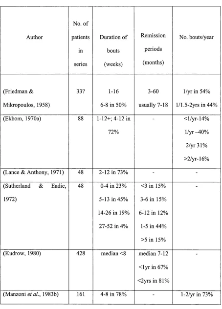
A seasonal preponderance of cluster headache bouts has suggested with onset of the bouts reported to occur mainly in the spring and autumn (Friedman & Mikropoulos, 1958; Homabrook, 1964; Symonds, 1956). However, as with the predictability of cluster headache attacks within the bouts, Sutherland and Eadie encountered patients in whom the bouts seemed to have a regular seasonal incidence initially but where the patients were followed-up over a period of several years, the season of occurrence had often changed. Retrospective collection of reliable data from this series showed that although fewer bouts begin in the Australian autumn and early winter, there was no significant seasonal predilection for the onset of the bouts (Sutherland & Eadie, 1972). Manzoni commented from his series o f 180 patients that no specific month seemed to predominate for the onset of the bouts; however, the most involved months seem to be Febmary (24 patients) and March (20 patients). In those with season-bound bouts, these would recur mostly in the spring or autumn, or both, in 40% of the cases, and in summer or winter, or both, in 24% (Manzoni et al., 1983b). Lance and Anthony did not find that the periodicity of the bouts occurred consistently upon the time of year (Lance & Anthony, 1971). Ekbom found that in 56% of cases the bouts mainly started in the autumn or spring (Ekbom, 1970a). A retrospective examination by Kudrow of the onset of 892 bouts from 404 male episodic cluster headache patients over a period of 1-10 years, showed that the frequency of the onset of the bouts increased with gradual increase or decrease of daylight throughout the year. Two significant peaks occurred 7- 10 days after the longest and shortest days of the year (July and January). The gradual rise was interrupted twice showing a significant drop in frequency of bout onsets 7-10 days after the clocks were reset for Daylight Saving and Standard times in April and October (Kudrow, 1987).
2.5 Chronic Cluster Headache
Chronic cluster headache occurs in 10-20% of individuals with cluster headache. The current definition of chronic cluster headache is of ‘attacks occurring for more than one year without remission or with remissions lasting less than 14 days’. This group of patients are further subdivided into primary chronic cluster headache, whereby the symptoms are unremitting from the onset of the condition, and secondary chronic cluster headache, where a chronic pattern of symptoms has developed following a period of episodicity (Headache Classification Committee of the International Headache Society, 1988). The original criterion for chronicity was proposed by Ekbom and de Fine Olivarius in 1971 (Ekbom & de Fine Olivarius, 1971). The relative frequency of primary and secondary chronic cluster headache is relatively consistent in different series. A slightly greater number of cases are seen in the primary chronic cluster headache group (ratios of primary to secondary 1.1:1 (Ekbom & de Fine Olivarius,
1971), 1.1:1 (Manzoni, 1998), and 1.5:1 (Kudrow, 1980).
2.6 Differences Between Episodic and Chronic Cluster Headache
1983b) and head injury with and without loss o f consciousness (Manzoni, 1999). With regard to the former the patient numbers were markedly disproportionate with only 19 chronic sufferers from a total of 180, and, most other studies addressing this issue (see Chapter 1) did not differentiate between the chronic and episodic form of the disorder. Kudrow studied 5 episodic and 5 chronic (1 female) cluster headache sufferers with nocturnal polysomnography (Kudrow et al., 1984). All 5 of the episodic group were found to have sleep apnoea, 3 with central and 2 with obstructive sleep apnoea. One in the chronic group was found to have central sleep apnoea. In the sleep apnoea group the nocturnal attacks were more likely to be associated with REM sleep and preceding oxygen desaturation than the non-sleep apnoea group (all of whom had chronic cluster headache). This was consistent with a similar observation in another small chronic group (n=4) of no evidence of sleep apnoea and no differences in sleep pattern during a symptom-free and symptomatic night; but there was a decrease in total time of REM sleep and increase in stage 3 and 4 sleep (Dexter, 1984). It was commented upon by Kudrow at al. that sleep apnoea is observed in up to 62% of males in the population aged over 55 years; all but one (age 37 years) of the episodic group was aged over 55 years. In addition the number of patients and the number of attacks studied were small. HLA typing has been done in 42 patients with cluster headache, 37 with the episodic and 5 with the chronic form. The histocompatibility antigen A l was found in all the chronic sufferers but not in the episodic group (Cupyers & Altenkirch, 1979) as demonstrated in an earlier study (Kudrow, 1978); whether the patients had primary or secondary cluster headache was not mentioned. Support for a genetic predisposition to cluster headache has been discussed in Chapter 1 ; HLA antigen subtyping may provide insights into those individuals who have a genetic susceptibility to the development of chronic cluster headache.
2.7 Symptomatic Cluster Headache
Cluster headache associated with intracranial pathology has been reported for different lesions at different anatomical sites. These include pituitary tumours (Greve & Mai, 1988; Hannerz, 1989; Porta-Etessam et al., 2001; Tfelt-Hansen et al., 1982) meningioma of the cervical canal (Kuritzky, 1984), aneurysm o f the anterior communicating artery (Greve & Mai, 1988), posterior communicating artery (McBeath & Nanda, 2000) arteriovenous malformations (Mani & Deeter, 1982; Munoz et al.,
2.8 Post-traumatic cluster headache
There have been a number of reports of cluster headache associated with trauma. Lance and Anthony reported 8 patients of their 60-patient series (13%) to have experienced a head injury (Lance & Anthony, 1971). Although in 4 patients the site of the original injury was the same as the side of the cluster headaches, the temporal relationship was not close (21 months, 7, 8 and 30 years). The range in the 8 patients was 2 months to 30 years. Friedman commented upon a past history of ‘some type of head injury with or without loss of consciousness in 16%’ of his series (Friedman & Mikropoulos, 1958). Symonds reported head injury in 2 of his 17 (12%) cases; again although the site of the injury in both cases coincided with the side of the cluster headache attacks, the onset occurred 5 and 25 years later (Symonds, 1956). Manzoni found that 36.9% of patients in his series of 374 male sufferers, gave a past history of head injury, 13.4% with loss of consciousness (Manzoni, 1999). However head injury followed the onset of cluster headache in 22% of the cases. In the rest of the cases the average time elapsed between the head injury and cluster headache onset was 10 years. The frequency of head injury was higher in the chronic group, particularly secondary chronic cluster headache. Kudrow did not find a correlation with head injury in his large series; he reported a history of significant head injury in 5.2% of patients (Kudrow, 1980). Reik described 3 cases o f cluster headache onset in close temporal relation to head injury (from immediately to 1 month after the injury); 2 patients gave a history of primary chronic cluster headache and one patient gave a history of bouts of variable duration (from 1 to 18 months) interspersed by remission periods (Reik, 1987). Turkewitz reported on the almost immediate onset of cluster headache following minor head injury without loss of consciousness; the symptoms were consistent with primary chronic cluster headache with cessation of attacks after 2 years, the time at which the report was written (Turkewitz et al., 1992). Mathew and Rueveni presented a series of patients with onset
o f cluster headache attacks with significant temporal relation to head injury or a surgical procedure (Mathew & Rueveni, 1988). The side of the attacks corresponded to the side o f maximal trauma, the symptoms were of the primary chronic type and response to treatment was less satisfactory compared to the idiopathic syndrome.
Attack duration and frequency
Table 2.3
Author No. of
patients in series
Duration o f attacks (minutes)
Attack frequency
(Friedman & Mikropoulos, 1958)
50 30-120 in 86% range - ‘some minutes
up to 360’
2/week to 4/day 1-3/day in 90%
(Sutherland & Eadie, 1972)
58 <30- 11% 30 to 120-50% 120 to 180-17% 180 to 360-13% over 360 - 9%
<1/day-2 8% l/day-40% 2-3/day-30%
4-6/day-2%
(Lance & Anthony, 1971) 60 Shortest 10 longest 480 usually 10-120
1-3/day up to 8/day
(Ekbom, 1970a) 105 <30-180
30-120 in 63%
<1 to >3/day 1-2/day in 78% (Manzoni et a l, 1983b) 180 30-120 in 73% Usually 1-2; range <1-5
(Kudrow, 1980) 500 30-120 2/week to 8+/day
Bout duration and frequency
Table 2.4
Author
No. of patients in series Duration of bouts (weeks) Remission periods (months) No. bouts/year (Friedman & Mikropoulos, 1958)
33? 1-16
6-8 in 50%
3-60 usually 7-18
1/yr in 54% l/1.5-2yrs in 44% (Ekbom, 1970a) 88 1-12+; 4-12 in
72%
< 1/yr-14% 1/yr-40% 2/yr31% >2/yr-16%
(Lance & Anthony, 1971) 48 2-12 in 73% -
-(Sutherland & Eadie, 1972)
48 0-4 in 23% 5-13 in 45% 14-26 in 19%
27-52 in 4%
<3 in 15% 3-6 in 15% 6-12 in 12%
1-5 in 44% >5 in 15% (Kudrow, 1980) 428 median <8 median 7-12
< ly r in 67% <2yrs in 81%
CHAPTER 3
The Pathophysiology o f Cluster Headache
3.1 The pain phenomenon
Work done in the early part of the twentieth century (Feindel et al., 1960; Penfleld & McNaughton, 1940; Wolff, 1963) identified the principal intracranial pain-producing structures to be the blood vessels, particularly the proximal cerebral and durai arteries, and the large veins and venous sinuses. Stimulation (Wolff, 1963) or distension (Martins et al., 1993; Nichols et al., 1990) of different vessels will refer pain to different parts of the head. These studies formed part of the basis for the concept of a vascular origin to the primary headache syndromes of migraine and cluster headache. The throbbing quality of pain and relief from the vasconstrictive agent, ergotamine, has lent further weight to this view (Duvoisin et al., 1961; Graham & Wolff, 1938). However migraine and cluster headache are clinically, epidemiologically and in terms of management very distinct (Lance & Goadsby, 1998). Moreover clinical studies (Drummond & Lance, 1983; Drummond & Lance, 1984) have suggested a dissociation between the vascular phenomena and clinical manifestations.
Surrounding the large cerebral vessels, meningeal vessels, large venous sinuses and dura mater is a plexus of largely unmyelenated fibres which arise mainly from the ophthalmic division of the trigeminal nerve with some contribution from the maxillary division o f the trigeminal nerve, and in the posterior fossa from the upper cervical dorsal roots. Branches of the vagal and glossopharyngeal nerves contribute to the innervation of the posterior circulation and dura mater o f the posterior fossa (Moskowitz, 1990; Penfield & McNaughton, 1940). The trigeminal fibres arise from neurons in the trigeminal ganglion. The central axon of the cell body in the trigeminal
ganglion descends in the spinal tract of the trigeminal nerve to the second segment of the spinal cord. Here fibres from the posterior part of the head traverse the dorsal root ganglion to converge on second order neurons in the dorsal horn and subsequently ascend in the contralateral spinothalamic tract to connections in the brainstem (nucleus gigantocellularis, midbrain reticular formation, periaqueductal gray) and diencephalon (hypothalamus), and thence ultimately to the thalamic nuclei and the cerebral cortex. Functionally the trigeminal nucleus extends beyond the traditional nucleus caudalis to the dorsal horn of the high cervical region. In the cat, stimulation of either the superior sagittal sinus or the greater occipital nerve results in increased metabolism in both the trigeminal nucleus and the dorsal horn at Cl and C2 (Goadsby et al., 1997; Kaube et a l,
1993). Exteroceptive sensation from the concha of the ear is carried by the vagus nerve to the jugular ganglion and also terminates within the spinal nucleus of the trigeminal nerve. Transmission of pain is controlled by the endogenous pain control system descending from the region of the periaqueductal gray, raphe nuclei and locus coeruleus, to the spinal intemeurones in the spinal trigeminal nucleus and dorsal horn of the upper cervical cord (Basbaum & Fields, 1978).
VIP, a marker for parasympathetic activity (Edvinsson, 2001) during the acute cluster headache attack. Both levels returned to normal after treatment of the attack.
3.2 The autonomic phenomena
The autonomic features of cluster headache are characterised by a parasympathetic discharge and sympathetic deficit on the side ipsilateral to the pain during the acute attacks.
Parasympathetic fibres originate in the superior salivatory nucleus. The fibres traverse the facial nerve and the greater superficial petrosal nerve to join the vidian nerve (the greater superficial petrosal nerve and greater deep petrosal nerve join to form the vidian nerve) and synapse in the pterygopalatine ganglion. Post ganglionic fibres loop back as orbital rami to the cavernous sinus and internal carotid artery where they form a plexus with sympathetic and trigeminal fibres (ophthalmic and maxillary division fibres), before advancing to supply the lacrimal glands and circulation of the forehead. Sympathetic fibres arise from the superior cervical ganglion and course along the internal carotid artery, through the cavernous sinus to the long ciliary nerve which innervates the dilator pupillae. Some fibres follow the external carotid artery and innervate Müller’s muscle of the eyelid, and the blood vessels and sweat glands of the face except for sweating of the medial aspect of the forehead, which follows the internal carotid artery (Drummond & Lance, 1992).
The parasympathetic discharge in cluster headache is manifested by lacrimation, conjunctival injection, nasal secretion, nasal congestion and facial swelling (see Chapter 2). Saunte has quantified lacrimation during the acute attack and found this to be increased on the side ipsilateral to the pain by a factor of about two. Lacrimation also occurred on the non-symptomatic side but by a factor of 1.4. Similarly nasal secretion
was increased bilaterally during the acute attack, but this was greater on the symptomatic side compared to the asymtomatic side (Saunte, 1984). The conjunctival injection, nasal congestion and facial swelling are suggestive of extracranial vasodilatation. This is supported by an increase in comeal temperature, comeal indentation pulse amplitudes and intraocular pressure during the acute attack. The changes seen in comal indentation pulse amplitues and intraocular pressure were bilateral but most pronounced on the symtomatic side, while comeal temperature increases occurred only on the side ipsilateral to the pain during the attack. No such changes have been observed in migraineurs during and between attacks of pain (Sjaastad, 1992; Sjaastad et a l, 1985). In addition, during spontaneous and induced attacks of cluster headache thermographic measurments have shown an increase in temperature of the affected orbital and maxillary region (Dmmmond & Lance, 1984). Salivation is diminished during the attack; it has been suggested that sympathetic activity dominates over parasympathetic action on the salivary glands but is unable to override its lacrimal and vascular functions (Saunte, 1984). This may be part of a generalised sympathetic arousal distinct from the observed local sympathetic deficit. It is most likely that the parasympathetic discharge is mediated via the greater superficial petrosal nerve (Dmmmond, 1988). Increase in cutaneous blood flow has been observed to follow the onset of pain and has been therefore considered to be a secondary phenomenon; a vasodilator reflex mediated by the trigeminal nerve as the afferent arm and the greater superficial petrosal nerve as the efferent arm (Goadsby & Lance, 1988).
permanent sympathetic deficit on the asymptomatic side has also been reported (Fanciullacci et a l, 1982; Micieli et a l, 1988). In attempt to determine the site of the sympathetic deficit - 1®‘ (central), 2"^* (preganglionic) or 3'^* (postganglionic) order neuron Fanciullaci et a l studied pupil responses to thymoxamine (selective a l blocker), 4% cocaine (a re-uptake inhibitor), 2% tyramine (releases noradrenaline from the nerve terminals) and 1% phenylephrine (direct sympathomimetic agent), in individuals with cluster headache whilst asymptomatic, during and out of the active bout (Fanciullacci et a l, 1982). Cocaine dilatation has been shown to be reduced in any Homer’s syndrome regardless of site. Tyramine will dilate the pupil normally in cases with a central or preganglionic lesion, but the reaction is reduced in a postganglionic lesion. When sympathetic innervation is impaired, sympathetically innervated effector cells become supersensitive to directly acting sympathomimetic agents at weak concentrations ordinarily ineffective; a denervated muscle (postganglionic lesion) is more sensitive to such drugs than a decentralised lesions (central and preganglionic) (Thompson & Mencher, 1971). Baseline pupil diameters were not statistically significant. There was no asymmetry in response to thymoxamine. There was bilateral mydriasis in response to topical cocaine and tyramine, but this was significantly reduced on the symptomatic side compared to the asymptomatic side. Although this study found an insignificant aniscoria with smaller dilatation on the symptomatic side with phenylephrine, other studies have shown excessive dilatation of the pupil suggestive of denervation supersensitivity of the receptors (Salveson & Sjaastad, 1987; Thompson & Mencher, 1971). Deficient pupillary dilatation to hydroxy-amphetamine (which also releases noradrenaline from the nerve terminals) on the symptomatic side has been shown to be accompanied by a similar deficiency of sweating of the medial aspect of the forehead, with overactivity o f the sweat glands in this region in response to circulating pilocarpine (Salveson & Sjaastad, 1987). The studies therefore suggest the
lesion to be postganglionic. But it has been aknowledged that the pupil responses to the different agents studied is not as clear cut as defined above (Fanciullacci et al., 1988; Salveson et al., 1987; Thompson & Mencher, 1971). The matter is not further clarified by the response to intravenous clonidine, an inhibitor o f central sympathetic activity; this resulted in more marked miosis bilaterally (compared to the control group) in cluster headache sufferers (ECH during active bout but outside an attack) but more marked on the symptomatic side. Tyramine alone caused bilateral mydriasis significantly less on the symptomatic side. Clonidine administration before tyramine resulted in an increase in mydriasis compared to tyramine alone on the aymptomatic side (Fanciullacci et al., 1988). In another study Fanciullacci and colleagues showed that lithium induced a symmetrical response to tyramine, postulating this to be a correction o f abnormal asymmetries in central neuronal systems which regulate autonomic function (Fanciullacci et al., 1983).
3.3 Pathophysiological hypotheses
There have been two main theories regarding the pathophysiology of cluster headache. These have addressed pathology in the region of the superior pericarotid cavernous sinus plexus and the hypothalamus. Dysfunction of the carotid body chemoreceptors has also been postulated.
3.3.1 A pathophysiological focus in the region of the pericarotid cavernous sinus
division of the trigeminal nerve, the maxillary division, the superior cervical ganglion, and the pterygopalatine ganglion, all join together (Hardebo, 1990; Moskowitz, 1988). Orbital phlebographic findings during cluster headache attacks have been found to be abnormal on the symptomatic side only, and bilaterally but with greater changes on the symptomatic side. The most frequent findings were of narrowing of the entire superior opthalmic vein or of the third segment of the vein; two of the 8 patients with abnormal imaging (from a total of 13) had partial occlusion of the cavernous sinus and collateral veins in the region o f the superior orbital fissure (Hannerz et al., 1987). The changes observed were similar to those seen in patients with Tolosa-Hunt syndrome (Hannerz et a l, 1986). Magnetic resonance angiography in a single case report o f a spontaneous attack of cluster headache has shown dilatation of the ipsilateral opthalmic artery, with normal repeat angiographic findings during remission (Waldenlind et a l, 1993). Dilatation of the ipsilateral opthalmic artery with localised narrowing of the extradural part of the internal carotid artery has been shown on carotid angiography during a cluster headache attack occurring 15 minutes after left internal carotid artery puncture (Ekbom & Greitz, 1970). Cluster headache has therefore been attributed to an inflammatory process occurring in the cavernous sinus and tributary veins. It has been proposed that inflammation obliterates the venous outflow from the cavernous sinus on one side, thus injuring traversing sympathetic fibres o f the intracranial internal carotid artery and its branches. It is further theorised that the active period ends when the inflammation is suppressed and the sympathetic fibres fully or partially recover (Hardebo, 1994). This hypothesis was supported by Gawel et a l who used gallium single photon emission computerised tomography (SPECT) to study patients with cluster headache, on the basis that the tracer is picked up in areas of inflammation such as sarcoid and allergic alveolitis. In 3 of 6 patients imaged with gallium SPECT during the active bout, increased activity was seen in the parasellar region and faded in one
narrowed during the acute attack (Moskowitz, 1988).
However activation in the region of the cavernous sinus during gallium SPECT has been observed in chronic cluster headache, episodic cluster headache in and out of the bout and also in migraineurs (Sianard-Gainko et aL, 1994). Increased activity in this region imaged with PET has been observed during capsaicin-induced first division trigeminal pain (May et al., 1998b). The abnormal findings on orbital phlebography are not specific to cluster headache and have been observed in migraine, tension-type headache and ‘cervicogenic headache’ (Bovim et al., 1992). Symptomatic cluster headache has been reported not only for lesions in the region of the cavernous sinus but lesions at different anatomical sites such as the upper cervical canal (Kuritzky, 1984), the occipital lobe (Mani & Deeter, 1982), the vertebral artery (West & Todman, 1991) and the skull base (Mathew, 1992). Moreover an inflammatory hypothesis would have to account for longstanding recurrent episodes of inflammation with precise onset of bouts, spontaneous resolution and no residual morbidity or mortality. The periodicity of cluster headache is a key feature of the syndrome. The precise circadian and circannual pattern of symptoms are more suggestive of primary dysfunction o f the central nervous system.
3.3.2 Central pathophysiological mechanisms involving the hypothalamus
The onset of cluster headache bouts has been suggested to have a seasonal preponderance, occurring maximally in autumn and spring. Most o f the reported observations have been retrospective (see Chapter 2). The largest of these was reported by Kudrow who showed that two significant peaks o f onset o f cluster headache bouts occurred 7-10 days after the longest and shortest days o f the year (July and January). The gradual rise was interrupted twice showing a significant drop in frequency of bout
onsets 7-10 days after the clocks were reset for Daylight Saving and Standard times in April and October (Kudrow, 1987). These results were interpreted to indicate that the frequency of cluster bout onsets increased as photoperiods lengthened or shortened. Artificial interruption of the photoperiods such as changing of the clocks, resulted in sharp decreases in the frequency of cluster bout onsets, ruling out temperature influences. It was speculated that the onset of cluster bouts was due to an inability to synchronise the internal circannual pacemaker, the hypothalamus, to environmental light cues. Patients with bipolar affective disorders demonstrate excacerbation of symptoms associated with changes in photoperiods and respond to resetting of the internal pacemaker with bright-light therapy (Lewy et al., 1987).
headache with predominantly nocturnal symptoms (Bono et al., 1985). The same group showed that sleep deprivation had a short and long term effect in preventing attacks of cluster headache.
Disturbance of hypothalamic function during the active cluster bout is supported by a number of studies which have shown abnormalities of hypothalamic-determined functions:
Cortisol: Cortisol shows a diurnal pattern of secretion with trough nocturnal levels and high morning levels on waking. The mean 24 hour cortisol levels are higher during the active bout compared to out of the bout (Waldenlind et al., 1987) and to non-headache controls (Facchinetti et al., 1986). Facchinetti et al. attributed this to stress experienced during the active bout, but also showed a phase-advance in circadian pattern of secretion of about 2 hours. In a study of 11 patients during the active bout (10 males, 1 female) compared to 8 controls, the typical circadian pattern of secretion was noted to be blunted, and again there was a significant phase-advance in the circadian pattern of secretion (Chazot et al., 1984).
Melatonin: Melatonin has a strong circadian rhythm and is produced in the pineal gland. It is converted from serotonin to melatonin. This is largely regulated by N- Acetyltransferase. Regulation of enzymatic activity responsible for the circadian oscillations of melatonin is by the suprachiasmatic nucleus of the hypothalamus which is innervated by retino-hypothalamic fibres. Melatonin is produced at night and its production ceases in response to retinal stimulation by sunlight via the retino- hypothalamic fibres. Melatonin secretion is therefore very sensitive to light-dark cycles and therefore photoperiodicity (Brezinski, 1997). Chazot et al. showed that in 11
patients during the active bout compared to 8 controls nocturnal melatonin levels were blunted with a slightly advanced phase of secretion; one patient showed complete abolition of the circadian pattern of secretion. The 3 patients who had the lowest melatonin plasma levels had the largest cortisol phase-shift (Chazot et a l, 1984). Waldenlind et a l confirmed lower nocturnal melatonin levels during the active bout compared to the inactive period (Waldenlind et a l, 1987). The main metabolite of melatonin is 6-sulphatoxymelatonin. Leone et a l have shown higher nocturnal than day-time urine levels o f the metabolite in controls (n= 14), while no difference between night and day-time secretion was seen in 20 cluster headache sufferers during the active bout and 13 of this group re-tested in the remission period. Moreover nocturnal secretion in and out of the active bout was lower than nocturnal secretion in the control group (Leone et a l, 1998).