Article
1
Comparative study of physicochemical and
2
antibacterial properties of ZnO nanoparticles prepared
3
by laser ablation of Zn target in water and air
4
Ekaterina A. Gavrilenko1, Daria A. Goncharova1, Ivan N. Lapin1, Anna L. Nemoykina2,
5
Valery A. Svetlichnyi1,*, Ali A. Aljulaih 3,4, Neli Mintcheva3,5 and Sergei A. Kulinich3,4,6,*
6
1 Siberian Physical Technical Institute, Tomsk State University, Lenina, 36, Tomsk, 634050, Russia
7
2 Laboratory of Biopolymers and Biotechnology, Tomsk State University, Lenina, 36, Tomsk, 634050, Russia
8
3 Institute of Innovative Science and Technology, Tokai University, Hiratsuka, Kanagawa 259-1259, Japan
9
4 Department of Mechanical Engineering, Tokai University, Hiratsuka, Kanagawa 259-1259, Japan
10
5 Department of Chemistry, University of Mining and Geology, Sofia 1700, Bulgaria
11
6 Research Institute of Science and Technology, Tokai University, Hiratsuka, Kanagawa 259-1259, Japan
12
* Correspondence: [email protected] (V.A.S.); [email protected] (S.A.K.)
13
Tel.: +7-3822-531-591 (V.A.S.); +81-463-58-1211 (ext.4893) (S.A.K.)
14
15
Abstract: Here, we report on ZnO nanoparticles (NPs) generated by nanosecond pulsed laser
16
(Nd:YAG, 1064 nm) through ablation of metallic Zn target in water and air and their comparative
17
analysis as potential nanomaterials for biomedical applications. The prepared nanomaterials were
18
carefully characterized in terms of their structure, composition, morphology and defects. It was
19
found that in addition to the main wurtzite ZnO phase, which is conventionally prepared and
20
reported by others, the sample laser-generated in air also contained some amount of monoclinic
21
zinc hydroxynitrate. Both nanomaterials were then used to modify model wound dressings based
22
on biodegradable poly-L-lactic acid. The as-prepared model dressings were tested as biomedical
23
materials with bactericidal properties towards S. aureus and E.coli strains. The advantages of the
24
NPs prepared in air over their counterparts generated in water found in this work are discussed.
25
Keywords: Pulsed laser ablation in water; pulsed laser ablation in air; ZnO nanoparticles;
26
biomedical materials; PLLA-scaffold; antibacterial properties
27
28
1. Introduction
29
Because of their unique physicochemical properties, nanomaterials have recently attracted a lot
30
of research interest as materials and components for various applications. Zinc oxide (ZnO) is a
wide-31
bandgap semiconductor of the II-VI group (Eg = 3.3 eV) [1,2]. Of all known metal-oxide
32
semiconductors, it is probably the most extensively studied material with applications in numerous
33
fields, such as optoelectronics, piezoelectronics, spintronics, solar energy, gas sensing, bio-sensing,
34
UV-blue diodes, and photocatalysis, just to name several [1-6]. Because of its high surface energy, its
35
nanoparticles (NPs) are able to generate various reactive oxygen species (ROS), which makes such
36
NPs efficient bacteria inhibitors attractive for biomedical use as well. Having low toxicity, ZnO
37
nanomaterials, unlike those of many other semiconductor oxides, can be applied to problems such as
38
water, working surface and wound disinfection, as well as in food industry, and hence the number
39
of studies on potential use of ZnO nanomaterials keep growing nowadays quite fast [1-6].
40
There are many approaches to prepare ZnO NPs, such as, for example, hydrothermal methods,
41
chemical deposition from gas phase, microwave and sonochemical approaches, numerous sol-gel
42
based methods, and so on [1-3]. One of attractive methods to produce so-called “pure” ZnO
43
nanostructures (i.e. with surface free of any impurities or stabilizers) for biomedical use is based on
44
pulsed laser ablation (PLA) of metallic Zn target in different media (mainly in liquids) [4-10]. The
45
method uses pulsed lasers (with different pulse energy and pulse width) to ablate zinc target and
46
produce species which then react with the surrounding liquid and form ZnO NPs. The formation
47
mechanisms are quite complex, depending on laser pulse parameters and composition of liquid
48
medium, and involve several fast stages that overlap and compete with each other, such as:
49
absorption of irradiation, melting and evaporation on the target, expansion of the formed
50
plasma/vapor, chemical reactions and quenching by the liquid, formation of clusters and primary
51
NPs, and secondary irradiation of the formed NPs by laser beam, just to name the main ones. Thus,
52
the method combines both “top-down” and “bottom-up” synthesis approaches, as the target gets
53
destroyed by laser beam into very small clusters (atoms, radicals, ions), after which self-assembly of
54
such species into ZnO NPs follows (accompanied by oxidation processes and quenching to ambient
55
temperatures) [7,9]. In addition to PLA in liquids [4-10], more recently the PLA approach in gases or
56
vacuum was also applied to nanomaterial preparation, including Zn-based NPs [11,12].
57
The characteristics of laser-produced ZnO NPs were found to depend on both laser parameters
58
(wavelength, frequency, pulse energy, energy fluence, and pulse duration) and on target and ablation
59
medium (vacuum, gas, or liquid). The effect of liquid on the physicochemical properties of generated
60
ZnO NPs was extensively studied, the main media used being based on water, water-ethanol
61
mixtures, H2O2, alkali and acidic solutions, salts and various surfactants [4-10,13-15]. The produced
62
NPs were reported to have different shapes, such as rods, spheres, flakes, dendrites, spindles, also
63
including additional phases, such as β-Zn(OH)2, metallic zinc, zinc peroxide ZnO2, or having
64
core@shell morphology Zn@ZnO as a result of incomplete oxidation of metallic zinc [8,13-15].
65
The PLA-generated NPs were reported to demonstrated high reactivity, unique optical, catalytic
66
and antibacterial properties, which is often explained by their structural and surface defects
[4-67
8,10,13-15]. The defectiveness of various ZnO NPs was extensively analyzed as various defects are
68
known to provide different functionalities to ZnO materials [6,14,16,17]. Similar to other II-VI
69
semiconductors, ZnO tends to have deficiencies in its anion sub-lattice, which leads to the formation
70
of oxygen vacancies (VO) with low formation energy, interstitial zinc (Zni) and zinc atoms in oxygen
71
sub-lattice (ZnO), as well as defects such as interstitial oxygen (Oi) and oxygen in the zinc sub-lattice
72
(OZn), the latter defects requiring high energies [14,16]. In their comparative study, Goto and
co-73
workers showed that PLA in pure water mainly generated ZnO NPs with Oi defects, while NPs
74
prepared in pure ethanol were rich in Zni–VZn defects, which was explained by stronger oxidation
75
ability of water [14]. At the same time, no information on defect composition of ZnO NPs
PLA-76
prepared in air was reported thus far.
77
The present work aimed at preparing ZnO NPs by means of PLA in water and air and comparing
78
their composition, structure, and properties. The materials were then incorporated into polymeric
79
tissues based on poly-L-lactic acid (PLLA) used as scaffold, where their antibacterial behavior against
80
two different bacteria strains was evaluated and compared.
81
2. Materials and Methods
82
2.1. Preparation of ZnO NPs using PLA method
83
In this study, all nanomaterials were obtained by means of a nanosecond pulsed laser (Nd:YAG
84
type, TII LS-2131M-20, LOTIS model, Belarus) that ablated Zn targets (purity 99.5 %) using the
85
following parameters: 1064 nm, 20 Hz, 7 ns, and 150 mJ/pulse (as wavelength, frequency, pulse width
86
and pulse energy, respectively). Two somewhat different setups were used, as presented in Fig.1.
87
For experiments in air, target with a diameter of 30 mm and thickness of 10 mm was fixed onto
88
the back wall of the cylindrical quartz reactor filled with ambient air (see Fig.1a). The reactor was 200
89
mm in length, its internal diameter being 45 mm, and volume being ~ 300 cm3. The wall of the reactor
90
was made of polyethylene membrane which is transparent for the irradiation used. The laser beam
91
was focused on the target surface by a long-focus (F= 500 mm) collecting lens. The produced NPs
92
precipitated on the reactor walls forming a powdery layer. The whole ablation procedure took 3 h,
during which the target was slowly moved along the vertical direction. The powder was then
94
mechanically removed from the walls, and the collected sample was denoted as ZnO_air.
95
To prepare ZnO nanostructures in liquid phase, a zinc plate target (40×15×10 mm3 in size) was
96
immersed into a cylindrical glass reactor filled with distilled water. The laser irradiation was focused
97
by a short-focus (F = 50 mm) lens, with the beam entering the reactor through its sidewall, as seen in
98
Fig.1b. To enhance NP oxidation, air was bubbled through the water by a compressor. The ablation
99
time was 2 h, after which the produced colloid was centrifuged and dried in air at 50°C. The ablation
100
and separation procedures were repeated several times to collect a sufficient amount of nanomaterial.
101
Hereafter, the sample produced in water is denoted as ZnO_water.
102
In both cases, the initial power density on the target surface was estimated to be around 250
103
MW/cm2. To maintain uniform irradiation of the target surface, thus providing uniform and constant
104
ablation, the target was automatically moved both horizontally and vertically in the XY plane normal
105
to incident beam. In both cases, the average material production rate observed after experiments was
106
~40 mg/h. More detailed description of similar experiments both in liquid and air were previously
107
published elsewhere [17].
108
(a)
(b)
Figure 1.Setups used in the present study for PLA in air (a) and water (b). 1: laser beam; 2: focusing lens; 3:
109
movement direction for target; 4: Zn target; 5: polyethylene membraine; 6: cylindric reactor: 7: distilled water; 8:
110
target holder.
111
2.2. Preparation of ZnO-PLLA composites
112
As a model material for bandage tissues with antibacterial properties, we chose biodegradable
113
polymer poly-L-lactic acid (PLLA) which easily decays upon hydrolysis and fermenting processes
114
giving rise to non-toxic compounds. PLLA is also known to be biocompatible, which makes it an
115
excellent candidate for pharmaceutical and biomedical applications [18]. Using the previously
116
reported methodology based on electrospinning [19], PLLA scaffold was prepared and then kindly
117
supplied to us by Drs. S. Tverdokhlebov and E. Bolbasov (Tomsk Polytechnic University).
118
The above mentioned nanopowders of ZnO (samples ZnO_air and ZnO_water) were dispersed
119
in distilled water by means of sonication, so that the concentration of materials was 1 g/L in both
120
cases. Pieces of PLLA scaffold, 10×10 cm2 in size and 250 μm-thick, were placed on a net where they
121
were homogenously fed with colloidal solutions with ZnO NPs by means of a pump. The NP-loaded
122
scaffolds were then dried with air at 20 oC, after which the procedures were repeated until the loading
123
level of 1 mg/cm2 was achieved. Thus, two samples were obtained which consisted of PPLA scaffolds
124
loaded with ZnO NPs of two types with same loading (composite samples referred to as
125
ZnO_water_PLLA and ZnO_air_PLLA below).
126
2.3. Characterization of nanopowders and ZnO-PLLA composites
127
The crystal structure of the samples was analyzed by X-ray diffractometry (XRD), for which a
128
XRD 6000 model (from Shimadzu) was used. The samples were analyzed with CuKα irradiation (λ =
129
1.54056 Å) at sampling rate of 0.02 o/s and in the range of 2θ from 10 to 70°. Phase identification and
130
quantitative analysis of XRD patterns were conducted using the database PDF4 and PowderCell 2.4
software (from BAM, Germany). The morphology and size of NPs were studied by transmission
132
electron microscopy (TEM, model CM 12 from Philips, Netherlands) with accelerating voltage of 120
133
kV. Drops of freshly prepared dispersions were placed onto copper grids coated with carbon film
134
and then dried at room temperature. The surface morphology of PLLA scaffolds loaded with ZnO
135
NPs was studied by scanning electron microscopy (SEM, model VEGA 3 SBH from Tescan, Czechia).
136
The specific surface area of the prepared powder samples was evaluated by using a standard
137
Brunauer–Emmett–Teller procedure (low-temperature adsorption/desorption of nitrogen) in a
138
TriStar II 3020 analyzer (Micromeritics, USA). The samples were degassed at 200 °С for 2 h prior to
139
the measurements in a VacPrep 061 station (Micromeritics, USA).
140
UV-Vis absorption spectra of both powder samples and ZnO-PLLA composites were examined
141
by the diffuse-reflection spectroscopy (DRS) technique on a Cary 100 spectrophotometer (Varian,
142
Australia) with accessory DRA-CA-30I (Labsphere, USA) in the range of 200-800 nm. As reference
143
samples, MgO powder and as-supplied (i.e., unloaded with ZnO) PLLA scaffold were used.
144
Reflection spectra were converted using the Kubelka–Munk transformation approach. The obtained
145
absorption spectra were used to evaluated the bandwidth values (Eg) of the ZnO nanomaterials, for
146
which they were replotted in the (F(R)hν)2 versus E(eV) coordinates and the absorption edge was
147
then extrapolated onto the absciss axis. Photoluminescence (PL) spectra of the samples were recorded
148
at room temperature by means of a Fluorolog 3-22 spectrometer (Horiba, Jobin Yvon, USA) with an
149
excitation wavelength being 350 nm. Fourier-transform infrared (FTIR) spectra were registered by a
150
Nicolet 6700 spectrometer (Thermo Fisher Scientific, USA). Raman shift spectra were collected by an
151
InVia (Renishaw, UK) Raman spectrometer, while the second harmonic of Nd:YAG laser (λ=532 нм)
152
was used as excitation source.
153
Zeta potential values of the NPs were determined through electrophoretic light scattering with
154
phase analysis (PALS) using a Nano Brook Omni instrument (Brookhaven, USA). To determine the
155
isoelectric point (рНiep), a BI-ZTU autotitrator was utilized (Brookhaven, USA). Titration was done
156
with aqueous KOH, prior to which ZnO dispersions in water (0.1 g/L) were sonicated for 10 min.
157
2.4. Antibacterial activity of NPs
158
The antibacterial activity of the prepared inorganic-organic composites as model wound
159
dressing materials was tested in accordance with the standard ISO 20743:2013 [20] and by using two
160
bacteria strains: (1) gram-positive Staphylococcus aureus (S. aureus, test strain ATCC 25923) and (2)
161
gram-negative Escherichia coli (E.coli, test strain В-6954, Russian Collection of Microorganisms). Both
162
loaded with NPs and as-supplied (ZnO-free) PLLA scaffolds were cut to samples with sizes 3 × 5 cm2
163
for testing. Bacteria cultures (0.2 mL) with concentration of 105 CFU/mL in nutrient broth (diluted 20
164
times with distilled water) were placed onto each PLLA sample. Immediately after contact, as well
165
as after 24 h, the tested objects were rinsed with 20 mL of physiological solution for 5 min. The
166
obtained liquid was placed into a Petri dish containing 100 mL of beef-extract agar (also known as
167
meat-and-peptone agar, МPA), and the obtained material was then cultivated at 37 °С for 24 h. The
168
quantity of grown microorganisms was counted in cultures extracted after the contact with both
169
control (ZnO-free) and NP-modified PLLA samples after cultivation, which permitted to determine
170
the death percentage of tested microorganisms. The values of antibacterial activity A were
171
determined using formula (1):
172
A = (lgCt – lgC0) – (lgTt – IgT0) = F - G, (1)
where F = (lgCt – lgC0) is the growth rate on the control (ZnO-free) PLLA sample; lgCt is the average
173
decimal logarithm of the number of bacteria found on three control samples incubated for 24 h; lgC0
174
the average decimal logarithm of the number of bacteria observed on three control samples
175
immediately upon seeding with bacteria; G = (lgTt – lgT0) the growth rate on the sample loaded with
176
antibacterial NPs; lgTt the average value of decimal logarithm of the number of bacteria observed
177
after incubation for 24 h on three treated samples; lgT0 the average decimal logarithm of bacteria
178
number observed immediately after bacteria seeding on three PLLA samples loaded with ZnO.
3. Results and discussion
180
3.1 XRD data
181
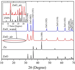
Figure 2 exhibits XRD patterns of the laser-produced samples ZnO_water (blue line) and
182
ZnO_air (red) and compares them with XRD data for metallic Zn and ZnO, both taken from databases
183
(black lines). Both samples are well-seen to demonstrate intense peaks at 2θ = 31.68°, 34.34°, 36.17°,
184
47.48°, 56.51°, 62.78°, 66.27°, 67.85° and 69.00°, implying the presence of wurtzite ZnO (PDF4 Card
185
no. 04-007-9805) as dominating phase. This is consistent with previous reports on ZnO NPs prepared
186
via PLA in liquids [5,6,10,14,15], confirming that highly-active Zn species generated by laser pulse
187
react with oxygen and/or water molecules, giving rise to ZnO nanomaterial. Small amounts of
188
metallic zinc were also detected in the samples, as very weak peaks at 2θ = 43.19° (PDF4 Card no.
01-189
071-4620), the signal being somewhat stronger in the sample produced in air than in its counterpart
190
generated in water. This observation is also in agreement with previous studies as small amounts of
191
metallic-phase inclusion was previously reported by different groups for various ZnO NPs produced
192
in liquids [6,14,15].
193
194
Figure 2. XRD patterns of PLA-prepared samples ZnO_water (blue), ZnO_air (red), and Zn and ZnO patterns
195
from database (black). Inset shows pattern of sample ZnO_air and that of ZHN phase in a narrower range of
196
2θ between 12o and 30°.
197
In addition to the diffraction peaks of ZnO, the sample produced in air exhibited a series of small
198
peaks in the range 2θ = 10-30° (see inset in Fig.2). The latter peaks were indexed as monoclinic zinc
199
hydronitrate (ZHN), Zn5(OH)8(NO3)2×2H2O, whose pattern (PDF4 Card no. 01-072-0627) is also given
200
in the inset for convenience. Previously, this phase was reported in the NPs generated via laser
201
ablation of Zn foil immersed into aqueous solution of zinc nitrate [21]. In the present study, the
202
formation of ZHN is explained by interaction of laser-induced plasma with Zn species (atoms, ions,
203
radicals, clusters) and air components, such as molecules (N2, O2, H2O) and excited species (N*, NO*,
204
NO2*, OH*). It is very likely that zinc nitrate Zn(NO3)2 is one of intermediate products of such
205
reactions. It should be noted here that no formation of ZHN is observed as a result of atmospheric
206
corrosion of metallic Zn, which supports the efficiency of PLA in preparing metastable phases. More
207
detailed phase composition of the ZnO samples, as well as their specific surface area and NP sizes,
208
are presented in Table 1.
Table 1. Structural characteristics and specific surface area values of prepared samples.
210
Sample
Phase composition Surface area (m2/g)
NPs average size parameters (nm)
Name % Diameter
(nm)
Length (nm)
Width (nm)
ZnO_air
ZnO 92
36±4 18-26 - -
ZHN 7 Zn 1
ZnO_water ZnO > 99 20±2 12-21 30-100 14-20
Zn < 1
Thus, the XRD measurements indicate that the nanomaterial obtained in water was mainly ZnO
211
with trases of metallic Zn, while the NPs prepared in air had a few per cent of ZHN.
212
3.2 Microscopic observations
213
3.2.1. TEM images
214
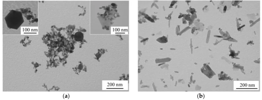
According to TEM image of sample ZnO_air in Fig.3a, the material prepared in air consisted of
215
NPs with different shapes: spheres, nanocubes, and polyhedrons (see insets in Fig.3a), with average
216
sizes being 18-26 nm. Meanwhile, sample ZnO_water had mainly nanorods as its main component
217
(Fig.3b). Similarly shaped ZnO NPs were previously reported by Honda et al. [15] who ablated Zn in
218
water with millisecond pulsed laser and explained nanorod formation by an increase in temperature
219
during PLA. Another group also produced ZnO nanorods by PLA of Zn in water by means of
220
nanosecond pulsed laser followed by heat treatment of the formed colloid at 60-80 oC [22]. In this
221
study, we also used no surfactant when ablating in water, and thus some increase in temperature
222
during PLA (and secondary irradiation of the initial ZnO NPs) could lead to their recrystallization
223
into nanorods. ZnO NPs are known to have various shapes as the phase has no symmetry center in
224
its crystal lattice [23]. This may explain the variety of shapes observed in Fig.3a. At the same time,
225
when small ZnO NPs had a chance to recrystallize, they tended to form rod-like shapes, as well seen
226
in Fig.3b.
227
(a) (b)
Figure 3. TEM images of samples: ZnO_air (a) and ZnO_water (b).
228
The sizes and shapes of NPs exhibited in Fig.3 correlate well with the results of BET
229
measurements. The specific surface area of the smaller NPs produced in air is seen in Table 1 to be
230
1.8 times larger compared with their counterparts obtained in water. It is also worth noting one more
231
time that the rod-shaped morphology and somewhat bigger sizes of the NPs presented in Fig.3b are
232
results of secondary growth processes taking place in water after their initial formation.
233
3.2.2. SEM images of ZnO-PLLA scaffolds
235
The surface morphology of PLLA scaffolds, both as-supplied and modified by ZnO NPs, was
236
studied by SEM, with images of samples ZnO_water_PLLA (a) and ZnO_air_PLLA (b) being
237
presented in Fig.4. It is clearly seen in Fig.4 that both types of ZnO NPs are distributed quite
238
uniformly over the scaffold surface. At the same time, the NPs obtained in water are seen in panel (a)
239
to be somewhat more agglomerated, while those prepared in air cover the PLLA matrix somewhat
240
more homogeneously. This can be explained by better dispersibility of sample ZnO_air in solvent
241
prior to its loading onto the PLLA scaffold.
242
(a) (b)
Figure 4. SEM images of model wound dressing tissues based on PLLA scaffold loaded with ZnO_water (a) and
243
ZnO_air (b) NPs.
244
The uniform NP distribution on the PLLA fibers and good adhesion were believed to be
245
achieved owing to electrostatic interaction forces between the fibers and ZnO NPs. Polymer fibers of
246
PLLA produced by electrospinning are known to have a high negative surface charge [24], while the
247
laser-generated ZnO NPs possess positive surface charge, as will be described below in section 3.4.
248
3.3 Spectroscopic data
249
3.3.1. UV-Vis spectra
250
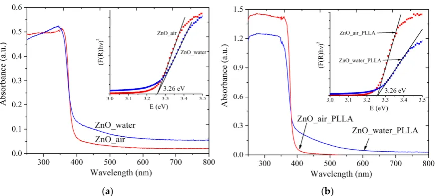
The absorption spectra of both powder samples are presented in Fig.5a. Similarly, Fig.5b exhibits
251
absorption spectra of PLLA scaffolds loaded with the same NPs. The wurtzite ZnO phase is known
252
to possess a characteristic shoulder of exciton absorption in the UV range below ~400 nm, which
253
results from the electron transfer from the valence zone (formed by Zn(3d) and О(2p) orbitals) to the
254
conductance zone (formed by Zn(4s) orbitals) [1,23]. This absorption band is observed at room
255
temperature because of the high binding energy of excitons [1,16]. Both samples, ZnO_water (blue
256
curve) and ZnO_air (red curve) are seen in panel (a) to have very weak absorption in visible range,
257
which is believed to result from admixtures (such as metallic Zn, carbonate species, and so on). When
258
the powders were loaded onto a polymer scaffold, the obtained composite materials are seen in panel
259
(b) to demonstrate very similar spectra (compare blue and red curves in Figs.5a,b).
260
The absorption spectra were used to evaluate the bandwidth of the materials (see insets in Fig.5).
261
Interestingly, both samples gave the same value of ~ 3.26 eV, in both forms, i.e. as-prepared and after
262
loading onto PLLA matrix. This value agrees well with literature values for ZnO nanomaterials where
263
Eg was reported to be of 3.2-3.4 eV [1,25,26]. No quantum-confinement effects are observed as the
264
obtained ZnO NPs are relatively large (over 10 nm).
(a) (b)
Figure 5. UV-Vis absorption spectra of powder samples (a) and ZnO-PLLA composites with NPs (b). Insets
266
present how Eg values were evaluated. Red and blue lines represent data for ZnO NPs produced in air and water,
267
respectively.
268
3.3.2. Photoluminescence spectra
269
Figure 6 presents PL spectra of as-prepared samples. Both samples (ZnO_air, red curve, and
270
ZnO_water, blue curve) are seen to have two bands in their PL spectra: a narrow UV peak around
271
380 nm and a wide band in the visible range, with maxima at 612 nm (red spectrum) and 625 nm
272
(blue spectrum). The two bands are known to have the exciton (UV) and defect (visible range) nature
273
[6,14,15,27,28]. The maximum of defect-related visible PL of sample ZnO_water is less intense and
274
red-shifted compared with that of sample ZnO_air.
275
276
Figure 6. PL spectra of samples PLA-generated in air (red line) and water (blue). Excitation source use
277
had λex=350 nm.
278
It should be mentioned that there is no full agreement in the literature on interpreting
defect-279
related PL in ZnO [14,15,27-30]. On one hand, the nature of defects is obviously one of main factors,
280
while on the other hand, the NP size and morphology were also reported to play their role
281
[6,14,29,30]. Experimentally, it was found that during aging of ZnO colloids prepared via PLA in
282
liquid, red-shift of the defect-related PL band occurred [31]. According to work [30], the redshift in
283
ZnO PL is owing to the presence of defects such as Zni, Oi, and OH groups, and especially surface
284
defects that are caused by all the peculiarities of PLA in liquid medium as preparation technique. All
285
this taken into account, as well as the results of XRD and TEM presented above, one can conclude
that the redshift of the PL band of sample ZnO_water could result from the following two factors:
287
excess of Oi defects and the rod-shaped morphology of the NPs.
288
Altogether, the PL spectra in Fig.6 demonstrate that both powders produced by PLA are rich in
289
defects, which should make them attractive for applications in catalysis or as antibacterial
290
nanomaterials.
291
3.3.3. IR and Raman spectra
292
The IR spectra of the PLA-prepared samples are presented in Fig. 7a. The band observed for
293
both samples (red and blue curves) at 500 cm-1 is characteristic of valence vibrations of the Zn-O band
294
[13]. Apart from Zn-O vibrations, two wide and intense bands in the range of 3750-2800 cm-1 and
295
1630-1200 cm-1 are observed for both samples, as well as a number of less pronounced bands between
296
~1000 and 600 cm-1.
297
Sample ZnO_water demonstrates a wide and featureless band at ~3400 см-1, similar to what was
298
previously reported by others for ZnO NPs prepared via PLA in water [9]. This band originates from
299
the valent vibrations of OH groups [32], OH groups bound to H atoms [33], and from Zn-OH species
300
[9]. In particular, there is a narrow peak at 3740 cm-1 which can be assigned to vibrations of isolated
301
OH group of water on the [10 ̅10] facet of ZnO [32]. The sample prepared in air exhibits more features
302
in this spectral region (see red spectrum in Fig.7a).
303
The second intense band of sample ZnO_water is believed to be related to carbonate species. For
304
instance, the peak seen at 1372 cm-1 is most likely from the carbonate group of hydrozincite, which is
305
also supported by another band at 1515 cm-1 [28]. The bands at 1049 and 825 cm-1 are also related to
306
the υ1 and υ2 vibrations of carbonate groups from hydrozincite [34]. In this region, the sample
PLA-307
prepared in water demonstrates a more intense band at 1609 cm-1 which is related to deformational
308
vibrations of intercalated H2O molecules. In addition, the vibration band υ3 of nitric group (1374 cm
-309
1) belonging to Zn5(OH)8(NO3)2×2H2O is also expected to manifest itself in this region [35].
310
The Raman spectra of samples are exhibited in Fig.7b. The wurtzite ZnO (space group 𝐶 ) is
311
known to demonstrate the following optical modes: polar А1 and Е1 that are active in the IR and
312
Raman ranges, doubly-degenerate Е2 mode with frequencies Е2(low) and Е2(hight), and inactive
313
“silent” В1 mode. The spectrum of sample ZnO_air (red line in Fig.7b) is seen to have more features,
314
demonstrating, e.g., bands at 205, 408, and 437 cm-1 which are related to the main optical vibrations
315
of the crystal lattice 2E2(low), transversal E1(TO) and E2(hight) vibrations, respectively. Since sample
316
ZnO_air exhibits a more pronounced band E2(hight) at 437 cm-1, this implies that its NPs are better
317
crystallized than those obtained in water (compare red and blue curves in Fig.7b). The band at 577
318
cm-1 can be assigned to either the A1(LO) or to E1(LO) lateral vibrations. This spectral range can also
319
be linked to a mixed quasi-LO mode with E1 and A1 symmetries that are closely related to the presence
320
of defects [36]. Because PLA-generated NPs are known to be rich in defects [7,9,14], it is safe to assume
321
that the peak at 577 cm-1 originates from the Е1(LO) mode. Finally, the bands below 150 cm-1 are hard
322
to identified and are most likely to be related to defects as well.
323
The distinctive feature of sample ZnO_water (blue spectrum in Fig.7b) is its wide dominating
324
A1(LO) mode around 560 cm-1, which is characteristic of lattice vibrations parallel to the growth
325
direction. This agrees well with the above presented TEM images where rod-like morphology of the
326
NPs prepared in water was revealed.
327
In agreement with the XRD results, where the presence of ZHN phase was revealed, the Raman
328
spectrum of sample ZnO_air also demonstrates additional modes of this phase (see peaks at 273, 506
329
and 639 cm-1 in Fig.7b) that correspond to the B1(low), 2B1(low) and B1(hight)+TA vibrations,
330
respectively. In addition, the intense peak at 1050 cm-1 belonging to υ1(NO3-) vibrations of nitric group
331
is also observed [37]. The weak band at 713 cm-1 is also due to the nitric group, presening its υ4(NO
3-332
) vibration mode. Finally, the wide band between 1050 and 1150 cm-1 observed for both samples is
333
related to multi-phonon vibrations of ZnO crystal lattice.
(a) (b)
Figure 7. FTIR (a) and Raman (b) spectra of powder samples. Red and blue lines present data for samples
335
prepared in air and water, respectively.
336
Thus, the FTIR and Raman spectroscopic data correlate well with the above presented XRD, PL
337
and TEM results. In particular, they confirm: (i) the presence of ZHN phase in sample ZnO_air; (ii)
338
the rod-shaped morphology of NPs generated in water; and (iii) the highly-defective nature of both
339
samples as previously revealed by their PL spectra.
340
3.4. Zeta potential
341
The value of ζ- potential is known to determine the stability of a colloidal solution, the system
342
being stable when it is over ±30 mV. Information on surface charge of NPs permits to predict their
343
interaction with bacterium cell membraine, the strength of which also depends on the bacteria type.
344
Figure 8 exhibits the values of ζ-potential of the two samples dispersed in water as they were titrated
345
with KOH solution to determine their рНiep.
346
347
Figure 8. Zeta potential of NPs evaluated at different pH values. Blue and red symbols stand for samples
348
prepared in water and air, respectively.
349
It is well-seen in Fig.8 that the ζ-potential values of both samples are positive, being +33 mV (red
350
curve) and +28 mV (blue curve) for NPs produced in air and water, respectively. Because of the larger
351
value of its ζ-potential, the dispersion of sample ZnO_air is more stable in water, which agrees with
352
the above discussion in section 3.2.2. While the initial pH of water was ~6.3, the values were shifted
353
to pH 7.3 (red curve) and pH 7.8 (blue curve) when the tested powders were dispersed. The values
354
of the isoelectric point obtained for the dispersions were found to be in the basic range, agreeing well
355
with the literature [38]. Thus, the surface of both types of NPs is positively charged at pH below this
value as protons from water are transferred onto the NPs and form surface Zn-OH2+ groups [39]. The
357
difference between the two curves in Fig.8 and thus in the values of рНiep implies that the surface
358
chemistry of the two powders prepared in different media is different, which was previously
359
revealed by both IR and Raman spectroscopy measurements.
360
As previously mentioned in section 3.2.2, the positive ζ-potential of both laser-generated
361
samples is an important factor that contributed to good surface adhesion and uniform deposition of
362
their NPs onto PLLA scaffolds, the latter being negatively charged.
363
3.4. Antibacterial activity
364
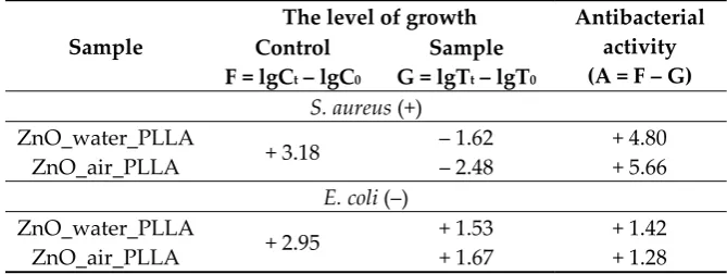
Table 2 shows the results of antibacterial tests obtained on the model PLLA tissues loaded with
365
the two laser-generated ZnO powders. It is clearly seen that both samples demonstrated good
366
antibacterial activity towards S. aureus (А>3, according to ISO 20743: 2013), the activity of sample
367
ZnO_air_PLLA being significantly higher. The latter finding could be explained by either the larger
368
specific surface area of its NPs or their surface chemistry, both assumptions needing additional
369
studies.
370
Table 2. Antibacterial activity of prepared samples towards S. aureus and E.coli.
371
Sample
The level of growth Antibacterial activity (А = F – G) Control
F = lgCt – lgC0
Sample G = lgTt – lgT0
S. aureus (+) ZnO_water_PLLA
+ 3.18 – 1.62 + 4.80
ZnO_air_PLLA – 2.48 + 5.66
E. coli (–) ZnO_water_PLLA
+ 2.95 + 1.53 + 1.42
ZnO_air_PLLA + 1.67 + 1.28
At the same time, the antibacterial activity of both samples towards E. coli, though exhibiting
372
strong inhibitory effect, was found to be very close to each other and lower than in case of S. aureus.
373
This agrees well with our previous results reported for PLA-prepared ZnO NPs deposited onto cotton
374
fabrics [10]. The observed difference in bactericidal action between the two strains tested is believed
375
to be caused by their wall structures. While the S. aureus is known to be protected by its wall made
376
of peptidoglikan (which has teichoic and lipoteichoic acids), the E. coli bacteria have an outer
377
membraine that has a more complex chemical composition and contains lipopolysaccharide
378
surrounded by a thin layer of peptidoglycan. The latter more complex structure provides the E. coli
379
higher resistivity. It should be noted, however, that for wound dressing it is the bactericidal action
380
against the S.aureus that is of higher importance than that against E. coli.
381
There are several models explaining the antibacterial action of ZnO NPs. One possible
382
mechanism proposed suggests that their bactericidal effect can be related to reactive oxygen species
383
generated by such particles [40]. It cannot be excluded that the inhibiting action could also be due to
384
the Zn2+ cations that are released as a result of partial dissolution of ZnO NPs [41] and result in
385
membraine disfunction [42] and NP internalization with the bacterium surface [43]. More details on
386
possible bactericidal mechanisms of ZnO NPs can be found elsewhere [2, 44].
387
The antibacterial tests carried out in this study confirm a high bactericidal potential of
nano-388
sized ZnO loaded onto biodegradbalbe PLLA matrix as a model dressing for wound treatment.
389
Besides, the novel ZnO powder produced via PLA in air was found to demonstrate higher
390
antibacterial activity, which can be explained by its more uniform distribution over matrix fibers and
391
by a larger specific surface area of its NPs.
392
393
4. Conclusions
395
In this study, we prepared ZnO nanoparticles (NPs) via ablating Zn target with nanosecond
396
pulsed laser in water and air media and compared their properties. It was found that the NPs
397
produced in air were of spherical shape, while their counterparts produced in water were somewhat
398
larger and rod-shaped. Both samples were based on hexagonal wurtzite ZnO phase, while because
399
of interaction with atmosphere nitrogen, the sample generated in air also had some fraction of
400
monoclinic zinc hydroxynitrate.
401
The NPs prepared in air were more stable in colloid (having zeta potential ζ > 30 mV) and
402
demonstrated better dispersibility in water. After characterization by various structural and
403
spectroscopic techniques, both powders were loaded onto polymeric matrix of biodegradable
poly-404
L-lactic acid, thus forming model biomedical composite materials for wound dressing. The
405
antibacterial behavior of the two model dressings was tested against S. aureus and E. coli strains,
406
showing promising bactericidal action against the former.
407
Upon comparing ZnO NPs produced in water and air, this work demonstrates that the latter
408
NPs had better dispersibility in water, while their antibacterial behavior was at least comparable with
409
that of the former ones. Thus, when it comes to using ZnO NPs as powders, for example to disperse
410
them and load onto/into biodegradable polymer matrix, nanoparticles produced via laser ablation in
411
air should be considered as real promising alternatives to their counterparts prepared in water.
412
Author Contributions: conceptualization, V.A.S. and S.A.K.; methodology, D.A.G.; validation, A.A.A. and
413
N.M.; investigation, E.A.G., D.A.G., I.N.L. and A.L.N.; visualization, E.A.G.; writing and editing, E.A.G., V.A.S.
414
and S.A.K.
415
Funding: This research was funded by the Tomsk State University competitiveness improvement program, and
416
by the Japan Society for the Promotion of Science (JSPS, grant no. 16K04904). D.A.G was supported by the
417
scholarship program of the President of the Russian Federation for young scientists and post-graduate students
418
(SP-2018 Competition).
419
Acknowledgments: The authors thank Dr. T.S. Kharlamova for zeta potential measurements and Dr.
420
M.A. Gerasimova for photoluminescence measurements. Characterization was carried out using equipment of
421
the Tomsk Regional Common Use Centre, Tomsk State University.
422
Conflicts of Interest: The authors declare no conflict of interest.
423
References
424
1. Özgür, Ü.; Alivov, Ya.I.; Liu, C.; Teke, A.; Reshchikov, M.A.; Doğan, S.; Avrutin, V.; Cho, S.J.; Morkoç, H.
425
A comprehensive review of ZnO materials and devices. J. Appl. Phys. 2005, 98, 041301,
426
doi:10.1063/1.1992666
427
2. Król, A.; Pomastowski, P.; Rafińska, K.; Railean-Plugaru, V.; Buszewski, B. Zinc oxide nanoparticles:
428
Synthesis, antiseptic activity and toxicity mechanism. Adv. Colloid Interface Sci. 2017, 249, 37–52,
429
doi:10.1016/j.cis.2017.07.033
430
3. Gomez, J.L.; Tigli, O. Zinc oxide nanostructures: From growth to application. J. Mater. Sci. 2013, 48,
612-431
624, doi:10.1007/s10853-012-6938-5
432
4. Mintcheva, N.; Aljulaih, A.A.; Wunderlich, W.; Kulinich, S.A.; Iwamori, S. Laser-ablated ZnO nanoparticles
433
and their photocatalytic activity toward organic pollutants. Materials 2018, 11, 1127,
434
doi:10.3390/ma11071127
435
5. Kondo, T.; Sato, Y.; Kinoshita, M.; Shankar, P.; Mintcheva, N.; Honda, M.; Iwamori, S.; Kulinich, S.A. Room
436
temperature ethanol sensor based on ZnO prepared via laser ablation in water. Jpn. J. Appl. Phys. 2017, 56,
437
080304, doi:10.7567/jjap.56.080304
438
6. Kulinich, S.A.; Kondo, T.; Shimizu, Y.; Ito, T. Pressure effect on ZnO nanoparticles prepared via laser
439
ablation in water. J. Appl. Phys. 2013, 113, 033509, doi: 10.1063/1.4775733
440
7. Zeng, H.; Du, X.W.; Singh, S.C.; Kulinich, S.A.; Yang, S.; He, J.; Cai, W. Nanomaterials via laser ablation/
441
8. Liang, C.; Shimizu, Y.; Masuda, M.; Sasaki, T.; Koshizaki, N. Preparation of layered zinc
443
hydroxide/surfactant nanocomposite by pulsed-laser ablation in a liquid medium. Chem. Mater. 2004, 16,
444
963–965, doi:10.1021/cm034706e
445
9. Yan, Z.; Chrisey, D.B. Pulsed laser ablation in liquid for micro-/nanostructure generation. J. Photochem.
446
Photobiol. C. 2012, 13, 204–223, doi:10.1016/j.jphotochemrev.2012.04.004
447
10. Svetlichnyi, V.; Shabalina, A.; Lapin, I.; Goncharova, D.; Nemoykina, A. ZnO nanoparticles obtained by
448
pulsed laser ablation and their composite with cotton fabric: Preparation and study of antibacterial activity.
449
Appl. Surf. Sci. 2016, 372, 20–29, doi:10.1016/j.apsusc.2016.03.043
450
11. Yang, X.C.; Riehemann, W.; Dubiel, M.; Hofmeister, H. Nanoscaled ceramic powders produced by laser
451
ablation. Mater. Sci. Eng., B 2002, 95, 299–307, doi:10.1016/S0921-5107(02)00291-X
452
12. Niu, K.Y.; Kulinich, S.A.; Jing, Y.; Zhu, A.L.; Du, X.W. Galvanic replacement reactions of active-metal
453
nanoparticles. Chem. Eur. J. 2012, 18, 4234–4241, doi:10.1002/chem.201102544
454
13. Gondal, M.A.; Drmosh, Q.A.; Yamani, Z.H.; Saleh, T.A. Synthesis of ZnO nanoparticles by laser ablation in
455
liquid and their annealing transformation into ZnO nanoparticles. Appl. Surf. Sci. 2009, 256, 298–304,
456
doi:10.1016/j.apsusc.2009.08.019
457
14. Goto, T.; Honda, M.; Kulinich, S.A.; Shimizu, Y.; Ito, T. Defects in ZnO nanoparticles laser-ablated in water–
458
ethanol mixtures at different pressures. Jpn. J. Appl. Phys. 2015, 54, 070305, doi:10.7567/JJAP.54.070305
459
15. Honda, M.; Goto, T.; Owashi, T.; Rozhin, A.G.; Yamaguchi, S.; Ito, T.; Kulinich, S.A. ZnO nanorods
460
prepared via ablation of Zn with millisecond laser in liquid media. Phys. Chem. Chem. Phys. 2016, 18, 23628–
461
23637, doi:10.1039/C6CP04556A
462
16. Janotti, A.; Van de Walle, C.G. Fundamentals of zinc oxide as a semiconductor. Rep. Prog. Phys. 2009, 72,
463
126501, 1–29, doi:10.1088/0034-4885/72/12/126501
464
17. Svetlichnyi, V.A.; Shabalina, A.V.; Lapin, I.N.; Goncharova, D.A.; Kharlamova, T.S.; Stadnichenko, A.I.
465
Comparative study of magnetite nanoparticles obtained by pulsed laser ablation in water and air. Appl.
466
Surf. Sci. 2019, 467–468, 402–410, doi:10.1016/j.apsusc.2018.10.189
467
18. Albertsson, A.-C.; Varma, I.K. Degradable aliphatic polyesters. In Advances in Polymer Science; A.-C.
468
AlbertssonEd; Springer: Berlin, Germany, 2002; Volume. 157, pp 1-40, ISBN 978-3-642-10576-0.
469
19. Badaraev, A.D.; Nemoykina, A.L.; Bolbasov, E.N.; Tverdokhlebov, S.I. PLLA scaffold modification using
470
magnetron sputtering of the copper target to provide antibacterial properties. Resour.-Effic. Technol. 2017,
471
3, 204–2011, doi:10.1016/j.reffit.2017.05.004
472
20. ISO 20743:2013 Textiles - Determination of antibacterial activity of textile products. 2013 (2nd Edition)
473
21. Roske, C.W.; Lefler, J.W.; Müller, A.M. Complex nanomineral formation utilizing kinetic control by PLAL.
474
J. Colloid Interface Sci. 2017, 489, 68–75, doi:10.1016/j.jcis.2016.08.079
475
22. Ishikawa, Y.; Shimizu, Y.; Sasaki, T.; Koshizaki N. Preparation of zinc oxide nanorods using pulsed laser
476
ablation in water media at high temperature. J. Colloid Interface Sci. 2006, 300, 612–615,
477
doi:10.1016/j.jcis.2006.04.005
478
23. Panda, D.; Tseng T.Y One-dimensional ZnO nanostructures: fabrication, optoelectronic properties, and
479
device applications. J. Mater. Sci. 2013, 48, 6849–6877, doi:10.1007/s10853-013-7541-0
480
24. Croisier, F.; Aqil, A.; Malherbe, C.; Gilbert, B.; Detrembleur, C.; Jerome, C. Charged Poly(D,L-lactide)
481
nanofibers: Towards customized surface properties, Macromol. Symp. 2011, 309–310, 20–27,
482
doi:10.1002/masy.201100037
483
25. Pearton, S.J.; Norton, D.P.; Ip, K.; Steiner, T.; Heo; Y.W. Recent advances in processing of ZnO. J. Vac. Sci.
484
Technol., B. 2004, 22, 932–948, doi:10.1116/1.1714985
485
26. Yi, G.C.; Wang, C.; Park, W. ZnO nanorods: Synthesis, characterization and applications. Semicond. Sci.
486
Technol. 2005, 20, 22–34, doi:10.1088/0268-1242/20/4/003
487
27. Vempati, S.; Mitra, J; Dawson P. One-step synthesis of ZnO nanosheets: a blue-white fluorophore. Nanoscale
488
Res. Lett. 2012, 7, 470, 1–10, doi:10.1186/1556-276X-7-470
489
28. Mao, J.; Chen, X.; Ling, T.; Du X. Strong blue emission from zinc hydroxide carbonate nanosheets. J. Lumin.
490
2016, 177, 242–248, doi:10.1016/j.jlumin.2016.05.006
491
29. Anantachaisilp, S.; Smith, S.M.; Ton-That, C.; Pornsuwan, S.; Moo, A.R.; Nenstiel, C.; Hoffmann, A.;
492
Phillips, M.R. Nature of red luminescence in oxygen treated hydrothermally grown zinc oxide nanorods.
493
30. Kurudirek, S.V.; Pradel ,K.C.; Summers, C.J. Low-temperature hydrothermally grown 100 mm vertically
495
well-aligned ultralong and ultradense ZnO nanorod arrays with improved PL property. J. Alloys Compd.
496
2017, 702, 700–709, doi:10.1016/j.jallcom.2017.01.273
497
31. Svetlichnyi, V.A.; Lapin, I.N. Structure and properties of nanoparticles fabricated by laser ablation of Zn
498
metal targets in water and ethanol. Russ. Phys. J. 2013, 56, 581-587, doi:10.1007/s11182-013-0071-z
499
32. Wöll, C. The chemistry and physics of zinc oxide surfaces. Prog. Surf. Sci. 2007, 82, 55–120,
500
doi:10.1016/j.progsurf.2006.12.002
501
33. Barsukov, D.V.; Subbotina, I.R. IR-study of hydrated surface of oxide photocatalysts. Russ. Chem. Bull. 2017,
502
66, 1847–1853, doi:10.1007/s11172-017-1956-8
503
34. Japi, D.; Bitenc, M.; Marinšek, M.; Orel, Z.C. The impact of nano-milling on porous ZnO prepared from
504
layered zinc hydroxide nitrate and zinc hydroxide carbonate. Mater. Res. Bull. 2014, 60, 738–745,
505
doi:10.1016/j.materresbull.2014.09.061
506
35. Li, P.; Xu, Z.P.; Hampton, M.A.; Vu, D.T.; Huang, L.; Rudolph, V.; Nguyen, A.V. Control preparation of
507
zinc hydroxide nitrate nanocrystals and examination of the chemical and structural stability. J. Phys. Chem.
508
C. 2012, 116, 10325−10332, doi:10.1021/jp300045u
509
36. Klingshirn, C.F.; Meyer, B.K.; Waag, A.; Hoffmann A.; Geurts, J. Zinc oxide: From fundamental properties
510
towards novel applications.In Springer Series in Materials Science; Hull R., Jagadish, C., Osgood, R.M, Jr.,
511
Parisi, J., Wang Z., Warlimont, H., Eds; Springer: New York, USA, 2010; Volume. 120; pp. 233-266. ISBN
512
978-3-642-10576-0.
513
37. Liu, X.; Wang, C.; Liu, X.; Ouyang, L.; You, Z.; Lu, Y.; Chen, X. Understanding the factors that control the
514
formation and morphology of Zn5(OH)8(NO3)2×2H2O through hydrothermal route. J. Nanomater. 2013, 2013,
515
938370, doi:10.1155/2013/938370
516
38. Degen, A.A.; Kosec, M. Effect of pH and impurities on the surface charge of zinc oxide in aqueous solution.
517
J. Eur. Ceram. Soc. 2000, 20, 667–673, doi:10.1016/S0955-2219(99)00203-4
518
39. Lee, K.M.; Lai, C.W.; Ngai, K.S.; Juan, J.C. Recent developments of zinc oxide based photocatalyst in water
519
treatment technology: A review. Water Res. 2016, 88, 428–448, doi:10.1016/j.watres.2015.09.045
520
40. Xia, T.; Kovochich, M.; Liong, M.; Mädler, L.; Gilbert, B.; Shi, H.; Yeh, J.I.; Zink, J.I.; Nel, A.E. Comparison
521
of the mechanism of toxicity of zinc oxide and cerium oxide nanoparticles based on dissolution and
522
oxidative stress properties. ACS Nano 2008, 2, 2121–2134, doi:10.1021/nn800511k
523
41. Li, M.; Pokhrel, S.; Jin, X.; Mädler, L., Damoiseaux, R., Hoek, E.M. Stability, bioavailability, and bacterial
524
toxicity of ZnO and iron-doped ZnO nanoparticles in aquatic media. Environ. Sci. Technol.2011, 45, 755–
525
761, doi:10.1021/es102266g
526
42. Kavitha, T.; Gopalan, A.I.; Lee, K.-P.; Park, S.-Y. Glucose sensing, photocatalytic and antibacterial
527
properties of graphene–ZnO nanoparticle hybrids. Carbon 2012, 50, 2994–3000,
528
doi:10.1016/j.carbon.2012.02.082
529
43. Huang, Z.; Zheng, X.; Yan, D.; Yin, G.; Liao, X.; Kang, Y.; Yao, Y.; Huang, D.; Hao, B. Toxicological effect of
530
ZnO nanoparticles based on bacteria. Langmuir 2008, 24, 4140–4144, doi:10.1021/la7035949
531
44. Sirelkhatim, A.; Mahmud, S.; Seeni, A.; Kaus, N.H.M.; Ann, L.C.; Bakhori, S.K.M.; Hasan, H.; Mohamad,
532
D. Review on zinc oxide nanoparticles: Antibacterial activity and toxicity mechanism. Nano-Micro Lett.