C A S E S E R I E S
Peripheral hypertrophic subepithelial corneal
degeneration: clinical aspects related to in vivo
confocal microscopy and optical coherence
tomography
This article was published in the following Dove Press journal:
International Medical Case Reports Journal
Jeanne Martine Gunzinger Nafsika Voulgari
Aleksandra Petrovic Kattayoon Hashemi Georgios Kymionis
Lausanne University, Cornea and Refractive Surgery Department, Jules-Gonin Eye Hospital, Lausanne, Switzerland
Purpose:To report thefindings of anterior segment optical coherence tomography (AS-OCT) and in vivo confocal microscopy (IVCM) in two patients with peripheral hypertrophic sub-epithelial corneal degeneration (PHSD).
Methods:Case series by restrospective chart review and imaging analysis of AS-OCT and IVCM. Results: Slit lamp examination of the two patients revealed a bilateral subepithelial-elevatedfibrous tissue of the superior-nasal quadrant, as well as inferior-nasal in one of the patients. Best corrected visual acuity ranged from 20/25 to 20/15. AS-OCT showed continuous, homogenous, well-demarked hyperreflective subepithelial band associated with hyperreflectivity in the anterior stroma. IVCM demonstrated normal epithelial cell morphology and arrangement and a fibrous structure subepithelial and in the anterior stroma.
Conclusion: AS-OCT and IVCM can facilitate the diagnosis of PHSD and differentiate it from other corneal entities that present peripheral opacifications.
Keywords:peripheral hypertrophic subepithelial corneal degeneration, anterior segment optical coherence tomography, in vivo confocal microscopy, peripheral corneal opacification
Introduction
Peripheral hypertrophic subepithelial corneal degeneration (PHSD) is a clinical
entity of unknown etiology, first reported in 2003,1that more commonly needs to
be differentiated from Salzmann’s nodular degeneration (SND). It is characterized
by predominantly bilateral, gradually progressing, perilimbal, subepithelial corneal
fibrosis with adjacent superficial neovascularization in the absence of concurrent or
preceding clinically visible ocular surface inflammation.1–3Terrien marginal
degen-eration and corneal intraepithelial neoplasia (CIN) can equally present with
periph-eral corneal opacification sharing many similarities with PHSD but otherwise
exhibiting distinct clinicopathological properties. We herein present two cases of PHSD that manifested as Terrien marginal degeneration and CIN, respectively. The
distinguishable features that allow identification of this uncommon entity are
high-lighted. To our knowledge, this is the first report of anterior segment optical
coherence tomography (AS-OCT) and in vivo confocal microscopy (IVCM) char-acteristics of PHSD.
Correspondence: Georgios Kymionis Lausanne University, Cornea and Refractive Surgery Department, Jules-Gonin Eye Hospital, Avenue de France 15, 1004 Lausanne, Switzerland
Email [email protected]
International Medical Case Reports Journal
Dove
press
open access to scientific and medical researchOpen Access Full Text Article
International Medical Case Reports Journal downloaded from https://www.dovepress.com/ by 118.70.13.36 on 24-Aug-2020
Case series
Case 1
An 88-year-old man was referred to the cornea department of the University of Lausanne, Jules Gonin Eye Hospital for suspicion of a CIN or carcinoma in situ due to a
superficial corneal lesion in the right eye. According to
the referring ophthalmologist, this lesion was first
described 10 years ago, showing gradual progression to the paracentral region.
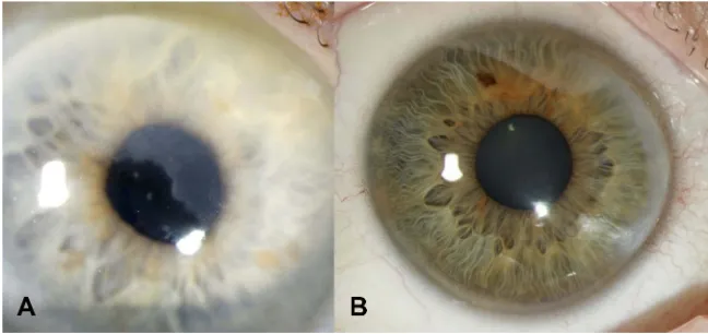
The patient complained of slightly blurred vision in his right eye for a few months. Best corrected visual acuity (BCVA) was 20/25 in the right eye and 20/20 in the left eye. Slit lamp biomicroscopy revealed subepithelial-elevated
opacities andfibrosis of the superior-nasal quadrant of the
right eye, with otherwise no other ocular surface lesion or
scar (Figure 1A). Topography (Pentacam®, Oculus,
Wetzlar, Germany) showed a topographic astigmatism of 0.1 diopters at 167° in the right eye. AS-OCT
(Avanti-Optovue, Fermont, California, 70,000 scans/second, scan beam wavelength 840 nm) showed a mostly continuous,
homogenous, well-demarcated hyperreflective
(qualita-tively, compared to surrounding structures as the epithelium or the stroma) subepithelial band respecting the Bowman layer while rarely breaking through and expanding slightly
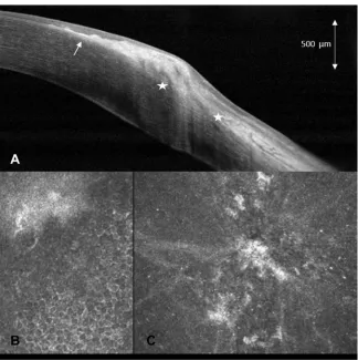
into the anterior stroma (Figure 2AandB). IVCM (Rostock
Cornea Module for Heidelberg Retina Tomograph,
Heidelberg Engineering Inc., Heidelberg, Germany, 670 nm diode laser, axial resolution of approximately 1 µm) showed normally structured epithelium with underlying
hyperreflective,fibrotic appearing structures comprising of
oval-shaped cells (Figure 2C). Bowman layer was mostly
smooth and continuous but showed a reduction of nerve
axons. Few images showed breakthrough of fibrosis and
slight expansion into the anterior stroma.
On close examination of the left eye an identical, but smaller, only peripheral opacity was also found in the
Figure 1Slit lamp photographs of (A) patient 1 (masquerading as carcinoma in situ) and (B) patient 2 (masquerading as Terrien marginal degeneration), showing peripheral corneal opacities of the superior-nasal quadrant or the nasal quadrant, respectively.
Figure 2(A) Anterior segment optical coherence tomography (AS-OCT) of the right eye horizontally and (B) vertically in patient 1, demonstrating mostly continuous, homogenous, well-demarcated hyperreflective subepithelial band respecting the Bowman layer (arrow) while rarely breaking through and expanding slightly into the anterior stroma (star) (C) oblique in vivo confocal microscopy (IVCM) of patient 1 demonstrating normal structured epithelium, underlyingfibrotic structures, intact Bowman layer (arrowhead) and normal anterior stroma.
International Medical Case Reports Journal downloaded from https://www.dovepress.com/ by 118.70.13.36 on 24-Aug-2020
superior-nasal quadrant. Systemic evaluation was normal, there was no sign of paraproteinemia.
Due to the preserved BCVA of 20/25 and thefindings of
AS-OCT and IVCM, keratectomy was not recommended.
Case 2
A 42-year-old woman was referred for suspicion of Terrien marginal degeneration in both eyes. The patient complained of red eyes for 3 months prior to the consultation.
BCVA was 20/20 in the right eye and 20/15 in the left eye. Slit lamp biomicroscopy revealed symmetric slightly ele-vated, nodular, peripheral, subepithelial, and anterior stromal
fibrosis in the nasal quadrant of both eyes with a non-nodular
zone toward the limbus giving the false impression of
thin-ning (Figure 1B). Superficial new vessels were present from
to limbus toward the basis of the fibrosis. Topography
showed astigmatism of 1.0 diopter at 13° in the right eye and 1.1 diopters at 126° in the left eye. Pachymetry did not show any sign of peripheral corneal thinning. AS-OCT demonstrated a continuous, homogenous, well demarcated,
slightly nodular hyperreflective subepithelial band. This
band respects the Bowman layer central to the lesion, while showing an inhomogeneous structure breaking through the Bowman layer and expanding into the anterior stroma
per-ipheral to the lesion (Figure 3A). IVCM showed normally
structured epithelium with underlying hyperreflective,
fibrous structures with rare cells (Figure 3B and C).
Bowman layer was smooth and continuous centrally, but
not well demarcated peripherally. Expansion offibrosis into
the anterior stroma was visible on peripheral images. Systemic evaluation was normal, there was no sign of paraproteinemia.
The patient was treated with ciclosporin 0.05% eye drops
twice daily andfluorometholone 0.1% one drop every second
day. Six and nine months follow-ups showed no evolution of the lesion and topical treatment was slowly tapered.
Discussion
We describe herein two cases of PHSD, resembling CIN and Terrien marginal degeneration, respectively.
Figure 3(A) anterior segment optical coherence tomography (AS-OCT) of the right eye in patient 2, demonstrating mostly continuous, slightly nodular, well-demarcated hyperreflective subepithelial band partially breaking through the Bowman layer (arrow) and expanding into the anterior stroma (star). (B) Slightly oblique in vivo confocal microscopy (IVCM) of patient 2 demonstrating normal structured epithelium, and adjacentfibrotic structures (C) tangential IVCM on the level of thefibrotic structures.
International Medical Case Reports Journal downloaded from https://www.dovepress.com/ by 118.70.13.36 on 24-Aug-2020
Recently proposed diagnostic criteria for PHSD
include perilimbal subepithelial fibrosis with thickening
of the cornea, possible extension to the mid-periphery but
no central cornea involvement.2,3Superficial
neovascular-ization up to the base of the fibrous tissue and two or
more diopters of topographic astigmatism represent addi-tional distinguishable features. Typical symptoms consist of ocular surface irritation and decreased visual acuity due to regular and irregular astigmatism. PHSD must be differentiated from other corneal diseases such as SND, auto-immune diseases, paraproteinemic keratopathy due
to monoclonal gammopathy,4 CIN or other entities such
as Terrien marginal degeneration as noted in one of our cases.
CIN is a squamous cell in situ carcinoma showing dysplastic epithelial cells which can involve less than 25% (Grade I) or up to more than 75% (Grade III) of
epithelial thickness.5 Clinical presentation,
indistinguish-able from PHSD, includes ocular surface irritation, red-ness, foreign body sensation or decreased vision. Slit lamp examination, similar to PHSD, reveals a mostly well
demarcated, slightly elevated superficial corneal lesion.
However, in contrast to PHSD, it presents as a translucent corneal clouding rarely accompanied by leukoplakia,
lack-ing subepithelialfibrosis and accompanied by feeder
ves-sels expanding into the lesion.6The lesion tends to evolve
rapidly. As CIN is an epithelial disorder, AS-OCT will
show a typical pattern of hyperreflective, thickened
epithe-lium (normal epithelial thickness is 50–52 µm) and an
abrupt transition from normal to abnormal epithelium.7
In ourfirst patient, epithelium was clearly normal on
AS-OCT and thus rule out the diagnosis of CIN.
If the clinical presentation is in favor of PHSD, AS-OCT is
invaluable in identifying a hyperreflective, strictly
subepithe-lial lesion with intact overlying epithesubepithe-lial structures1,8–and
absence of epithelial destruction or largefibrovascular
struc-tures, as noted in CIN.9IVCM can further help to clarify the
diagnosis of PHSD by showing regularly structured epithelial cells with no sign of enlargement, irregularity, anisocytosis, or
anisonucleosis as seen in CIN.9If there is doubt about the
diagnosis, further investigations must be performed to exclude CIN.
Terrien marginal degeneration is a slowly progressive degeneration of unclear origin, usually starting in the
super-onasal quadrant.10 Clinical presentation often includes
vision loss due to induced astigmatism. Slit lamp
examina-tion typically shows peripheral thinning and superficial
vascularization with lipid deposition at the leading edge.
Similar to PHSD, it presents as a perilimbal opacity, how-ever, in contrast to PHSD, it is often accompanied by yellow lipid deposits and peripheral thinning. AS-OCT is helpful to differentiate between the two entities. Whereas in Terrien
marginal degeneration, findings include corneal thinning
(normal peripheral corneal thickness is 612–640 µm11) but
no associated reflectivity changes in subepithelial
structures12 in contrast to the typical findings in PHSD
which include a hyperreflective subepithelial band and
might be associated with hyperreflective areas in the
ante-rior stroma, but does not show corneal thinning. To our
knowledge there are only two case reports of IVCMfi
nd-ings in Terrien marginal degeneration, both showing lipid deposits, activated keratocytes and needle-like material or honeycomb-like pattern respectively, and in one case
inflammatory cells,13,14 whereas in our case IVCM
pre-sentedfibrous subepithelial structures with expansion into
the anterior stroma, but no lipid deposits.
In conclusion, our cases suggest that PHSD might masquerade as Terrien marginal degeneration or CIN and highlight the importance of considering this pathology in
the differential diagnosis of peripheral corneal opacifi
ca-tions not fitting the criteria of other broadly known
enti-ties. In both cases, the diagnosis of PHSD was made by
slit lamp examination and confirmed by AS-OCT and
IVCM, demonstrating their value as complementary tools in the diagnosis of PHSD.
Ethics statement
Written informed consent has been provided by both patients for the use of retrospective chart data as well as publication of images. Institutional approval was granted for the publication of the cases.
Acknowledgments
None of the authors have any commercial or propriety interest related to the case series.
Disclosure
The authors report no conflicts of interest in this work.
References
1. Maust HA, Raber IM. Peripheral hypertrophic subepithelial corneal degeneration.Eye Contact Lens.2003;29(4):266–269. doi:10.1097/01. icl.0000087489.61955.82
2. Järventausta PJ, Tervo TMT, Kivelä T, Holopainen JM. Peripheral hypertrophic subepithelial corneal degeneration –clinical and histo-pathological features. Acta Ophthalmol. 2014;92(8):774–782. doi:10.1111/aos.12394
International Medical Case Reports Journal downloaded from https://www.dovepress.com/ by 118.70.13.36 on 24-Aug-2020
3. Gore DM, Iovieno A, Connell BJ, Alexander R, Meligonis G, Dart JK. Peripheral hypertrophic subepithelial corneal degeneration: nomencla-ture, phenotypes, and long-term outcomes.Ophthalmology.2013;120 (5):892–898. doi:10.1016/j.ophtha.2012.10.037
4. Skalicka P, Dudakova L, Palos M, et al. Paraproteinemic keratopathy associated with monoclonal gammopathy of undetermined signifi -cance (MGUS): clinicalfindings in twelve patients including recur-rence after keratoplasty. Acta Ophthalmol. 2019:aos.14123. doi:10.1111/aos.14123
5. Hamam R, Bhat P, Foster CS. Conjunctival/corneal intraepithelial neoplasia. Int Ophthalmol Clin. 2009;49(1):63–70. doi:10.1097/ IIO.0b013e3181924ec3
6. Honavar SG, Manjandavida FP. Tumors of the ocular surface: a review. Indian J Ophthalmol. 2015;63(3):187–203. doi:10.4103/ 0301-4738.156912
7. Atallah M, Joag M, Galor A, et al. Role of high resolution optical coherence tomography in diagnosing ocular surface squamous neo-plasia with coexisting ocular surface diseases.Ocul Surf.2017;15 (4):688–695. doi:10.1016/j.jtos.2017.03.003
8. Rommel F, Grisanti S, Ranjbar M. Peripheral hypertrophic subepithe-lial corneal degeneration.JAMA Ophthalmol.2017;135(6):e170664. doi:10.1001/jamaophthalmol.2017.0664
9. Xu Y, Zhou Z, Wang M, Liu F, Qu H, Hong J. The clinical value of in vivo confocal microscopy for diagnosis of ocular surface squamous neoplasia.Eye (Lond). 2012;26(6):781–787. doi:10.103 8/eye.2012.15
10. Chan AT, Ulate R, Goldich Y, Rootman DS, Chan CC. Terrien marginal degeneration: clinical characteristics and outcomes.Am J Ophthalmol.2015;160(5):867–872.e1. doi:10.1016/j.ajo.2015.07.031 11. Sridhar MS. Anatomy of cornea and ocular surface. Indian J
Ophthalmol.2018;66(2):190–194. doi:10.4103/ijo.IJO_646_17 12. Rodriguez M, Yesilirmak N, Chhadva P, Goldhagen B, Karp C, Galor
A. High-resolution optical coherence tomography in the differentia-tion of inflammatory versus noninflammatory peripheral corneal thin-ning. Cornea. 2017;36(1):48–52. doi:10.1097/ICO.00000000000 01023
13. Ceresara G, Migliavacca L, Orzalesi N, Rossetti L. In vivo con-focal microscopy in terrien marginal corneal degeneration: a case report. Cornea. 2011;30(7):820–824. doi:10.1097/ICO.0b013e3 1820143ed
14. Ferrari G, Tedesco S, Delfini E, Macaluso C. Laser scanning in vivo confocal microscopy in a case of Terrien marginal degenera-tion. Cornea. 2010;29(4):471–475. doi:10.1097/ICO.0b013e31 81b46aa3
International Medical Case Reports Journal
Dove
press
Publish your work in this journal
The International Medical Case Reports Journal is an international, peer-reviewed open-access journal publishing original case reports from all medical specialties. Previously unpublished medical posters are also accepted relating to any area of clinical or preclinical science. Submissions should not normally exceed 2,000 words or 4
published pages including figures, diagrams and references. The manuscript management system is completely online and includes a very quick and fair peer-review system, which is all easy to use. Visit http://www.dovepress.com/testimonials.php to read real quotes from published authors.
Submit your manuscript here:https://www.dovepress.com/international-medical-case-reports-journal-journal
International Medical Case Reports Journal downloaded from https://www.dovepress.com/ by 118.70.13.36 on 24-Aug-2020