Clinical and Experimental Gastroenterology
Dove
press
R E V I E W
open access to scientific and medical research
Open Access Full Text Article
Natural orifice specimen extraction in colorectal
surgery: patient selection and perspectives
Kevin M Izquierdo Ece Unal
John H Marks
Division of Colorectal Surgery, Lankenau Medical Center, Wynnewood, PA, USA
Abstract: Over the past 30 years, colorectal surgery has evolved to include minimally invasive surgical techniques. Minimally invasive surgery is associated with reduced postoperative pain, reduced wound complications, earlier return of bowel function, and possibly shorter length of hospital stay. These benefits have been attributed to a reduction in operative trauma compared to open surgery. The need to extract the specimen in colorectal operations through a “mini-laparotomy” can negate many of the advantages of minimally invasive surgery. Natural orifice specimen extraction (NOSE) is the opening of a hollow viscus that already communicates with the outside world, such as the vagina or distal gastrointestinal tract, in order to remove a speci-men. The premise of this technique is to reduce the trauma required to remove the specimen with the expectation that this may improve outcomes. Reduction in postoperative analgesic use, quicker return of bowel function, and shorter length of hospital stay have been observed in colorectal operations with NOSE compared to conventional specimen extraction. While the feasibility of NOSE has been demonstrated in colorectal surgery, failures of this technique have also been described. Selection of patients who can successfully undergo NOSE needs further investigation. This review aims to guide surgeons in appropriately selecting patients for NOSE in colorectal surgery. Patient and specimen characteristics are reviewed in order to define patient populations in which NOSE is likely to be successful. Randomized trials comparing NOSE to conventional specimen extraction in colorectal surgery tend to enroll patients with favorable
characteristics (body mass index <30, American Society of Anesthesiologists class ≤3, specimen
diameter <6.5 cm) and demonstrate improved outcomes. Adopters of NOSE should restrict using
this technique to the populations in which feasibility has been defined in the literature. Wider
application to other populations, particularly patients with body mass index >30 and those with
significant comorbidities, requires further study.
Keywords:natural orifice specimen extraction, colorectal, minimally invasive surgery, patient selection, rectum, feasibility of NOSE, target organ
History and feasibility
Colorectal minimally invasive surgery is associated with improved outcomes and
fewer complications when compared to open surgery.1–3 Despite these advantages,
there is significant morbidity that is associated with abdominal wall incisions made for colorectal specimen extraction. In minimally invasive colorectal surgery, the length of the longest incision is dependent upon that which is needed to extract the specimen: the so-called mini-laparotomy. Mini-laparotomies used for specimen extraction are
associated with increased wound infection, hernia, and postoperative pain.4–7 Natural
orifice specimen extraction (NOSE) eliminates extraction site trauma, which is other-wise always needed. NOSE is defined as the removal of a surgical specimen by opening
Correspondence: John H Marks Division of Colorectal Surgery, Lankenau Medical Center, 100 East Lancaster Avenue, Medical Science Building Suite 375, #330w, Wynnewood, PA, USA Tel +1 610 645 9093
Email [email protected]
Journal name: Clinical and Experimental Gastroenterology Article Designation: REVIEW
Year: 2018 Volume: 11
Running head verso: Izquierdo et al
Running head recto: Clinical and experimental gastroenterology DOI: http://dx.doi.org/10.2147/CEG.S135331
Clinical and Experimental Gastroenterology downloaded from https://www.dovepress.com/ by 118.70.13.36 on 20-Aug-2020
For personal use only.
Dovepress
Izquierdo et al
a hollow viscus that already communicates with the outside world, such as the gastrointestinal tract or the vagina. Rather than obtaining the specimen through an abdominal incision, a viscerotomy is used for specimen extraction, which allows patients to completely avoid the morbidity associated with larger abdominal incisions. Feasibility of NOSE in colorectal surgery is well documented.
Stewert et al8 and Nezhat9 were among the first to report
extraction of a colectomy specimen through the vagina
in 1991 and 1992. In Nezhat’s series,9 colectomies with
transvaginal specimen extraction were performed for the treatment of endometriosis, though this technique has now been successfully implemented in the treatment of
inflam-matory bowel disease, diverticulitis, and malignancy.10 The
first report of partial colectomy with NOSE via the anus was
described in 1993 by Franklin et al.11 To date, there have been
many documented cases in which either the colon, rectum, anus, or vagina has been used to remove both malignant
and benign pathology from the cecum to the distal rectum.10
Additionally, successful total mesorectal excision has been
performed with NOSE, as first described by Person et al12
in 2006.
A barrier to wider adoption of NOSE is technical diffi-culty. The mini-laparotomy, in some cases, is used to perform a majority of the operation, as in hand-assisted laparoscopy. Adoption of NOSE by surgeons who typically perform colec-tomies in this fashion would be faced with a steeper learning curve than surgeons who use the mini-laparotomy solely as a specimen extraction site. On that note, intracorporeal anastomosis is a prerequisite skill for those adopting NOSE. Removal of more proximal specimens, as in a right colectomy, requires the presence of a skilled endoscopist who can snare and pull the specimen endoluminally through the length of the distal gastrointestinal tract. Specimen extraction via the vagina requires a posterior colpotomy, an operative maneuver that is not typically performed by general or colorectal sur-geons. Furthermore, these technical challenges are amplified by a lack of standardization of the technique.
The demands for these technical skills are more impor-tant in removing right-sided colon pathology as compared to left-sided pathology. There are inherent anatomic factors that make NOSE for right-sided colon pathology more dif-ficult. Right colectomy specimens extracted through the lower gastrointestinal tract via distal colotomy must travel the length of the remaining transverse, descending, and sig-moid colon, through the rectum and out of the anus using an endoscope. While this was demonstrated to be feasible in 2010 by Eshuis et al,13 it is inherently difficult due to the
anatomically narrow and torturous sigmoid colon. In that series, extraction via colotomy failed in 2 of 10 patients due to the bulk of the specimen. This technique is still performed in some centers, though limitations related to the size of the
specimen are stricter than for left-sided colon lesions.14 This
approach has little practicality due to its significant technical challenges, hence its limited use.
The vaginotomy, also called a colpotomy, is a safe technique commonly employed by gynecologists and has been applied in NOSE for both gynecologic and colorectal pathologies. The first recorded vaginal specimen extractions in colorectal surgery were in the 1990s. This technique con-tinues to be implemented, particularly for right-sided colon pathology, given the difficulty of using the distal colon for specimen extraction. A more recent series of right colecto-mies with NOSE via the vagina was described by Franklin
et al15 in 2013. In this series that included 26 transvaginal
specimen extractions, feasibility of this method is demon-strated with a complication rate of 7.7%, a rate comparable
to conventional specimen removal. Karagul et al14 found that
specimens that were too large for transrectal and transanal extraction were successfully removed via the vagina. The obvious restriction of this technique is that it is only an option for female patients.
A greater volume of literature focuses on NOSE for
left-sided colon pathology10 and is typically performed through a
proctotomy. The distal transection point for specimens in the left colon can serve as the viscerotomy in these cases. The distensible rectum lends itself well to passage of specimens for extraction, and anal dilation for retrieval is a relatively simple task. A step-wise description of this technique is
described by Wolthuis et al.16 Transluminal endoscopic
operation (TEO) ports (TEO platform; Storz, Tuttlingen,
Germany) have been used in long rectal stumps (>10 cm)
with double-ringed wound protectors (Alexis wound retrac-tor; Applied Medical, Rancho Santa Margarita, CA, USA) used for shorter rectal stumps to facilitate the removal of
the specimen.17 Other groups have demonstrated removal of
specimens without any wound protection.10 Theoretically,
there exists the possibility of wound implantation with cancer cells in malignant cases. The long-term implications of this, particularly in removing malignant tumors, remain to be seen.
In the treatment of rectal cancer, the transanal total mesorectal excision (taTME) has been well described with promising oncologic results. NOSE, in these cases, is the most practical route of specimen extraction as the rectum or anus serves as the viscerotomy site and needs to be transected
Clinical and Experimental Gastroenterology downloaded from https://www.dovepress.com/ by 118.70.13.36 on 20-Aug-2020
Dovepress Clinical and experimental gastroenterology
as a mandatory step in the operation. In December 2008, we performed our first taTME using transanal endoscopic microsurgery equipment (Richard Wolf, Knittlingen, Ger-many). We now routinely perform NOSE as part of any laparoscopic or robotic transanal-abdominal transanal proc-tosigmoidectomy and have reported perioperative morbidity and mortality of 13.4% and 0.3%, respectively, in our series
of 373 consecutive patients.18
Quality of studies that report on colorectal NOSE out-comes has substantially increased in recent years. Larger series and randomized controlled trials now demonstrate some benefits with NOSE as compared to conventional specimen extraction, particularly in regard to postoperative analgesic use, time to first bowel function, cosmesis, and
length of hospital stay.17,19–21 With the growing experience
in NOSE in colorectal surgery, failures in the technique that ultimately required conventional specimen extraction through an abdominal incision have been reported. More recently, literature about NOSE in colorectal surgery has begun to focus on patient selection and the specific factors that may prevent successful NOSE. Technical feasibility of this technique relies upon careful patient selection, which will be subsequently discussed in greater detail.
Potential benefits
Several studies suggest some benefit to NOSE over laparos-copy with conventional abdominal wall specimen extraction. Superior outcomes in terms of postoperative pain control, time to first bowel function, hospital length of stay, reduced incisional complications, and improved cosmesis have
been demonstrated.17,19–22 It should be noted that studies
demonstrating these superior outcomes had strict inclusion criteria. Careful examination of patient characteristics in these studies can be used to extrapolate themes that can aid in selecting patients who can most benefit from NOSE in colorectal surgery.
Improved postoperative analgesia in colorectal surgery patients undergoing NOSE is demonstrated by large case series and randomized controlled trials. Several larger case series report lower analgesic use and better pain scores after
NOSE.17,19–21,23,24 Wolthuis et al20 performed a randomized
controlled, single-blinded study to compare postoperative analgesic use between NOSE and conventional specimen extraction groups. The study was powered to show differ-ences in analgesic use after surgery. The NOSE group used significantly less acetaminophen and patient-controlled epidural analgesia. Pain scores remained significantly lower after 1 week.
Quicker return of bowel function, measured by time to first passage of flatus or first bowel movement following surgery, has been described when comparing NOSE to
con-ventional specimen extraction.17,19,21 One possible explanation
is that postoperative ileus is less common due to decreased manipulation of the bowel and the elimination of bowel exposure to the extracorporeal environment. Earlier bowel function may contribute to shorter length of hospital stay.
Shorter hospital stays have been reported in case-matched
studies.17,21 Saurabh et al17 showed no increase in
readmis-sion rates, in addition to shorter length of hospital stay. Some discrepancy exists, however, as several other studies in the literature show no difference in length of hospital stay
compared to conventional specimen extraction.20,23,24 A
meta-analysis performed by Ma et al19 did show reduced length of
hospital stay in patients undergoing NOSE, but heterogeneity in operative technique and recovery pathways suggests that more definitive conclusions about shorter length of hospital stay still require higher-quality clinical trials. A properly powered randomized controlled trial has yet to report on the length of hospital stay in colorectal NOSE.
Incisional-related complications in colorectal surgery have similar rates when comparing open and conventional
laparoscopic surgery. Hackert et al4 reported a wound
infec-tion rate of 9% in laparoscopic colorectal surgery in which
a mini-laparotomy was used. Kuhry et al25 showed in a
meta-analysis that incisional hernia rates in colorectal cancer operations are 10.9% for open and 7.9% for laparoscopic surgery with no statistically significant difference between them. These incision-related complications are thought to be linked to the mini-laparotomy. NOSE addresses this by eliminating the need for mini-laparotomy. In a study that compared postoperative complications between NOSE and conventional specimen extraction in sigmoid or rectal cancer, patients having NOSE had significantly lower perioperative complication rates. This was attributed to a significant
reduc-tion in incisional wound infecreduc-tion.21 While this study was
retrospective, with patients assigned to NOSE or conventional specimen extraction based on their own personal choice, the two groups were well matched in their characteristics, particularly in regard to sex, body mass index (BMI), age, American Society of Anesthesiologists (ASA) class, and tumor characteristics. Incisional hernias associated with mini-laparotomies may be avoided by NOSE as well.
Improved cosmesis has been assumed to be superior in patients undergoing NOSE compared to conventional
specimen extraction; however, Wolthuis et al22 investigated
this more objectively in a case-matched series. They found
Clinical and Experimental Gastroenterology downloaded from https://www.dovepress.com/ by 118.70.13.36 on 20-Aug-2020
Dovepress
Izquierdo et al
that cosmesis was significantly better in the NOSE group as evaluated by a body image questionnaire and the Patient Scar Assessment Questionnaire. This cosmetic advantage can be factored into selecting patients who could benefit from NOSE in colorectal surgery.
While several benefits of NOSE in colorectal surgery have been demonstrated, the patient populations in which these outcomes have been reported largely include only non-obese
patients (BMI <30 kg/m2) with ASA class of III or less in
the setting of elective operations. In this ideal population, superior outcomes are described without significant increases
in morbidity.10,17 While some studies show benefits of
NOSE when directly comparing the method to conventional specimen extraction, much of the data are retrospective from centers that routinely perform NOSE. Wolthuis published a randomized controlled trial that demonstrates significantly reduced pain with NOSE compared to conventional
speci-men extraction,20 but higher-quality studies looking at time
to first bowel function, hospital length of stay, and morbidity are lacking. Randomized controlled trials should assess these outcome measures, as initial results in large series of care-fully selected populations are encouraging. It is important to note that even in these carefully selected patient cohorts, there are several reports in the literature in which NOSE attempts failed and required extraction of the specimen by conventional means. This highlights the importance of patient selection in utilizing this technique.
Potential pitfalls
As with any new technique, several concerns are raised with NOSE. Particularly, these issues include infection associated with viscerotomy, breakdown in the closure of the organ used for specimen extraction, pain or functional consequences of disturbing an otherwise healthy organ for specimen extrac-tion, and the potential for seeding unaffected organs in the extraction of malignancy.
Contamination associated with opening a hollow viscus for NOSE, particularly the colon or rectum, has been inves-tigated. It should be noted that bacterial contamination of the operative field is common in laparoscopic surgery, even when
specimens are removed by a mini-laparotomy.26 Costantino
et al23 reported in a case–control study that 100% of patients
who underwent NOSE for sigmoidectomy had peritoneal contamination, compared to 88% in cases with conventional specimen extraction. However, the study reports no statisti-cal differences in clinistatisti-cal infectious outcomes. The NOSE group of 17 patients reported one patient who developed a 2 cm pelvic abscess and another who had an anastomotic leak.
Fever of unknown source was reported in two other patients. The matched conventional specimen extraction group of nine patients had no infectious complications. While conclusions about clinically significant infectious risk are difficult to make based on this study, it is worth noting that this study, and several other series, report infectious complication rates that are similar to those reported for laparoscopic colorectal surgery with conventional specimen extraction. In each of these experiences, bowel preparations were performed preoperatively.
In transvaginal NOSE, some have raised concern about the effects of a posterior colpotomy. No complications related to the colpotomy incision have been reported in colorectal
surgery.27 It is interesting to note that many gynecologists
will routinely leave a posterior colpotomy open without any issues in healing. The vagina has many properties that are ideal for specimen extraction, including elasticity, redundant vascular supply lending to excellent healing, clean nature, and
relatively easy access.28 Concerns about sexual dysfunction
after colpotomy have been raised, but there are no reports of
this in the literature. Palanivelu et al29 found no dyspareunia
in any of the 11 patients in their series after nearly 1 year of
follow-up. Kim et al30 demonstrated no rectovaginal fistulas
and no dyspareunia at 3-months follow-up in 58 patients who underwent transvaginal specimen extraction. In trans-anal NOSE, concerns have been raised about incontinence
and anal dysfunction; however, both Zhang41 and Wolthuis
et al20,31 found no anal dysfunction or fecal incontinence
associated with this.
Evaluating oncologic outcomes when implementing new surgical techniques is of paramount importance. Short-term outcomes have been favorable. Margin status has been
reported to be similar to conventional specimen extraction.21
Nodal harvest has been reported to be well above 12 lymph nodes in several studies.17,21 In our series of 373 patients with
low rectal cancer who underwent a taTME procedure, many of whom underwent NOSE, we reported 96% complete/near-complete TME specimens, with negative circumferential and distal margins in 94% and 98.6%, respectively. Long-term oncologic outcomes in our series are encouraging with 7.4% overall local recurrence, 19.5% distant metastasis, and 90% overall survival at 5 years.18
Tumor implantation upon specimen removal via natural orifice is another concern that has been raised. Reports of laparoscopic port site metastasis in the past raise concerns of removing malignant specimens through healthy viscera such as the rectum, anus, or vagina. Variants in wound protection including laparoscopic bags, duel-ring wound protectors, and
Clinical and Experimental Gastroenterology downloaded from https://www.dovepress.com/ by 118.70.13.36 on 20-Aug-2020
Dovepress Clinical and experimental gastroenterology
TEO ports have all been used for this purpose. It has been reported that use of these devices can reduce the chance of successful NOSE. Two of 110 such patients of the Wolthuis
et al’s31 experience required removal of wound protectors in
order to remove specimens from the anus. Karagul et al14
describe washing the anus and rectum with povidone-iodine before and after specimen removal without using a wound protector. With a mean follow-up of 21.1 months, there has been no reported extraction site metastasis.
Concerns about infection, pain, or functional conse-quences of the organ used for specimen extraction, and oncologic outcomes in NOSE have been addressed in the literature. While no clear disadvantages have been demon-strated in using organs for specimen extraction, infection rates and oncologic outcomes seem to be similar to conventional specimen extraction.
Patient selection
The feasibility of NOSE is well documented, but reported failures to complete NOSE suggest that feasibility is highly reliant on patient selection. In the literature, the two prevailing themes that seem to be associated with feasibility of NOSE are patient characteristics, particularly BMI and male sex, and specimen bulk, measured by maximum diameter and size of tumor.
Patient factors
Patient characteristics including BMI, sex, and comorbidity may influence the feasibility and potential benefit of NOSE in colorectal surgery. Most studies of NOSE exclude patients
with BMI >30, ASA class greater than III, and those having
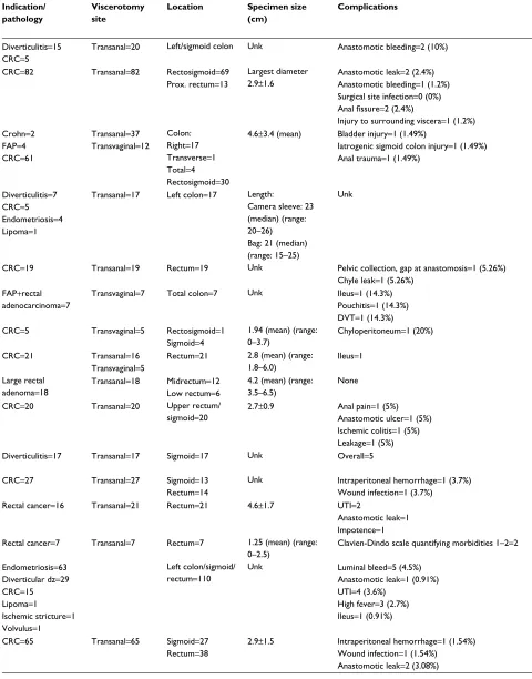
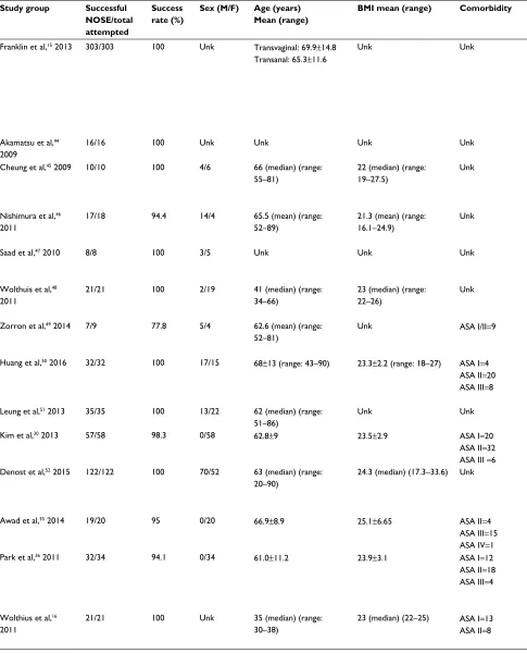
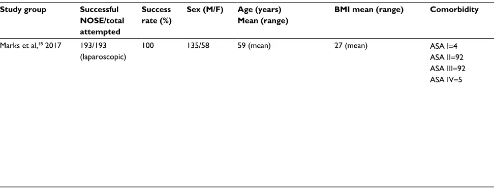
emergency surgery. Patient characteristics and overall suc-cess rate for completion of NOSE are displayed in Table 1. Failure of NOSE and the reason for failure are reported in many of these studies; yet, patient characteristics in this subset are not commonly provided by authors. Table 2 lists the available patient characteristics in cases of failed NOSE from the literature.
Sex
Bulky specimens are the most commonly cited reason for NOSE failure in colorectal surgery. The option of NOSE via the vagina appears to be the most significant factor contrib-uting to higher NOSE success in women compared to men. It has been well established that the elastic properties of the vagina allow removal of more bulky specimens when com-pared to proctotomies. This is best demonstrated by Karagul
et al,14 in which 12 of 15 women who failed NOSE via the
anus were able to have successful NOSE via the vagina. In this series, there was a statistically significant difference in the mean size of extracted specimens via vagina and anus (5.4±1.4 and 3.5±3.1 cm, respectively).14 Further
demonstrat-ing the elastic properties of the vagina, Yagci et al32
dem-onstrated transvaginal removal of a 9-cm right-sided colon mass. Independent of specimen size, transvaginal specimen extraction is more technically feasible for removing right colectomy specimens compared to the alternative of moving the right-sided specimen endoscopically through the entire length of the colon.
The question of pelvic shape and anatomy differences between men and women is worth mentioning. Some studies compare transanal NOSE between men and women. While pelvic geometry has not been cited as a reason for NOSE failure, it is unclear how much this may contribute to NOSE failure due to a “bulky specimen”. When transanal NOSE for laparoscopic colorectal resections is compared between men and women, the success rates are similar (40.5% in men and
50% in women).14 It should be pointed out that in these studies
comparing men and women, most patients have a BMI <30
kg/m2 and pelvic geometry may become a more significant
factor for larger patients, particularly if a higher BMI is cor-related to an increase in mesenteric bulk.
Body mass index
NOSE in colorectal surgery for the obese patient poses unique technical challenges. Increased BMI is associated with
increased visceral fat,33 which may be associated with
speci-men bulk. It is well recognized that one of the advantages of minimally invasive surgery is the improved visualization and postoperative outcomes in patients with higher BMI, but as we discussed earlier, the benefit of minimally invasive sur-gery is diminished by mini-laparotomies made for specimen
extraction. While most series exclude patients with BMI >30,
successful colorectal NOSE has been reported in patients
with BMI >30. Yagci reported one case in a patient with BMI
of 31 kg/m2, but NOSE required transection of the mesentery
– a technique that raises concerns about long-term oncologic
outcomes. In Zhang et al’s34 series of 27 patients, the highest
BMI reported with successful NOSE was 28.3 kg/m2. They
report a failure in a patient with BMI 30.2 kg/m2. Wolthuis
et al31 reported successful NOSE in six patients with BMI
>30, as part of their series of 110 patients. The postoperative outcomes for these patients were not discussed separately. As the literature is highly selective in terms of BMI, this should be considered a significant factor in assessing whether or not NOSE will be technically feasible when planning an
Clinical and Experimental Gastroenterology downloaded from https://www.dovepress.com/ by 118.70.13.36 on 20-Aug-2020
Dovepress
Izquierdo et al
Table 1 Patient and specimen characteristics for NOSE in colorectal surgery
Study group Successful
NOSE/total attempted
Success rate (%)
Sex (M/F) Age (years)
Mean (range)
BMI mean (range) Comorbidity Indication/
pathology
Viscerotomy site
Location Specimen size
(cm)
Complications
Wolthuis et al,20 2015
19/20 95 5/15 54 (median)
(31–72)
23.5 (18–29) ASA I=5
ASA II=15 Diverticulitis=15CRC=5 Transanal=20
Left/sigmoid colon Unk Anastomotic bleeding=2 (10%)
Saurabh et al,17 2017 77/82 93.9 47/35 63.3±13.9 24.4±4.2 ASA I=59
ASA II/III=23 CRC=82 Transanal=82 Rectosigmoid=69Prox. rectum=13
Largest diameter
2.9±1.6 Anastomotic leak=2 (2.4%)Anastomotic bleeding=1 (1.2%) Surgical site infection=0 (0%)
Anal fissure=2 (2.4%)
Injury to surrounding viscera=1 (1.2%)
Karagul et al,14 2017 49/67 73.1 40/27 57.9±13.4 Unk Unk Crohn=2
FAP=4 CRC=61
Transanal=37 Transvaginal=12
Colon: Right=17 Transverse=1 Total=4 Rectosigmoid=30
4.6±3.4 (mean) Bladder injury=1 (1.49%)
Iatrogenic sigmoid colon injury=1 (1.49%) Anal trauma=1 (1.49%)
Wolthuis et al,37 2015
17/17 100 3/15 Camera sleeve: 63
(median) (range: 47–69) Specimen bag: 51 (median) (range: 40–56)
Camera sleeve: 23 (median) (range 21–25) Specimen bag: 22 (median) (range: 20–24)
ASA I=4 ASA II=11 ASA III=2
Diverticulitis=7 CRC=5 Endometriosis=4 Lipoma=1
Transanal=17 Left colon=17 Length:
Camera sleeve: 23 (median) (range: 20–26) Bag: 21 (median) (range: 15–25)
Unk
Pai et al,38 2015 19/19 100 13/6 48 (median) (range:
23–78)
Unk Unk CRC=19 Transanal=19 Rectum=19 Unk Pelvic collection, gap at anastomosis=1 (5.26%)
Chyle leak=1 (5.26%) Palanivelu et al,29
2008
7/7 100 0/7 49.5 (mean) 25.3 Unk FAP+rectal
adenocarcinoma=7 Transvaginal=7 Total colon=7
Unk Ileus=1 (14.3%)
Pouchitis=1 (14.3%) DVT=1 (14.3%) Nishimura et al,39
2013
5/5 100 0/5 67.4 (mean) (range:
54–84)
21.3 (range: 16.2–27.3) Unk CRC=5 Transvaginal=5 Rectosigmoid=1
Sigmoid=4
1.94 (mean) (range: 0–3.7)
Chyloperitoneum=1 (20%)
Wang et al,40 2013 21/21 100 4/17 62 (median) (range: 50–80)
23.6 (mean) (range: 18–30)
Unk CRC=21 Transanal=16
Transvaginal=5
Rectum=21 2.8 (mean) (range: 1.8–6.0)
Ileus=1
Zhang et al,41 2014 18/18 100 10/8 56.6 (mean) (range: 48–69)
22.6 (mean) (range: 19.7–26.4)
Unk Large rectal
adenoma=18 Transanal=18 Midrectum=12Low rectum=6
4.2 (mean) (range: 3.5–6.5)
None
Hisada et al,24 2014 20/20 100 12/8 63.7±9 Unk Unk CRC=20 Transanal=20 Upper rectum/
sigmoid=20 2.7±0.9 Anal pain=1 (5%)Anastomotic ulcer=1 (5%) Ischemic colitis=1 (5%) Leakage=1 (5%) Costantino et al,23
2012
16/17 94.1 6/11 60.1±9.42 25.47±3.02 1.47±0.51 Diverticulitis=17 Transanal=17 Sigmoid=17 Unk Overall=5
Zhang et al,34 2014 24/27 88.9 16/11 54.8 (mean) (range: 37–77)
22.11 (mean) (range: 18.4–30.2)
Unk CRC=27 Transanal=27 Sigmoid=13
Rectum=14
Unk Intraperitoneal hemorrhage=1 (3.7%) Wound infection=1 (3.7%)
Han et al,42 2013 21/21 100 12/9 45.4±3.6 23.1±2.8 Unk Rectal cancer=16 Transanal=21 Rectum=21 4.6±1.7 UTI=2
Anastomotic leak=1 Impotence=1
Meillat et al,43 2017 7/7 100 2/5 Unk <25 Unk Rectal cancer=7 Transanal=7 Rectum=7 1.25 (mean) (range:
0–2.5)
Clavien-Dindo scale quantifying morbidities 1–2=2 Wolthuis et al,31
2015
110/110 100 13/97 38 (median) (range:
32–56)
23 (median) (range: 21–25)
ASA I=45 ASA II=56 ASA III=9
Endometriosis=63 Diverticular dz=29 CRC=15 Lipoma=1
Ischemic stricture=1 Volvulus=1
Left colon/sigmoid/ rectum=110
Unk Luminal bleed=5 (4.5%)
Anastomotic leak=1 (0.91%) UTI=4 (3.6%)
High fever=3 (2.7%) Ileus=1 (0.91%) Xingmao et al,21
2014
65/65 100 32/33 56.1±9.3 23.7±2.9 ASA I=10
ASA II=50 ASA III=5
CRC=65 Transanal=65 Sigmoid=27
Rectum=38
2.9±1.5 Intraperitoneal hemorrhage=1 (1.54%) Wound infection=1 (1.54%)
Anastomotic leak=2 (3.08%)
Clinical and Experimental Gastroenterology downloaded from https://www.dovepress.com/ by 118.70.13.36 on 20-Aug-2020
Dovepress Clinical and experimental gastroenterology
Table 1 Patient and specimen characteristics for NOSE in colorectal surgery
Study group Successful
NOSE/total attempted
Success rate (%)
Sex (M/F) Age (years)
Mean (range)
BMI mean (range) Comorbidity Indication/
pathology
Viscerotomy site
Location Specimen size
(cm)
Complications
Wolthuis et al,20 2015
19/20 95 5/15 54 (median)
(31–72)
23.5 (18–29) ASA I=5
ASA II=15 Diverticulitis=15CRC=5 Transanal=20
Left/sigmoid colon Unk Anastomotic bleeding=2 (10%)
Saurabh et al,17 2017 77/82 93.9 47/35 63.3±13.9 24.4±4.2 ASA I=59
ASA II/III=23 CRC=82 Transanal=82 Rectosigmoid=69Prox. rectum=13
Largest diameter
2.9±1.6 Anastomotic leak=2 (2.4%)Anastomotic bleeding=1 (1.2%) Surgical site infection=0 (0%)
Anal fissure=2 (2.4%)
Injury to surrounding viscera=1 (1.2%)
Karagul et al,14 2017 49/67 73.1 40/27 57.9±13.4 Unk Unk Crohn=2
FAP=4 CRC=61
Transanal=37 Transvaginal=12
Colon: Right=17 Transverse=1 Total=4 Rectosigmoid=30
4.6±3.4 (mean) Bladder injury=1 (1.49%)
Iatrogenic sigmoid colon injury=1 (1.49%) Anal trauma=1 (1.49%)
Wolthuis et al,37 2015
17/17 100 3/15 Camera sleeve: 63
(median) (range: 47–69) Specimen bag: 51 (median) (range: 40–56)
Camera sleeve: 23 (median) (range 21–25) Specimen bag: 22 (median) (range: 20–24)
ASA I=4 ASA II=11 ASA III=2
Diverticulitis=7 CRC=5 Endometriosis=4 Lipoma=1
Transanal=17 Left colon=17 Length:
Camera sleeve: 23 (median) (range: 20–26) Bag: 21 (median) (range: 15–25)
Unk
Pai et al,38 2015 19/19 100 13/6 48 (median) (range:
23–78)
Unk Unk CRC=19 Transanal=19 Rectum=19 Unk Pelvic collection, gap at anastomosis=1 (5.26%)
Chyle leak=1 (5.26%) Palanivelu et al,29
2008
7/7 100 0/7 49.5 (mean) 25.3 Unk FAP+rectal
adenocarcinoma=7 Transvaginal=7 Total colon=7
Unk Ileus=1 (14.3%)
Pouchitis=1 (14.3%) DVT=1 (14.3%) Nishimura et al,39
2013
5/5 100 0/5 67.4 (mean) (range:
54–84)
21.3 (range: 16.2–27.3) Unk CRC=5 Transvaginal=5 Rectosigmoid=1
Sigmoid=4
1.94 (mean) (range: 0–3.7)
Chyloperitoneum=1 (20%)
Wang et al,40 2013 21/21 100 4/17 62 (median) (range: 50–80)
23.6 (mean) (range: 18–30)
Unk CRC=21 Transanal=16
Transvaginal=5
Rectum=21 2.8 (mean) (range: 1.8–6.0)
Ileus=1
Zhang et al,41 2014 18/18 100 10/8 56.6 (mean) (range: 48–69)
22.6 (mean) (range: 19.7–26.4)
Unk Large rectal
adenoma=18 Transanal=18 Midrectum=12Low rectum=6
4.2 (mean) (range: 3.5–6.5)
None
Hisada et al,24 2014 20/20 100 12/8 63.7±9 Unk Unk CRC=20 Transanal=20 Upper rectum/
sigmoid=20 2.7±0.9 Anal pain=1 (5%)Anastomotic ulcer=1 (5%) Ischemic colitis=1 (5%) Leakage=1 (5%) Costantino et al,23
2012
16/17 94.1 6/11 60.1±9.42 25.47±3.02 1.47±0.51 Diverticulitis=17 Transanal=17 Sigmoid=17 Unk Overall=5
Zhang et al,34 2014 24/27 88.9 16/11 54.8 (mean) (range: 37–77)
22.11 (mean) (range: 18.4–30.2)
Unk CRC=27 Transanal=27 Sigmoid=13
Rectum=14
Unk Intraperitoneal hemorrhage=1 (3.7%) Wound infection=1 (3.7%)
Han et al,42 2013 21/21 100 12/9 45.4±3.6 23.1±2.8 Unk Rectal cancer=16 Transanal=21 Rectum=21 4.6±1.7 UTI=2
Anastomotic leak=1 Impotence=1
Meillat et al,43 2017 7/7 100 2/5 Unk <25 Unk Rectal cancer=7 Transanal=7 Rectum=7 1.25 (mean) (range:
0–2.5)
Clavien-Dindo scale quantifying morbidities 1–2=2 Wolthuis et al,31
2015
110/110 100 13/97 38 (median) (range:
32–56)
23 (median) (range: 21–25)
ASA I=45 ASA II=56 ASA III=9
Endometriosis=63 Diverticular dz=29 CRC=15 Lipoma=1
Ischemic stricture=1 Volvulus=1
Left colon/sigmoid/ rectum=110
Unk Luminal bleed=5 (4.5%)
Anastomotic leak=1 (0.91%) UTI=4 (3.6%)
High fever=3 (2.7%) Ileus=1 (0.91%) Xingmao et al,21
2014
65/65 100 32/33 56.1±9.3 23.7±2.9 ASA I=10
ASA II=50 ASA III=5
CRC=65 Transanal=65 Sigmoid=27
Rectum=38
2.9±1.5 Intraperitoneal hemorrhage=1 (1.54%) Wound infection=1 (1.54%)
Anastomotic leak=2 (3.08%)
(Continued)
Clinical and Experimental Gastroenterology downloaded from https://www.dovepress.com/ by 118.70.13.36 on 20-Aug-2020
Dovepress
Izquierdo et al
Study group Successful
NOSE/total attempted
Success rate (%)
Sex (M/F) Age (years)
Mean (range)
BMI mean (range) Comorbidity Indication/
pathology
Viscerotomy site
Location Specimen size
(cm)
Complications
Franklin et al,15 2013 303/303 100 Unk Transvaginal: 69.9±14.8 Transanal: 65.3±11.6
Unk Unk Unk Transanal=277
Transvaginal=26
Unk Unk Transvaginal: none
Transanal:
Wound infection=2 (0.7%) Ileus=7 (2.5%)
UTI=6 (2.2%)
Bowel obstruction=1 (0.4%) Fecal incontinence=6 (2.2%) Anastomotic leak=3 (1.1%) Akamatsu et al,44
2009
16/16 100 Unk Unk Unk Unk CRC=16
T1/T2
Transanal=16 Sigmoid/ rectosigmoid=16
Unk Wound infection=1 (6.25%)
Cheung et al,45 2009 10/10 100 4/6 66 (median) (range: 55–81)
22 (median) (range: 19–27.5)
Unk CRC=10 Transanal=10 Rectosigmoid=5
Sigmoid=4 Descending colon=1
Unk None (0%)
Nishimura et al,46 2011
17/18 94.4 14/4 65.5 (mean) (range:
52–89)
21.3 (mean) (range: 16.1–24.9)
Unk CRC=18 Transanal=18 Sigmoid=18 1.84 (mean) (range:
0–4.0)
Anastomotic leak=1 (5.56%) Wound infection=1 (5.56%) Enteritis=1 (5.56%)
Saad et al,47 2010 8/8 100 3/5 Unk Unk Unk Diverticulitis=5 CRC=3 Transanal=8 Left colon=8
Length of specimen: range 16–34
None (0%)
Wolthuis et al,48 2011
21/21 100 2/19 41 (median) (range:
34–66)
23 (median) (range: 22–26)
Unk Endometriosis=13
Diverticulitis=5 CRC=3
Transanal=21 Sigmoid=21 Length of specimen: 20 (median) (range: 13–25)
Anastomotic leak=1 (4.8%)
Zorron et al,49 2014 7/9 77.8 5/4 62.6 (mean) (range: 52–81)
Unk ASA I/II=9 CRC=9 Transanal=9 Rectum=9 Unk Transitory feet neuralgia=1
Inadequate TME, tumor rupture=1 Anastomotic leak=1
Huang et al,50 2016 32/32 100 17/15 68±13 (range: 43–90) 23.3±2.2 (range: 18–27) ASA I=4 ASA II=20 ASA III=8
CRC=32 Transanal=32 Sigmoid=15
Rectosigmoid=9 Upper rectum=6 Mid rectum=2
3.3±1.8 (range: 1.3–6.2)
Ileus=2 (6.24%)
Wound infection=1 (3.12%)
Leung et al,51 2013 35/35 100 13/22 62 (median) (range: 51–86)
Unk Unk CRC=35 Transanal=35 Left colon=35 2 (median) (range:
2–4)
None (0%)
Kim et al,30 2013 57/58 98.3 0/58 62.8±9 23.5±2.9 ASA I=20
ASA II=32 ASA III =6
CRC=58 Transvaginal=58 Sigmoid=21
Rectosigmoid=8 Rectum=29
3.4±1.8 Bleeding=1 (1.7%)
Intra-abdominal abscess=1 (1.7%) Denost et al,52 2015 122/122 100 70/52 63 (median) (range:
20–90)
24.3 (median) (17.3–33.6) Unk CRC=122 Transanal=122 Low rectum=122 3.9 (median) (range:
1–10)
Overall morbidity=42 (34%)
Surgical morbidity (Clavien-Dindo scale quantifying morbidities III)=18 (15%)
Mortality=1 (0.8%)
Awad et al,35 2014 19/20 95 0/20 66.9±8.9 25.1±6.65 ASA II=4
ASA III=15 ASA IV=1
CRC=20 Transvaginal=20 Right colon=20 4.735±3.61 Ileus=2 (10%)
Park et al,36 2011 32/34 94.1 0/34 61.0±11.2 23.9±3.1 ASA I=12
ASA II=18 ASA III=4
CRC=34 Transvaginal=34 Cecum=10
Ascending colon=16 Proximal transverse colon=8
3.8±1.3 Hemorrhage requiring transfusion=2 (5.88%) Ileus=1 (2.94%)
Urinary retention=1 (2.94%)
Wolthius et al,16 2011
21/21 100 Unk 35 (median) (range:
30–38)
23 (median) (22–25) ASA I=13 ASA II=8
Bowel
endometriosis=21 Transanal=21 Sigmoid=21
Length of specimen: 21 (median) (range: 17–24)
UTI=1 (4.76%)
Table 1 (Continued)
Clinical and Experimental Gastroenterology downloaded from https://www.dovepress.com/ by 118.70.13.36 on 20-Aug-2020
Dovepress Clinical and experimental gastroenterology
Study group Successful
NOSE/total attempted
Success rate (%)
Sex (M/F) Age (years)
Mean (range)
BMI mean (range) Comorbidity Indication/
pathology
Viscerotomy site
Location Specimen size
(cm)
Complications
Franklin et al,15 2013 303/303 100 Unk Transvaginal: 69.9±14.8 Transanal: 65.3±11.6
Unk Unk Unk Transanal=277
Transvaginal=26
Unk Unk Transvaginal: none
Transanal:
Wound infection=2 (0.7%) Ileus=7 (2.5%)
UTI=6 (2.2%)
Bowel obstruction=1 (0.4%) Fecal incontinence=6 (2.2%) Anastomotic leak=3 (1.1%) Akamatsu et al,44
2009
16/16 100 Unk Unk Unk Unk CRC=16
T1/T2
Transanal=16 Sigmoid/ rectosigmoid=16
Unk Wound infection=1 (6.25%)
Cheung et al,45 2009 10/10 100 4/6 66 (median) (range: 55–81)
22 (median) (range: 19–27.5)
Unk CRC=10 Transanal=10 Rectosigmoid=5
Sigmoid=4 Descending colon=1
Unk None (0%)
Nishimura et al,46 2011
17/18 94.4 14/4 65.5 (mean) (range:
52–89)
21.3 (mean) (range: 16.1–24.9)
Unk CRC=18 Transanal=18 Sigmoid=18 1.84 (mean) (range:
0–4.0)
Anastomotic leak=1 (5.56%) Wound infection=1 (5.56%) Enteritis=1 (5.56%)
Saad et al,47 2010 8/8 100 3/5 Unk Unk Unk Diverticulitis=5 CRC=3 Transanal=8 Left colon=8
Length of specimen: range 16–34
None (0%)
Wolthuis et al,48 2011
21/21 100 2/19 41 (median) (range:
34–66)
23 (median) (range: 22–26)
Unk Endometriosis=13
Diverticulitis=5 CRC=3
Transanal=21 Sigmoid=21 Length of specimen: 20 (median) (range: 13–25)
Anastomotic leak=1 (4.8%)
Zorron et al,49 2014 7/9 77.8 5/4 62.6 (mean) (range: 52–81)
Unk ASA I/II=9 CRC=9 Transanal=9 Rectum=9 Unk Transitory feet neuralgia=1
Inadequate TME, tumor rupture=1 Anastomotic leak=1
Huang et al,50 2016 32/32 100 17/15 68±13 (range: 43–90) 23.3±2.2 (range: 18–27) ASA I=4 ASA II=20 ASA III=8
CRC=32 Transanal=32 Sigmoid=15
Rectosigmoid=9 Upper rectum=6 Mid rectum=2
3.3±1.8 (range: 1.3–6.2)
Ileus=2 (6.24%)
Wound infection=1 (3.12%)
Leung et al,51 2013 35/35 100 13/22 62 (median) (range: 51–86)
Unk Unk CRC=35 Transanal=35 Left colon=35 2 (median) (range:
2–4)
None (0%)
Kim et al,30 2013 57/58 98.3 0/58 62.8±9 23.5±2.9 ASA I=20
ASA II=32 ASA III =6
CRC=58 Transvaginal=58 Sigmoid=21
Rectosigmoid=8 Rectum=29
3.4±1.8 Bleeding=1 (1.7%)
Intra-abdominal abscess=1 (1.7%) Denost et al,52 2015 122/122 100 70/52 63 (median) (range:
20–90)
24.3 (median) (17.3–33.6) Unk CRC=122 Transanal=122 Low rectum=122 3.9 (median) (range:
1–10)
Overall morbidity=42 (34%)
Surgical morbidity (Clavien-Dindo scale quantifying morbidities III)=18 (15%)
Mortality=1 (0.8%)
Awad et al,35 2014 19/20 95 0/20 66.9±8.9 25.1±6.65 ASA II=4
ASA III=15 ASA IV=1
CRC=20 Transvaginal=20 Right colon=20 4.735±3.61 Ileus=2 (10%)
Park et al,36 2011 32/34 94.1 0/34 61.0±11.2 23.9±3.1 ASA I=12
ASA II=18 ASA III=4
CRC=34 Transvaginal=34 Cecum=10
Ascending colon=16 Proximal transverse colon=8
3.8±1.3 Hemorrhage requiring transfusion=2 (5.88%) Ileus=1 (2.94%)
Urinary retention=1 (2.94%)
Wolthius et al,16 2011
21/21 100 Unk 35 (median) (range:
30–38)
23 (median) (22–25) ASA I=13 ASA II=8
Bowel
endometriosis=21 Transanal=21 Sigmoid=21
Length of specimen: 21 (median) (range: 17–24)
UTI=1 (4.76%)
(Continued)
Clinical and Experimental Gastroenterology downloaded from https://www.dovepress.com/ by 118.70.13.36 on 20-Aug-2020
Dovepress
Izquierdo et al
operation. Feasibility and complication rates for colorectal
NOSE in patients with BMI ≥30 kg/m2 require further study.
Medical comorbidities
NOSE in colorectal surgery has been successful in patients with ASA class III. Complication rates for this specific population are not reported. In a randomized clinical trial by
Wolthuis et al,20 inflammatory response between conventional
specimen extraction and NOSE was compared using post-operative C-reactive protein. C-reactive protein levels were found to be higher in the NOSE group, but ultimately, this was not associated with an increased length of hospital stay. The implications of this are unclear, but it could be suggested that patients with more comorbidities are less likely to toler-ate a more robust inflammatory response. Conversely, use of pain scores in the same study was significantly lower in the NOSE group. In more frail patients with comorbidities, the advantage of decreased narcotic use is obvious.
Coagulopathy was set as the exclusion criterion in some NOSE colorectal studies. This should be considered as there is a reported rate of postoperative intraluminal hemorrhage
of 4.5%31 and intraperitoneal hemorrhage of 1.5%–3.7%.21,34
Transfusion was required in some of these patients, but all cases of postoperative bleeding resolved without another operation.
Other potential patient factors
History of prior operations and radiation exposure may influ-ence the feasibility of any operation. While such history can make operations more challenging, there are no reported
NOSE failures that cite this as the reason for failure. In the context of rectal cancer treatment and taTME, neoadjuvant radiation is the standard of care. NOSE in this context has not been reported to be a significant issue. In our series of 373 patients with low rectal cancer treated with transanal abdominal transanal taTME, 100% had successful NOSE via the anus when this approach was chosen.
Specimen factors
Review of the literature suggests that specimen factors such as size and shape are the most important factor in determining feasibility of successful NOSE in colorectal surgery. Preoperative assessment for this can be performed with computerized tomography scan or barium enema. The influential factors include length, location, and pathology. Table 1 lists the specimen-related factors with NOSE success rates for reviewed studies. Table 2 shows the specimen factors associated with failed NOSE in cases where the information was available.
Size
A majority of the literature regarding NOSE in colorectal surgery will describe specimen features by specimen length, width, and/or tumor size. Specimen traits that failed NOSE are often not discussed in detail and were only reported in
one study.14 Success rates >90% are seen in specimens with
smaller diameter. Common exclusion criteria restrict NOSE
to specimens of <6.5 cm width. Karagul et al14 included
patients with a wide range of specimen size (width 4.6±3.4
cm, length 25.2±22.6 cm). The authors point out other aspects
Study group Successful
NOSE/total attempted
Success rate (%)
Sex (M/F) Age (years)
Mean (range)
BMI mean (range) Comorbidity Indication/
pathology
Viscerotomy site
Location Specimen size
(cm)
Complications
Marks et al,18 2017 193/193 (laparoscopic)
100 135/58 59 (mean) 27 (mean) ASA I=4
ASA II=92 ASA III=92 ASA IV=5
CRC=193 Transanal=193 Low rectum=193 Unk Anastomotic leak=4 (1.07%)
Pelvic abscess=3 (0.8%) Bleeding=2 (0.54%) Ileus=8 (2.14%)
Small bowel obstruction=3 (0.8%) Acute kidney injury=1 (0.27%) Urinary sepsis=1 (0.27%)
DVT/pulmonary embolism=3 (0.8%) Arrhythmia=2 (0.54%)
Wound infection=1 (0.27%) Ischemic bowel=2 (0.54%)
(open and laparoscopic pooled complications reported only)
Abbreviations: ASA, American Society of Anesthesiologists; BMI, body mass index; DVT, deep vein thrombosis; NOSE, natural orifice specimen extraction; prox., proximal;
TME, total mesorectal excision; unk, unknown; UTI, urinary tract infection; CRC, colorectal cancer; FAP, familial adenomatous polyposis.
Table 1 (Continued)
Clinical and Experimental Gastroenterology downloaded from https://www.dovepress.com/ by 118.70.13.36 on 20-Aug-2020
Dovepress Clinical and experimental gastroenterology
of the specimen which contribute to the feasibility of NOSE on a patient by patient basis: shape, location, and consistency. Reducing the bulk of specimens by transecting the mesentery has been described by Yagci et al.32 In this case report, a
speci-men with width 12 cm was removed via posterior colpotomy by partially dividing the mesentery. With this maneuver, the maximum width of the specimen was 9 cm compared to the initial width of 12 cm. NOSE was successful in this patient. The tumor size for this right-sided colon adenocarcinoma was 5 cm. This technique raises concerns about the long-term oncologic outcomes related to tumor implants. Laparoscopic bags and povidone-iodine have been used in attempts to mini-mize this risk. Long-term oncologic outcomes for mesentery transection are not available. It is unclear if this technique affects oncologic outcomes, but would be a safe option in the treatment of benign pathologies such as endometriosis, diverticulitis, and inflammatory bowel disease.
Length of the specimen also appears to be an important
factor in NOSE feasibility. In Karagul et al’s14 series, there
was a statistically significant difference between specimen
lengths in successful NOSE (21.2±19.4 cm) and failed NOSE
(36.5±27.6 cm). Despite this, Palanivelu et al29 have
dem-onstrated that total proctocolectomies via vaginal specimen extraction can be performed successfully. While specimen length contributes to NOSE feasibility, it must be considered in conjunction with specimen width, pathology, and location in the distal colorectum.
Pathology
In the context of colorectal malignancies, large tumors have been shown to contribute to NOSE failure. Benign colorectal
pathologies associated with more inflammation, such as diverticulitis and inflammatory bowel disease, can also add to the bulk of specimens. This increased bulk, in turn, can theoretically increase the failure rate of NOSE, though this has not yet been demonstrated in the literature. NOSE has been demonstrated as feasible for diverticulitis and inflam-matory bowel disease, though in each case, operations were performed on an elective basis and presumably had, at most, mild inflammation at the time of operation.
Anatomic location
Distal lesions are most amendable to successful NOSE. This is demonstrated by the high success rates of NOSE in
taTME operations for rectal cancer. Karagul et al14 reported a
96.7% NOSE success rate for pathologies in the rectum and 54.5% for pathologies located in the colon. This is a 13.7-fold increase in the chance of NOSE failure with colonic resections as compared to rectal resections. Specimens of the rectum require a proctotomy as part of the distal margin tran-section that is already a required step in the operation. After the specimen is extracted, the proctotomy is then completely resected with another firing of a stapler in preparation for use of an end-to-end anastomosis (EEA) stapler, as demonstrated
by Wolthuis et al.16 In our experience with low rectal cancers,
the distal transection point of the specimen is at the dentate line and specimens were removed transanally in 100% of laparoscopic cases. A coloanal anastomosis using this already existing viscerotomy was created after specimen extraction.
As distance of the specimen from the anal verge increases, NOSE via the transanal route becomes more difficult. In right-sided colon pathology, many series have addressed this
Study group Successful
NOSE/total attempted
Success rate (%)
Sex (M/F) Age (years)
Mean (range)
BMI mean (range) Comorbidity Indication/
pathology
Viscerotomy site
Location Specimen size
(cm)
Complications
Marks et al,18 2017 193/193 (laparoscopic)
100 135/58 59 (mean) 27 (mean) ASA I=4
ASA II=92 ASA III=92 ASA IV=5
CRC=193 Transanal=193 Low rectum=193 Unk Anastomotic leak=4 (1.07%)
Pelvic abscess=3 (0.8%) Bleeding=2 (0.54%) Ileus=8 (2.14%)
Small bowel obstruction=3 (0.8%) Acute kidney injury=1 (0.27%) Urinary sepsis=1 (0.27%)
DVT/pulmonary embolism=3 (0.8%) Arrhythmia=2 (0.54%)
Wound infection=1 (0.27%) Ischemic bowel=2 (0.54%)
(open and laparoscopic pooled complications reported only)
Abbreviations: ASA, American Society of Anesthesiologists; BMI, body mass index; DVT, deep vein thrombosis; NOSE, natural orifice specimen extraction; prox., proximal;
TME, total mesorectal excision; unk, unknown; UTI, urinary tract infection; CRC, colorectal cancer; FAP, familial adenomatous polyposis.
Clinical and Experimental Gastroenterology downloaded from https://www.dovepress.com/ by 118.70.13.36 on 20-Aug-2020
Dovepress
Izquierdo et al
Table 2
Description of NOSE failures in colorectal surgery
Study group
Number failed/ number attempted Failure rate (%)
Reason for failure
Sex (M/F)
Age in years, mean Range
BMI
Comorbidity
Pathology
Viscerotomy site
attempted
Location
Specimen size
Wolthuis et al,
20 2015
1/20 5 Bulky specimen Unk Unk Unk Unk Unk Transanal = 1
Left/sigmoid colon
Unk Saurabh et al, 17 2017 5/82 6.1
Bulky specimen Difficult purse-string suturing of rectal stump in deep, narrow pelvis
Unk Unk Unk Unk Unk Transanal = 5 Unk Unk Karagul et al, 14 2016 18/67 26.9%
Sex: males more likely to fail than females (3.3-fold) Bulky specimen Anatomic location: colonic resections more likely to fail than rectal resections (13.7-fold)
15/3 60.5 ± 13.3 Unk Unk CRC = 16 FAP = 2 Transanal = 15 Transvaginal = 3 Right colon = 8
Transverse colon
= 1 Left colon = 6 Rectosigmoid = 1 Total colon = 2
Tumor width 6.5±
4.2 cm
Specimen length 36.5
±
27.6 cm
Constantino et al,
23 2012
1/17 5.9 Bulky specimen Unk Unk Unk Unk Diverticulitis = 1 Transanal = 1 Sigmoid = 1 Unk Zhang et al, 34 2014 3/27 11.1
Bulky specimen Mesenteric fat too thick Unable to close rectal stump Position of opening too low, narrow pelvis
1/2
50.7 Range: 48–53 24.8 (median) Range: 23.6–30.2
Unk
CRC
=
3 (all T3)
Transanal = 3 Sigmoid = 2 Rectum = 1
Tumor width: (range: 3.0–7.5 cm)
Nishimura et al,
46 2011
1/18
5.56
Mesenteric fat too thick
1/0 72 23.9 Unk CRC = 1 (T1N0) Transanal = 1 Sigmoid = 1
Tumor size: 2.5 cm
Zorron et al, 49 2014 2/9 22.2
Bulky specimen in proximal rectum (10 cm from anal verge) Anatomic location: proximal rectum (12 cm from anal verge)
+
unable to
dissect specimen free
2/0
64.5 Range: 62–67
Unk ASA I/II = 2 CRC = 2 (pT2N0 = 1) (pT3N1 = 1) Transanal = 2 Rectum = 2 Unk Kim et al, 30 2013 1/58 1.72
Bulky specimen in the setting of vaginal atrophy
0/1 Unk Unk Unk Unk Transvaginal = 1 Left-sided colon Unk Awad et al, 35 2014 1/20 5 Bulky specimen 0/1 Unk Unk Unk Unk Transvaginal = 1 Right colon
Tumor size 8.0 cm
Park et al, 36 2011 2/34 5.88 Bulky specimen + colpotomy
opening was inadequate
0/2 Unk Unk Unk Unk Transvaginal = 1 Right colon Unk Abbreviations:
ASA, American Society of Anesthesiologists; BMI, body mass index; NOSE, natural orifice specimen extraction; unk, unknown; CRC, colorectal cancer; FAP, familial adenomatous polyposis.
Clinical and Experimental Gastroenterology downloaded from https://www.dovepress.com/ by 118.70.13.36 on 20-Aug-2020
Dovepress Clinical and experimental gastroenterology
issue by removing the specimen transvaginally.11,35,36 This
option is obviously available only to women, as discussed earlier.
Discussion/future directions
Beyond its established feasibility, short-term retrospec-tive data and early randomized controlled trials do show advantages of NOSE compared to conventional specimen extraction in defined populations. While more randomized controlled trials are needed to compare the two techniques, potential benefits, including less postoperative analgesia, better cosmesis, quicker return of bowel function, and shorter hospital length of stay, can be significant. While this explains the motivation to utilize the NOSE technique, these favorable outcomes need to be considered in the context of their strictly selected patient populations.
Several patient and specimen factors are involved in determining the feasibility of NOSE for a given patient. It is not likely that any one of these factors would independently prevent NOSE unless the specific factor is an extreme devia-tion from the established patient populadevia-tion that is amend-able to NOSE. Important patient factors are sex and BMI. Specimen factors are pathology, anatomic location, and size (width and length).
The role of NOSE in patients with BMI >30 requires more
study. As incision-related complications are more common in obese patients, this population would stand to gain the most from avoiding a larger abdominal incision for specimen extraction. Feasibility of NOSE in this population is not well established and it has seldom been attempted. Higher BMI is associated with increased visceral fat, sometimes manifested as increased bulk of the mesentery. In the case of NOSE, a more bulky mesentery may prevent the removal of specimens by means of natural orifices altogether.
Another population that would benefit from avoidance of an abdominal incision for specimen extraction would be elderly patients and those with significant comorbidities. In general, as the burden of comorbidity increases, the more a patient would stand to gain by avoiding a larger abdominal incision. While reported postoperative morbidity in colorectal NOSE appears to be similar to that of conventional specimen removal, it is unclear if patients with less physiologic reserve would do as well. It is possible that the documented increased inflammatory response associated with NOSE would trans-late into inferior clinical outcomes. Future research should investigate the role of NOSE in these sicker patients, as a majority of the current literature involves patients with ASA classes I and II.
An attempt at NOSE does not prevent specimen removal via conventional means, thus, attempts at NOSE can be made with relatively low consequence. An algorithmic approach to attempting NOSE seems most reasonable. This is best
demonstrated in the study design by Karagul et al.14 NOSE
is first attempted by transanal means. If this fails and if the patient is a female, transvaginal specimen extraction is attempted, and in the experience of Karagul, it is often (80%) successful. If this also fails, traditional specimen extraction can still be performed. As with any new surgical technique, if a surgeon attempts to add NOSE to their armamentarium, they should start with the easiest cases, selecting the most ideal patient. Surgeons performing NOSE should already be technically facile laparoscopically – in particular, intracorpo-real anastomosis should already be a part of their skill set. As experience with NOSE increases for an individual surgeon, they can then select more difficult cases.
One of the greatest difficulties in reviewing the literature for NOSE in colorectal surgery is the wide heterogeneity in
the operative technique10 between studies. Variances in
tech-nique include single-stapled versus double- or triple-stapled anastomosis and use of a specimen bag or wound protector for specimen removal. These variables can confound out-comes significantly. Introducing and implementing any new operative technique involve standardization, so that the skill can be taught and disseminated to achieve wider adoption. More recently, larger series including some randomized controlled trials have been performed in a consistent, clearly described, step-wise manner. As some of these studies have shown benefits in outcome, the operations as described in these studies would be the best framework for standardizing NOSE in colorectal surgery.
Conclusion
While the feasibility and benefits of NOSE have been dem-onstrated in the literature, patient selection plays a significant role in taking full advantage of this evolution in minimally invasive surgery. The benefits of NOSE need further study, particularly to see if the reported benefits can be general-ized to larger populations. The ideal patient in whom NOSE
would be feasible and beneficial is a non-obese (BMI <30)
woman with pathology involving the distal colon or rectum
where the specimen has a maximum width <6.5 cm. While
each of these factors individually contributes to feasibility of NOSE, patient selection must continue to be on a case-by-case basis. Conversely, the patients who stand to benefit the most from avoidance of any abdominal wall extraction incision are morbidly obese and frail patients. While NOSE
Clinical and Experimental Gastroenterology downloaded from https://www.dovepress.com/ by 118.70.13.36 on 20-Aug-2020
Dovepress
Izquierdo et al
surgery offers an exciting option, careful study and adoption of this approach is mandated to ensure optimal patient benefit.
Disclosure
The authors report no conflicts of interest in this work.
References
1. Clinical Outcomes of Surgical Therapy Study Group, Nelson H, Sargent DJ, Wieand HS, et al. A comparison of laparoscopically assisted and open colectomy for colon cancer. N Engl J Med. 2004;350(20):2050–2059. 2. Colon Cancer Laparoscopic or Open Resection Study Group, Buunen M, Veldkamp R, Hop WC, et al. Survival after laparoscopic surgery ver-sus open surgery for colon cancer: long-term outcome of a randomised clinical trial. Lancet Oncol. 2009;10(1):44–52.
3. Kennedy GD, Heise C, Rajamanickam V, Harms B, Foley EF. Lapa-roscopy decreases postoperative complication rates after abdominal colectomy: results from the national surgical quality improvement program. Ann Surg. 2009;249(4):596–601.
4. Hackert T, Uhl W, Buchler MW. Specimen retrieval in laparoscopic colon surgery. Dig Surg. 2002;19(6):502–506.
5. Kaminski JP, Pai A, Ailabouni L, et al. Role of epidural and patient-controlled analgesia in site-specific laparoscopic colorectal surgery.
JSLS. 2014;18(4):e2014.00207.
6. Ihedioha U, Mackay G, Leung E, Molloy RG, O’Dwyer PJ. Laparoscopic colorectal resection does not reduce incisional hernia rates when com-pared with open colorectal resection. Surg Endosc. 2008;22(3):689–692. 7. Winslow ER, Fleshman JW, Birnbaum EH, Brunt LM. Wound
complications of laparoscopic vs open colectomy. Surg Endosc. 2002;16(10):1420–1425.
8. Stewert EA, Liau AS, Friedman AJ. Operative laparoscopy followed by colpotomy for resecting a colonic leiomyosarcoma: a case report. J Reprod Med. 1991;36(12):883–884.
9. Nezhat F. Laparoscopic segmental resection for infiltrating endometrio-sis of rectosigmoid colon: a preliminary report. Surg Laparosc Endosc Percutan Tech. 2001;11(1):67–68.
10. Wolthuis AM, de Buck van Overstraeten A, D’Hoore A. Laparoscopic natural orifice specimen extraction-colectomy: a systematic review.
World J Gastroenterol. 2014;20(36):12981–12992.
11. Franklin ME Jr, Ramos R, Rosenthal D, Schuessler W. Laparoscopic colonic procedures. World J Surg. 1993;17(1):51–56.
12. Person B, Vivas DA, Wexner SD. Totally laparoscopic low anterior resec-tion with transperineal handsewn colonic J-pouch anal anastomosis for low rectal cancer. Surg Endosc. 2006;20(4):700–702.
13. Eshuis EJ, Voermans RP, Stokkers PC, van Berge Henegouwen MI, Fockens P, Bemelman WA. Laparoscopic resection with transcol-onic specimen extraction for ileocaecal Crohn’s disease. Br J Surg.
2010;97(4):569–574.
14. Karagul S, Kayaalp C, Sumer F, et al. Success rate of natural orifice specimen extraction after laparoscopic colorectal resections. Tech Coloproctol. 2017;21(4):295–300.
15. Franklin ME Jr, Liang S, Russek K. Natural orifice specimen extrac-tion in laparoscopic colorectal surgery: transanal and transvaginal approaches. Tech Coloproctol. 2013;17(Suppl 1):S63–S67.
16. Wolthuis AM, Meuleman C, Tomassetti C, et al. Laparoscopic sigmoid resection with transrectal specimen extraction: a novel technique for the treatment of bowel endometriosis. Human Reprod.
2011;26(6):1348–1355.
17. Saurabh B, Chang SC, Ke TW, et al. Natural orifice specimen extraction with single stapling colorectal anastomosis for laparoscopic anterior resection: feasibility, outcomes, and technical considerations. Dis Colon Rectum. 2017;60(1):43–50.
18. Marks JH, Myers EA, Zeger EL, Denittis AS, Gummadi M, Marks GJ. Long-term outcomes by a transanal approach to total mesorectal exci-sion for rectal cancer. Surg Endosc. 2017;31(12):5248–5257.
19. Ma B, Huang XZ, Gao P, et al. Laparoscopic resection with natural ori-fice specimen extraction versus conventional laparoscopy for colorectal disease: a meta-analysis. Int J Colorectal Dis. 2015;30(11):1479–1488. 20. Wolthuis AM, Fieuws S, Van Den Bosch A, de Buck van Overstraeten A,
D’Hoore A. Randomized clinical trial of laparoscopic colectomy with or without natural-orifice specimen extraction. Br J Surg. 2015;102(6):630–637. 21. Xingmao Z, Haitao Z, Jianwei L, Huirong H, Junjie H, Zhixiang Z.
Totally laparoscopic resection with natural orifice specimen extraction (NOSE) has more advantages comparing with laparoscopic-assisted resection for selected patients with sigmoid colon or rectal cancer. Int J Colorectal Dis. 2014;29(9):1119–1124.
22. Wolthuis AM, Meuleman C, Tomassetti C, et al. How do patients score cosmesis after laparoscopic natural orifice specimen extraction colec-tomy? Colorectal Dis. 2015;17(6):536–541.
23. Costantino FA, Diana M, Wall J, Leroy J, Mutter D, Marescaux J. Prospective evaluation of peritoneal fluid contamination following transabdominal vs. transanal specimen extraction in laparoscopic left-sided colorectal resections. Surg Endosc. 2012;26(6):1495–1500. 24. Hisada M, Katsumata K, Ishizaki T, et al. Complete laparoscopic resec-tion of the rectum using natural orifice specimen extracresec-tion. World J Gastroenterol. 2014;20(44):16707–16713.
25. Kuhry E, Schwenk WF, Gaupset R, Romild U, Bonjer HJ. Cochrane Database Syst Rev. 2008;(2):CD003432.
26. Saida Y, Nagao J, Nakamura Y, et al. A comparison of abdominal cavity bacterial contamination of laparoscopy and laparotomy for colorectal cancers. Dig Surg. 2008;25(3):198–201.
27. Park JS, Choi GS, Kim HJ, Park SY, Jun SH. Natural orifice specimen extraction versus conventional laparoscopically assisted right hemico-lectomy. Br J Surg. 2011;98(5):710–715.
28. Sehgal R, Cahill RA. Advanced laparoscopic surgery for colorectal disease: NOTES/NOSE or single port? Best Pract Res Clin Gastroen-terol. 2014;28(1):81–96.
29. Palanivelu C, Rangarajan M, Jategaonkar PA, Anand NV. An inno-vative technique for colorectal specimen retrieval: a new era of “natural orifice specimen extraction” (N.O.S.E). Dis Colon Rectum.
2008;51(7):1120–1124.
30. Kim HJ, Choi GS, Park JS, Park SY, Ryuk JP, Yoon SH. Transvaginal specimen extraction versus conventional minilaparotomy after laparo-scopic anterior resection for colorectal cancer: mid-term results of a case-matched study. Surg Endosc. 2014;28(8):2342–2348.
31. Wolthuis AM, de Buck van Overstraeten A, Fieuws S, Boon K, D’Hoore A. Standardized laparoscopic NOSE-colectomy is feasible with low morbidity. Surg Endosc. 2015;29(5):1167–1173.
32. Yagci MA, Kayaalp C, Novruzov NH. Intracorporeal mesenteric division of the colon can make the specimen more suitable for natural orifice specimen extraction. J Laparoendosc Adv Surg Tech A.
2014;24(7):484–486.
33. Camhi SM, Bray GA, Bouchard C, et al. The relationship of waist circumference and BMI to visceral, subcutaneous, and total body fat: sex and race differences. Obesity (Silver Spring). 2011;19(2):402–408. 34. Zhang X, Zhou H, Hou H, Hu J, Wang H, Zhou Z. Totally laparoscopic resection with natural orifice specimen extraction for carcinoma of sigmoid colon and rectum: a feasible and innovative technique. J Clin Gastroenterol. 2014;48(7):e57–e61.
35. Awad ZT, Griffin R. Laparoscopic right hemicolectomy: a comparison of natural orifice versus transabdominal specimen extraction. Surg Endosc. 2014;28(10):2871–2876.
36. Park JS, Choi GS, Kim HJ, Park SY, Jun SH. Natural orifice specimen extraction versus conventional laparoscopically assisted right hemico-lectomy. Br J Surg. 2011;98(5):710–715.
37. Wolthuis AM, De Buck Van Overstraeten A, D’Hoore A. Laparoscopic NOSE colectomy with a camera sleeve: a technique in evolution.
Colorectal Dis. 2015;17(5):O123–O125.
38. Pai VD, Desouza A, De Menezes JL, Saklani AP. Laparoscopic inter-sphincteric resection and hand-sewn coloanal anastomosis: a natural orifice specimen extraction technique. J Laparoendosc Adv Surg Tech A.
2015;25(5):396–400.
Clinical and Experimental Gastroenterology downloaded from https://www.dovepress.com/ by 118.70.13.36 on 20-Aug-2020