THE BIOENGINEERING OF NERVE
CONDUITS
Miss Pari-Naz Mohanna
MBBS BSc
FRCS(Eng.)
Doctor of Medicine
Blond Mclndoe Centre
Royal Free and University College Medical Schools
University o f London
ProQuest Number: U642544
All rights reserved
INFORMATION TO ALL USERS
The quality of this reproduction is dependent upon the quality of the copy submitted.
In the unlikely event that the author did not send a complete manuscript and there are missing pages, these will be noted. Also, if material had to be removed,
a note will indicate the deletion.
uest.
ProQuest U642544
Published by ProQuest LLC(2015). Copyright of the Dissertation is held by the Author.
All rights reserved.
This work is protected against unauthorized copying under Title 17, United States Code. Microform Edition © ProQuest LLC.
ProQuest LLC
789 East Eisenhower Parkway P.O. Box 1346
To my family
That knowledge which stops at what it does not know,
is the highest knowledge.
Chuang Tzu
Learning without thought is labour lost;
thought without learning is perilous.
Confucius
The difference between what the most and the least learned people
know is inexpressibly trivial in relation to that which is unknown.
Albert Einstein
Every great advance in science has issued from a
new audacity of imagination.
ABSTRACT
Poly-3-hydroxybutyrate (PHB) conduits are an alternative to nerve autografting and
support regeneration across long nerve gaps, although to suboptimal levels. The aim of
this study was to improve these results by combining PHB with glial growth factor
(GGF), enhancing nerve regeneration by contact guidance and an improved trophic
microenvironment. Two and 4cm gaps in the rabbit common peroneal nerve were
bridged using PHB-GGF conduits. The rate and quantity of axonal and Schwann cell
(SC) regeneration were assessed by quantitative immunohistochemistry at 21, 42 and 63
days, and compared to empty and alginate filled conduits. Addition of GGF improved
axonal and SC regeneration, which was sustained up to 63 days independent of gap
length. The distance and quantity of axonal regeneration were increased by up to 53%
and 4317% respectively. At 120 days axonal and SC regeneration within the PHB-GGF
grafts remained superior to the controls resulting in enhanced motor organ
reinnervation, as was demonstrated by an improved recovery of muscle mass compared
to the controls.
In both the short and long term studies the alginate filled conduits resulted in
regeneration inferior to both the GGF and empty tubes. As a result alginate fibres were
assessed in vitro and in vivo as an alternative to alginate hydrogel with a potential to
deliver GGF. However, regeneration in vivo in alginate fibre filled conduits was inferior
Polyhydroxyalkatone (PHA) was also evaluated as a conduit material, as GGF linkage
and release from its walls is a feasible option. Four different PHA configurations were
used to bridge a 1cm rat sciatic nerve gap. All 4 PHA configurations, accelerated axonal
regeneration rate to 1 mm/day versus 0.7mm/day with PHB conduits and resulted in a
quantity of axonal regeneration superior to that seen in the autograft repairs.
In conclusion, GGF improves axonal and SC regeneration across short and long gaps
through PHB conduits, but alginate hydrogel appears to limit the trophic effect of GGF.
Alginate fibres provide no improvement, however alginate’s limitations may be
overcome and regeneration further improved by using PHA as a bioconstruct releasing
ACKNOWLEDGEMENTS
Firstly I would like to express my heart felt appreciation and thanks to my family.
Without their unconditional love, support, encouragement, understanding patience and
prayers I could not have reached this stage of my career and completed this thesis.
I have the deepest of gratitude to my supervisor Dr. Giorgio Terenghi. This thesis would
not have been possible without his wisdom, enthusiasm, guidance and ability to show
me the light at the end of sometimes, a very long and dark tunnel. In addition to Dr.
Giorgio Terenghi I would like to thank Professor Mikael Wiberg, Department of
Anatomy, Umea University, Sweden for his advice, comments and original sense of
humour. I am grateful to Mr Richard Young and Mr Afshin Mosahebi, who in the initial
stages of my research passed on to me their expertise in both laboratory and
micro surgical techniques as well as computer skills, which proved to be invaluable. My
thanks to Mr. Paul Fuller for his technical assistance in the laboratory and his help in the
analysis of some of my work.
Many thanks to the staff of the Comparative Biology Unit at the Royal Free Hospital,
London for their dedication and eagerness to help, as well as the Electron Microscopy
Unit at the Royal Free Hospital, London who expertly prepared the electron microscopy
specimens. The statistical analysis was carried out in collaboration with Dr. Deborah
Ridout, Statistician, Institute of Child Health, Great Ormond Street Hospital, London. I
explained the appropriate statistical methods and their interpretation to a novice such as
myself.
I would like to thank the Royal College of Surgeons of England and the Royal Free
Special Trustees for the joint research grant they awarded to me. Their financial support
together with that of the East Grinstead Medical Research Trust gave me the opportunity
of carrying out the research presented in this thesis. I am grateful to Dr. David Gwynne,
Cambridge NeuroScience, Cambridge MA, USA, for the gift of recombinant human
glial growth factor, Astra Tech for the PHB material, Tepha for the PHA material and
Pronova for the alginate.
Last but by no means least my special gratitude to all my colleagues at the Blond
Mclndoe Centre. Their wide array of personalities and unique qualities has made for a
fun, happy and supportive environment, which has been conducive to carrying out this
research. I feel lucky to take away with me not only numerous fond memories and many
new friendships but more knowledge and skills than I thought would be possible to
CONTENTS
Abstract 3
Acknowledgements 5
Contents 7
Figures and Tables 15
Abbreviations 20
Chapter 1 INTRODUCTION
1.1
Morphology of the peripheral nerve
24
1.1.1 Neurons 24
1.1.2 Schwann cells 26
1.1.3 Extracellular matrix 29
1.2 Axonal injury
30
1.2.1 Classification of nerve injury 30
1.2.2 Wallerian degeneration 31
1.3 Axonal regeneration
33
1.3.1 Factors influencing axonal growth 3 3
1.4
Growth factors
39
1.4.1 Neurotrophic factors 39
1.4.2 Glial growth factor 42
1.5
Peripheral nerve repair
44
1.5.1 Historical perspective 45
1.5.2 Nerve grafting 47
1.5.3 Nerve conduits 50
1.5.4 Polyhydroxyalkatone conduits 54
1.5.5 Poly-3 -hydroxybutyrate conduits 57
1.5.6 Nerve conduits for long gap repair 59
1.6
Delivery of neurotrophic factors
62
1.6.1 Alginate 64
1.7
Hypothesis
70
1.8
Aims
71
Chapter 2 MATERIALS AND METHODS
2.1
Alginate gel
75
2.1.1 Preparation of alginate stock 75
2.1.2 Dilution of alginate stock 75
2.3
Glial growth factor (GGF)
76
2.4
Poly-3-hydorxybutyrate (PHB)
77
2.4.1 PHB conduit preparation 77
2.4.2 Loading of PHB conduit with alginate gel 78
2.4.3 Loading of PHB conduit with alginate fibres 81
2.4.4 In vitro PHB and alginate fibres 81
2.5
Polyhydroxyalkatone (PHA)
84
2.5.1 PHA conduit preparation 85
2.6
New Zealand white rabbit model
85
2.6.1 Anaesthesia, pre-operative preparation, recovery and euthanasia 86
2.6.2 Operative procedure 87
2.7
Sprague Dawley rat model
91
2.7.1 Anaesthesia, pre-operative preparation, recovery and euthanasia 91
2.7.2 Operative procedure 92
2.8 Tissue Collection
93
2.8.1 New Zealand white rabbit model 93
2.8.1.1 Short-term group 93
2.8.1.2 Long-term group 95
2.8.2 Sprague Dawley rat model 98
2.10 Morphological Assessment
100
2.10.1 Immunohistochemistry 100
2.10.2 Histological staining 100
2.10.3 Electron microscopy 101
2.11 Quantification
102
2.11.1 Regeneration distance 102
2.11.2 Regeneration area 104
2.12 Statistical analysis
107
2.12.1 Regeneration distance 107
2.12.2 Regeneration area 108
2.12.2.1 Short-term group 108
2.12.2.2 Long-term group 109
2.12.3 Motor Reinnervation 110
Chapter 3 SHORT-TERM ASSESSMENT OF SHORT AND LONG
NERVE GAP REPAIR WITH A PHB/GGF CONDUIT
3.1
Introduction
114
3.2
Aims
115
3.4
Results
116
3.4.1 Regeneration distance 119
3 .4.1.1 Comparison between gap lengths 122
3.4.1.2 Comparison between groups 122
3.4.1.2.1 S100 & PanNF regeneration distance across 2cm gaps 122
3.4.1.2.2 S100 & PanNF regeneration distance across 4cm gaps 123
3.4.1.3 Comparison between time points 124
3.4.1.3.1 S100& PanNF regeneration distance across 2cm gaps 124
3.4.1.3.2 SlOO & PanNF regeneration distance across 4cm gaps 125
3.4.2 Regeneration area 130
3.4.2.1 Axonal regeneration area 131
3.4.2.1.1 Area of PanNF staining 131
3.4.2.1.2 Percentage area of PanNF staining 134
3.4.2.2 SC regeneration area 136
3.4.2.2.1 Area of SlOO staining 136
3.4.2.2.2 Percentage area of S100 staining 138
3.5
Discussion
140
Chapter 4 LONG-TERM ASSESSMENT OF SHORT AND LONG
NERVE GAP REPAIR WITH A PHB/GGF CONDUIT
4.1
Introduction
149
4.3
Material and Methods
1^0
4.4
Results
4.4.1 General Observations 152
4.4.2 Regeneration area 156
4.4.2.1 Axonal regeneration area 157
4.4.2.1.1 Area of PanNF staining 157
4.4.2.1.2 Percentage area of PanNF staining 161
4.4.2.2 SC regeneration area 165
4.4.2.2.1 Area of SlOO staining 165
4.4.2.2.2 Percentage area of SlOO staining 169
4.4.2.3 Axonal and SC regeneration area at 63 vs 120 days 171
4.4.3 PHB conduit transverse semithin sections 177
4.4.4 PHB conduit transverse ultrathin sections 181
4.4.5 Distal nerve stump semithin Sections 185
4.4.6 Muscle reinnervation 189
4.5
Discussion
192
Chapter 5 SHORT-TERM ASSESSMENT OF A PERIPHERAL
NERVE GAP REPAIR WITH A PHA CONDUIT
5.1
Introduction
198
5.3
Material and Methods
199
5.4
Results
200
5.4.1 Electron micrographs of the PHA material 201
5.4.2 Regeneration distance 206
5.4.3 Regeneration area 209
5.5
Discussion
212
Chapter 6 IN VITRO AND IN VIVO ASSESSMENT OF ALGINATE
FIBRES
6.1
Introduction
218
6.2 Aims
219
6.3
Material and Methods
219
6.4 Results
221
6.4.1 IN VITRO 221
6.4.2 IN VIVO 229
6.5 Discussion
234
Chapter 7 GENERAL DISCUSSION
Bibliography 247
Chapter 1
FIGURES AND TABLES
Introduction
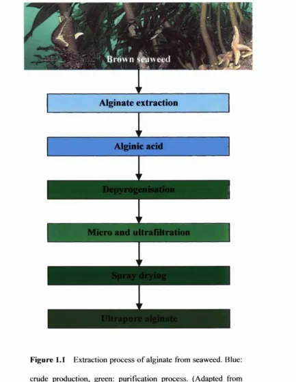
Figure 1.1 Extraction process of alginate 68
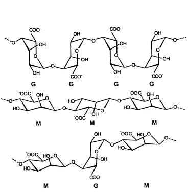
Figure 1.2 Molecular structure of alginate 69
Chapter 2 Materials and Methods
Figure 2.1 Separating PHB sheets 79
Figure 2.2 PHB conduit preparation 79
Figure 2.3 Scanning electron micrographs of PHB 80
Figure 2.4 Loading of PHB conduits with alginate fibres 83
Figure 2.5 Rabbit common peroneal and tibial nerves 89
Figure 2.6 Common peroneal nerve repair with a PHB conduit 89
Figure 2.7 Detail of the proximal anastomosis 90
Figure 2.8 PHB conduit at the end of implantation 90
Figure 2.9 Harvested PHB conduit 94
Figure 2.10 Long-term group nerve repair tissue collection 97
Figure 2.11 Quantification of axonal and SC regeneration 103
Chapter 3 Short-term assessment of short and long nerve gap repair
with a PHB/GGF conduit
Figure 3.1 PHB conduit 42 days after implantation 118
Figure 3.2 Pseudocapsule surrounding PHB conduit 118
Table 3.1 Axonal regeneration distances in the conduits 120
Table 3.2 SC regeneration distances in the conduits 121
Figure 3.3 Axonal regeneration distance for the 2cm nerve repairs 126
Figure 3.4 SC regeneration distance for the 2cm nerve repairs 127
Figure 3.5 Axonal regeneration distance for the 4cm nerve repairs 128
Figure 3.6 SC regeneration distance for the 4cm nerve repairs 129
Figure 3.7 Total area of PanNF staining in the conduits 133
Figure 3.8 Percentage area of PanNF staining in the conduits 135
Figure 3.9 Total area of SlOO staining in the conduits 137
Figure 3.10 Percentage area of SlOO staining in the conduits 139
Chapter 4 Long-term assessment of short and long nerve gap repair
with a PHB/GGF conduit
Figure 4.1a Pseudocapsule surrounding PHB conduit 154
Figure 4. lb PHB conduit in situ after explantation from pseudocapsule 154
Figure 4.2 Long-term harvest of PHB conduits 155
Figure 4.3 Total area of PanNF staining in the 2 and 4cm conduits 158
Figure 4.4 Total area of PanNF staining in the 2cm conduits 159
Figure 4.6 Percentage area of PanNF staining in the 2 and 4cm conduits 162
Figure 4.7 Percentage area of PanNF staining in the 2cm conduits 163
Figure 4.8 Percentage area of PanNF staining in the 4cm conduits 164
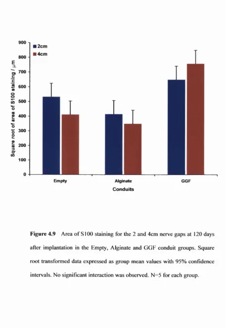
Figure 4.9 Total area of SlOO staining in the 2 and 4cm conduits 166
Figure 4.10 Total area of SlOO staining in the 2cm conduits 167
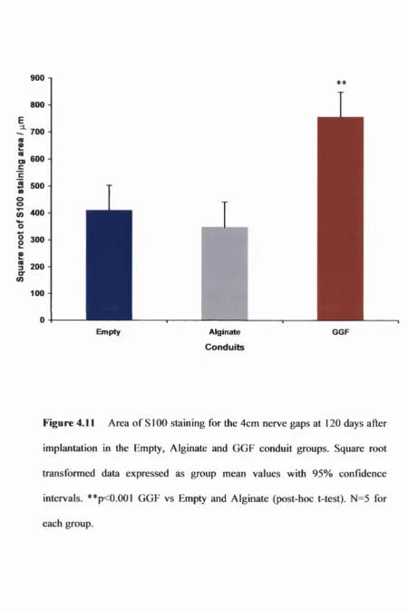
Figure 4.11 Total area of SlOO staining in the 4cm conduits 168
Figure 4.12 Percentage area of SlOO staining in the 2 and 4cm conduits
combined 170
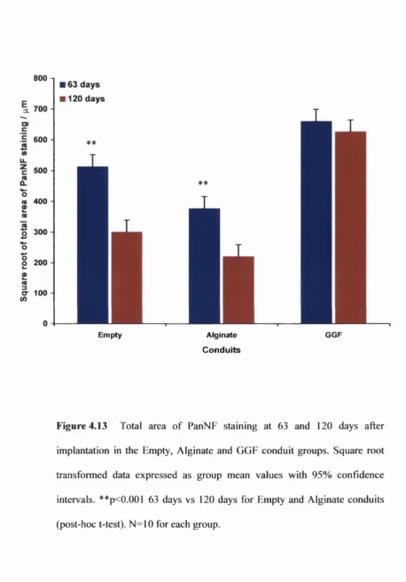
Figure 4.13 Total area of PanNF staining in the conduits at 63 and 120 days 173
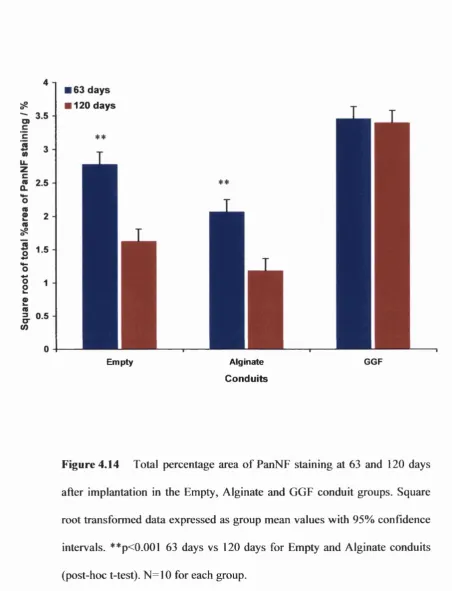
Figure 4.14 Percentage area of PanNF staining in the conduits at 63 and
120 days 174
Figure 4.15 Total area of SlOO staining in the conduits at 63 and 120 days 175
Figure 4.16 Percentage area of SlOO staining in the conduits at 63 and 120 days 176
Figure 4.17 Transverse semithin sections of the 2 and 4cm PHB-GGF
conduits at 120 days 178
Figure 4.18 Transverse semithin sections of the 2 and 4cm PHB-ALG
conduits at 120 days 179
Figure 4.19 Transverse semithin sections of the 2 and 4cm E-PHB
conduits at 120 days 180
Figure 4.20 Transmission electron micrograph from a PHB-GGF conduit 182
Figure 4.21 Transmission electron micrograph from an E-PHB conduit 183
Figure 4.22 Transmission electron micrograph from a PHB-ALG conduit 183
Figure 4.24 Transverse semithin sections of the distal nerve stumps of the
2 and 4cm PHB-ALG conduit repairs 186
Figure 4.25 Transverse semithin sections of the distal nerve stumps of the
2 and 4cm PHB-GGF conduit repairs 187
Figure 4.26 Transverse semithin sections of the distal nerve stumps of the
2 and 4cm E-PHB conduit repairs 188
Figure 4.27 TA/EDL muscle complex at harvest 190
Figure 4.28 Percentage loss of muscle mass at 120 days after 2 and 4cm
conduit nerve repairs 191
Chapter 5 Short-term assessment of a peripheral nerve gap repair
with a PHA conduit
Table 5.1 Groups studied 200
Figure 5.1 Low and high power electron micrographs of PH A-A 202
Figure 5.2 Low and high power electron micrographs of PHA-B 203
Figure 5.3 Low and high power electron micrographs of PHA-C 204
Figure 5.4 Low and high power electron micrographs of PHA-D 205
Figure 5.5 Axonal staining through a PHA-D conduit 207
Figure 5.6 SC staining through a PHA-D conduit 208
Table 5.2 Percentage axonal regeneration area 10 and 20 days after PHA
and autograft nerve repairs 210
Figure 5.7 Percentage axonal regeneration area 10 and 20 days after PHA
Chapter 6 In vitro and in vivo assessment of alginate fibres
Figure 6.1 PHB-Sorbasan alginate conduit on day 2 221
Figure 6.2 PHB-Comfeel alginate conduit on day 2 222
Figure 6.3 PHB-AMS alginate conduit on day 2 222
Figure 6.4 PHB-LVM alginate conduit on day 2 223
Figure 6.5 PHB-LVG alginate conduit on day 2 223
Figure 6.6 PHB-Sorbasan alginate conduit on day 7 224
Figure 6.7 PHB-Comfeel alginate conduit on day 7 224
Figure 6.8 PHB-AMS alginate conduit on day 7 225
Figure 6.9 PHB-LVM alginate conduit on day 7 225
Figure 6.10 PHB-LVG alginate conduit on day 7 226
Figure 6.11 PHB-Sorbasan alginate conduit on day 10 226
Figure 6.12 PHB-Comfeel alginate conduit on day 10 227
Figure 6.13 PHB-AMS alginate conduit on day 10 227
Figure 6.14 PHB-LVM alginate conduit on day 10 228
Figure 6.15 PHB-LVG alginate conduit on day 10 228
Figure 6.16 Axonal staining through the proximal part of a PHA-LVG conduit 232
ABBREVIATIONS
AG Autografl
ANOVA Analysis of variance
B Band of Biingner
BDNF Brain-derived neurotrophic factor
BSA Bovine serum albumin
°C Celsius
CNTF Ciliary neurotrophic factor
cm Centimetre
D Distal
EDL Extensor digitorum longus muscle
G Gauge
GDNF Glial-derived neurotrophic factor
GGF Glial growth factor
g Gram
i.m. Intramuscular
Kda Kilodalton
kg Kilograms
1 Litre
LIF Leukaemia inhibitory factor
M Myelinated nerve fibre
ml Millilitre
ni Microlitre
mm Millimetre
pm Micrometer
NGF Nerve growth factor
NT-3/4/5 Neurotrophin-3/4/ 5
o.d Once a day
P Proximal
PBS Phosphate buffered saline
PDGF Platelet-derived growth factor
PHA Polyhydroxyalkatone
PHB Poly-3 -hydroxybutyrate
s.c. Subcutaneously
SC Schwann cell
TA Tibialis anterior muscle
UM Unmyelinated nerve fibre
CHAPTER 1
INTRODUCTION
1.1 MORPHOLOGY OF THE PERIPHERAL NERVE
1.1.1 Neurons
1.1.2 Schwann cells
1.1.3 Extracellular matrix
1.2 AXONAL INJURY
1.2.1 Classification of nerve injury
1.2.2 Wallerian degeneration
1.3 AXONAL REGENERATION
1.3.1 Factors influencing axonal growth
1.3.2 Role of Schwann cells and basal lamina
1.4 GROWTH FACTORS
1.4.1 Neurotrophic factors
1.5 PERIPHERAL NERVE REPAIR
1.5.1 Historical perspective
1.5.2 Nerve grafting
1.5.3 Nerve conduits
1.5.4 Polyhydroxyalkatone conduits
1.5.5 Poly-3 -hydroxybutyrate conduits
1.5.6 Nerve conduits for long gap repair
1.6 DELIVERY OF NEUROTROPHIC FACTORS
1.6.1 Alginate
1.7 HYPOTHESIS
1.1 MORPHOLOGY OF THE PERIPHERAL NERVE
Hippocrates provided the first written description of the peripheral nervous system as
early as the 4^*^ century B.C., and later Herophilus identified nerves as such,
distinguishing them from tendons and tracing them to the spinal cord. The peripheral
nervous system is comprised of the peripheral nerve fibres and their associated tissues,
which include glial cells, sensory organs, muscle motor end plates, intraneural
vasculature and related extracellular matrix (Landon 1976; Dyck & Thomas 1984).
1.1.1 Neurons
The functional unit of the nervous system upon which all structures are dependent is the
neuron. The neuronal cell body is the site of all metabolic activities, which are then
transported along the axon. Motoneurons innervate striated muscles, and their cell
bodies are located in the anterior horn of the spinal cord, the axons pass through the
anterior root to join the peripheral nerve bundle. These axons are myelinated except at
their origins and terminations, where they divide into numerous branches to form
neuromuscular synapses. Motoneurons innervating smooth muscles and glands belong
to the autonomic nervous system. Their cell bodies are found in the peripheral
autonomic ganglia and their axons are unmyelinated. Sensory neurons are located
outside, but close to the spinal cord in the dorsal root ganglia of the spinal nerves. They
spinal cord. The sensory fibres may be myelinated or unmyelinated according to the
sensory modality.
A nerve fibre is generally described as any process of the nerve cell, either dendrite or
axon. It is a cylindrical extension of the cell soma, surrounded by a specialised
membrane, called the axolemma that maintains a resting action potential. The nerve
fibres can be divided into motor, sensory, sympathetic and parasympathetic, and are
classified according to their fibre diameter and conduction velocity into A, B and C
fibres (Gasser & Grundfest 1939). The principle content of the fibres are axoplasm,
microfilaments, neurofilaments and neurotubules. Microfilaments are made up of actin
proteins and may play a role in intra-axonal transport (Varon & Bunge 1978).
Neurofilaments are larger and can be distinguished by immunohistology (Lee et al
1982) and disappear following damage. Neurotubules give cytoskeletal support.
Axons are unable to synthesise proteins and are dependent on the neuronal cell body or
the glial cells for their maintenance. Macromolecules are transported along both
directions of the axon concurrently at various speeds (Tytell et al 1981). The slow
anterograde transport (0.1-3mm/day) is for the bulk movement of neurofilaments and is
equivalent to the rate of axonal outgrowth sprouting (Di Giamberardino et al 1973). An
important function of fast retrograde transport (100-400mm/day) is the uptake of
exogenous growth factors produced by the end organs and transport back to the neuronal
Nerve fibres with their supporting Schwann cells (SC), form the bulk of the peripheral
nerves. These are surrounded by an outer epineurium, a perineurium which surrounds
the nerve fascicles and an inner endoneurium. The endoneurium contains the supporting
elements of the nerve fibres, which includes SC, fibroblasts, occasional mast and fat
cells and extracellular matrix (ECM) (Thomas et a l 1993). Although the fascicular
pattern of the nerve fibre is variable, there is a high degree of segregation of afferent and
efferent fascicles of pure motor or sensory nerves within each fascicle and nerve type
(Terzis & Smith 1990a).
1.1.2 Schwann cells
These are the principal glial cells of the peripheral nerve, and they were first described
by Theodor Schwann (1810-1882), who thought that they developed from the axons.
However, it is now clear that SC originate form the neural crest and to a lesser extent
from the neural tube and they grow with or slightly ahead of the neurites, which arise
from the primitive dorsal root ganglia and ventral horn cell bodies (lessen & Mirsky
1998). The irreversible step fi*om neural crest cells to SC precursors is observed in cells
that express high levels of mRNA for PO, which is a major myelin protein (Mirsky &
lessen 1999). SC precursors do not express S I00 marker but within days and early
during gestation the typical SI00 antigen is expressed in the cells (lessen & Mirsky
1991). The precursors of SC undergo a proliferative phase that might partly be regulated
by the developing embryonic neurons as they express high levels of neuregulins, in
(Marchionni et a l 1993) and it interacts with SC heterodiameric receptors composed of
c-erbB2, c-erbB3 and c-erbB4 (Grinspan et a l 1996). Deficiency of SC is observed in
mice with the deletion of neuregulin-l (Meyer & Birchmeier 1995). Further evidence on
the role of the neurons is evident when SC isolated from axons become quiescent in
vitro. SC mitosis continues until there is a 1:1 ratio between axons and myelinating SC,
and the segment of axon that is myelinated by a single SC is termed the node of
Ranvier. As myelination proceeds, there is a down regulation of GGF receptors, erbB2
and erbB3 (Mirsky & lessen 1999). Therefore SC proliferate in three distinct
circumstances: during development, following loss of contact with axons as a result of
axonal degeneration and during the re-establishment of axonal contact that follows
axonal regeneration. In all three cases, axon-SC contact, or its loss, has a central role in
the induction of the SC proliferative phase (Reynolds & Woolf 1993).
There are two distinct categories of SC, myelinating and non-myelinating. Axons,
whether myelinated or unmyelinated are ensheathed by SC along their entire length. A
feature of SC is the deposition of a basal lamina, a component of the extracellular
matrix, which surrounds the outer surface of the SC and isolates them from the
surrounding matrix. Axonal contact has been shown to be a factor for differentiation of
SC to myelin forming phenotype (lessen et al 1987). Axonal diameter is also important,
as only axons with a diameter larger than 0.7pm undergo myelination (Windebank et al
1985). On loss of axon contact the SC dedifferentiate and will redifferentiate to express
the appropriate ensheathment phenotype on contact with small or large axons. However,
s c synthesise progesterone, which stimulates myelin formation (Chan et a l 2000) and
blocking of progesterone receptors also inhibits myelination (Koenig et a l 2000).
The node of Ranvier is the site of interruption of the myelin sheath, where the axon is
covered only by microvilli of SC and sodium channels are accumulated at the node of
Ranvier. Cajal named it the “germinative territory” because following trauma to the
nerve axonal regeneration appeared at the node In contrast unmyelinated fibres have
continuous SC sheaths with no gap or node between adjacent SC. The basal lamina
however, is continuous for the length of both myelinated and unmyelinated axons
(Thanos et a l 1998).
The phenotypic markers expressed by SC seem to be dependent on their related axon
rather than an inherent difference in SC. This is evident as after loss of axonal contact,
SC readily revert back to the expression of markers characteristic of immature non
myelinating SC (lessen & Richardson 1996) and this phenomenon is irrespective of SC
type. Some of the phenotypic markers are common to all mature SC, such as SI00,
vimentin and laminin (Scherer 1997). Other markers like PO, myelin basic protein and
myelin associated glycoprotein are restricted to myelin forming SC (Scherer 1997).
Some markers are exclusive to non-myelinating SC such as intermediate glial fibrillary
acid protein (GFAP), p75 (low affinity nerve growth factor receptor), neural cell
adhesion molecule (NCAM) and LI (lessen & Mirsky 1992). Thus it is possible to
ascertain the SC phenotype using immunohistochemical stains for the distinctive
1.1.3 Extracellular matrix
Neuronal axons and SC are enmeshed in a complex extracellular matrix (ECM), whose
two major components are basal lamina and fibrillar matrix. The fibrillar matrix is made
up mostly of type I and IQ collagen, while the basal lamina contains types IV and V
collagen, the glycoproteins laminin, fibronectin and tenascin, as well as heparin sulphate
(Chemousov & Carey 2000), The ECM components act together to provide structural
support for the cellular elements and affect their behaviour during development and
maturity.
Tissue culture studies have defined the ECM products that are produced by SC, how
their production is controlled, and how endoneurial and perineural fibroblasts co-operate
and are regulated by SC in organising ECM. Fibroblasts can also induce synthesis of
ECM via SC (Obremski et al 1993), and in turn the SC signal to the surrounding
connective tissue, possibly via Desert Hedgehog molecules, to organise perineural
fibroblasts (Mirsky & lessen 1999). ECM is not produced and organised by neurons
cultured alone, whereas cultured SC can express basal lamina as identifiable by electron
microscopy (Baron-Van Evercooren et a l 1986). It is in co-cultures of neurons and SC
that a basal lamina is formed on the axonal surface of SC, and a continuous basal lamina
production depends on direct contact with axons (Bunge et a l 1990). Thus SC have a
critical role in the production of ECM, which is essential for the ensheathing of the
axons. The importance of ECM was demonstrated in tissue culture studies where
ascorbic acid (Eldridge et al. 1989). An important effect of the basal lamina is to
polarise the myelinating SC, as the surface free of the basal lamina is responsible for
axonal adhesion (Chemousov & Carey 2000). Laminin and fibronectin are potent
promoters of cell adhesion, while migration, adhesion and signalling functions of ECM
are mediated by integrin receptors on SC (Chemousov & Carey 2000). Antibodies to p 1
integrin block migration on lamininl and 2, while av integrin may be involved in
migration of SC on fibronectin (Mirsky & Jessen 1999).
1.2 AXONAL INJURY
Axonal degeneration is the biological response which follows axonopathy. Although
there are many causes of axonopathy, either hereditary or acquired, the focus of this
section is on traumatic axotomy.
1.2.1 Classification of nerve injury
A scheme for classification of traumatic nerve injuries was first developed by Seddon
(Seddon 1954) and subsequently modified by Sunderland (Sunderland 1978).
Sunderland classified nerve injuries into five degrees of increasingly severe injury
pattems. In first degree injuries (neuropraxia) the gross nerve stmcture is intact and the
decreased function is reversible. In second degree injuries (axonotmesis) the
epineurium, perineurium and basal lamina are intact but there is complete axonal
interrupted but the epineurium and perineurium are intact. In fourth degree injuries only
elements of the epineurium are left intact. Finally in fifth degree injuries (neurotmesis)
the nerve is completely transected with all the elements of the nerve being separated. It
is this latter type of injury that has been experimentally studied in this thesis.
1.2.2 Wallerian degeneration
Following nerve injury, complex changes occur throughout the neuron, both in the
proximal and distal nerve segments, extending to the muscle motor end plates and
sensory receptors distally, and the cell bodies proximally. Dorsal root ganglia cell death
after peripheral nerve injury ranges from 7-50%, with the diversity of experimental
models and techniques involved accounting for the wide variation in estimates (Hart et
al. 2002). However, motor neurones survive unless the lesion is very proximal at the
level of the nerve root (Ranson 1909; Himes & Tessler 1989; Liuzzi & Tedeschi 1991).
Neuronal cell death occurs as a result of the insult produced by nerve injuries, loss of
neurotrophic support from the target organ, abnormal retrograde electrical activity and
release of toxic cytokines such as interlukin-6 and tumour necrosis factor a produced by
inflammatory cells at the site of injury (Liuzzi & Tedeschi 1991). The closer the injury
to the cell body the more likely cell death may occur.
The sequence of events occurring in the distal and the proximal stumps following nerve
injury were described by Waller (Waller 1850), these being known under the term
whole of the distal segment and up to the first node of Ranvier in the proximal stump. In
the myelinated fibres, the axons and the myelin sheaths in the distal stump break down.
The debris is phagocytosed partly by SC but predominantly by macrophages. An influx
of macrophages and activation of resident macrophages (Griffin et a l 1993) results in
the removal of axonal and myelin debris to leave the basal lamina tubes and SC of the
distal nerve, these cordons of SC are termed ‘bands of Bimgner’ (Cajal 1928; Perry &
Brown 1992). In addition to their phagocytic role the macrophages also secrete
cytokines that promote axonal regeneration directly or via their interaction with SC
(Brown 1991).
Loss of the intimate contact between axons and glial cells causes SC to de-differentiate
and to alter their phenotypic marker expression (Nikam et a l 1995), thereby becoming
mitotic and starting to proliferate. The glial growth factor (GGF) subfamily of axon-
associated neuregulins and their putative erbB-tyrosine kinase receptors on SC
(Carraway & Burden 1995) play an important role in signalling this event (cf 1.4.2).
Carroll et a l demonstrated that both GGF and erbB2 and erbB3 receptors were
coordinately upregulated and expressed by SC in the distal stump, suggesting an
autocrine mechanism in addition to paracrine stimulation of SC proliferation (Carroll et
al 1997). Following proliferation, the SC align themselves longitudinally within the
basal lamina tubes forming bands of Büngner, ready to receive regenerating fibres that
have grown out of the proximal stump and to guide them to their target (Fawcett &
Keynes 1990; Hall 1997). The changes in unmyelinated fibres are similar, but there is
In the proximal stump the SC also de-differentiate, proliferate and start to migrate along
the internal surface of the basal lamina tube from the initial regeneration zone located at
the first node proximal to the site of injury (Thanos et a l 1998).
1.3 AXONAL REGENERATION
1.3.1 Factors influencing axonal growth
Axonal regeneration is governed by three main factors; contact guidance, neurotropism
and neurotrophism. Contact guidance describes the topographical and microgeometric
cues and physical support provided for the regenerating and maturing axons as well as
their interaction with cell surface adhesion molecules, which are present along the
appropriate pathways. Neurotropism refers to the process by which the distal stump and
end organ positively influence and direct regeneration of the proximal stump by
chemical signals. Lastly neurotrophism is the support provided to the regenerating axons
by the growth factors produced by the surrounding cells and end organs, allowing the
neurons to survive the injury and mature following regeneration (cf 1.4).
After nerve gap injury, the process of successful regeneration begins with the formation
of a fibrin matrix containing fibroblasts, fibronectin, macrophages, leukocytes and
erythrocytes, which bridges the proximal and distal stumps (Lundborg et a l 1982a;
Lundborg et a l 1982b; Lundborg et a l 1982c; Lundborg et al 1982d; Longo et al
the migration of fibroblasts, SC and axons (Weiss 1944). SC migrate out from the
proximal and distal stumps and meet approximately two-thirds of the way along the
nerve gap to form a continuous scaffold.
A “latent period” exists between nerve injury and the commencement of axonal
regeneration (Gutmann et al 1942). Regeneration rate varies as a function of injury
type, age, and species especially during the early stage of regeneration (Gutmann et al
1942; Black & Lasek 1979; Pestronk et al 1980). Outgrowth of axonal sprouts is more
rapid after cmsh injury in which the continuity of nerve sheaths and basement
membrane is preserved than after nerve section (Thomas 1964). Regeneration is also
more rapid in young mammals and varies across species (Gutmann et al 1942; Black &
Lasek 1979). In the rat sciatic nerve model the rate of regeneration through a PHB
conduit is O.lSmm/day for the first 7 days, increasing to 0.36mm/day between 7 and 14
days (Hazari et al 1999a). Using the same conduit in the rabbit common peroneal nerve
model the rate of regeneration is 0.26mm/day in the first 21 days, which then increases
to 1.6mm/day between 21 and 42 days (Young et a l 2002). Other researchers have
estimated the rate of regeneration to vary between 1 and 3mm/day (Gutmann et al
1942; Black & Lasek 1979).
Within 3 hours of injury, axonal sprouting begins at the node of Ranvier immediately
proximal to the site of the injury (Torigoe et a l 1996). This happens before any change
in the cell body becomes apparent; thus the ability to grow sprouts may be intrinsic to
maximise the chances for each neuronal cell reaching it’s target organ. Some of these
sprouts will ‘die back’ through axonal pruning (Brushart 1993) because of insufficient
survival signal fi*om the target organ, most probably in the form of growth factors.
The terminal tips of the regrowing axons, or growth cones, respond to contact guidance
cues and extend along the inner surface of SC regenerating ahead of them, or through
the bands of Büngner (Martini et al 1990). Diffusible tropic factors from the distal
stump generate a concentration gradient along which the growth cone moves (Lundborg
et al 1994a). Therefore through contact guidance and neurotropism the regrowing axons
extend into the distal nerve segment. In addition, neurons have been shown to
demonstrate tissue specificity, by recognising a distal nerve stump rather than other
tissues (Lundborg et al 1986; Mackinnon et a l 1986), motor and sensory specificity,
separating motor and sensory nerve fibres (Brunelli et a l 1987; Brushart & Seiler 1987;
Rath & Green 1991; Brushart 1993) and topographic specificity, which manifests in the
return of axons to the topographic area they previously served (Zhao et al 1992; Hefii
1994). Preferential motor reinnervation has been demonstrated, its mechanism is still
obscure although it has been suggested that it is due to the presence of recognition
molecules in motor SC tubes as opposed to sensory SC tubes (Brunelli et a l 1987;
Brushart 1987; Brushart 1988; Brushart 1993). Other hypotheses include the chemical
coding of individual neurons for their target, the regeneration of axons to appropriate SC
tubes by diffusible factors and the random regrowth of regenerating axons with survival
of only specific and appropriate axons (Rende et a l 1991). Longo and coworkers
essential if nerve regeneration was to occur beyond the first few mm (Longo et al
1983). However, there is debate about the distance over which the distal stump can
influence the proximal regenerating stump, and this may be as little as 5-10mm in the rat
(Lundborg et a l 1982b; Politis et al 1982; Longo et a l 1983; Brunelli et al 1994; Hefli
1994; Frey et a l 1996; Kiyotani et al 1996; Strauch et al 1996).
Lundborg et a l demonstrated that fluid taken from a silicone or mésothélial chamber
bridging a 1cm gap in the rat sciatic nerve contained trophic factors, which ensured the
in vitro survival and growth of sensory neurons from rodent dorsal root ganglia
(Lundborg et a l 1982a; Lundborg et a l 1982c). In their in vivo rat sciatic nerve studies,
they observed that the growth-promoting influences from the distal stump on the
regenerating proximal stump acted over a limited distance of 6-10mm and increasing the
gap length to 15mm resulted in no regeneration (Lundborg et a l 1982b). Brushart et al
demonstrated that this influence of the distal stump on proximal regeneration also has a
high degree of specificity and when the distance between the proximal and distal stumps
is too short the benefits of the distal stump directing appropriate regeneration may be
lost (Brushart & Seiler 1987). They observed that when a 2mm gap existed between the
ends of the peripheral nerve, selective motor reinnervation was not demonstrated, yet
when the gap distance was increased to 5mm selective motor reinnervation occurred
(Brushart & Seiler 1987). This has resulted in the concept that entubulation with the
deliberate creation of a small nerve defect of 3-5mm might be superior to “state of the
art” microsurgical fascicular nerve repair even when the nerve ends can be brought
1.3.2 Role of Schwann cells and basal lamina
SC migration into the site of injury from the proximal and distal stumps is independent
of accompanying axons (Andeson et ah 1991), but SC are pivotal to peripheral nerve
regeneration as inhibition of SC proliferation has shown to severely retard this process
(Millesi 1984; Hall 1986a; Hall 1997). Indeed intraneural injection of mitomycin C
proximal to the site of peripheral nerve transection inhibited SC proliferation, resulting
in absence of axonal regeneration when the proximal stump was anastomosed to an
acellular graft. However, axonal outgrowth occurred when the proximal stump was
sutured to a viable nerve graft rich in SC (Hall 1986a).
SC have multiple roles in peripheral nerve regeneration. During Wallerian degeneration
the ensheathing SC assist in the removal of the debris of both axons and myelin sheaths.
Following injury, SC upregulate the synthesis of many known growth factors, including
nerve growth factor (NGF) (Heumann et a l 1987), brain-derived neurotrophic factor
(BDNF) (Acheson et al. 1991), platelet-derived growth factor (PDGF) (Eccleston et al.
1993), insulin-like growth factor (Cheng et al. 1996), ciliary neurotrophic factor
(CNTF) (Friedman et al. 1992) and leukaemia inhibitory factor (LIF) (Matsuoka et al.
1997), all of which play a role in nerve regeneration (Terenghi 1999). SC express a
number of cell adhesion molecules on their surface which are known to influence
neurite growth (Jessen & Mirsky 1992; Jessen & Richardson 1996). They are
segments that characterize myelinated nerve fibres (Bunge 1993). They also produce
basal lamina and fibrillar matrix (Bunge 1993).
The basal laminae associated to SC also play a major role in supporting nerve
regeneration (Thanos et a l 1998), forming the main scaffold for regenerating axons and
surrounding the external aspect of the SC-axon unit of both myelinated and
unmyelinated nerves. The basal lamina contains types IV and V collagen, the
glycoproteins laminin, fibronectin and tenascin, as well as heparin sulphate (Chemousov
& Carey 2000). The basal lamina has been demonstrated to be a suitable conduit for
peripheral nerve regeneration on its own (Ide et a l 1983; Hall 1986b). In addition anti-
laminin antibodies have been shown to reduce the rate of regeneration (Wang et a l
1992; Bryan et al 1993).
Both neuronal and glial elements express a number of cell adhesion molecules such as
NCAM, LI and laminin (Jessen & Mirsky 1992). These are upregulated following
denervation, and by two weeks after injury SC display LI and NCAM phenotype
(Martini et al 1994). The presence of adhesion molecules on SC and basal lamina are
essential for adequate axonal regrowth as they mediate interaction with counterpart
molecules present on the growth cone providing stmctural guidance (Jessen &
1.4 GROWTH FACTORS
1.4.1 Neurotrophic factors
Neurotrophic factors are endogenous soluble proteins synthesised by the neurons, glial /
cells, muscles and glands and delivered to the neuronal soma via retrograde transport.
They regulate survival, growth, morphological plasticity and synthesis of proteins for
the differential functions of neurons (Purves 1986; Oppenheim 1991; Hefli 1994). They
are present in the mature peripheral nervous system at low concentrations, but after
nerve injury altered gene expression of trophic factors occurs resulting in their
upregulation in a specific order.
Their existence was first proposed by Forssman (Forssman 1898) and confirmed in 1953
when nerve growth factor (NGF) was discovered (Levi-Montalcini & Hamburger 1953).
NGF is part of the neurotrophin family of which other members include brain derived
neurotrophic factor (BDNF) (Barde et al 1978), neurotrophin-3 (NT-3) (Emfors et al
1990) and neurotrophin 4/5 (NT-4/5) (Berkemeier et a l 1991). The neurotrophin family
share a low-affmity receptor p75 (Chao et a l 1986), which interacts with high affinity
receptors belonging to the tyrosine kinase (trk) receptor family (Barbacid 1994). Three
trk receptors have been identified, each specific for a different neurotrophin: trkA binds
NGF (Kaplan et al 1991), trkB is specific for BDNF and NT-4/5 (Klein et a l 1989)
while NT-3 binds preferentially to trkC (Lamballe et al 1991). All 3 trk receptors are
(McMahon et al. 1994; Bennett et al. 1996), while trkB and trkC are also present in
spinal motomeurons (Emfors et al. 1993).
NGF is specific for a subset of primary sensory neurons and sympathetic neurons. It is
produced by the target organs (Bandtlow et al. 1987), firom where it is delivered to the
neuronal cell bodies by retrograde axonal transport following binding to trkA receptors
and internalisation by the nerve terminals (Lewin & Barde 1996). Following cmsh or
transection of the peripheral sciatic nerve, there is dramatic increase in NGF, which is
sustained over the period of axonal regeneration (Hengerer et al. 1990; Seniuk 1992).
The high affinity trkA receptors are down regulated whilst there is a massive
upregulation of low-affinity p75 receptors and NGF mRNA in SC (Taniuchi et al. 1986;
Seniuk 1992). This is induced by the lack of normal axonal contact with SC and is later
suppressed by contact with regenerating axons (Taniuchi et al. 1988). It is postulated
that normal, intact, sensory and sympathetic nerves are the usual targets of NGF, but
denervated SC may be the primary target of NGF following nerve injury (Raivich et al.
1991; Seniuk 1992). After axotomy exogenous administration of NGF has resulted in
sustained axonal regeneration (Terenghi 1999).
BDNF is synthesised in adult primary sensory neurons (Emfors et al. 1990; Thompson
et al. 1999). Axotomy increases the synthesis and anterograde transport of BDNF from
sensory neurons (Tonra et al. 1998) acting as an anterograde trophic messenger. Skeletal
muscle also expresses BDNF mRNA and the released BDNF binds to a trk receptor
motomeuron death after axotomy in neonatal animals (Yan et a l 1992) and in adults
after ventral root avulsion (Novikov et a l 1995). Administration of exogenous BDNF to
the cut sciatic nerve has been shown to improve ftmctional recovery (Lewin et a l 1997).
NT-3 is ftmctionally distinguished from NGF and BDNF by its weak activity on
sympathetic neurones. NT-3 induces survival and differentiation in sensory and
parasympathetic neurones (Henderson et a l 1993). It is present in significant amounts in
adult skeletal muscle (Griesbeck et al 1995), exerting a trophic role for sensory neurons
innervating muscle spindles and for motomeurons (Emfors et a l 1994). Following
axotomy targeted administration of NT-3 to the cut sciatic nerve using fibronectin mats
improves the rate and amount of axonal regeneration (Steme et a l 1997a).
NT-4/5 binds to the trkB receptor expressed by most rat retinal ganglion cells and
motomeurons (Escandon et al 1994) and following nerve injury SC from the distal
stump upregulate the synthesis of NT4/5. NT-4/5 enhances the survival of injured retinal
ganglion cells (Sawai et a l 1996), acts as an extremely potent survival factor for
motomeurons (Henderson et a l 1993) and increases the ability of motomeurons to
innervate skeletal muscle fibres in co-cultures of rat spinal cord and human muscle
(Braun e/a/. 1996).
In addition to the neurotrophin family a growing number of neurotrophic factors have
been identified including ciliary neurotrophic factor (CNTF), glial derived neurotrophic
cytokines and the neuregulins (Longo et al. 1984; Glasby et a l 1986a; Liuzzi &
Tedeschi 1991; Seniuk 1992; Tham et a l 1997; Thanos et a l 1998; Yin et a l 1998;
Bryan et a l 2000).
1.4.2 Glial growth factor
Glial growth factor (GGF) is a trophic factor specific for SC rather than neurons, but it
has a significant role in the reciprocal neuron-glia interaction (Terenghi 1999). GGF was
originally described in brain and pituitary extracts (Raff et a l 1978; Brockes 1983) and
was later identified as a basic, heat-resistant 31-kDa protein (Lemke & Brockes 1984).
Three forms of GGF have been isolated; GGF-I, a 34-kDa species with properties
similar to the previously identified molecule, GGF-II, and GGF-in, of 59kDa and
45kDa, respectively (Goodearl et al 1993). Subsequent purification showed that GGF is
a group of proteins encoded by differentially spliced transcripts of a single gene
(Goodearl et a l 1993; Marchionni et a l 1993), which belong to a larger family
including the structurally related heregulins (Holmes et al 1992), the neu differentiation
factor (Wen et al 1992) and the acetylcholine receptor inducing activity protein (ARIA)
(Falls et a l 1993). These proteins are known collectively as neuregulins and they
stimulate phosphorylation of the heterodimers of erbB2, erbB3 and erbB4 receptors
(Carraway & Burden 1995). Neuregulin mRNA is expressed by primary sensory
During development GGF is trophic for SC precursors, stimulates SC proliferation
(Dong et a l 1995) and is critical for the survival of SC in developing neuromuscular
junctions (Trachtenberg & Thompson 1996). All three isoforms of GGF are potent
mitogens for rat SC in vitro at nanomolar concentrations, whereas at lower
concentrations they promote SC survival, in the absence of cAMP elevating agents
(Glasby er a/. 1986a).
The continued expression of neuregulins in adults suggests an additional role in
peripheral nerve maintenance and/or regeneration (Chen et a l 1994). Mahanthappa et
a l demonstrated that rhGGF-H exerted multiple effects on mature SC in vitro. At doses
submaximal for proliferation (2.5ng/ml), rhGGF-U solely promoted SC migration. At
higher doses (500ng/ml) rhGGF-II drove SC proliferation. At concentrations greater
than necessary to saturate the mitotic response, there was induction of neurotrophic
factor secretion by SC (Mahanthappa et a l 1996). Hence GGF may promote neuronal
survival and proliferation indirectly, by promoting glial cell-neuron interaction
(Reynolds & Woolf 1993). Indeed, following rat sciatic nerve crush, treatment with
rhGGF-U promoted nerve regeneration, as measured by functional recoveiy and by
qualitative assessment of nerve morphology (Chen et a l 1998).
After axotomy the expression of erbB2 and erbBS receptors on SC in the distal stump is
rapidly upregulated and correlates with the induction of mRNAs encoding GGF in the
nerve and the onset of SC DNA synthesis (Li et a l 1997; Bryan et a l 2000). However,
poor regeneration when an acutely transected proximal stump is anastomosed to the
denervated distal stump (Li et al 1997). Hence these results underline the axonal-glial
interdependence after nerve injury, as the persistence of responsive SC in the distal
stump is dependent on axonally derived trophic signalling and in turn the responsive SC
provide trophic support for the regrowing axons.
1.5 PERIPHERAL NERVE REPAIR
Peripheral nerve injuries are common and the injury may be traumatic, iatrogenic,
inflammatory, metabolic or infectious. These differing aetiological factors lead to a
common pathway involving disruption in the continuity or function of axons and hence
the loss of conduction of nerve impulses. In traumatic injury this problem maybe further
compounded by the actual loss of axonal tissue resulting in the presence of an
irreducible gap between the ends of the proximal and distal stumps. The clinical
outcome fi*om nerve repair and reconstruction today is not much different ft"om 25 years
ago, with complete recovery of sensory and motor function being a rarity.
Functional recovery following nerve injury depends upon the number of new nerve
fibres reaching the periphery and undergoing maturation; the speed by which the new
fibres reach the periphery, thereby limiting the amount of end organ atrophy; and the
cortical reorganizational processes in the somatosensory and motor brain cortex. The
aim of nerve repair is to optimise the first two conditions, hence minimising the
dependent on whether the two ends of the severed nerve can be brought together in a
tension free manner. Nerve gaps are generally divided into short gaps of up to 2cm and
long gaps greater than 2cm (Terzis & Smith 1990b). With short nerve gaps the proximal
and distal stumps can be mobilised and a tension free end-to-end coaptation can be
performed. Long nerve gaps require the use of additional material to bridge the gap
between the proximal and distal stumps. As discussed previously three main factors are
required for nerve regeneration. The first requirement is a channel to grow down, the
second is the presence of appropriate cellular elements such as SC and macrophages and
the third is the provision of neurotrophic and neurotropic factors.
1.5.1 Historical perspective
For many centuries surgeons avoided touching the nerve stumps of divided nerves, for
the fear of causing convulsions and some tried to achieve indirect closure by
approximating the surrounding soft tissue (Omer et a l 1997). The first recorded nerve
repair took place in the thirteenth century (Browne 1951; McCarthy 1998). The initial
results were very variable but some degree of success was claimed in isolated cases. The
subsequent five centuries saw a steady increase in the knowledge and understanding of
the structure and function of the peripheral nervous system. During the nineteenth
century researchers published accurate descriptions of peripheral nerves and their
response to injury that are still valid today (Waller 1850; Bimgner 1891; Cajal 1928;
Snyder 1980). As knowledge increased the repair of divided nerves became more
available materials in terms of sutures and instrumentation (Huber 1895). Many of the
suture materials produced a foreign body reaction that is now known to be detrimental
to nerve regeneration (Sunderland 1984; Glasby et a l 1990). Also the lack of operating
microscopes and accompanying microinstruments meant that the accuracy of coaptation
and the placement of sutures were not optimal (Orgel 1984).
In the nineteenth century as surgeons became more confident they went to great lengths
to achieve end-to-end coaptation, perhaps unaware of the consequences. Mobilisation of
the proximal and distal stumps followed by stretching of the nerve and hence suturing
under tension was common. When the proximal and distal nerve stumps could not be
brought together by mobilisation alone various methods, some very drastic, were
developed to allow the surgeon to repair the nerve directly. These included flexion of
adjacent joints, bulb suture, nerve re-routing and even bone shortening (Richardson
1886; Fields et a l 1989; Huber 1895; Seddon 1947; Sunderland 1978). Post-operative
immobilisation of adjacent joints in flexion followed by gradual extension with
concomitant elongation of the nerve failed because scars formed at the site of the repair
(Highet & Sanders 1943; Seddon 1963). This precluded good axonal regeneration and
hence a useful functional outcome (Millesi 1984; Zhao et a l 1992). Even if a direct
repair could not be performed by these early methods, the attempts to regain axonal
continuity were conceptually similar to those utilised by surgeons today. These fall into
two broad categories. The first is the use of a nerve graft to bridge the gap. The second
is the use of a nerve conduit to guide the regenerating axons from the proximal to the
1.5.2 Nerve grafting
Nerve grafts may either be full-thickness grafts, where a nerve of equal or larger
diameter than the host nerve is used, or cable grafts, where a graft of equal diameter to
the host nerve is attained by the use of a bundle of small diameter nerve pieces Nerve
autografts are currently used for clinical applications whereas allografts and xenografts
have been explored experimentally.
The first successful nerve graft was performed in 1870 by Philipeaux and Vulpain when
they grafted the hypoglossal nerve of a dog (Huber 1895; Millesi et a i 1972). Albert in
1878 carried out the first human nerve graft, when he used an allograft to bridge a defect
created by tumour resection (Huber 1895; Browne 1951). Over the next century more
surgeons began to use nerve grafting for the management of long gaps although the
results varied greatly (Huber 1895; Huber 1919). The outcome depended on whether
xenografts, allografts or autografts were used (Sanders 1942; Seddon 1947). Sanders in
1942 concluded that autografts were the most successful type of nerve graft, after
observing an acute tissue reaction around allografts (Sanders 1942; Sanders & Young
1942). Although autografts produced better results than allografts and xenografts the
outcome was still considered to be poor and the benefits did not outweigh the loss of
ftmctioning donor nerve, which resulted in some strong opposition (Platt 1919; Stopford
However, as more reports were received of consistent and beneficial outcomes after
peripheral nerve autografting, the technique began to gain favour after the publication of
a successful series (Huber 1919). Soon after, Bunnell reported success in digital nerve
grafting (Bunnell 1927), shortly followed by the description of successful facial nerve
grafting (Ballance & Duel 1932). During World War II nerve autografting was
increasingly used (Seddon 1954).
In 1947, Seddon published a report on a series of 58 peripheral nerve autografts and
concluded that nerve autografting was the best option for the management of long gaps
and that the recovery was as good as direct end-to-end suture in about half of his cases
(Seddon 1947). After introduction of microsurgery techniques in the 1960s and the
improvement of suture materials, the results of nerve autografting improved, indicating
that suboptimal coaptation and suture line foreign body reaction and scarring
contributed to the poor results. (Kurze 1964; Millesi et al. 1972; Millesi 1973; Orgel
1984).
Pioneering work by Millesi developed the concept of interfascicular and group
fascicular nerve grafting which are in routine clinical practice today (Millesi et a l 1972;
Millesi 1981; Millesi 1984). As well as these free non-vascularised grafting techniques,
pedicled nerve grafts and free vascularised nerve grafts have been described and
developed for particular situations (Strange 1947; Taylor & Ham 1976). Vascularised
nerve grafts have been used when the vascular bed is poor and when long gaps have to
is better than a fascicular repair and a non-vascularised graft is better than a vascularised
graft (Orgel 1984; Mani et a l 1992; Best & Mackinnon 1994).
Independent of the repair technique the recovery following nerve grafting depends on a
number of factors: the type of nerve involved, the level of the injury, the delay at the
time of repair, a healthy soft tissue bed, an adequate trimming of the proximal and distal
stumps, a graft longer than the defect, a graft with a cross-sectional area at least as large
as the recipient nerve, an accurate and tension free coaptation, avoidance of sepsis,
associated morbidities and the age of the patient (Seddon 1947; Vanderhooft 2000).
Generally the results of nerve grafts in the upper limb are better than those in the lower
limb (Donzelli et al 1998).
There has been increasing interest in developing an alternative method to nerve
autografting in order to overcome the associated problems. The harvesting of a nerve
graft results in co-morbidity for the patient, including a further wound site, loss of
sensation and the possibility of painful neuronal formation. There are limited cutaneous
sensory nerves available for harvesting and often they are of a smaller calibre requiring
the autograft to be cabled. Despite advances in microsurgical techniques,
instrumentation and sutures, full functional recovery is never achieved (Mackinnon &
Dellon 1988a). Surveys of the clinical literature have shown that in patients undergoing
either a direct end-to-end coaptation or a nerve autograft for the repair of the median
nerve at the level of the wrist, less than 25% recovered complete voluntary motor
Dellon & Jabaley 1982; Beazley et al 1984; Mackiimon & Dellon 1988a). These results
are significantly worse the more proximal the injury.
1.5.3 Nerve Conduits
The concept of nerve conduits and tubulisation is an alternative to nerve autografling.
The materials used are either naturally tubular or can be fashioned into the form of a
tube. They can be divided into biological and non-biological, degradable and non-
degradable materials. Theoretically tubulisation techniques offer several advantages,
such as reducing invasion by connective tissue and scarring of the nerve, reducing
adhesions, aiding guidance of growing fibres along appropriate paths by mechanical
orientation, discouraging the formation of neuromas, confining trophic factors to the site
of regeneration, directing vascularizaiton longitudinally firom intraneural vessels,
preventing displacement or rotation of nerve stumps, providing a means of
administering adjuncts to promote regeneration, providing a quicker surgical procedure
and the possibility to have “off-the-shelf’ prostheses (Fields et a l 1989). Currently
however, only a few of these can be achieved in practice.
The experimental use of nerve conduits was first reported by Gluck in 1880 followed by
Vanlair in 1882 (Fields et a l 1989). Both were unsuccessful and used decalcified hone
grafts as conduits for axonal regrowth. A decade later Büngner used a segment of artery
to repair the hypoglossal nerve of a dog with some success (Büngner 1891; Huber 1895;