Molecular epidemiological studies of defined variants of
hepatitis B and TT viruses
Rachel Louise Hallett
Submitted for the degree of Doctor of Philosophy
Department of Virology
ProQuest Number: U644202
All rights reserved
INFORMATION TO ALL USERS
The quality of this reproduction is dependent upon the quality of the copy submitted. In the unlikely event that the author did not send a complete manuscript and there are missing pages, these will be noted. Also, if material had to be removed,
a note will indicate the deletion.
uest.
ProQuest U644202
Published by ProQuest LLC(2016). Copyright of the Dissertation is held by the Author. All rights reserved.
This work is protected against unauthorized copying under Title 17, United States Code. Microform Edition © ProQuest LLC.
ProQuest LLC
789 East Eisenhower Parkway P.O. Box 1346
Abstract
During surveillance of viral hepatitis in England, three particular viral variants,
two of hepatitis B virus (HBV) and the other of TT virus (TTV), were identified.
Molecular biological and phylogenetic analysis tools, largely developed to study
eukaryotic genes, were adapted to define the genetic characteristics of the three
variants, to estimate their relative prevalences and to study their relationships
with other similar viral strains. The hypothesis investigated in this thesis is that
such studies permit the epidemiological and evolutionary significance of
particular viral variants to be elucidated.
One of the HBV variants, HBV^^, was discovered while analysing clusters of
acute hepatitis B infection in northern English prisons. A high throughput assay
using denaturing gradient gel electrophoresis (DGGE) was developed to screen
HBV PGR products for sequence variation. This approach revealed that while
the prevalence of HBV^^ was similar in communities in the north and south east
of England in 1990, over the next 6 years it dramatically increased in the north,
but not the south east. The spread of HBV^^ into the community thus appeared to
be linked to its prior dissemination among prison inmates.
The other HBV variant, HBV^^, was shown by DGGE to infect a large number
of patients attending an alternative therapy clinic. Unlike HBV^^, it was not
circulating commonly in the community, further implicating it as the cause o f the
point source outbreak.
DNA from the TTV-like variant, PM virus (PMV), was isolated while
conducting a search for possible viral causes of acute non-A to E viral hepatitis.
The entire PMV genome was characterised and several phylogenetic analysis
tools were applied to study the evolutionary and taxonomic relationships
between PMV and other TTVs. These approaches permitted clarification of the
status of TTVs as viruses that belong to a new genus of the Circoviridae family
rather than variants of a single species, and PMV itself as the prototype of a
Acknowledgements
Many thanks to my supervisors Dr Chong Gee Teo and Dr Peter Balfe, and also
to Dr Jonathan Clewley, for their help in the preparation of this thesis.
Table of contents
Title page--- 1
Abstract--- 2
Acknowledgements--- 3
Table o f contents--- 4
List o f figures---7
List o f tables---9
Abbreviations--- 10
Chapter 1 Introduction--- 12
1.1 Viral causes of hepatitis---13
1.2 Hepatitis B V irus--- 15
1.2.1 Virion structure, genetic organisation and proteins--- 15
1.2.2 Replication--- 17
1.2.3 Subtypes and genotypes--- 19
1.2.4 Epidemiology and transmission--- 19
1.2.5 Outcomes of infection--- 21
1.2.6 Prevention and treatm ent--- 22
1.2.7 Variation in H B V ---24
1.3 TT V irus--- 27
1.3.1 Genetic organisation--- 27
1.3.2 Genotypes--- 29
1.3.3 Epidemiology and transmission--- 30
1.3.4 Disease association--- 31
1.4 Molecular epidemiological tools--- 32
1.4.1 Gel-based mutation screening--- 32
1.4.2 Phylogenetic analysis--- 35
1.4.3 Recombination detection--- 41
1.5 Aims--- 41
1.5.1 Molecular epidemiology o f two variants of HBV--- 41
1.5.2 Molecular characterisation o f a variant o f T T V --- 42
Chapter 2 Materials and M ethods--- 43
2.1.1 DNA extraction--- 44
2.1.2 PCR conditions--- 44
2.1.3 Detection of PCR products--- 45
2.2 Single strand conformation polymorphism (SSCP) assay---45
2.2.1 Radioisotopic PCR--- 45
2.2.2 Preparation of non-denaturing polyacrylamide g e l---46
2.2.3 Loading and electrophoresis--- 46
2.2.4 Development o f the autoradiograph---46
2.3 Sequencing PCR products---47
2.3.1 Automated sequencing--- 47
2.3.2 Analysis of sequence d a ta --- 47
2.4 Denaturing gradient gel electrophoresis (DGGE) screening--- 47
2.4.1 GC-clamp PCR---47
2.4.2 Preparation of denaturing gradient polyacrylamide gels--- 48
2.4.3 Loading and electrophoresis--- 48
2.4.4 Staining the gels--- 49
2.5 Statistical analysis... ... ... 49
2.6 Amplification of TTV N22 and non-coding region (NCR) fragments — 49 2.6.1 DNA extraction---49
2.6.2 PCR conditions---50
2.6.3 Detection of PCR products--- 51
2.7 Cloning N22 PCR products---51
2.8 Sequencing PCR products---52
2.8.1 Automated sequencing--- 52
2.8.2 TTV genome sequencing--- 52
2.8.3 Analysis of sequence data —... 53
Chapter 3 The molecular epidemiology of two variants of H BV---57
3.1 Introduction--- 5 8 3.2 The emergence and spread o f the HBV prison variant (HBV^^)--- 60
3.2.1 HBV outbreaks in prisons in the north o f England--- 60
3.2.2 HBV^^ in the community: W arrington--- 65
3.2.3 HBV^^ in the community: Hull---68
3.2.4 HBV^^ in the community: London—--- 68
3.3 Tracking the spread of HBV^^--- 69
3.3.1 Choice of DGGE as the molecular screening m ethod---69
3.3.2 Samples used for screening--- 70
3.3.3 Optimisation o f DGGE conditions---71
3.3.4 DGGE results--- 77
3.4 The emergence and spread of the HBV autohaemotherapy variant--- 86
3.4.1 HBV outbreak in an alternative therapy clinic--- 86
3.4.2 Identification and tracking the spread o f H B V ^^--- 87
3.5 Discussion--- 93
Chapter 4 The molecular characterisation o f a variant o f T T V --- 100
4.1 Introduction---101
4.2 Detection of a divergent TTV-like sequence--- 102
4.2.1 Survey o f TTV sequences present in two patient groups--- 102
4.2.2 Assessment of the stability of TTV D N A --- 105
4.2.3 Cloning PM PCR products---107
4.2.4 Detection of PM sequence in archived liver tissue--- 107
4.3 Characterisation of the PM virus (PMV) genome---107
4.3.1 Initial sequence analysis and comparison with TTV genomes 107 4.3.2 Open reading frames--- 116
4.3.3 Non-coding region--- 120
4.4 Relationship of PMV and other TTVs to TLMV and CAV--- 125
4.5 Recombination in P M V --- 128
4.6 Screening for PMV-like sequences--- 128
4.7 Discussion---131
Chapter 5 Conclusions and Further W ork--- 140
5.1 HBV'''' and H B V * ''--- 141
5.2 PM V --- 142
5.3 Ethical considerations--- 145
References--- 148
List of figures
Figure 1.1 The genetic organisation of the HBV genome
Figure 1.2 The genetic organisation of the TTV genome
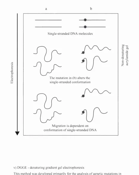
Figure 1.3 Schematic diagram demonstrating the principle of SSCP analysis
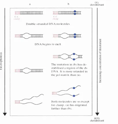
Figure 1A Schematic diagram demonstrating the principle of DGGE analysis
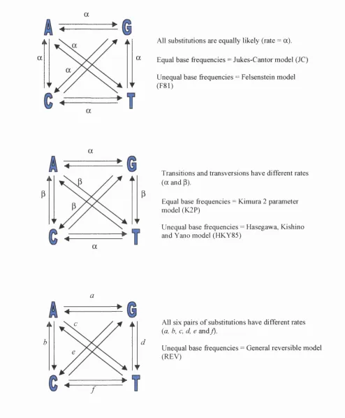
Figure 1.5 Different models of nucleotide substitution
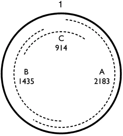
Figure 2.1 Positions of the 3 overlapping PCR products in the PMV genome
Figure 3.1a Nucleotide alignment of prison inmates' surface gene sequences
Figure 3.1b Nucleotide alignment of prison inmates' core gene sequences
Figure 3.2 Autoradiograph of HBV SSCP assay on samples from a
Liverpool prison
Figure 3.3 Dendrograms of HBV sequences from prison inmates
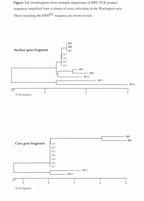
Figure 3.4 Dendrograms of HBV sequences from patients in Warrington
Figure 3.5 Graph showing the number of acute cases o f HBV infection
occurring in Hull
Figure 3.6a Nucleotide alignment of optimisation panel surface gene
sequences
Figure 3.6b Nucleotide alignment of optimisation panel core gene sequences
Figure 3.7a DGGE gel picture and dendrogram of optimisation panel surface
gene PCR products
Figure 3.7b DGGE gel picture and dendrogram of optimisation panel core
gene PCR products
Figure 3.8 DGGE results o f PCR products from Hull samples, 1992-1996
Figure 3.9a DGGE results of PCR products from SE England samples, 1990
Figure 3.9h DGGE results of PCR products from SE England samples, 1996
Figure 3.10 DGGE results of PCR products from autohaemotherapy outbreak
samples
Figure 3.11a Nucleotide alignment o f clinic attendees' surface gene sequences
Figure 3.11b Nucleotide alignment of clinic attendees' core gene sequences
Figure 3.12 DGGE results of PCR products from randomly selected samples
Figure 3.13 Dendrogram of HBV surface sequences from autohaemotherapy
Figure 4.1 Dendrogram o f TTV N22 region sequences
Figure 4.2 Deduced amino acid alignment of TTV N22 region sequences
Figure 4.3 Nucleotide alignment of PCR product sequences from sample PM
Figure 4.4 Nucleotide sequence of PMV genome
Figure 4.5 Organisation o f the PMV genome
Figure 4.6 Phylogenetic tree of PMV and 23 TTV sequences from GenBank
Figure 4.7 Phylogenetic trees of PMV and 7 representative TTV sequences
Figure 4.8 Genetic distances used to produce the Fitch tree in Figure 4.7
Figure 4.9a Nucleotide similarity plot of an alignment of PMV with 7 TTVs
Figure 4.9b Amino acid similarity plots of alignments o f ORFs 1 and 2 from
PMV and 7 TTVs
Figure 4.10 Phylogenetic trees of the NCR of PMV and 5 TTVs
Figure 4.11 Predicted secondary structure of the TTV NCR region
downstream o f the polyadenylation signal
Figure 4.12 Predicted secondary structure of the TTV GC-rich region
Figure 4.13 Amino acid alignment of a conserved ORF-2 motif in TTV,
TLMV and CAV
Figure 4.14 Phylogenetic tree of the conserved ORF-2 motif sequences
Figure 4.15 Similarity plot output from Recombinant Identification Program
List of tables
Table 2.1 Oligonucleotide primers used for the amplification of HBV
surface and core fragments
Table 2.2 Oligonucleotide primers used for the amplification of TTV N22
and NCR fragments
Table 3.1 Summary of DGGE results from Hull and SE England samples
Table 3.2 Summary o f DGGE results from Hull samples, analysed
according to risk groups
Table 3.3 Summary o f serological results on HBV DNA positive samples
from autohaemotherapy outbreak
Table 4.1 Full and near full-length TTV sequences
A bbreviations
ALT alanine aminotransferase
BFDV beak and feather disease virus
BLAST basic local alignment search tool
bp base pair
CAV chicken anaemia virus
cccDNA covalently closed circle DNA
CDSC Communicable Disease Surveillance Centre
CPHL Central Public Health Laboratory
CTL cytotoxic T lymphocyte
DGGE denaturing gradient gel electrophoresis
DHBV duck hepatitis B virus
DNA deoxyribonucleic acid
dNTP deoxynucleotide triphosphate
EDTA disodium ethylenediaminetetra acetate
EMBL European Molecular Biology Laboratory
8 encapsidation signal
HBV hepatitis B virus
HBV*'' hepatitis B virus autohaemotherapy variant
HBV’’'' hepatitis B virus prisoner variant
HBcAg hepatitis B core antigen
HBeAg hepatitis B e antigen
HBsAg hepatitis B surface antigen
HBIG hepatitis B immunoglobulin
HCC hepatocellular carcinoma
HCV hepatitis C virus
HIV human immunodeficiency virus
HLA human leukocyte antigen
IDU injecting drug user
IFN-a alpha interferon
kb kilobase
MHR mRNA NCR nt ORF P PCR PCV PHLS PHYLIP PMV RCR RDA RIP RNA SSCP S proteins TAB TBE TLMV Tm TTV UPGMA
major hydrophilic region
messenger RNA
non-coding region
nucleotide
open reading frame
polymerase
polymerase chain reaction
porcine circovirus
Public Health Laboratory Service
phytogeny inference package
PM virus
rolling circle replication
representational difference analysis
recombinant identification program
ribonucleic acid
single strand conformation polymorphism
surface proteins
tris-acetate-EDT A
tris-borate-EDTA
TTV-like mini virus
melting temperature
TT virus
Chapter 1
1.1 Viral causes of hepatitis
Knowledge of the existence of hepatitis can be traced back to the fifth century
BC, when Hippocrates first described epidemic jaundice. Throughout history,
outbreaks of hepatitis have been described in association with wars and natural
disasters (Mahoney, 1999; Paver and Mortimer, 1996). In 1885, Lurman
recorded 191 cases of jaundice in workers at a shipyard in Bremen who had all
received a smallpox vaccine preparation containing glycerinated human lymph.
This report provided the first evidence that hepatitis could be transmitted from
blood-containing products. Similar outbreaks have been documented in
diabetics, in patients treated for syphilis, and following the administration of
yellow fever vaccines. However, it was only after World War II that a distinction
could be made between hepatitis A (infectious hepatitis) which was transmitted
via the faecal-oral route, and hepatitis B (serum hepatitis) which resulted from
percutaneous exposure. Descriptions of their different epidemiologies resulted
from human volunteer studies (Havens, 1946; MacCallum and Bauer, 1947) and
Krugman and colleagues later verified that homologous immunity occurred after
infection with each type (Krugman et a l, 1967).
At about this time, Blumberg et al. (1967) described an immunoprecipitin that
was present in the blood of a leukaemic Australian Aborigine, which they named
Australia antigen. Subsequent studies found that this antigen was highly
prevalent in multiply transfused patients and a link with hepatitis B was
hypothesised. Its viral origin was established when particles that reacted with
antisera to Australia antigen were visualised using immune electron microscopy
(Dane et a l, 1970). The various components that make up the hepatitis B “Dane
particle” were rapidly dissected. Hepatitis B virus (HBV) is now classified as a
hepadnavirus and several related mammalian and avian members have since
been characterised. HBV will be further described in Section 1.2.
The discovery of the hepatitis A virus, also by the use o f immune electron
microscopy, was reported later (Feinstone et al., 1973). It could be
experimentally transmitted to primates, and was finally isolated in cell culture in
Diagnostic assays for both viruses quickly became available. In the early 1970s,
many developed countries introduced blood donor screening for the Australia
antigen, now renamed hepatitis B surface antigen (HBsAg), in an effort to reduce
post-transfusion jaundice. However, this condition did not disappear, and
hepatitis was still observed in approximately 10% o f blood transfusion
recipients. It thus became apparent that at least one further bloodbome hepatitis
agent remained to be found.
Hepatitis D virus (delta virus) was first described in 1977, and was initially
thought to be a new antigen of hepatitis B (Rizzetto et ah, 1977). However, it
was later shown to be a separate transmissible, but defective, virus. Its 1.7 kb
RNA genome encodes only one antigen and it requires HBV to replicate and to
coat its genome with an envelope consisting of hepatitis B surface proteins.
A further enterically transmitted hepatitis virus, hepatitis E virus, was detected
when clinical samples, collected during water-borne epidemics o f hepatitis in
India, were serologically tested for HAY and HBV. It was found that most
patients had evidence of anti-HAV IgG and so were immune to infection by this
virus, which had initially been postulated as the causative agent (Wong et al.,
1980). HEV was visualised in 1983 and later experimental transmission studies
demonstrated its aetiologic role in hepatitis (Balayan et a i, 1983; Krawczynsky
and Bradley, 1989).
Meanwhile, the principal cause of post-transfusion non-A non-B hepatitis cases
remained undiscovered until 1989, when the hepatitis C virus (HCV) genome
was characterised using molecular cloning techniques (Choo et a i, 1989).
Around 170 million people worldwide are thought to be chronically infected
with this flavivirus.
It was found, however, that some cases of post-transfusion non-A non-B
hepatitis still occurred that could not be linked to HCV infection. Efforts
continue to isolate infectious agents that are responsible for these residual cases,
and for sporadic community-acquired non-A to E hepatitis. In 1995, two groups
to be isolates of the same virus, named hepatitis G virus (HGV) or GB virus C
(GBV-C) (Simons et a l, 1995; Linnen et al., 1996). It is related to HCV, and is
transmitted via bloodbome routes. A few studies have demonstrated a higher
prevalence of HGV/GBV-C in patients with acute or chronic hepatitis than in
healthy controls (Linnen et al., 1996; Fiordalisi et al., 1996) and linked it to
some cases of fulminant hepatitis (Heringlake et al., 1996). However,
chronically infected patients have been found with normal aminotransferase
levels, and HGV/GBV-C appears to have no effect on the progression to liver
disease in patients who already carry another hepatitis vims (Alter et al., 1997).
Recent evidence suggests it replicates in lymphoid tissue, but not in hepatocytes
(Hoffman et al., 1998; Kobayashi et al., 1999). Thus its role as a hepatitis agent
is questionable.
The most recent hepatitis vims candidate was discovered in 1997, using the
technique of representational difference analysis (RDA) (Nishizawa et al., 1997).
It was named TT vims (TTV) after the initials o f the post-transftision hepatitis
patient from whom viral DNA was first isolated. TTV is discussed further in
Section 1.3.
1.2 Hepatitis B Virus
1.2.1 Virion stmcture, genetic organisation and proteins
The HBV virion (Dane particle) consists of a 42nm double-shelled stmcture. The
viral genome and DNA polymerase are encapsulated by a nucleocapsid made up
of hepatitis B core antigen (HBcAg), which in turn is surrounded by an envelope
comprising the predominant HBsAg (also known as the small S protein), and the
middle and large S proteins.
Subviral stmctures (spheres and filaments), composed of the three hepatitis B S
proteins, are usually present in the blood of HBV carriers at much higher
concentrations than the infectious Dane particles. The exact function of these
empty particles is unknown, but it has been postulated that they serve as
they are able to enhance intracellular viral replication at the early stage of
infection (Bruns et al., 1998).
HBV has a circular, partially double-stranded DNA genome. Its 3200 bp are
economically organised, with four overlapping open reading frames (ORFs) and
regulatory sequences contained within the genes (Figure 1.1). This genomic
arrangement allows HBV to code for more protein per nucleotide than any other
known virus, and imposes constraints on its evolution (Mizokami et al., 1997).
The longer minus strand has a 5’ covalently liked terminal protein, or ‘primase’,
which is part of the viral polymerase and is necessary for priming minus strand
synthesis (Bosch et al., 1988; Weber et al., 1994). The 5’ end of the plus strand
has an overlap o f around 200 complementary bases with the 5’ end of the minus
strand, enabling the DNA to circularise. This is helped by two 1 Ibp direct repeat
sequences, DRl and DR2, which are also the initiation sites for minus strand and
plus strand DNA synthesis respectively (Will et al., 1987; Seeger and Maragos,
1990). The length of the 3’ end of the plus strand is variable and is still attached
to the viral polymerase, indicating that hepatitis B virions are secreted from
infected hepatocytes before plus strand synthesis is completed (Landers et al.,
1977).
The four ORFs encode seven known viral proteins. Other than the three envelope
(S) proteins, these are HBcAg, hepatitis B e antigen (HBeAg), viral polymerase
(P) and the X protein. Four promoters and two enhancers provide transcriptional
control. The preSl promoter regulates transcription of a 2.4 kb mRNA, which
codes for the large S protein, and contains binding sites for liver specific
transcription factors. The preS2/S promoter is embedded within the preSl region
and controls transcription o f a 2.1 kb mRNA, encoding both the middle S protein
and HBsAg. The core promoter directs the production of two supergenomic
transcripts: the 3.5kb pregenomic RNA, from which the HBcAg and polymerase
are translated, and which serves as the RNA template during replication, and the
precore mRNA, which codes for HBeAg, a secreted protein. The P ORF is the
largest, encompassing about 80% of the genome. As well as the amino terminal
primase protein and DNA polymerase it contains a reverse transcriptase domain
X promoter controls transcription and translation from the smallest ORF, producing the X protein, which has transactivating activity and many other regulatory functions (Murakami, 1999). All transcripts use the same polyadenylation site and are therefore coterminal.
Figure 1.1 The genetic organisation o f the HBV genome. The partially double
stranded DNA is shown innermost, with promoters (prom), direct repeats (DR), enhancer regions (En) and the glucocorticoid regulatory element (GRE). Shaded arrows denote positions o f ORFs. Outer circles show the transcribed mRNAs and pregenomic RNA.
S2/S
r e S f
2307
833
Gene X
1.2.2 Replication
Extensive experimental studies o f duck hepatitis B virus (DHBV) have provided important insights into the key stages o f replication o f Hepadnaviridae. The initial host and organ-specitîc attachment o f HBV to a hepatocyte is largely mediated by the large S protein, although cellular receptors have not been
defined (Bruss et al., 1996). Similarly, the passage o f the virion into the cell is
not well understood. A recent study demonstrated that under conditions of low
pH, a conformational change in the large S protein of DHBV occurs, which
enables virions to bind to liposomes and intact cells (Grgacic and Schaller,
2000). However, previous studies have shown that uptake o f the virus by
hepatocytes is not dependent on an acidic endocytotic vesicle (Kock et al., 1996;
Hagelstein et a l, 1997).
The nucleocapsid is uncoated and directed to the nucleus where the DNA is
released. The plus strand is filled by the DNA polymerase and the genome
converted into a covalently closed circle (cccDNA) for transcription of genomic
and subgenomic RNAs by cellular RNA polymerase II (Summers and Mason,
1982; Rail et a l, 1983; Miller et a l, 1984). These are transported to the
cytoplasm for translation into the virion proteins and, in the case of the
pregenomic RNA, for reverse transcription into the minus strand o f the genome.
This occurs following encapsidation of the pregenome. The HBV polymerase is
thought to bind to a stem-loop structure at the 5’ end (encapsidation signal e) and
this complex is then recognised by the self-assembling nucleocapsid (Knaus and
Nassal, 1993). Due to its terminal redundancy, the pregenome also has an
encapsidation signal structure at its 3' end. It has been hypothesised that the HBV
polymerase must also require the 5' cap structure for encapsidation to occur
(Jeong et a l, 2000). The pregenome is degraded by endogenous RNaseH activity
during reverse transcription (Radziwill et a l, 1990).
Once plus strand synthesis has begun, the mature capsid is packaged in the
envelope within the endoplasmic reticulum and disulphide bridges between the
core and surface proteins maintain the virion structure. Again, the large S protein
contains a sequence that is essential for virion assembly (Bruss, 1997). The
mature capsids can follow a different route, back to the nucleus, if the large S
protein is in short supply, thus increasing the pool of cccDNA molecules
(Lenhoff and Summers, 1994). Virions are transported via the Golgi apparatus to
1.2.3 Subtypes and genotypes
The possible existence of antigenic heterogeneity within HBsAg was recorded
soon after its discovery, as a result of immunodiffusion experiments (Raunio et
al., 1970). In addition to the a determinant, which is common to all variants, two
pairs o f mutually exclusive antigenic determinants were identified {d/y and w/r),
giving rise to four defined subtypes (Le Bouvier, 1971; Bancroft et a l, 1972).
This was increased to nine with the description of ftirther subdeterminants. These
are aywj.4, ayr, adw2, adw4, adr q~ and adrq-^ (Magnius et al., 1975; Couroucé et
al., 1976; Couroucé-Pauty et al., 1978). DNA sequencing o f the S gene has
allowed the genetic basis of the subtypes to be determined (Magnius and Norder,
1995). However, antigenic subtypes do not reflect the phylogenetic relationships
between HBV genomes and, therefore, classification into six genotypes (A-F) is
now common (Okamoto et al., 1988; Norder et al., 1994). The designations are
based on an intergroup divergence o f at least 8%. Recently, using this criterion, a
new genotype (G) has been identified (Stuyver et al., 2000). Both genotypes and
subtypes show clear differences in their geographical distribution, with
genotypes A and D being the most prevalent in Europe (Magnius and Norder,
1995).
1.2.4 Epidemiology and transmission
As one o f the most common infections in the world, hepatitis B is responsible for
considerable levels of morbidity and mortality. It is currently estimated that there
are 350 million chronic carriers of HBV, and one fifth of these are likely to die
as a result of HBV induced liver disease (Kane, 1996). The global distribution is
varied and, based on the prevalence o f chronic carriers, countries are
conventionally divided into areas of high (>8%), medium (2-7%) and low (<2%)
endemicity (Evans and London, 1998).
The predominant modes of HBV transmission are perinatal, parenteral and
sexual. Although HBsAg or DNA has been detected in saliva, breast milk, urine
and faeces, these are not thought to represent important vehicles of transmission
(Boag, 1991). The relative importance of each transmission route depends upon
endemicity, perinatal transmission maintains the pool o f chronic carriers, and
individuals have a greater than 60% lifetime risk of acquiring HBV (Alter,
1996). In the UK, where the prevalence is estimated to be <1%, sexual
transmission and injecting drug use are the main routes of acquisition (Balogun
et a l, 1999), with young adults aged 15-24 being at highest risk (Zuckerman,
1999a).
Needle exchange programmes and health campaigns encouraging safer sex were
implemented in the mid-1980s in England and Wales to control the HIV
epidemic; these measures also served to reduce the incidence of HBV infection.
A recent survey o f laboratory confirmed acute HBV infections revealed a large
decrease from 1761 reported cases in 1985 to 583 in 1989 (Balogun et al., 1999).
The incidence remained stable until 1996, but has since risen significantly, with
719 cases reported in 1999. This rise has largely been attributed to an increase in
transmissions amongst injecting drug users (IDUs) (Communicable Disease
Surveillance Centre, CDSC, 1999). The true number o f acute infections cannot
be accurately assessed, as a large proportion of cases remain asymptomatic.
Although rare, nosocomial transmission continues to occur in the UK. Lapses in
cross-infection control have resulted in the transmission of HBV in
haemodialysis and organ transplantation units (Wreghitt, 1999; Tedder et al.,
1995). Similarly, surgeon to patient transmissions have been described, despite
routine vaccination of all healthcare workers (The Incident Investigation Team
and Others, 1997; Scottish Centre for Infection and Environmental Health,
SCIEH, 1999).
Since the introduction of blood donation screening for HBsAg in the early 1970s,
the risk o f post-transfusion hepatitis caused by this agent has become very low
(Soldan and Barbara, 1999). The introduction of diagnostic PCR has generated
debate as to whether donations should now be screened for nucleic acids in an
1.2.5 Outcomes of infection
The incubation period of HBV ranges from 45 to 120 days. The time between
exposure and the first detectable serological response is known as the window
period. This has been estimated to be in the region o f 59 days to HBsAg
positivity, with PCR to detect HBV DNA reducing it by around 1 week (Gerlich
and Caspari, 1999; K. Soldan, pers. comm.). However, it is likely to vary widely
between individuals. An acute phase follows, when HBsAg, HBeAg, HBV DNA
and anti-HBc IgM antibodies are detected. The levels of aminotransferases, eg.
alanine aminotransferase (ALT), in the blood are also raised, which provides a
marker of liver injury. HBV infection can have a variety of clinical outcomes.
Perinatally infected infants are generally asymptomatic, and only about one third
of adults have specific symptoms. These include jaundice, malaise, nausea, and
the excretion of dark urine and pale stools. HBV is not directly cytopathic: liver
damage largely results from the cytotoxic T lymphocyte (CTL) response to
infected hepatocytes (Chisari and Ferrari, 1995). In rare cases of acute HBV
infection, liver necrosis is extensive and fulminant hepatitis develops.
Chronic HBV infection is operationally defined as the presence of HBsAg in
serum for over 6 months. The major determinant of progression to chronic
disease is the age of HBV acquisition. Around 90% of infants become chronic
carriers, possibly due to neonatal tolerance to HBeAg epitopes. The risk of
developing chronic infection drops to between 25 and 50% o f children infected
at age 1 to 5 years, and only 5 to 10% if infected as adolescents or adults
(Beasley et ah, 1981; McMahon ef a/., 1985).
The mechanisms for viral persistence in adults are under investigation. It has
been noted that in adult immunocompetent patients who clear HBV after an
acute infection there is a vigorous, polyclonal cellular immune response,
involving CD4+ and CD8+ cells. This appears to be weak or interrupted in those
who progress to chronic carriage. Other host and viral factors have been
postulated to effect viral persistence, such as allelic differences in the HLA
complex and the circulating HBV DNA level in early acute infection (Thio et a l,
1999; Roingeard. et a l, 1997). Mayerat et a l (1999) examined patients with
predominate, and found that infection with genotype A was significantly
associated with chronic outcome. However, no biological basis for this finding
has been described.
The sequelae o f HBV chronic infection can vary widely in severity, from chronic
active hepatitis to an asymptomatic carrier, or tolerant, state. In general, carriers
with detectable HBeAg have actively replicating HBV and are more likely to
develop cirrhosis and hepatocellular carcinoma (HCC). Spontaneous
seroconversion to anti-HBe positive has been reported in a varying proportion of
carriers and is often associated with clearance o f HBsAg and HBV DNA from
the blood and remission o f disease (Fattovich, 1996). However, using PCR, HBV
DNA is still detectable in the liver of a proportion of chronic carriers who have
cleared HBsAg, indicating that low level replication may continue (Mason et al.,
1998).
1.2.6 Prevention and treatment
Both passive and active immunisation are available for the prevention of HBV
infection. Hepatitis B immunoglobulin (HBIG) was first prepared in the early
1970s by fractionation of pooled serum containing high titres of anti-HBs
antibodies, and is effective in neutralising circulating virions if given soon after
exposure (Grady et al., 1978). Currently, owing to high costs, HBIG is given to
specific risk groups only, such as infants bom to HBeAg positive mothers
(Wheeley et al., 1991). The first plasma-derived vaccine of purified HBsAg
subviral particles became commercially available in the UK in 1982, and was
followed five years later by a recombinant yeast-derived HBsAg vaccine. When
the recommended three doses are given at months 0, 1 and 6, a protective
antibody response is induced in >90% of adults younger than 40 years (Andre,
1989).
In order to further improve the efficacy of vaccination and reduce the number of
doses required, several new vaccine strategies are being examined. Small animal
studies and clinical trials have shown that the inclusion of preS epitopes may be
beneficial in terms of increased anti-HBs production, earlier onset of antibody
(Suzuki et al., 1994; Shouval et a l, 1994; Zuckerman et al., 1997). Davis (1998)
demonstrated the feasibility o f DNA vaccines using eukaryotic expression
vectors in mice and chimpanzees, and a novel approach using transgenic potatoes
to produce oral HBV vaccine was recently reported (McNamee, 1999).
In 1992, the WHO recommended that all countries, regardless of endemicity,
should introduce universal HBV vaccination into their national programmes of
immunisation by 1997 (WHO, 1992). Over 80 countries have achieved this,
although coverage varies widely (Kane, 1996). In the UK, control o f HBV is
based on the selective vaccination of high risk groups, such as injecting drug
users, haemophiliacs, haemodialysis patients, homosexual men with multiple
partners, prostitutes, infants bom to HBsAg positive mothers, health care
workers and contacts of patients with acute or chronic HBV infection
(McMenamin, 1996). However, there is much evidence to suggest that this
policy is failing as at-risk groups, particularly IDUs in the community and in
prisons, are not being reached (Goldberg and McMenamin, 1998; Lamagni et al.,
1999). Universal antenatal screening for HBsAg is now recommended in an
effort to reduce the pool of chronic carriers from whom perinatal transmission
can ensue (Ramsay et al., 1998).
Treatment strategies that aim to clear chronic carriers of the virus have had only
limited success. Recombinant alpha interferon (IFN-a) was the first dmg to be
licensed for the treatment of chronic HBV infection. As a cytokine, it functions
as an immune modulator and after 4-6 months of treatment can lead to a HBeAg
to anti-HBe seroconversion rate of 30-40% (Wong et al., 1993). However, it is
associated with significant side effects, and a limited response rate in carriers
who are in the tolerant (anti-HBe positive) phase (Brook et al., 1989).
Several nucleoside analogues are currently under investigation for anti-HBV
activity. Lamivudine has recently been licensed for clinical use. Lamivudine (or
3TC) is an enantiomer of 2'-deoxy-3'-thiacytidine, and thus competes with dCTP
for incorporation into extending DNA chains. Once introduced, chain
new virions and a decrease in intracellular cccDNA (Severini et ah, 1995).
Although less toxic than IFN-a, HBeAg seroconversion is only seen in
approximately 16% of patients after 12 months of treatment, compared to 4%
with placebo (Lai et a l, 1998). Combination therapy with other nucleoside
analogues, such as famciclovir and adefovir dipivoxil, may improve outcome,
although these agents act only to suppress viral replication and are therefore
unlikely to be curative. Emerging therapies may prove to be more successful. For
example, therapeutic vaccines may stimulate a HBV-specific CTL response,
which is weak in chronic carriers (Pianko and McHutchison, 1999).
1.2.7 Variation in HBV
Many studies of HBV infection have focused on genomic variants in recent years
in the hope that knowledge o f their cause, prevalence and functional significance
will aid the design of new therapies. Due to its unique method of replication, via
an RNA intermediate, the HBV genome mutates at a higher rate than do those of
other DNA viruses. The rate of nucleotide substitution per site per annum has
been calculated to be 2 x 10'^ (Okamoto et aL, 1987), although this is likely to
vary from patient to patient depending on their stage of infection. Also,
variability is not seen randomly throughout the genome, due to the constraints
imposed by the overlapping ORFs and the functions of the encoded proteins
(Lauder et al., 1993).
The observation that G->A hypermutation occurs suggests that mutations
generally arise from a lack o f proofreading by the P protein reverse transcriptase
(Gunther et a l, 1997). Hypermutation is a common feature associated with
retroviral reverse transcriptases.
In addition to point mutations that occur during replication, variation can be
generated in a number of ways, such as insertion and deletions due to RNA
splicing (Terre et al., 1991), template switching during reverse transcription, and
recombination between different genotypes (Bollyky et al., 1996; Bowyer and
Certain point mutations are epidemiologically significant. One o f the most
common is a G -^A at position 1896, which produces a stop codon at residue 28
of the precore protein and thus prevents the translation of HBeAg. It was first
detected in patients who had appeared to seroconvert to anti-HBe, but had severe
liver disease (Carman et al., 1989). Since then, the mutation has been found
frequently in patients with long-standing infection, with and without active
hepatitis. In such patients, the mutant often becomes the dominant circulating
type after seroconversion (Hamasaki et al., 1994). Many cases of acute fulminant
hepatitis have been linked to infection with this variant, although it can also
cause a self-limiting hepatitis, suggesting that other factors must be involved in
the production o f severe acute disease (Tanaka et al., 1995; Karayiannis et al.,
1995; Mphahlele et al., 1997).
That the G 1896A mutation emerges at all is still not understood; possible
explanations are enhanced replication, immune selection and hypermutation
(Carman et al., 1998; Ngui et al., 1999). It appears mainly in genotypes B, C, D
and F, due to structural constraints imposed by the RNA encapsidation signal,
and its prevalence is therefore geographically restricted (Lok et al., 1994).
HBV carrying the G1896A mutation has been associated in recent years with
several transmissions of HBV from surgeons to patients in the UK (The Incident
Investigation Team and Others, 1997; SCIEH, 1999). Recognition that anti-HBe
positive carriers can have a high level of circulating virus has led to proposals
that new guidelines be introduced to restrict practices of HBV positive surgeons.
The challenge is to decide at which titre they become effectively uninfectious
(Ballard and Boxall, 1999). New guidance from the Department of Health
recommends that health care workers whose HBV DNA concentration in the
blood exceeds 1000 genome equivalents per ml should not perform exposure-
prone procedures (Department of Health, 2000).
Mutations in the S gene, which result in vaccination failure in infants bom to
HBsAg positive mothers, have been studied intensively in recent years. The most
resulting in a G to R change at amino acid 145 (Carman et al., 1990), Several
other mutations have now been described within the major hydrophilic region
(MHR) o f HBsAg that also appear to result in vaccine failure, with concurrent
circulation in the infected vaccinees of HBsAg and anti-HBs antibodies (Wallace
and Carman, 1997; Chong-Jin et ah, 1999). On examination of maternal blood,
the mutant is often absent, or present in very low amounts, suggesting that its
appearance in the infant is due to a rapid selection (Gunther et al., 1999a; Ngui
etal., 1997).
Transmission studies have demonstrated that the G145R mutant is stably
transmitted and does not revert to wild type during chronic carriage (Ogata et al.,
1997), thus raising concerns that vaccine-escape mutants may emerge.
Mathematical modelling predicts that this will be the case given certain different
assumptions about the infectivity of the mutant strains, and the extent of
protection afforded against them by current vaccines. However, it is forecast that
emergence would not occur for at least 50 years (Wilson et ah, 1998). In order to
monitor the prevalence of HBsAg mutants, modifications to current serological
tests may be necessary, particularly where monoclonal antibodies are used, as
altered epitopes can result in false negative results (Carman et al., 1995;
Zuckerman and Zuckerman, 1999).
The introduction of nucleoside analogues for the treatment o f chronic HBV
infection has been followed swiftly by the description of resistant mutants.
Lamivudine resistance is mediated by mutations in the polymerase gene, most
commonly in the YMDD locus. This locus falls within the catalytic region of the
RTase/DNA polymerase domain of the HBV P protein, and is well conserved in
all viral reverse transcriptases (Bartholomeusz et al., 1998). The M residue,
which is at codon 552, is substituted by V or, more rarely, I (Hussain and Lok,
1999). Identical mutations at the YMDD locus of the HIV reverse transcriptase
have also been reported, in variants that are resistant to lamivudine (Tisdale et
al., 1993). In HBV, a further substitution at codon 528 from L to M has been
Transfection studies have confirmed that the YMDD locus mutations bestow
lamivudine resistance in both HIV and HBV, but they also reduce replication
competence, which may explain why wild type virus re-emerges on withdrawal
of therapy (Back et al., 1996; Ling and Harrison, 1999; Ono-Nita et al., 1999).
Das et al. (2000) have predicted that steric hindrance occurs between the
p-methyl group o f V or I, at codon 552 of HBV in the mutant polymerase, and the
sulphur atom of lamivudine. This would account for the observed cross
resistance to another nucleoside analogue, emtricitabine, which has a similar
oxathiolane ring.
The L528M mutation has also been detected in patients with breakthrough
infection following famciclovir therapy, suggesting a degree of cross resistance
with lamivudine (Tillman et al., 1999). The importance of other reported
famciclovir-induced substitutions has not yet been established, although the
YMDD locus does not appear to be involved (Gunther et al., 19996; Seignères et
al., 2000). Further work will be needed in order to devise effective combination
therapies for the control of HBV replication in long term carriers.
1.3 TT Virus
1.3.1 Genetic organisation
TTV has been characterised as an non-enveloped single-stranded DNA virus
with a caesium chloride density of 1.31-1.34 g/cm^ (Mushahwar et ah, 1999).
Filtration studies indicate that the virion is 30-50 nm in diameter, although it has
yet to be visualised (Mushahwar et ah, 1999). Several complete TTV DNA
sequences have now been published, revealing a circular genome that ranges
from 3808 to 3852 nucleotides in length (Erker et ah, 1999; Okamoto et ah,
19996; Hijikata et ah, 1999). Nuclease protection assays show that the genome is
negative-stranded (Mushawar et ah, 1999).
A number of regulatory sequences and two conserved OREs have been identified
on the positive strand of the genome-length sequences described so far (Miyata
ORF-1, codes for approximately 770 amino acids, with a smaller ORF upstream that
overlaps ORF 1 by around 130 nucleotides (0RF2). Two putative third ORFs
have been proposed, also on the positive strand (Erker et ah, 1999; Takahashi et
al., 20006). Searches of protein databases have not revealed any significant
similarities to known amino acid sequences. It is assumed therefore that TTV
does not encode a polymerase and must rely on host cell polymerases for
replication. The protein products of TTV have yet to be expressed in cell culture.
TTV was initially reported to share some physical characteristics with the
Parvoviridae, although no nucleotide similarity was demonstrated. A
resemblance between TTV and chicken anaemia virus (CAV), a circovirus, has
been described by Takahashi et al. (1998a) on the basis of the presence of
arginine-rich regions at the N-terminal of ORF-1 from both viruses. As ORF-1 is
thought to code for the 51.7 kDa capsid protein in CAV, this similarity has led to
the suggestion that ORF-1 of TTV is the capsid gene. Furthermore, CAV
contains motifs at the carboxyl end of ORF-1 that may be involved in rolling
circle replication, two of which have been identified in ORF-1 of TTV (Niagro et
al., 1998; Mushahwar et al., 1999). A likeness in the genomic arrangements of
the two viruses has also been noted (Miyata et al., 1999). However, no
significant sequence similarity between TTV and any other member o f the
Circoviridae has been shovm, and this has led Mushahwar et al. (1999) to
propose that TTV be placed in a new virus family called Circinoviridae.
Recently, three novel human DNA virus sequences (CBD203, CBD231 and
CBD279) have been described that appear to be intermediately related to TTV
and CAV (Takahashi et al., 2000a). They have been named TTV-like mini virus
(TLMV). It is not clear from these reports whether TTV, CAV and TLMV
Figure 1.2 The genetic organisation o f the TTV genome. The N22 region was the first TTV DNA to be discovered using RDA (Nishizawa et al., 1997).
Conserved features shown are ORFs 1 and 2 (shaded arrows), TATA box (TATA), polyadenylation signal (polyA) and GC-rich region (GC).
ORF 2
poly A
ORF I
1.3.2 Genotypes
Considerable genetic variability o f TTV has been demonstrated, with a 222-bp fragment o f the genome from ORF-1 showing as much as 65% divergence between sequences. Several groups have assigned up to 16 genotypes based on the sequence variability in short TTV PCR products (Y. Tanaka et al., 1998; Okamoto et a i, 1999c; IChudyakov et al., 2000). In contrast, phylogenetic analysis o f full-length, or near full-length TTV sequences reveals division into three main types, represented by the prototype TA278 in group 1, US35 in group 2 and JAIO in group 3 (Erker et al., 1999). Most o f the sequence variability
occurs in the coding regions. In addition to these three TTV groups there are
other highly divergent complete sequences (Okamoto et al., 19996; Hijikata et
al., 1999; Takahashi et al., 20006; Ukita et al., 2000). PCR cloning studies have
found mixed infections, with up to seven different genetic types, to be a common
event (Takayama et al., 1999; Niel et al., 2000). Different TTV sequences do not
appear to show geographical segregation, as is seen with other bloodbome
hepatitis viruses (Mushahwar et al., 1999).
1.3.3 Epidemiology and transmission
TTV was initially presented as a candidate hepatitis virus, as TTV DNA titres
were found to correlate closely with aminotransferase levels in three post
transfusion non A-G hepatitis patients (Nishizawa et al., 1997). TTV DNA has
since been amplified from a high percentage of patients with post-transfusion
hepatitis and those with chronic liver disease (Charlton et al., 1998; H. Tanaka et
al., 1998). It has also been detected in the liver of infected patients at 10-100
times the concentration in serum, suggesting that the virus is hepatotropic
(Okamoto et a l, 1998a). However, more extensive studies have revealed that
TTV is highly prevalent in healthy individuals in many parts of the world, so its
ability to cause disease is far from certain (Prescott and Simmonds, 1998; Hsieh
et al., 1999; Maggi et a l, 1999; Abe et al., 1999). PCR assays with primers
directed against more conserved genomic regions have led to reports of
prevalences approaching 100% in some population groups (Takahashi et al.,
19986; Leary et al., 19996). In the UK, the prevalence of TTV DNA in blood
donors was 18% using two primer systems (F. Bobet, pers. comm.).
TTV DNA has been detected in blood products that have undergone viral
inactivation procedures. Thus, its prevalence in haemophilia patients is high
(Gerolami et a l, 1998). Transmission by blood transfusion has also been
reported (Fujiwara et al., 1998). However, TTV DNA has been found in faeces
(Okamoto et al., 19986), breast milk (Toyoda et al., 1999), semen (Inami et al.,
2000) and saliva (Ross et al., 1999) and the age-distribution o f infected
individuals suggests that TTV is mainly a community-acquired virus (Yamada-
Osaki et al., 1998; Davidson et al., 1999). Evidence for transplacental
modes o f transmission may therefore account for its high global prevalence
(Zuckerman, 19996). The detection of human-like TTV sequences in domestic
farm animals raises the possibility that they may serve as a source of human
infection (Leary et al., 1999a). TTV-like DNA sequences have also been
identified in nonhuman primates (Verschoor et al., 1999; Okamoto et al., 2000a;
Abe et al., 2000; Romeo et al., 2000; Cong et al., 2000).
Both acute and chronic TTV infections have been documented, with DNA
persisting for up to 22 years in one study (Matsumoto et al., 1999; Leffere et al.,
2000). On examining the sequence o f fragments o f the genome, several groups
have found it to remain very stable over a number of years (Biagini et ah, 1999;
Ball et al., 1999). This would be expected if TTV were using host cell
polymerases to replicate, due to the low mutation rate of human DNA
polymerase enzymes. However, it suggests that in order to generate the observed
extensive genetic variability, TTV must have been present in the human
population for many thousands of years. This has led to the consideration that
TTV is part of the normal human flora (Simmonds et a l, 1999).
1.3.4 Disease association
It is now accepted that TTV is unlikely to cause liver disease in most infected
individuals. Numerous studies have failed to find any association between TTV
DNA positivity and acute sporadic hepatitis, fulminant hepatitis, HCC, or
exacerbations of chronic hepatitis (Parquet et al., 1999; Skelton et al., 2000;
Huang et al., 2000; Giménez-Barcons et al., 1999). Preliminary attempts to
detect anti-TTV antibodies have used immunoprécipitation techniques, and, as
antigen, an expressed truncated ORF-1 protein product (Tsuda et al., 1999;
Handa et al., 2000). These methods indicate that antibody is formed and can be
detected in patients with and without TTV viraemia. In an attempt to find the site
of replication of TTV, Okamoto et al. (20006 and c) have identified double
stranded forms of the viral genome in hepatocytes and bone marrow cells, which
they suggest are the replicative intermediates. TTV DNA has also been detected
in liver biopsy material by in situ hybridisation (Rodriguez-Ifiigo et ah, 2000). A
types are able to cause disease, or that certain hosts, eg. immunocompromised
patients, are susceptible to potential pathogenic effects of the virus.
1.4 Molecular epidemiological tools
1.4.1 Gel-based mutation screening
Numerous gel-based assays have been developed in order to screen PCR
products for mutations, without the need for sequencing. These have many
applications, but in virology they are employed for genotyping, investigating
transmission events, screening for epidemiologically important mutants, and for
the examination o f quasispecies evolution (Cotton, 1993; Arens, 1999). Some
methods are sensitive to single base pair mutations. The most commonly used
are described here:
i) RFLP - restriction fragment length polymorphism
This method employs restriction endonucleases to cleave PCR products and the
resulting fragments are separated by gel electrophoresis. It is often used for
genotyping, where a specific pattern of digested fragments is obtained from each
genotype. For this reason it is important to amplify a relatively conserved region
of the viral genome where the restriction sites are likely to remain stable
(Pohjanpelto et al., 1996; Lindh et al., 1998) A large amount of sequence data is
required in order to design a RFLP assay, and the panel of endonucleases must
be customised. RFLP can also be used to screen for a known mutation.
ii) HMA - heteroduplex mobility assay
When a mixed population of PCR products are denatured and allowed to
reanneal, a mixture of homoduplex (both strands have the same sequence) and
heteroduplex (there is one or more mismatch) double strands are formed. When
electrophoresed in a non-denaturing gel, the heteroduplexes are retarded in
relation to the homoduplexes and the distance between the two species is related
to the number o f mismatched base pairs (Barlow et al., 2000). This technique has
been used to detect single base pair mutations in the human cystic fibrosis gene
(White et al., 1992), although in other applications, only about 80% of mutations
genotyping HIV, where tester PCR products of known genotype are mixed with
a PCR product amplified from a sample of unknown genotype (Tatt et al., 2000).
iii) BESS - base excision sequence scanning
This is a relatively new technique that uses automated sequencing technology.
PCR products are amplified with one fluorescently labelled primer in a PCR mix
containing dUTP. They are then subjected to digestion with uracil-N-glycosylase
and endonuclease IV, generating a defined series o f fragments which can be
separated and detected on a standard sequencing gel (Hawkins and Hoffman,
1997). Approximately 95% o f mutations can be detected and localised when
both strands of the PCR product are analysed. The data are suitable for
phylogenetic analysis (see section 1.4.2), which is useful for grouping specimens
eg. from an outbreak or from different geographical locations (Charrel et al.,
1999).
iv) SSCP - single strand conformation polymorphism
Many studies of sequence polymorphisms in eukaryotic and prokaryotic genes
have utilised this method, which is based on the principle that in a non
denaturing gel single-stranded PCR products will fold into distinct secondary
structures depending on their nucleotide sequence. The differences in
conformation alter electrophoretic mobility, which is detected as different
banding patterns (Orita et al., 1989). This is depicted in Figure 1.3. SSCP assays
have been used to investigate nosocomial outbreaks of HBV (Yusof et al., 1994;
Hardie et al., 1996), although detection of single base pair mutations is again not
Figure 1.3 Schem atic diagram dem onstrating the principle o f SSCP analysis.
Q . O
UJ
Single-stranded DNA molecules
The mutation in (b) alters the single-stranded conformation
Migration is dependent on conformation o f single-stranded DNA T
S s
IS
v) DGGE - denaturing gradient gel electrophoresis
This method was developed primarily for the analysis o f genetic mutations in human disease and can be used to detect single base pair mutations. It is based on the differential migration o f double stranded DNA through a gel containing
increasing concentrations of the denaturing chemicals formamide and urea
(Myers et al., 1987). As electrophoresis progresses, each DNA fragment will
reach a position where the portion of the fragment with the lowest melting
temperature (Tm) will begin to unravel, causing a significant decrease in
electrophoretic mobility. The Tm of any domain is dependent on its nucleotide
sequence and therefore mutations are detected as differences in band positions on
the gel. If a mutation is present in the domain with the highest Tm, it will not be
detected, as sequence-dependent migration is lost when the strands become
completely dissociated. To improve sensitivity a 'GC clamp' is usually added to
one end of the PCR product to provide a domain with a very high Tm that will
not become denatured under DGGE conditions (Sheffield et al., 1989; Fodde and
Losekoot, 1994). The DGGE principle is shown in Figure 1.4. It has been used
recently in the study of JC virus molecular epidemiology, and to examine HCV
quasispecies (De Santis and Azzi, 2000; Harris and Teo, 2001).
1.4.2 Phylogenetic analysis
DNA sequencing inevitably provides more information about a viral genomic
fragment than any gel-based assay. Sequence data can be submitted to
phylogenetic analyses in order to answer questions about the genetic relatedness
of homologous DNA. In virology, it aids in the genotyping of virus families,
enables molecular epidemiological studies to be carried out, and allows the
evolution of viruses to be investigated (Holmes and Garnett, 1994). Of relevance
to public health is the application of phylogenetic analyses to the investigation of
microbial transmissions, such as that of HIV from a dentist (Crandall, 1995),
HBV from a blood donor (Zuckerman et al., 1995), and HCV from contaminated
Figure 1.4 Schem atic diagram dem onstrating the principle o f D G G E analysis.
0% denatiiiiun
mujtiii
D o u b le - s t r a n d e d D N A m o l e c u l e s
D N A b e e in s to melt
T h e m u t a tio n in ib ) h a s d e s t a b ilis e d a r e g io n ol the d s D N A . It is m o r e retarded in the g e l matrix than (a).
Both m o l e c u l e s are ss e x c e {’4 for c l a m p , (a) h as m ig r a t e d lurther than tb).
dénaturant
Phylogenetic methods were originally developed and applied to the study of eukaryotic evolution using data such as morphological characters and gene frequency data (Edwards and Cavalli-Sforza, 1964; Gamin and Sokal, 1965). The advent of DNA sequencing has resulted in a rapid accumulation of molecular data, which can be analysed in a similar way to answer questions about the relatedness of particular sequences to others, and to understand evolutionary relationships. The algorithms used to infer phylogenies are continually being refined and updated, and are now applied regularly to viral nucleotide and amino acid sequences.
There are several points to consider when drawing and interpreting a
phylogenetic tree, perhaps the most important being that all trees represent only
estimates of the real genetic relationship between sequences. In order to
maximise the accuracy of a tree, it is important to use models that approximate
the true behaviour of DNA. Similarly, the quality of the data entered will affect
the tree topology obtained. Sequences should come from homologous regions,
and be as similar in length as possible. If too short, or lacking in variation, there
will be insufficient ‘phylogenetic information’, but if sequences are too variable
the true relationships may be masked by background noise (Clewley, 1998).
The first stage in the analysis of nucleotide or amino acid data is to produce an
alignment of a set of homologous sequences. Several algorithms are available for
this purpose, the most useful for evolutionary studies being the global methods
(Weiller et al., 1995). These determine an optimal alignment, according to their
parameters, across the whole length of the sequences by carrying out pairwise
alignments, identifying the two most closely related sequences, and adding the
rest in order of similarity. Gaps are introduced where necessary to accommodate
insertions or deletions.
There are many methods for constructing a phylogenetic tree from a sequence
alignment. The most commonly used programs fall into three major categories:
i) Maximum Parsimony - These methods search for the tree that requires the
minimum number of changes at each character (nucleotide or amino acid) site to
explain the evolution of the group, and only phylogenetically informative sites
are considered. The main failure of parsimony is that it cannot easily allow for
homoplasy, i.e. convergent mutations, parallel mutations and reversals, which
result from the independent acquisition of the same properties along different
evolutionary lineages. This can lead to unrelated sequences being clustered
together (Stewart, 1993). To help address this problem, weights can be applied to
rare substitution events, or to different codon positions to improve the model
ii) Distance methods - These involve two main stages; firstly a distance matrix is
calculated by carrying out pairwise comparisons of the input sequences. There
are several available models to estimate genetic distances, many of which
consider transitions and transversions separately (2-parameter models), and can
add a correction for the possible occurrence of multiple substitutions at the same
site (Jukes and Cantor, 1969; Kimura, 1980; Felsenstein, 1981; Hasegawa et al.,
1985; Yang et al., 1994). The principles of some of the commonly used
nucleotide substitution models are represented in Figure 1.5.
Secondly, a clustering algorithm is applied to the data in the matrix to produce a
tree. The simplest program o f this type is UPGMA (unweighted pair group
method with arithmetic means), which looks for the smallest pairwise distance,
then considers these two most related sequences as a single taxonomic unit. The
distances are recalculated between this cluster o f two and each o f the remaining
sequences, and the process is repeated (Sneath and Sokal, 1973). The topology of
the tree is an arbitrary result o f the clustering algorithm. Other distance methods,
such as the Fitch and Margoliash method (1967) consider the difference between
the expected distance (that in the matrix) and the observed distance (the sum of
the branch lengths) between any two sequences, and search possible trees in
order to choose the one with the smallest difference. Another commonly used
method, the neighbour-joining method, uses a clustering algorithm which adds
pairs of sequences (neighbours) to a star-like tree, minimising the internal branch
length at each addition (Saitou and Nei, 1987). Thus, the most parsimonious tree
that fits the distance matrix is drawn.
iii) Maximum Likelihood - The starting point when using these methods is a
probabilistic model of sequence evolution. The programs then compute the
likelihood of any tree, which is the probability of the data, given the tree, and
therefore search for the tree with the maximum likelihood (Felsenstein, 1988).
These methods allow statistical comparisons between generated trees more easily
than the other methods, although they are not generally used for large data sets,
being computationally very demanding. For example, for ten sequences the
possible number of rooted trees is 34,459,425 (Li, 1997), and for each tree this
Figure 1.5 Different models of nucleotide substitution
a
ài
a
n a
All substitutions are equally likely (rate = a).
Equal base frequencies = Jukes-Cantor model (JC)
Unequal base frequencies = Felsenstein model (F81)
a
Ài Transitions and transversions have different rates (a and p).
Equal base frequencies = Kimura 2 parameter model (K2P)
Unequal base frequencies = Hasegawa, Kishino and Yano model (HKY85)
i i All six pairs of substitutions have different rates
(a, b, c, d, e and j).
Unequal base frequencies = General reversible model (REV)
Therefore, when more than 10 sequences are entered it is necessary to apply a
search mechanism to reduce the number of trees considered. As there is no
natural way to arrange trees in order to search, this is often carried using
‘perturbation’ methods, such as taxon swap or tree bisection and reconnection
(TBR). These take a possible tree, rearrange it and then recalculate the likelihood
to see if the new tree is an improvement. One downfall is the potential for ending
up with a locally optimum tree that is neither the true tree nor the most likely.
Most of the phylogenetic methods can be shown to provide the correct tree,
given a large amount o f data that does not breach any of the assumptions in the
model. However, real data sets often contain complexities that cannot be readily
handled by current phylogenetic methods. For example, there may be a base
composition bias, which would lead to sequences with a similar composition
being grouped together, even though they may not be the most closely related. A
difference in frequency of types of mutation events, and an unequal mutation
frequency at different sites are commonly seen in DNA data. It is therefore
important to use the weighting facilities in many programs to counteract these
occurrences. The simplest clustering methods, such as UPGMA, do not perform
well if the rate of evolution varies along different lineages in the same tree. Also,
the assumption o f independence among sites is likely to be inaccurate,
particularly in viral sequence data. Structures within DNA, such as stem-loops,
and the existence o f overlapping open reading frames mean that nucleotide
substitutions at affected sites will be constrained (Hillis et al., 1994).
Resampling methods, such as bootstrapping and jackknifing can be used to
examine the robustness of a tree, and statistical methods have been developed to
test the reliability o f a phylogeny (Felsenstein, 1988). More sophisticated
techniques are becoming available for inferring phylogenies. For example, the
use of split decomposition can take into account conflicting alternative groupings
by displaying results as a network, rather than the typical bifurcating tree
(Bandelt and Dress, 1992; Dopazo et a i, 1993). This technique has a particular
application when examining sequences arising from recombination events. To