Alopecia areata
Part 1: pathogenesis, diagnosis, and prognosis
Frank Spano
MD CCFPJeff C. Donovan
MD PhD FRCPCAbstract
Objective To provide family physicians with a background understanding of the epidemiology, pathogenesis, histology, and clinical approach to the diagnosis of alopecia areata (AA).
Sources of information PubMed was searched for relevant articles regarding the pathogenesis, diagnosis, and prognosis of AA.
Main message Alopecia areata is a form of autoimmune hair loss with a lifetime prevalence of approximately 2%. A personal or family history of concomitant autoimmune disorders, such as vitiligo or thyroid disease, might be noted in a small subset of patients. Diagnosis can often be made clinically, based on the characteristic nonscarring, circular areas of hair loss, with small “exclamation mark” hairs at the periphery in those with early stages of the condition. The diagnosis of more complex cases or unusual presentations can be facilitated by biopsy and histologic examination. The prognosis varies widely, and poor outcomes are associated with an early age of onset, extensive loss, the ophiasis variant, nail changes, a family history, or comorbid autoimmune disorders.
Conclusion Alopecia areata is an autoimmune form of hair loss seen regularly in primary care. Family physicians are well placed to identify AA, characterize the severity of disease, and form an appropriate differential diagnosis. Further, they are able educate their patients about the clinical course of AA, as well as the overall prognosis, depending on the patient subtype.
Case
A 25-year-old man was getting his regular haircut when his barber pointed out several areas of hair loss. The man booked an appointment with his family physician who noted mul-tiple circular areas of alopecia with small “exclamation mark” hairs at the periphery. During the assessment, the gentleman recalled some mild burning and itching at these sites in the pre-ceding months. A diagnosis of alopecia areata (AA) was made.
Primary care physicians are often called upon to assess and treat hair loss. Alopecia areata is an autoimmune form of hair loss seen regularly in primary care. At any given time, AA is found in 0.1% to 0.2% of the population, as established by the NHANES-I (first National Health and Nutrition Examination Survey).1 All
eth-nic backgrounds appear to be similarly affected. Further, a ret-rospective population-based study looking at incidence rates revealed no difference between the sexes, but identified an indi-vidual’s lifetime chance of developing AA to be 1.7%.2 The onset
of AA is typically before the age of 40 in 70% to 80% of those affected; however, a substantial proportion (48%) will show clini-cal signs during their first and second decades, making AA a com-mon cause of hair loss in children.3,4
Sources of information
The PubMed database was searched up to November 14, 2014, for relevant articles regarding the pathogenesis, diagnosis, and prognosis
Editor’s KEy Points
• Alopecia areata is an autoimmune form of hair loss with a lifetime prevalence of about 2%. Most cases can be recognized by well-defined patches of hair loss with “exclamation mark” hairs at the periphery.
• Scalp biopsy is typically not required to make the diagnosis; however, if one is performed, results will often show a lymphocytic infiltrate around the bulb region of the hair follicle.
• Alopecia areata has a genetic predisposition and can be associated with other autoimmune disorders.
This article is eligible for Mainpro-M1 credits. To earn credits, go to www.cfp.ca and click on the Mainpro link.
This article is eligible for Mainpro-M1 credits. To earn credits, go to www.cfp.ca and click on the Mainpro link.
This article has been peer reviewed.
Can Fam Physician2015;61:751-5
of AA. Search terms used included alopecia areata, alope-cia totalis, alopecia universalis, alopecia and review, alopecia areata and pathogenesis, and alopecia areata and prognosis.
Main message
Morphology. Although there are many clinical forms of AA, the condition typically presents as 1 or more well-defined round or oval patches of hair loss on the scalp
(Figure 1).5,6 Toward the periphery of the area of hair
loss, one can usually note characteristic, smaller 3- to 4-mm exclamation mark hairs (Figure 2).7,8 This
excla-mation mark appearance describes a damaged strand that is missing its distal end.8 It is thicker at its
dam-aged section and notably thinner proximally as it enters the scalp.8,9 Exclamation mark hairs occur only in acute
forms of AA and are not seen in patients with long-standing areas of hair loss.9
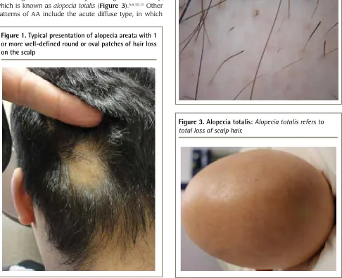
Although spontaneous resolution and regrowth of the patch might occur in up to 50% to 80% of individuals in certain subgroups, it is also possible for additional patches to form, resulting in multiple areas of hair loss coalescing into a larger lesion or eventually involving the entire scalp, which is known as alopecia totalis (Figure 3).5,6,10,11 Other
patterns of AA include the acute diffuse type, in which
there is sudden hair loss all over the scalp, or the ophiasis pattern, in which hair is lost on the posterior and lateral aspects of the scalp (Figure 4).5,12,13
Hair loss can present on any part of the body and might involve areas such as the eyelashes, eyebrows, beard, and pubic area.5,13 The disease can progress to
the point of global hair loss, after which it is known as alopecia universalis.5,13 Nail changes are present in
up to 7% to 66% of those with AA, and although many types of changes are possible, nail pitting (Figure 5) is classically described.6,14-18
Figure 1. Typical presentation of alopecia areata with 1
or more well-defined round or oval patches of hair loss
on the scalp
Figure 2. Characteristic “exclamation mark” hairs of
alopecia areata:
This exclamation mark appearance
describes a damaged strand that is missing its distal
end. It is thicker at its damaged section and notably
thinner proximally as it enters the scalp.
Histology. Hair follicles from patients with AA exhibit both abnormal hair cycling and inflammation. Normally, a hair follicle goes through 3 phases during its growth cycle: anagen, catagen, and telogen. The hair shaft elon-gates during the anagen phase. It can remain in this phase for a period of months to years.9 Next the
fol-licle enters a 2- to 4-week period known as the cata-gen phase, during which it prepares to enter the “resting phase” known as the telogen phase.9 This final part of the
follicular growth cycle takes approximately 3 months
to complete, after which the hair shaft is lost as a “club hair” and the hair follicle begins the cycle again.9
In AA, inflammation causes a large proportion of hair follicles to shift from the anagen phase to the telogen phase. In the acute stages of AA, most hair follicles are still in the anagen phase. If one were to perform a scalp biopsy at this stage, the histologic examina-tion would reveal an excessive amount of lymphocytes in and around the hair follicle. While the normal ratio of anagen to telogen hairs in the scalp is usually 80:20, patients with AA exhibit a 60:40 or even 50:50 anagen to telogen ratio, and in some cases the number of telo-gen hairs might dominate.8,9,19-22
Prognosis. The natural course of AA is unpredictable. Some patients might experience only a single episode of hair loss during their lifetimes, while others experience multiple recurrences. Further variation occurs during recovery. Some patients have full hair regrowth, yet oth-ers remain the same or experience further hair loss.10,11,23
Several epidemiologic studies have identified prog-nostic factors associated with poor regrowth in individu-als affected by AA (Box 1).23-28 In general, hair regrowth
is possible and known to be inversely correlated with the extent of hair loss. A study of 191 patients showed regrowth in roughly 40% to 70% of patients in those classified aslimited-patch stage.23 However, the greater
the area of involvement (alopecia totalis or universa-lis), the less likely the chance of complete regrowth, and the more likely AA will progress over time.23,24 One
exception would be individuals who rapidly lose their hair (acute diffuse and total alopecia variant). Despite substantial hair loss, chances of regrowth are high in this subtype.12 Other poor prognostic indicators include
those who experience the ophiasis variant or note coex-isting nail changes.25 Compared with adults, early onset
of AA in childhood often results in both a greater degree of and progression of AA.23,26 A history of atopy and the
coexistence of other autoimmune diseases also confer a poor outcome.25
Box 1. Prognostic factors associated with poor
outcomes in individuals with alopecia areata
The following factors are associated with poorer outcomes: • Extensive loss (especially alopecia totalis and universalis) • Ophiasis variant
• Nail changes • Early age of onset • Family history
• Concomitant autoimmune diseases (eg, atopy, Hashimoto thyroiditis)
Data from Tosti et al,23 Tan et al,24 De Waard-van der Spek et al,25 Yang
et al,26 Goh et al,27 and Barahmani et al.28
Figure 4. The ophiasis pattern of alopecia areata:
Hair
is lost on the posterior and lateral aspects of the scalp.
Pathogenesis. The exact cause of AA is still unknown. The current body of evidence supports an autoimmune origin and strong genetic contribution, further modified by unknown environmental influences.29
Genetic contribution: Multiple genetic factors con-tribute to the development of AA. A positive fam-ily history is evident in approximately 10% to 25% of cases.6,30,31 Further support for a genetic contribution
comes from a concordant twin study by Rodriguez et al, in which the percentage of monozygotes (42%) who both had AA was much greater than that of the dizygotes (10%).29
Molecular genetics: Extensive work has been done to determine the molecular basis of AA. Alopecia areata is considered to be a T cell–mediated autoimmune dis-ease.19,32 It is important to understand that the hair
fol-licle is fundamentally considered a site of immune privilege, whereby a number of mechanisms tightly con-trol immunologic access and prevent it from autoim-mune attack under normal conditions. The development of AA is thought to result from a breakdown of this immune-privileged site.33
A groundbreaking study from 2010 provided a deeper understanding of the genetic basis of AA. By compar-ing 1054 patient cases and 3278 controls, Petukhova et al discovered 139 single-nucleotide polymorphisms related to the development of AA.34 Further investigation
located 8 genes highly correlated with the risk of dis-ease. Interestingly, several of these “susceptibility loci” are thought to play a role in other autoimmune pro-cesses such as type 1 diabetes and rheumatoid arthri-tis, helping to once again strengthen the autoimmune hypothesis for AA.34
Evaluation and differential diagnosis. Multiple hair loss disorders can mimic AA. During examination one should consider possibilities such as trichotillomania, tinea capitis, scarring alopecia, and telogen effluvium.7
However, the diagnosis of AA can usually be made clini-cally owing to the characteristic appearance of the hair loss regions.7
Individuals with trichotillomania have broken hairs of different lengths and might or might not report a history of hair pulling when asked. Many also have concomitant psychiatric disorders.35 Tinea capitis is
a fungal infection of the scalp. Examination typically reveals hair loss associated with scale. A definitive diagnosis can be made by performing a scalp scrap-ing of the affected region and sendscrap-ing it for culture.36
Scarring alopecias, particularly a condition known as
pseudopelade, can closely mimic AA. However, the areas of hair loss are permanent, and in this particu-lar condition the hair follicle openings are not visible upon clinical examination. If there is any concern, a scalp biopsy can be performed looking for the
previously mentioned characteristics in order to dif-ferentiate the 2 conditions.37
Associated disorders. A review of the National Alopecia Areata Registry revealed that approximately 16% of AA patients had other coexisting autoimmune conditions.28 Specifically, rates of thyroid disease in
these patients are thought to range from approxi-mately 8% to 28% and vitiligo from 2.5% to 4.1%.24,27,38,39
Concomitant type 1 diabetes mellitus (0.6%) and lupus erythematosus (0.6%) were noted in a review of 513 patients performed by Goh et al.27 Atopy is also strongly
correlated with AA, showing comorbidity rates of up to 40%, compared with the background rate in the general population of 20%.28,40
Laboratory evaluation. A biopsy is not necessary in most cases of AA. Unusual forms of AA, including dif-fuse AA, might closely mimic other conditions, mak-ing a biopsy helpful in these specific situations. While we routinely check complete blood count and ferritin, thyroid-stimulating hormone, and 25-hydroxyvitamin D levels in AA, at present there are no evidence-based guidelines to direct the workup or studies highlight-ing the cost-effectiveness of screenhighlight-ing for concomitant disorders. Preliminary small studies show a possible increased risk of 25-hydroxyvitamin D deficiency in AA; however, more extensive research is still required.41,42
Therefore, we recommend a full and comprehensive review of systems be used to guide any further workup that might be required.
Revisiting the case
This 25-year-old gentleman had a classic presentation of AA in the multiple-patch stage. Further evaluation revealed a history of atopy, and examination revealed nail pitting. Findings from routine bloodwork did not reveal any other concomitant disorders. The man is upset by his hair loss and wants to understand his treatment options.
Conclusion
Hair loss is often encountered in primary care and it can be very distressing to patients. Family physicians are well positioned to identify AA, counsel patients, and ini-tiate treatment. Treatment options are discussed in part 2 of this review (page 757).43
Dr Spano is a dermatology resident at the University of Ottawa in Ontario.
Dr Donovan is a dermatologist, Director of the Women’s College Hospital Hair Loss Clinic, and Assistant Professor at the University of Toronto in Ontario.
Contributors
Both authors contributed to the literature review, analysis, and interpretation, and to preparing the manuscript for submission.
Competing interests
None declared
Correspondence
references
1. SafaviK. Prevalence of alopecia areata in the First National Health and Nutrition Examination Survey.Arch Dermatol1992;128(5):702.
2. SafaviKH, MullerSA, SumanVJ, MoshellAN, MeltonLJ3rd. Incidence of alo-pecia areata in Olmsted County, Minnesota, 1975 through 1989.Mayo Clin Proc1995;70(7):628-33.
3. ShellowWV, EdwardsJE, KooJY. Profile of alopecia areata: a questionnaire analysis of patient and family.Int J Dermatol1992;31(3):186-9.
4. KyriakisKP, PaltatzidouK, KosmaE, SofouriE, TadrosA, RachiotiE. Alopecia areata prevalence by gender and age.J Eur Acad Dermatol Venereol 2009;23(5):572-3. Epub 2008 Sep 8.
5. AndersonI. Alopecia areata: a clinical study.Br Med J1950;2(4691):1250-2.
6. MullerSA, WinkelmannRK. Alopecia areata. An evaluation of 736 patients. Arch Dermatol1963;88:290-7.
7. MessengerAG, McKillopJ, FarrantP, McDonaghAJ, SladdenM. British Association of Dermatologists’ guidelines for the management of alopecia areata 2012.Br J Dermatol2012;166(5):916-26.
8. EckertJ, ChurchRE, EblingFJ. The pathogenesis of alopecia areata.Br J Dermatol1968;80(4):203-10.
9. SperlingLC, LuptonGP. Histopathology of non-scarring alopecia.J Cutan Pathol1995;22(2):97-114.
10. IkedaT. A new classification of alopecia areata.Dermatologica 1965;131(6):421-45.
11. WalkerSA, RothmanS. A statistical study and consideration of endocrine influences.J Invest Dermatol1950;14(6):403-13.
12. LewBL, ShinMK, SimWY. Acute diffuse and total alopecia: a new sub-type of alopecia areata with a favorable prognosis.J Am Acad Dermatol 2009;60(1):85-93. Epub 2008 Nov 6.
13. FinnerAM. Alopecia areata: clinical presentation, diagnosis, and unusual cases.Dermatol Ther2011;24(3):348-54.
14. Kasumagic-HalilovicE, ProhicA. Nail changes in alopecia areata: frequency and clinical presentation.J Eur Acad Dermatol Venereol2009;23(2):240-1. Epub 2008 Jun 1.
15. GandhiV, BaruahMC, BhattacharayaSN. Nail changes in alopecia areata: incidence and pattern.Indian J Dermatol Venereol Leprol2003;69(2):114-5.
16. NandaA, Al-FouzanAS, Al-HasawiF. Alopecia areata in children: a clinical profile.Pediatr Dermatol2002;19(6):482-5.
17. SharmaVK, DawnG, MuralidharS, KumarB. Nail changes in 1000 Indian patients with alopecia areata.J Eur Acad Dermatol Venereol1998;10(2):189-91.
18. Baran R, Dawber RP. Alopecia areata. In: Diseases of nails and their manage-ment. Oxford, UK: Blackwell Scientific; 1984. p. 165-7.
19. WhitingDA. Histopathologic features of alopecia areata: a new look.Arch Dermatol2003;139(12):1555-9.
20. WhitingDA. The histopathology of alopecia areata in vertical and horizontal sections.Dermatol Ther2001;14(4):297-305.
21. Van ScottEJ. Morphologic changes in pilosebaceous units and anagen hairs in alopecia areata.J Invest Dermatol1958;31(1):35-43.
22. MessengerAG, SlaterDN, BleehenSS. Alopecia areata: alterations in the hair growth cycle and correlation with the follicular pathology.Br J Dermatol 1986;114(3):337-47.
23. TostiA, BellavistaS, IorizzoM. Alopecia areata: a long term follow-up study of 191 patients.J Am Acad Dermatol2006;55(3):438-41. Epub 2006 Jun 27.
24. TanE, TayYK, GohCL, Chin GiamY. The pattern and profile of alopecia areata in Singapore—a study of 219 Asians.Int J Dermatol2002;41(11):748-53.
25. De Waard-van der SpekFB, OranjeAP, De RaeymaeckerDM, Peereboom-WyniaJD. Juvenile versus maturity-onset alopecia areata—a comparative ret-rospective clinical study.Clin Exp Dermatol1989;14(6):429-33.
26. YangS, YangJ, LiuJB, WangHY, YangQ, GaoM, et al.The genetic epidemiol-ogy of alopecia areata in China.Br J Dermatol2004;151(1):16-23.
27. GohC, FinkelM, ChristosPJ, SinhaAA. Profile of 513 patients with alopecia areata: associations of disease subtypes with atopy, autoimmune disease and positive family history.J Eur Acad Dermatol Venereol2006;20(9):1055-60.
28. BarahmaniN, SchabathMB, DuvicM; National Alopecia Areata Registry.
History of atopy or autoimmunity increases risk of alopecia areata.J Am Acad Dermatol2009;61(4):581-91. Epub 2009 Jul 11.
29. RodriguezTA, FernandesKE, DresserKL, DuvicM; National Alopecia Areata Registry. Concordance rate of alopecia areata in identical twins supports both genetic and environmental factors.J Am Acad Dermatol2010;62(3):525-7.
30. FriedmannPS. Alopecia areata and auto-immunity.Br J Dermatol 1981;105(2):153-7.
31. BlaumeiserB, van der GootI, FimmersR, HannekenS, RitzmannS, Seymons K, et al. Familial aggregation of alopecia areata.J Am Acad Dermatol 2006;54(4):627-32. Epub 2006 Jan 23.
32. Todes-TaylorN, TurnerR, WoodGS, StrattePT, MorhennVB. T cell subpopu-lations in alopecia areata.J Am Acad Dermatol1984;11(2 Pt 1):216-23.
33. ChristophT, Müller-RöverS, AudringH, TobinDJ, HermesB, CotsarelisG, et
al.The human hair follicle immune system: cellular composition and immune
privilege.Br J Dermatol2000;142(5):862-73.
34. PetukhovaL, DuvicM, HordinskyM, NorrisD, PriceV, ShimomuraY, et al. Genome-wide association study in alopecia areata implicates both innate and adaptive immunity.Nature2010;466(7302):113-7.
35. WalshKH, McDougleCJ. Trichotillomania. Presentation, etiology, diagnosis and therapy.Am J Clin Dermatol2001;2(5):327-33.
36. PomeranzAJ, SabnisSS. Tinea capitis: epidemiology, diagnosis and manage-ment strategies.Paediatr Drugs2002;4(12):779-83.
37. AlzolibaniAA, KangH, OtbergN, ShapiroJ. Pseudopelade of Brocq.Dermatol Ther2008;21(4):257-63.
38. SeyrafiH, AkhianiM, AbbasiH, MirpourS, GholamrezanezhadA. Evaluation of the profile of alopecia areata and the prevalence of thyroid function test abnormalities and serum autoantibodies in Iranian patients.BMC Dermatol 2005;5:11.
39. CunliffeWJ, HallR, StevensonCJ, WeightmanD. Alopecia areata, thyroid dis-ease and autoimmunity.Br J Dermatol1969;81(12):877-81.
40. HordinskyM, EricsonM. Autoimmunity: alopecia areata.J Investig Dermatol Symp Proc2004;9(1):73-8.
41. MahamidM, Abu-ElhijaO, SamamraM, MahamidA, NseirW. Association between vitamin D levels and alopecia areata.Isr Med Assoc J
2014;16(6):367-70.
42. D’OvidioR, VessioM, d’OvidioFD. Reduced level of 25-hydroxyvitamin D in chronic/relapsing alopecia areata.Dermatoendocrinol2013;5(2):271-3. Epub 2013 Jan 1.