Electronic Thesis and Dissertation Repository
8-14-2012 12:00 AM
Effect of Menstrual Cycle Related Estrogen Fluctuations on
Effect of Menstrual Cycle Related Estrogen Fluctuations on
Working Memory
Working Memory
Mia Segal
The University of Western Ontario
Supervisor
Dr. Elizabeth Hampson
The University of Western Ontario Graduate Program in Neuroscience
A thesis submitted in partial fulfillment of the requirements for the degree in Master of Science © Mia Segal 2012
Follow this and additional works at: https://ir.lib.uwo.ca/etd
Part of the Cognition and Perception Commons
Recommended Citation Recommended Citation
Segal, Mia, "Effect of Menstrual Cycle Related Estrogen Fluctuations on Working Memory" (2012). Electronic Thesis and Dissertation Repository. 686.
https://ir.lib.uwo.ca/etd/686
This Dissertation/Thesis is brought to you for free and open access by Scholarship@Western. It has been accepted for inclusion in Electronic Thesis and Dissertation Repository by an authorized administrator of
i
EFFECT OF MENSTRUAL CYCLE RELATED ESTROGEN FLUCTUATIONS ON WORKING MEMORY
(Spine title: Menstrual Cycle and Working Memory)
(Thesis format: Monograph)
by
Mia Segal
Graduate Program in Neuroscience
A thesis submitted in partial fulfillment of the requirements for the degree of
Master of Science
The School of Graduate and Postdoctoral Studies The University of Western Ontario
London, Ontario, Canada
ii
THE UNIVERSITY OF WESTERN ONTARIO School of Graduate and Postdoctoral Studies
CERTIFICATE OF EXAMINATION
Supervisor
______________________________ Dr. Elizabeth Hampson
Supervisory Committee
______________________________ Dr. Peter-Klaus Ossenkopp
______________________________ Dr. J. Bruce Morton
______________________________ Dr. Stephen Lomber
Examiners
______________________________ Dr. Martin Kavaliers
______________________________ Dr. Jody Culham
______________________________ Dr. Susan Pigott
The thesis by
Mia T. Segal
entitled:
Effect of Menstrual Cycle Related Hormone Fluctuations on Working
Memory
is accepted in partial fulfillment of the requirements for the degree of
Master of Science
iii
Abstract
Working memory (WM) is a dynamic brain system which allows for the on-line
moment-to-moment maintenance, processing and monitoring of information involved in human
cognition. Behavioural and neuroimaging studies have shown that the prefrontal cortex
(PFC) plays an essential role in WM. Data suggest the PFC may be susceptible to
modulation by estrogen. Behavioural studies examining whether PFC-dependent WM
tasks exhibit estrogen sensitivity in postmenopausal women have shown a benefit of
estrogen. The present study used hormone changes associated with the menstrual cycle to
examine whether estrogen has a beneficial effect on WM function in reproductively aged
women. Thirty-six women completed a battery of cognitive tasks including 3 WM tests in
a repeated-measures design. The data showed that performance on the WM tasks was
significantly better when estrogen levels were high compared to when they were low,
suggesting that estrogen has the ability to modulate PFC-dependent WM function in
young women.
iv
Co-Authorship Statement
All research described in this thesis including the experimental design, data collect, data
analysis and editing was carried out by Mia Segal under the supervisor of Dr. Elizabeth
Hampson with the exception of the radioimmunoassays which were conducted by Bavani
Rajakumar under the supervision of Dr. Elizabeth Hampson. All experiments are original
v
Acknowledgments
First and foremost, I would like to thank my supervisor, Dr. Elizabeth Hampson. This
thesis would not have been possible without all of her patience, help and encouragement.
I would also like to thank my advisory committee Dr. Klaus-Peter Ossenkopp, Dr. Bruce
Morton and Dr. Stephen Lomber for their helpful comments and feedback on this project.
Also, a warm thank you to the rest of the Hampson lab for their assistance,
encouragement and friendship; Kelly Evans for her help with data analysis, Bavani
Rajakumar for her sample processing and analysis, Janani Sankar and Christina
Ivanowich for their support. Lastly, and most important I would like to thank my family
and friends for their endless and unconditional love and support. Thank you for keeping
me motivated, grounded and positive and for reminding me there is always a light at the
vi
Table of Contents
CERTIFICATE OF EXAMINATION ………...ii
Abstract ………..iii
Co-Authorship Statement ………..………iv
Acknowledgements ……….v
Table of Contents ………...…vi
List of Tables ………...…viii
List of Figures ………ix
List of Abbreviations ………..…x
Chapter 1: Introduction ………...1
Chapter 2: Method ………25
Chapter 3: Results ……….…40
Chapter 4: Discussion ………...60
References ……….72
Ethics Approval ………90
vii
List of Tables
Table 3.1: Correlations between estrogen and progesterone concentrations and
working memory errors ..………..51
Table 3.2: Partial correlations between estrogen concentrations and working memory
errors controlling for progesterone concentrations ………..53
Table 3.3: Partial correlations between progesterone concentrations and working
memory errors controlling for estrogen concentrations ...………54
Table 3.4: Means (±SD) on the control measures of WM span ……….………...55
viii
List of Figures
Figure 1.1: Schematic of the adapted version of the delayed-response task used with
nonhuman primates ………..………..…...3
Figure 1.2: Schematic example of the spatial version of the n-back task ………..….6
Figure 1.3: An example of the stimuli used by Petrides and Milner (1982) in the 6 item
abstract-design set of Self-Ordered Pointing Task stimuli…..………10
Figure 1.4: An illustration of the typical changes in progesterone and 17β-estradiol
concentrations that occur across a 28 day menstrual cycle ……….24
Figure 2.1: An photograph of a participant selecting a non-matching pair on the spatial
working memory board ………...29
Figure 2.2: The stimuli used for the 10 item set of the Self-Ordered Pointing Task…….32
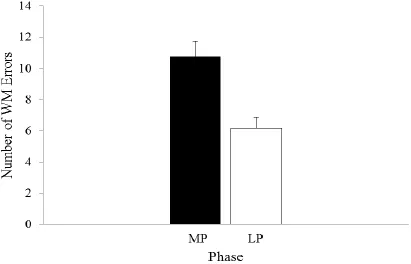
Figure 3.1: The mean number of working memory errors committed on the Spatial
Working Memory Task by women tested at the menstrual phase compared
to at the mid-luteal phase of their menstrual cycle ………...…..42
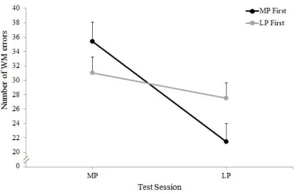
Figure 3.2: Mean number of working memory errors commited at each phase of the
cycle by women who were tested at the menstrual phase first compared to
those tested at the mid-luteal phase first ………...43
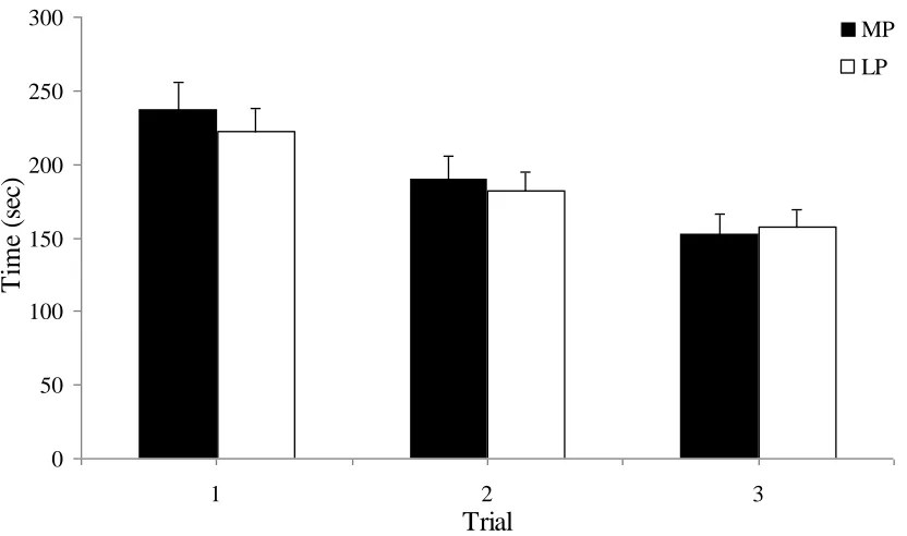
Figure 3.3: The mean amount of time required for women to complete Trials 1, 2 and 3
of the Spatial Working Memory Task at the menstrual phase compared to
the mid-luteal phase of their cycle ………..45
Figure 3.4: The mean number of working memory errors committed by women tested at
the menstrual phase of their cycle compared to their performance at the
ix
Figure 3.5: The mean amount of time required for women to perform the 10 trials of
the Digit Ordering task at the menstrual phase compared to the mid-luteal
phase of their cycle………...………...47
Figure 3.6:The mean number of working memory errors committed on the 4, 8, 10, 12
and two 14 item sets of the Self-Ordered Pointing Task when women were
tested at the menstrual phase of their cycle compared to their performance
at the mid-luteal phase ………..………...49
Figure 3.7:The mean amount of time required for women to complete the 4, 8, 10, 12
and two 14 item set sizes of the Self-Ordered Pointing Task at the menstrual
phase compared to the mid-luteal phase of their cycle ………..……..50
Figure 3.8:Mean time required for women to correctly identify the incomplete images
x
List of Abbreviations
125
I Iodine-125
5-HT Serotonin
ANCOVA Analysis of covariance
ANOVA Analysis of variance
BA Brodmann`s area
ChAT Choline acetyltransferase
dlPFC Dorsolateral prefrontal cortex
EIA Enzyme immunoassay
ERα Estrogen receptor alpha
ERβ Estrogen receptor beta
fMRI Functional magnetic resonance imaging
FSIQ Full scale IQ
GnRH Gonadotropin releasing hormone
NAART North American Adult Reading Test
OVX Ovariectomized
PET Positron emission tomography
PFC Prefrontal cortex
POMS Profile of Mood States
RIA Radioimmunoassay
SD Standard deviation
SEM Standard error of the mean
xi SWPM Spatial working memory task
Tukey HSD Tukey’s Honestly Significant Difference
WM Working memory
CHAPTER 1
INTRODUCTION
Working memory refers to the dynamic brain system which allows for the on-line
moment-to-moment maintenance, processing and monitoring of information involved in
human cognition (Baddeley, 1986; Goldman-Rakic, 1987). As such, the contents of
working memory are continuously manipulated and updated in order to accurately guide
subsequent actions. Working memory has been shown to play a fundamental role in an
array of complex cognitive activities such as reasoning, fluid intelligence, language
comprehension and mental calculation (Baddeley, 1986, 1994; Barett, Tugade, and
Engle, 2004; Goldman-Rakic, 1987). An integral feature of these and indeed all working
memory tasks, is their requirement for the temporary maintenance of task relevant
information in a form which is readily accessible while simultaneously allowing for the
performance of other cognitive decisions and operations on the stored material. For
example, the decoding and integration of text necessary for accurate reading
comprehension requires the reader to have access to semantic, pragmatic and syntactic
information from previously encountered text (Daneman, and Carpenter, 1980)
The temporary relevance and usefulness of the information in working memory is
the criterion which distinguishes the system from other forms of memory such as
semantic (Tulving, 1972) or procedural (Squire, and Cohen, 1984) that involve
longer-term representations. Additionally, unlike working memory, these other forms of memory
are associative in that their contents are acquired by the repeated contiguity between
stimuli and responses and/or consequences (Goldman-Rakic, 1995). In addition to their
scientific fields including neurobiology, neuropsychology, and cognitive neuroscience
suggests a distinction between working and other forms of memory is also evident in the
neuroanatomical structures which subserve them. Initial evidence for the neural basis of
working memory was obtained through studies of nonhuman primates.
The Neural Basis of Working Memory
Early nonhuman primate studies which attempted to elucidate the neural
underpinnings of working memory used conscious monkeys trained to perform a delayed
response task adapted for use with nonhuman primates by Jacobsen (1936) from that
originally devised by Hunter (1913) for human testing. On each trial of the task, the
monkey watches the experimenter hide a food reward in one of two spatial locations
(Figure 1.1). After a delay of several seconds, the monkey is allowed to retrieve the
reward. During the delay, the monkey is prevented from physically orienting itself to the
location of the target and from seeing the locations by an opaque screen. As such, the
monkey must remember in which of the two spatial locations the food was hidden in
order to be rewarded. The locations are identical in appearance eliminating the monkey’s
ability to use external cues to inform their decision. Therefore, the animal must rely
solely on information stored in memory to guide their choice. Importantly, the spatial
location of the hidden reward on each trial is varied and random. Thus in order to
accurately guide behavior, the animal must not only temporarily store information
regarding the spatial location of the target, but also update this information on a continual
basis and then use the updated, internally stored representation to guide their subsequent
actions.
Figure 1.1. Schematic of the adapted version of the delayed-response task used with
nonhuman primates. After watching the experimenter bait one of the two locations with a
food reward during the cue phase and a delay phase during which the monkey can no
longer see the locations, the monkey is allowed to make a response to retrieve the reward.
The two possible bait locations are identical in appearance, thus the monkey must rely
solely on information stored in memory to guide its response. Additionally, the location
of the hidden reward is random and varied on each trial, requiring the monkey to update
the information stored in memory on a trial-by-trial basis. (Adapted from
performance on the delayed response task in monkeys are analogous to those regarded as
working memory in humans. Electrophysiological studies utilizing the delayed response
task have consistently found neurons within the lateral prefrontal cortex (PFC) that
become activated during specifically the delay period of the task (Funahashi, Bruce, and
Goldman-Rakic, 1989; Fuster, and Alexander, 1971; Kubota, and Niki, 1971).
Pairing working memory tasks with a variety of neuroimaging techniques has
allowed researchers to distinguish the perceptual and mnemonic roles played by the
cortical regions involved in working memory. To date, a large number of such studies
have been conducted (see D'Esposito et al., 1998 for a review). Importantly, such studies
have consistently demonstrated that as in nonhuman primates, the PFC is engaged during
working memory tasks in humans. In humans, the engagement of the lateral PFC while
performing adapted versions of the delayed response task has been demonstrated using
both positron emission tomography (PET; Jonides et al., 1993) and functional magnetic
resonance imaging (fMRI; Courtney et al., 1997; Zarahn, Aguirre, and D’Esposito,
1999). These studies have used various methods to isolate those areas which play
predominant roles in the mnemonic portion of working memory rather than the
sensorimotor components of the tasks.
Researchers have also used fMRI and other neuroimaging techniques such as PET
paired with more complex measures of working memory such as the ‘n-back’ task to
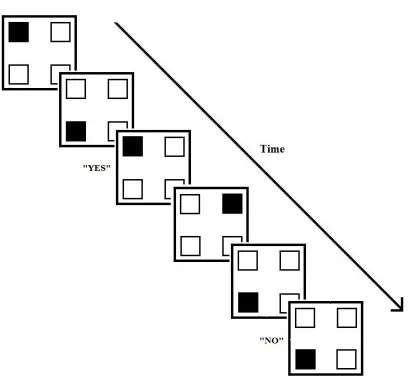
further elucidate the neural underpinnings of the system. The spatial ‘n-back’ task
involves participants indicating whether each item in a continuous series of visual stimuli
certain number (‘n’) steps earlier in the sequence of stimuli (Figure 1.2). The task
requires participants to not only store information related to the spatial location of the
stimuli, and to continually update this information as new items are incorporated, but also
to monitor the stored information in order to make accurate decisions to guide their
responses thus placing significant demands on working memory. McCarthy et al. (1994)
had participants complete both the spatial ‘n-back’ as well as a sensorimotor control task
which required participants to make simple perceptual judgements about a variety of
irregular shapes while in an fMRI magnet. The same irregular shapes also served as the
stimuli for the ‘n-back’ condition. The researchers found that compared to the control
condition, there was a significant signal increase in Brodmann's area (BA) 46, located in
the dorsolateral PFC (dlPFC), during the ‘n-back’ task. Similarly, using PET Smith et al.
(1996) found the ‘n-back’ task to elicit significant increases in blood flow bilaterally in
BA 46 when compared to a control task.
Both the delayed-response and the spatial ‘n-back’ tasks used in the previously
described studies are considered to be spatial working memory tasks as the information
which is required to be held within working memory pertains to the spatial locations of
the target stimuli. There are other kinds of working memory tasks however such as verbal
and visual which differ in the type of information that must be stored, manipulated and
updated in order to accurately guide performance.
Using PET, Petrides et al. (1993b) measured cerebral blood flow while human
participants performed both a control task and a digit randomization task which placed
significant demands on verbal working memory. The same verbal response of reciting
Figure 1.2. Schematic example of the spatial version of the n-back task. Depicted is a run
of N-2 trials in which the participant is to respond “yes” or “no” indicating whether the
filled square is in the same location as the stimulus that occurred two steps ago in the
asked to say out loud the numbers from 1 to 10 in a random, self-generated order without
repeating any numbers or leaving any numbers out. This task required participants to
maintain within working memory the digits which they had already generated and update
this information as they progressed through the task in order to accurately guide
performance and avoid making errors. For the control task, participants counted out loud
from 1 to 10 in ascending order. Thus the control task elicited the same processes of
generating and vocalizing the numbers, but without a significant working memory
component. When the researchers compared the patterns of activation elicited by the
performance of the two tasks, the digit randomization task was found to evoke significant
bilateral activation within the mid-dlPFC (BA 46/9) compared to the control task that was
perceptually and motorically similar, but lacked a working memory component. These
findings suggest that like spatial working memory tasks, the PFC is also essential to
performance of non-spatial working memory tasks (Petrides, 1995b; Petrides, and Milner
1982). These findings are consistent with findings from similar work done with
nonhuman primates (Petrides, 1995a; 2000a).
Other studies of working memory within the field of neurobiology have relied
heavily on the interpretation of deficits exhibited by humans and nonhuman primates
with either naturally-occurring or surgically-induced PFC lesions. PFC injury has been
shown to result in a variety of behavioural abnormalities and cognitive deficits including
non-generalized impairments in mnemonic functioning (Fuster, 1997; Goldman-Rakic,
1987; Miller, and Cummings, 1999). Of particular importance to the current investigation
observed following PFC lesions (Bauer, and Fuster, 1976; Milner, 1982; Petrides, 1989;
Yener, and Zaffos, 1999). A sampling of such studies is reviewed below.
A large number of monkey studies have linked the integrity of the PFC to
accurate performance of the delayed response task, beginning with the classic work by
Jacobsen (1936) in which he was the first to describe the significant impairment of
monkeys' ability to perform the delayed response task following bilateral excisions of the
PFC. Since Jacobsen`s seminal study, a number of other studies have demonstrated that
in addition to bilateral damage, unilateral damage to the PFC is sufficient to impair the
monkey`s capacity to perform the classic delayed response task (Fuster, 1997;
Goldman-Rakic, 1987). Such studies have also allowed for the source of the delayed response
deficit to be localized to the cortex lining the sulcus principalis, especially its middle and
caudal portions corresponding to BA 46 in the dlPFC (Butters, and Pandya, 1969;
Goldman, and Rosvold, 1970; Gross, and Weiskrantz, 1962; Mishkin, 1957).
Furthermore, research using rhesus monkeys has found that the deficits observed
following the focal lesioning of the cortex within the sulcus principalis were as severe as
those produced as a result of lesions to much larger portions of the PFC (Goldman, and
Rosvold, 1970; Goldman et al., 1971; Mishkin, 1957). The specificity of the delayed
response deficit to the dlPFC has been further supported by findings which show that
cortical lesions which spare the sulcus principalis either did not result in observable
deficits in delayed response task performance or only resulted in impairments when delay
periods were extremely long (Goldman et al., 1971; Bauer, and Fuster, 1976; Jacobsen,
1936). In addition to permanent surgical lesions, researchers using either direct electrical
in non-human primates to induce temporary disruptions of the activity of neurons in the
region have found these methods also induce errors in performance on delayed response
tasks.
In order to determine the role of the dlPFC in non-spatial working memory in
humans, Petrides and colleagues have studied the impact of unilateral frontal lobectomies
performed in order to relieve pharmacologically intractable epilepsy on human patient’s
performance on the Self-Ordered Pointing Task (SOPT; Petrides, and Milner, 1982).
Patients were presented with a set of stimuli consisting of either verbal (words) or visual
(representational drawings or abstract designs) stimuli. An example of the stimuli used
for this task are shown in Figure 1.3. The same set of stimuli were presented on a number
of different pages in different spatial arrangements and patients were required to point to
a different stimulus on each page until each item had been selected once but only once.
A working memory error was scored when a patient reselected a stimulus they had
already selected. The spatial locations of the stimuli were varied and random from page
to page so that patients were unable to use a spatial strategy to keep track of those items
which they had already selected. The patients were required to use memory for the
figures or words themselves to guide their performance. According to Petrides, and
Milner (1982), this task placed heavy demands on non-spatial working memory as it
required patients to constantly monitor and update the identity of multiple items held in
memory but did not require the maintenance or retrieval of spatial information. The
comparison of the performance of patients with PFC excisions to those with temporal
Figure 1.3. An example of the stimuli used by Petrides, and Milner (1982) in the 6 item
Specifically, the researchers observed that patients with PFC excisions that included the
dlPFC were markedly impaired on both the verbal and visual versions of the SOPT, while
patients with unilateral excisions of the temporal lobe that did not include extensive
damage to the hippocampal system were unimpaired. Patients with left frontal lobe
excisions were impaired on both the verbal and nonverbal tasks while those with lesions
of the right frontal lobe only showed deficits on the nonverbal tasks (Petrides, and
Milner, 1982). These findings strongly support the role of the PFC in non-spatial working
memory. The excisions experienced by these patients were made with the primary
purpose of relieving them of epilepsy symptoms thus in most cases the resulting neural
damage was not confined to a distinct neuroanatomical region within the PFC. For this
reason the researchers were unable to specify the critical regions within the PFC
responsible for the observed deficits on the self-ordering task.
In an attempt to more accurately isolate the critical prefrontal region Petrides
(1995a) studied the performance of monkeys with more focal lesions on a modified
version of the SOPT. It was found that those monkeys with lesions of the
mid-dorsolateral cortex at BA 46 were impaired on the task when compared to those animals
with lesions of the periarcuate cortex in the posterior frontal cortex. These findings led
Petrides (1995a) to conclude that as in humans, the dlPFC played a critical role in
working memory for non-spatial object information in the nonhuman primate brain.
Taken together, the results of the previously described animal, neuroimaging and
lesion studies strongly suggest that the PFC plays an important and essential role in
working memory function. Although the evidence is fairly new, recent
the adult primate PFC may be modulated by a number of metabolic factors including
circulating hormones. Notably, accumulating evidence suggests that estrogens may exert
regulatory effects on the adult primate PFC. This important metabolic influence has the
potential to impact functions subserved by this region such as working memory.
Modulation of PFC Function by Circulating Hormones
A number of studies have shown that estrogen receptors are transiently expressed
in the PFC of female rhesus monkeys during early brain development (Handa, Connolly,
and Resko, 1988; MacLusky, Naftolin, and Goldman-Rakic, 1986; Pomerantz et al.,
1985; Sholl, and Pomerantz, 1986). Specifically, MacLusky et al. (1986) observed levels
of estrogen binding in the dlPFC of both late prenatal and early infant (less than 6 days
postnatal) female rhesus monkeys to be comparable to the levels found in other cortical
areas such as the visual, motor and somatosensory cortices. Additionally, evidence
suggests that estrogen may also be present in the adult PFC (Hao et al., 2006). Expression
of both estrogen receptors alpha (ERα) and beta (ERβ) messenger RNA fragments have
been identified within the PFC of female rhesus monkey brain specimens (Pau, Pau, and
Spies, 1998). Furthermore, the analysis of both young and aged rhesus monkey brain
sections by electron microscopy indicates the presence of ERα in the dendritic spine and
axon terminals in layer III of BA 46 (Wang et al., 2010). The presence of estrogen
receptors within the PFC suggests that this region is a target of estrogens in adult
non-human primate brains.
Examination of human brain specimens has also revealed evidence of estrogen
binding within the PFC, although evidence is limited. Bixo et al. (1995) examined
of adult human females and found the PFC to represent one of the densest binding sites
for estradiol. Estrogen concentrations in the PFC were found to be approximately twice
as high as those found in the temporal neocortex and more than seven times as high as
those in the hippocampus. These findings suggest that the PFC is a principal location of
estrogen in the adult human brain (Bixo et al., 1995).
The analysis of estrone sulfatase levels in the human female brain provides further
evidence of estrogen activity in the PFC. Estrone sulfatase is the enzyme responsible for
converting estrone sulfate, the major conjugated estrogen in female plasma, to the more
biologically active estrogens, estrone and estradiol. Platia et al. (1984) found the levels of
estrone sulfatase activity in the PFC to be equivalent to or greater than those levels in the
endocrine hypothalamus. Taken together, these findings provide support for the
hypothesis that the PFC is a primary target for estrogens within the primate brain even
well into adulthood.
The presence of estrogen within the adult brain is clear evidence for the notion that the
hormone plays an important role in the maintenance and expression of sex-typical or
sexually differentiated behaviours into adulthood. Such effects of hormones (not limited
to estrogen) which have the ability to act in an acute manner and occur later in life after
the development of both the nervous system and sex organs are known as activational
affects (Arnold, 2009; Breedlove, Cooke, and Jordan, 1999; Cooke et al., 1998). Such
activational affects can be distinguished from the organizational effects of sex steroids
which influence physiology and behaviour through developmental mechanisms in which
sexually dimorphic development of brain morphology (LaCroix-Fralish, Tawfik, and
DeLeo, 2005).
A great number of animal and human studies have shown that experimentally
manipulating estrogen levels has significant effects on a wide variety of neurotransmitter
systems that project to the PFC including serotonin, norepinephrine, and dopamine
(Summer, and Fink, 1995; Kritzer, and Kohama, 1998). Many such studies have
investigated the effects of estrogen replacement in ovariectomized (OVX) rats. For
example, Summer, and Fink (1995) found the number of serotonin (5-HT) binding sites
in the anterior frontal cortex of OVX rats to increase 41% within 24 hours of the
administration of a single dose of estradiol. O’Malley et al. (1987) observed a significant
reduction in acetylcholine synthesis in the frontal cortex of female rats following
ovariectomy, an effect that was shown to be reversed within 5 days of estradiol
replacement. Similarly, 28 weeks post-ovariectomy a 56% reduction in the activity of
choline acetyltransferase (ChAT), one of the major enzymes responsible for acetylcholine
synthesis, was found in the frontal cortex of OVX female rats (Singh et al., 1994).
Importantly this reduction was prevented or reversed in OVX rats given exogenous
estradiol replacement. An effect of estrogen replacement on ChAT levels was also
observed in the basal forebrain of female rats, a major cholinergic afferent to the frontal
cortex such that enzyme levels were significantly increased within 6-24 hours of estradiol
treatment (McEwen, Luine, and Fischette, 1987). In both rodents and primates, activity of
the nigrostriatal and mesolimbic dopaminergic system both of which have projections
that terminate in the PFC has also been shown to be affected by estradiol at relatively
(1994) found an increase in both dopamine release and uptake within the mesolimbic
dopamine pathway projecting to the PFC in female rats. Estradiol administration has also
been found to decrease the activity of tyrosine hydroxylase, the enzyme responsible for
the conversion of tyrosine to dopamine’s precursor, L-DOPA, in the limbic forebrain of
OVX rats 24 hours after hormone injection (Hernandez et al., 1991).
Findings of the effects of estrogen on neurotransmitter systems are not limited to
rodent models. Evidence also suggests that estrogens have the ability to regulate
neurotransmitter activity in the PFC of nonhuman primates. Ovariectomized adult
monkeys have been found to display a dramatic decrease in the density of dlPFC fibers
which are immunoreactive for tyrosine hydroxylase and choline acetyltransferase,
enzyme markers for dopaminergic and noradrenergic fibers, and cholinergic fibers
respectively (Kritzer, and Kohama, 1998; 1999). An opposite effect of ovariectomy was
found for axons immunoreactive for dopamine β-hydroxylase and 5-HT such that there
was an increase in their density within the dlPFC. These findings suggests that ovarian
hormones might regulate various neurotransmitter afferents in the PFC. This hypothesis
was tested by examining the density of immunolabelling in the dlPFC of the same OVX
monkeys given either estrogen or estrogen plus progesterone hormone replacement. It
was found that monkeys receiving hormone replacements had labelling densities similar
to those observed in hormonally intact control animals. Specifically, estrogen was found
to be as effective as the combination of estrogen and progesterone in stimulating normal
prefrontal immunoreactivity for choline acetyltransferase and dopamine β–hydroxylase
(Kritzer, and Kohama, 1998; 1999). These findings suggest that estradiol was the critical
activity within the PFC of female primates. Taken together, these studies strongly suggest
that the modulatory effects of estrogen on various neurotransmitter systems active within
the PFC may be a mechanism by which circulating estrogens modulate the PFC and thus
the functions subserved by the region.
It is noteworthy that both dopamine and serotonin, and to some extent
norepinephrine and acetylcholine, play important roles in working memory processes
(Seamans, and Yang, 2004; Robbins, 2005). Experimental manipulation of dopamine
transmission in the dlPFC of nonhuman primates has been shown to affect the
performance of spatial working memory tasks (Luciana, Collins, and Depue, 1998).
Dopamine depletion in the PFC of both rats and monkeys, specifically from the sulcus
principalis, results in impaired performance on the spatial delayed response task
(Brozoski et al., 1979). Additionally, the administration of a selective 5-HT receptor
agonist has been shown to impair working memory on a radial arm maze task in rats
(Luciana, Collin, and Depue, 1998; Winter, and Petti, 1987).
A number of recent neuroimaging studies support the hypothesis that human PFC
function might be influenced by circulating estrogen levels. Such studies have found PFC
activation to change systematically with the hormonal, specifically estrogen status
although most existing studies have focused only on postmenopausal women (Berman et
al., 1997; Shaywitz et al., 1999; Smith et al., 2006). One of the few investigations to
study young women was conducted by Berman et al. (1997) and used PET to investigate
the changes in regional cerebral blood flow patterns elicited by the Wisconsin Card
Sorting Test associated with three pharmacologically controlled hormonal conditions.
PFC elicited during the performance of the Wisconsin Cart Sorting Test to be attenuated
in young women following the administration of a gonadotropin releasing hormone
(GnRH) agonist which supressed ovarian function resulting in estrogen and progesterone
levels which were comparable to those of postmenopausal women. Furthermore, a
normalization of the regional cerebral blood flow pattern was observed when estrogen
was administered in concert with the GnRH agonist.
More recently, Smith et al. (2006) used a randomized, double-blind,
placebo-controlled crossover study to find task-induced prefrontal activity as measured by fMRI
to change systematically with the hormonal status of female participants. The participants
consisted of a group of postmenopausal women whose ovaries had ceased functioning
leading to a significant decrease in endogenous estrogen levels. Each woman was tested
and scanned twice; once while receiving hormone replacement therapy and once while
receiving a non-hormone containing placebo. The researchers used a subtraction method
to isolate those areas which were more highly activated during the spatial working
memory task while participants were receiving hormone therapy than when they were
receiving the placebo. It was discovered that the hormone therapy was associated with a
more pronounced activation in the ventrolateral PFC bilaterally. Using a similar
experimental design, Shaywitz et al. (1999) found the differences in the patterns of
activation observed when the women were receiving either the hormone replacement or
the placebo treatment to be the same regardless of whether the working memory task was
verbal or spatial in nature. Specifically, the researchers observed the degree of PFC
activation in the superior frontal gyrus during the performance of the verbal working
received 21-days of hormone replacement therapy than the same length of a placebo
therapy. Taken together, these findings directly demonstrate that the estrogen status of
human females modulates cognition-related neural activity.
Estrogen Status Correlates with PFC-Dependent Cognitive Tasks on a Behavioural
Level in Both Nonhuman and Human Primates
The possibility that the estrogen status of a female may also correlate with her
level of performance on PFC-dependent cognitive tasks receives preliminary support
from behavioral studies with nonhuman primates. Roberts et al. (1997) compared the
performance of age-matched groups of pre-menopausal, menopausal and
post-menopausal female monkeys on the classic delayed response task. Both the post-menopausal
and post-menopausal groups were found to display significant impairments in
performance at a delay as short as 1-second. Furthermore, test performance was found to
be significantly correlated with the hormonal status of the animal such that those with
lowest estrogen metabolite levels also had the lowest performance scores (Roberts et al.,
1997).
In order to test whether surgical menopause and subsequent estrogen replacement
had an influence on cognitive outcomes of normal aging including working memory
decline, Rapp, Morrison, and Roberts (2003) compared the performance of two groups of
aged rhesus monkeys on a delayed response test of spatial working memory. The
researchers found the performance of OVX monkeys who had received a regimen of
low-dose, cyclic estradiol replacement to be substantially better than that of vehicle-injected
monkeys on the delayed response task. Additionally, the reversal of age-related
the PFC dependent delayed response task, as there was only a very modest recovery of
recognition memory which has been shown to be dependent on an intact medial temporal
lobe (Rapp, 1993). These findings clearly suggest that a nonspecific estrogen influence
on general performance factors such as motivation or perceptual ability is unlikely to
account for the benefits of estrogen treatment observed on the working memory task.
Behavioral investigations with human participants provide further support for the
existence of an association between estrogen levels and performance on tasks with
prominent working memory components. If in fact such an association exists, then it is
logical to hypothesize that performance on tasks which place significant demands on
working memory would differ between groups of individuals who are exposed to
different levels of estrogen. To date however, very few studies have been conducted in
order to test this hypothesis. One of the few studies which has investigated the potential
activational effects of estrogen on working memory performance was conducted by Duff,
and Hampson (2000). The researchers compared the performance of three groups of
post-menopausal women on the Spatial Working Memory Task (SPWM), a novel multi-trial
spatial working memory task that was designed to be sufficiently sensitive to detect small
to moderate individual differences within the normal range of performance, and Digit
Randomization, tasks with strong spatial and verbal working memory components
respectively. All three groups were post-menopausal. One group of women was not
receiving any hormone replacement therapy. The other two groups were taking hormone
replacement therapies consisting of either estrogen only, or a combination of estrogen
and a progestin. The researchers found that both groups of women receiving hormone
that had prominent working memory components, but not on control tasks. Furthermore,
no significant difference in performance between the two groups of women who received
hormone replacement was observed. These findings support the hypothesis that it was
specifically the estrogen component of the hormone replacement therapy that was
responsible for the improved performance of those women receiving exogenous
hormones.
Similarly, the results of a study conducted by Keenan et al. (2001) yielded
evidence for disruptions of a variety of pre-frontally mediated cognitive processes
including working memory in post-menopausal women not receiving hormone
replacement therapy as compared to aged matched controls receiving replacement
therapy. Specifically, untreated menopausal women were relatively impaired in inhibiting
inappropriate responses in the form of perseverative errors, and in the performance of an
auditory-verbal version of the N-back test of working memory. Further support for the
hypothesis that estrogens have the ability to modulate working memory function is
provided by a double-blind within-subject study by Krug, Born, and Rasch (2006). The
researchers found the performance of post-menopausal women on tasks involving PFC
dependent memory function to be significantly better after they had received transdermal
estrogen replacement for as little as 3 days as compared to a placebo treatment.
Importantly, estrogen was found not to affect hippocampus-dependent functions of
memory retention. Thus the findings of both Duff, and Hampson (2000) and Krug, Born,
and Rasch (2006) indicate that in postmenopausal women, a transient increase in plasma
estrogen concentration acutely improves PFC-dependent cognitive functions. These data
capable of influencing the function of the cognitive systems subserved by that region in
postmenopausal women.
Research by Grigorova, Sherwin, and Tulandi (2006) suggests that the same
association between estrogen levels and PFC mediated cognitive function could
potentially exist even in premenopausal women who have not undergone long-term
estrogen deprivation. The researchers investigated the influence of sex steroid hormone
suppression given to a group of premenopausal women to chemically suppress ovarian
function as a treatment for various benign gynecological disorders. The hormone
suppression treatment suppressed estrogen levels to the postmenopausal range whereas
progesterone levels fell to levels typical of the lower limit of the menstrual cycle range.
Each woman completed a battery of tasks including both a verbal and non-verbal version
of the N-back working memory task before and after 4 weeks of treatment. The
comparison of each woman’s pre-treatment baseline to their post-treatment performance,
revealed a decline in performance on both working memory measures. Importantly,
further analysis using post-treatment declines in progesterone, mood, or other
health-related symptoms as covariates revealed that the relative declines in working memory
performance were associated with post-treatment declines in estrogen concentrations, but
not these other factors. Thus these findings provide preliminary evidence that as was
observed in postmenopausal women, estrogen is also influential in the maintenance of
working memory function in premenopausal women. Such conclusions must be made
cautiously however as these data are preliminary and to date the research by Grigorova
effect of estrogen levels on PFC dependent cognitive functioning on a behavioural level
in young women.
Summary and Hypothesis
Numerous lines of research have demonstrated that the PFC, specifically its
dorsolateral region, is critical for working memory function. Furthermore, convergent
evidence from neuroendocrine and neuroimaging studies in both nonhuman and human
species suggests that the adult female PFC, including its dorsolateral region, may be
susceptible to the activational effects of estrogens. Thus, it appears that estrogens may
have the ability to modulate the function of the PFC. More recently, behavioural
investigations have begun to provide support for the notion that the activational effects of
circulating estrogens on the PFC may influence PFC-dependent working memory
processes at a functional level.
Two standard paradigms are commonly used by researchers to investigate the
potential activational effects of estrogens on cognitive functions in women. The first is
the study of surgically menopausal or naturally postmenopausal women. The second
paradigm takes advantage of the natural fluctuations in ovarian hormones across the
menstrual cycle. Such methods allow for the investigation of the effects of estrogen on
cognition in young women in a non-invasive and naturalistic manner.
The menstrual cycle refers to a recurrent sequence of ovarian changes and
hormone output that occurs cyclically in women of reproductive age under the guidance
of stimulation from the hypothalamus and pituitary, when conception has not occurred.
The exact timing of the endocrine events varies from woman to woman and cycle to
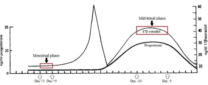
the luteal phase (Wilson et al., 1998). In women who have a 28-29 day cycle, the
follicular or proliferative phase is marked by the first day of menstruation (cycle Day 1)
and extends until cycle Day 14-15 when ovulation occurs (Figure 1.4). The beginning of
the follicular phase is characterized by low serum concentrations of both progesterone
and 17β-estradiol, the most abundant and potent form of estrogen. While progesterone
levels remain low, a preovulatory peak in estradiol occurs just prior to ovulation. The end
of the follicular phase is marked by ovulation. The luteal or secretory phase is
characterized by high endocrine concentrations of both estrogen and progesterone and
extends from cycle Days 15 to 28. The length of the luteal phase is relatively fixed at
13-15 days, regardless of the length of the overall menstrual cycle. An estimation of the
hormonal state of a female can be obtained by counting backward from cycle Day 1, the
first day of menstruation. This reverse counting method has been used in a number of
previous studies, however investigations of the validity of the approach have determined
it to be associated with error rates of 15% or higher (Stern, and McClintock, 1995). As
such, the confirmation of expected hormone concentrations for each menstrual cycle
phase by radioimmunoassay (RIA) or enzyme immunoassay (EIA) has become a critical
part of the methodology for studies using menstrual cycle related hormone fluctuations as
a variable. Such retrospective endocrine validation of hormone levels allows for greater
accuracy in determining menstrual phase at the time of testing.
The objective of the current study was to further extend the limited body of
research that has investigated estrogen's actions within the PFC and its ability to
Figure 1.4. An illustration of the typical changes in progesterone and 17β-estradiol
concentrations that occur across a 28 day menstrual cycle. Days +3 and +5 correspond to
the menstrual phase when estrogen concentrations are low. Days -5 to -10 correspond to
estrogen fluctuations associated with the female menstrual cycle as a noninvasive and
ecologically valid method of manipulating the hormone levels to which the participants
were exposed. Because previous evidence suggests that the PFC is susceptible to estrogen
and that estrogen has a facilitatory effect on working memory function, it was
hypothesized that women would perform selectively better on measures of working
memory (make fewer working memory errors) when tested under the influence of high
estrogen during the mid-luteal phase of the menstrual cycle than when tested under the
influence of low estrogen during the menstrual phase of the cycle. Such findings would
support previous theoretical findings of a beneficial effect of high estrogens on the PFC
and the functions it subserves.
CHAPTER 2
METHOD
Participants
The participants were 36 healthy female university staff, graduate and
undergraduate students ranging in age from 21 to 35 years (M = 24.75 years, SD = 3.93)
with a mean estimated IQ of 108.78 (SD = 7.12). All participants had regular menstrual
cycles between 21 (minimum) and 35 (maximum) days in length (M = 29.22, SD = 1.93).
Participants had not used oral or other types of hormonal contraceptives for at least 4
months. Participants who indicated histories of health conditions including attention
deficit disorder, seizures or epilepsy, major depression, schizophrenia, bipolar disorder,
over or underactive thyroid glands, diabetes, head injuries, or other serious neurological
or psychiatric conditions on a confidential health questionnaire were not eligible to
Procedure
Prospective participants were recruited via posters displayed throughout the
University of Western Ontario campus. After indicating their interest in participating,
volunteers completed a confidential online demographic and reproductive health
questionnaire in order to determine eligibility. Women meeting the above criteria were
contacted for testing at a later date. Eligible participants were tested on two separate
occasions the timing of which were individually targeted to coincide with either the
menstrual phase of the cycle when estrogen levels are lowest (cycle days +3 to +5
relative to the day of menstrual onset), or the mid-luteal phase of the cycle when estrogen
levels peak (cycle days -5 to -10). Cycle phase for the first session was randomly
assigned and counterbalanced across participants. Due to the fact that the menstrual cycle
is not perfectly reliable, each woman’s cycle stage at each test session was confirmed
retrospectively. Testing was considered successful if RIA of saliva collected during the
test sessions provided independent confirmation of the levels of progesterone and
17β-estradiol, the major estrogen in women of reproductive age (see Radioimmunoassays,
below). In the group of women reported here, the mean level of 17β-estradiol was
3.56pmol/L (SD = 1.76) during the menstrual phase and 6.20pmol/L (SD = 1.81) during
the mid-luteal phase. Associated levels of progesterone were 37.91pg/mL (SD = 12.22)
and 145.83pg/mL (SD = 63.31), respectively. Mean salivary hormone concentrations at
each phase of the cycle were in agreement with previous reports based on healthy women
with ovulatory menstrual cycles (Bao et al., 2003; Ellison, 1993; Gandara, Leresche, and
Mancl, 2007; Oinonen, and Mazmanian, 2007; Shirtcliff et al., 2000; Worthman,
Each woman was tested individually. Both sessions began and ended with the
collection of a saliva specimen for hormonal analysis. In order to ensure the purity of the
saliva samples, women were asked not to smoke, chew gum, brush their teeth, eat or
drink anything except plain water for 1 hour prior to their appointment time. For the
sample provided at the start of the session, approximately 7 mL of saliva were collected
into a polystyrene culture tube pre-treated with sodium azide. A stick of inert sugarless
gum was used as a saliva stimulant. For the sample provided at the end of the session,
approximately 2 mL of saliva were collected into a borosilicate tube by passive drool. No
saliva stimulant was used in order to permit analysis of the saliva using enzyme-linked
immunosorbent assay (not reported here). All specimens were frozen at −20°C until the
end of the study then analyzed in a single assay.
Each test session took approximately 1 hour during which the battery of tasks
described below was administered. Each task was administered once at each of the two
test sessions with the exception of the North American Adult Reading Test (NAART)
which was only administered at the first test session.
Cognitive and Memory Tasks
Working Memory Tasks
At each of the two test sessions, participants completed three tasks which had
significant spatial, verbal or figural working memory components:
Spatial Working Memory Task (SPWM; Duff, and Hampson, 2000). This task was
developed as a more cognitively demanding version of those spatial working memory
tasks which have been shown previously to depend on PFC in nonhuman (Passingham,
humans from a search task used by Passingham (1985), who found that rhesus monkeys
who had surgical lesions in the vicinity of the sulcus principalis (BA 46) were severely
impaired, relative to control animals (i.e., they produced high numbers of working
memory errors). The performance of younger adults on the SPWM has previously been
shown to correlate significantly with performance on Digit Ordering, a verbal WM task
(Duff, and Hampson, 2001) and with performance on SOPT (Hampson et al., 2010).
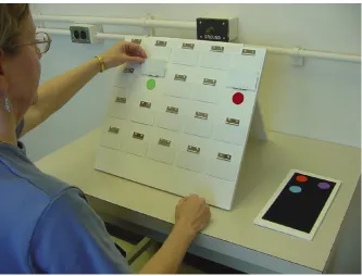
Participants were seated at a desk on which a board containing a 4 x 5 array of 10
different coloured dots (red, purple, green, blue, orange, pink, black, white, fuchsia,
yellow). Each dot was 3 cm in diameter and were arranged in a random order on a neutral
backing (45 cm long, 41 cm high). Figure 2.1 shows an illustration of the SPWM board.
The board containing the coloured dots was placed in front of participants at eye level.
Each item in the array was completely concealed beneath a hinged flap (8.0 cm long, 4.5
cm wide) and only became visible when the corresponding flap was temporarily lifted by
the participant. Two dots of each colour were hidden beneath the hinged flaps, and
participants were instructed to find all 10 matching pairs of dots, in as few choices as
possible, by lifting the flaps two at a time. When a flap was not being lifted by the
participant, it was closed, concealing the item beneath. As such, the participants were
required to maintain within WM the locations of the pairs of dots they had already found
and update this information iteratively as they found further pairs of dots.
Participants were familiarized with the complete set of colours before they began
Figure 2.1. A photograph of a participant selecting a non-matching pair on the spatial
working memory (SPWM) board. The board consists of 20 hinged doors under which 10
pairs of coloured dots are hidden. Participants are instructed that their goal is to find all
10 pairs of matching dots in as few tries as possible, lifting the doors two at a time.
Participants are required to update the locations stored in memory as they lift the doors
and find matching pairs. A working memory error is counted anytime the participant
selects a pair of doors that they have already selected, or a pair of doors which they
of the array. Before the participants began their search, the tokens were removed but
during the task, as each pair was discovered, the experimenter replaced the corresponding
token on the felt. By the end of each trial, a token of each of the 10 colours appeared on
the felt. The tokens provided visual feedback to the participants eliminating the need for
them to remember which colours they had already found. As such, the participants were
only required to remember the locations of the matched or not matched dots as the task
progressed.
Participants were told that they would be timed on the task, but that their primary
objective was to locate the matching pairs in the fewest tries possible. Each participant
completed three consecutive trials of the same array. Alternate forms of the task were
presented on each of the two testing days and which arrangement of the dots the women
received at their first test session was counterbalanced within each phase of cycle. A trial
was considered complete when all 10 matching pairs of dots were found. The dependent
variables were: the number of working memory errors and the time to completion of each
trial in seconds. A working memory error was committed anytime a participant chose a
pair of locations that had already been searched but did not match, or re-searched an
already matched pair.
Digit Ordering (Petrides et al., 1993b). Previous PET studies have used this task
with the aim of mapping the patterns of neural activation elicited by verbal working
memory tasks. The task has been shown to elicit significant activation in the mid-dlPFC
bilaterally (Petrides et al., 1993a, b).
In the present study, participants were asked to say out loud the numbers from 1
10 different randomization trials during each of the two test sessions. The dependent
variables were: the total time to complete each trial as well as the total number of
working memory errors committed. For the purpose of statistical analysis, the total
number of working memory errors summed across the 10 trails was used. A working
memory error was defined as the omission or repetition of any digit within a trial.
Self-Ordered Pointing Task (SOPT; adapted from Petrides, and Milner, 1982).
Performance on the SOPT has been shown in studies of neurological patients to be
extremely sensitive to frontal lobe damage (de Zubicaray, 1997; Petrides, and Milner,
1982; Wiegersma, van der Scheer, and Hijman, 1990). Imaging work in humans or
nonhuman primates has confirmed activation in the right mid-dorsolateral frontal cortex
(BA 46) as well as a weaker response within this same region in the left hemisphere,
consistent with its recruitment of the working memory system (Petrides, 2000a; Petrides,
2000b; Petrides, 1989; Petrides et al., 1993).
The task required participants to complete a series of trials in which the goal was
to point to all items in a set of stimuli once and only once. Participants completed a 4
item practice set, followed by 8, 10, 12 and two 14 item sets. Different visual stimuli
were used for each set size. All stimuli, other than the 4 item practice stimuli which
depicted food items, were abstract, and were chosen to be easily visually distinguishable
from one another but difficult to verbally encode. All items measured 5 cm in height and
4.1 cm in width. Each set of stimuli was printed on large-sized photopaper that ranged in
size from 21.59 x 27.94 cm to 29.72 x 41.91 cm. An example of one of the pages from
the 10 item set is shown in Figure 2.2.
Figure 2.2. The stimuli used for the 10 item set of the Self-Ordered Pointing Task. Each
of the 10 pages in the set contained the same 10 images, but the spatial location of each
image was random and varied on each page. The participant's goal was to point to one
item on each page of the stimulus set but to point to each item once and only once. This
required the participants to continually update the object identity information but not
spatial information stored in working memory in order to accurately guide their
participant by the experimenter after the completion of the previous set. Each set had a
number of pages corresponding to the number of items in the set (e.g., 12 pages for the 12
item set). On each page the items were arranged in a fixed two-column layout. Each
page in a given set contained the complete set of stimuli at that set size, and thus had the
same number and identity of items, but the spatial location of each item within the
two-column layout was randomized and varied unpredictably on each page.
The participant was required to initiate and execute a sequence of pointing
responses. In order to avoid making errors, the participant had to maintain an internal
representation of the responses made and continually update this record as they
monitored their own performance. Due to the fact that the spatial location of items on
each page of the set varied randomly, it was not possible to perform the SOPT using a
spatial strategy. Instead, participants had to maintain the figural identity of the items
which they had already selected in working memory. Thus the task required the
temporary maintenance and updating of object identity but not spatial information
throughout the task.
Subjects were instructed that they could point to the items in any order they
wished, but that they were not to point to any items they had selected on previous pages.
Before beginning each set size, the participants were given between 5 and 30 seconds
depending on the set size to familiarize themselves with the appearance of the visual
stimuli. The demand on working memory was increased by having participants complete
three trials at each set size consecutively during each testing session. The same set of
The dependent variables were: the time to completion (in seconds) at each set size
and the number of working memory errors at each set size. A working memory error was
counted anytime a participant reselected an item they had already selected within a trial.
Span Control Tasks
Because it is the frontally mediated executive functions of working memory that
were hypothesized to be influenced by estrogen, control tasks were used that required the
passive short-term retention and retrieval of verbal, spatial and visual information similar
to that used in the working memory tasks but without monitoring, active maintenance,
manipulation or reworking of the stored information. These control tasks enabled the
alternative possibility that estrogen-related effects on the passive storage processes of
working memory are the basis for any facilitative effect of estrogen observed on the
working memory tasks described above to be tested.
Digit Span (Wechsler Adult Intelligence Scale – Revised, Wechsler, 1981). This
task required only the passive retention of the digits in memory without any active
manipulation of the stored information. Previous neuroimaging research has shown the
type of short-term storage of verbal information required for performance of this task to
involve the recruitment of the posterior perisylvian cortex, but not the PFC (Postle,
Berger, and D'Esposito, 1999; Shallice, and Vallar, 1990). Furthermore, patient studies
have shown performance on the forward digit span task to be unaffected by excisions or
lesions of the PFC (Canavan et al., 1989a; D'Esposito, and Postle, 1999; Petrides, 1995b).
The Forward Digit Span portion of the WAIS-R Digit Span subtest was
administered in the standard manner. Participants were asked to repeat a sequence of
examiner. A maximum of two tries were allowed at each sequence length. The digit span
score was the maximum number of digits repeated correctly. This measure allowed for
the quantification of any changes in the participant's ability to temporarily store and
retrieve verbal information throughout the menstrual cycle which could contribute to or
account for any differences on the Digit Ordering task at the two cycle phases
investigated.
Corsi Block-Tapping (Milner, 1971). This task is a visuospatial analogue of the
Forward Digit Span task. Patient research indicates that performance on the Corsi
block-tapping task is unaffected by lesions to the PFC and implicated other more posterior
cortical areas such as the inferior parietal cortex as being essential for accurate task
performance (Baldo, and Dronkers, 2006; D'Esposito, and Postle; 1999). The task
involved participants observing the examiner tap a progressively longer spatial sequence
on a set of 10 identical 3 cm cubes painted black and randomly arranged on a 27.7 x 22.8
cm wooden platform. Immediately after each sequence was shown to the participant, they
were required to reproduce the exact sequence demonstrated by the examiner, in order.
The score was the maximum sequence length that a participant could reproduce in the
correct order. A maximum of two tries were allowed at each sequence length. The task
required participants to temporarily store a sequence of spatial locations but did not
require the active manipulation or on-line maintenance of the stored information. This
measure allowed for the quantification of any changes in the participant's ability to
temporarily store and retrieve spatial information throughout the menstrual cycle which
could contribute to or account for any differences on the SPWM at the two cycle phases