Scholarship@Western
Scholarship@Western
Electronic Thesis and Dissertation Repository
8-26-2015 12:00 AM
Acute Sprint Interval Exercise Induces a Greater FGF-21 Response
Acute Sprint Interval Exercise Induces a Greater FGF-21 Response
in Comparison to Work-Matched Continuous Exercise
in Comparison to Work-Matched Continuous Exercise
Blair M. Segsworth
The University of Western Ontario
Supervisor Dr. Peter Lemon
The University of Western Ontario Graduate Program in Kinesiology
A thesis submitted in partial fulfillment of the requirements for the degree in Master of Science © Blair M. Segsworth 2015
Follow this and additional works at: https://ir.lib.uwo.ca/etd
Recommended Citation Recommended Citation
Segsworth, Blair M., "Acute Sprint Interval Exercise Induces a Greater FGF-21 Response in Comparison to Work-Matched Continuous Exercise" (2015). Electronic Thesis and Dissertation Repository. 3254.
https://ir.lib.uwo.ca/etd/3254
This Dissertation/Thesis is brought to you for free and open access by Scholarship@Western. It has been accepted for inclusion in Electronic Thesis and Dissertation Repository by an authorized administrator of
(Thesis format: Monograph)
by
Blair Mackay Segsworth
Graduate Program in Kinesiology
A thesis submitted in partial fulfillment of the requirements for the degree of
Master of Science
The School of Graduate and Postdoctoral Studies The University of Western Ontario
London, Ontario, Canada
ii
Abstract
Sprint interval training (SIT) has been associated with substantial reductions in body
fat. Recent evidence suggests that myokines (small protein compounds produced in
muscle) may promote the fat loss with SIT. The purpose of this project was to
compare the plasma accumulation of three myokines (IL-15, Irisin and FGF-21) with
sprint interval exercise (SIE) vs work-matched continuous exercise (CE). Nine male
subjects completed an acute SIE session consisting of four-30 second sprints and a
work-matched CE session. Both exercise sessions were completed on an
electromagnetically braked cycle ergometer. Blood samples were collected before
and at 5, 30, 90, and 180 min post exercise to determine any changes in plasma
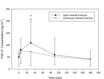
myokine concentration. Plasma FGF-21 was increased at five (P=0.04) and 30
(P<0.001) min with SIE vs baseline and was increased at 30 min (P=0.03) when
compared to CE. Neither Il-15 nor Irisin were altered significantly although there
were some methodology concerns and intersubject variability was substantial so a
Type II Error might have occurred. These findings suggest that exercise intensity is
a key determinant of plasma FGF-21 accumulation and that FGF-21 may serve as a
surrogate measure for both sympathetic activation and exercise-induced lipolysis.
Keywords
iii
Acknowledgements
None of the proceeding thesis could have been accomplished without the help of
three exceptional individuals: AJ Anstett, Rachael Thomas and Arash Bandegan. AJ
and Rachael; I would have been lost without your nursing expertise and deeply
appreciate the time you volunteered to me for the blood sampling within this project.
You both made the study participants feel at ease and are consummate
professionals. Arash; you have a keen scientific eye and are an incredible critical
thinker. I appreciate the advice you have given me throughout the development of
this project and you have helped me avoid many pitfalls throughout this process.
The hours running ELISA analysis were grueling and I cannot thank you enough for
your commitment to the project.
Dr. Lemon, I would like to thank you for giving me the opportunity to study here at
the University of Western Ontario. You afforded me the academic freedom and
flexibility to explore my own personal research interests. I believe that to be a key
tenet of innovation and its value has not gone unnoticed.
I would be no where today without the support of my friends and family. To all of you
from home, to Guelph and to now at Western, you have each given me something to
learn and grow from and develop into the person I am today. I am deeply indebted to
you all. For this thesis, I would like to make a special thanks to Kole Abbott. Most of
this work has its foundation in how you conducted your own research and I cannot
thank you enough for the time spent teaching me how to operate lab equipment and
providing an ear for ideas.
To my brother; you are my closest friend and once again you have helped me
through an education in a magnitude you may not realize. To my father; you have
always been a voice of reason and positivity in my life that has helped shape the
way I view the world. To my mother; you are the strongest person I have ever met
and words cannot express the appreciation I have for the sacrifices you have made
iv
Table of Contents
Abstract ... ii
Acknowledgments ... ii
Table of Contents ... iv
List of Tables ... vii
List of Figures ... viii
List of Appendices ... ix
List of Abbreviations ……….x
1 Introduction ... 1
2 Literature Review ... 6
2.1 Sprint Interval Exercise in Humans ... 6
2.2 Irisin ... 9
2.2.1 Cell Studies ... 10
2.2.2 Animal Studies ... 12
2.2.3 Human Studies ... 14
2.3 Fibroblast Growth Factor-21 ... 19
2.3.1 Cell Studies ... 19
2.3.2 Animal Studies ... 20
2.3.3 Human Studies ... 22
2.4 Interleukin-15 ... 25
2.4.1 Cell Studies ... 25
2.4.2 Animal Studies ... 26
2.4.3 Human Studies ... 27
v
3.1 Participants ... 30
3.2 Preliminary Visits ... 30
3.4.1 Continuous Exercise Trial ... 33
3.4.2 Sprint Interval Exercise Trial ... 34
3.5.1 Body Composition ... 35
3.5.2 Aerobic Capacity ... 35
3.5.3 Blood Sampling and Hematocrit ... 35
3.5.4 Blood Analysis ... 37
3.6 Statistical Analysis ... 37
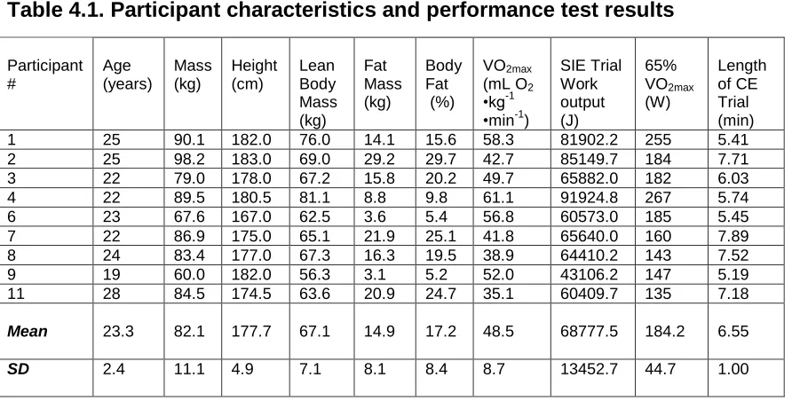
4 Results ... 38
4.1 Descriptive Statistics ... 38
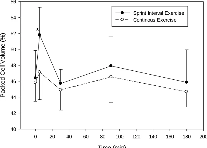
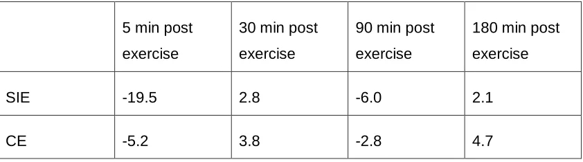
4.2 Hematocrit and Hemoconcentration ... 40
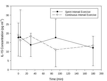
4.3 IL-15 ... 42
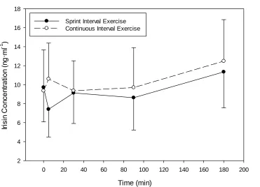
4.4 Irisin ... 43
4.4.1 Irisin and Body Composition ... 44
4.5 FGF-21 ... 46
5 Discussion ... 48
5.1 IL-15 ... 48
5.2 Irisin ... 49
5.3 FGF-21 ... 52
5.4 Limitations ... 55
5.5 Future Directions ... 56
5.6 Summary and Conclusion ... 57
References ... 58
vi
vii
List of Tables
Table 4.1:Participant Physical and Performance Characteristics ... 40
viii
List of Figures
Figure 4.1. Hematocrit changes after SIE or work-matched CE ... 40
Figure 4.2. Change in plasma IL-15 concentration with SIE or work-matched CE.. . 42
Figure 4.3. Change in plasma Irisin concentration with SIE or work-matched CE. ... 43
Figure 4.4. Correlation between lean body mass and resting plasma Irisin
concentration.. ... 44
Figure 4.5. Correlation between lean body mass and plasma Irisin 5 minutes
post-exercise under either SIE or CE paradigm.. ... 45
Figure 4.6.Correlation between lean body mass and plasma Irisin 180 minutes
post-exercise under either SIE or CE paradigm.. ... 46
Figure 4.7. Change in plasma FGF-21 concentration with SIE or work-matched CE..
ix
List of Appendices
Appendix A:Human Ethics Approval ... 70
Appendix B: Letter of Information and Informed Consent ... 72
Appendix C: Participant Health Information Form ... 78
x
List of Abbreviations
FGF-21 Fibroblast Growth Factor-21
PPAR-α Peroxisome Proliferator-Activated Receptor Alpha
FFA Free Fatty Acids
mRNA Messenger Ribonucleic Acid
CI Continuous Interval
SIE Sprint Interval Exercise
TFAM Transcription Factor Alpha, Mitochondrial
AMP Adenosine Monophosphate
UCP-1 Uncoupling Protein 1
UCP-3 Uncoupling Protein 3
IGF-1 Insulin Growth Factor 1
GLP-1 Glucagon-Like Protein 1
ADP Adenosine Diphosphate
SIT Sprint Interval Training
AMPK AMP Activated Protein Kinase
P38MAPK P38 Mitogen-Activated Protein Kinase
SIRT-1 Sirtuin 1
PGC-1α Peroxisome Proliferator-Activated Receptor Gamma Coactivator 1-Alpha
xi
IL-6 Interleukin 6
GLUT-4 Glucose Transporter 4
NRF-1 Nuclear Respiratory Factor 1
BDNF Brain Derived Neurotrophic Factor
ERK Extracellular Signal Regulator Kinase
PCr Phosphocreatine
1
Introduction
The current state of global health is at a crossroads between improvements in
longevity and infant survival and the looming crisis associated with the rising
rates of obesity. At this juncture, about 937 million people worldwide are
considered to be overweight and ~396 million considered obese. Further, these
numbers are projected to grow so that by the year 2030, it is expected that a
staggering 1.12 billion people will be obese (Kelly et al., 2008). Many
pharmacological interventions have been developed to battle this issue and its
comorbidities such as hypertension, dyslipidemia and diabetes; however, it may
be that the universal remedy to the obesity epidemic could simply involve
consistent physical activity. For example, regular exercise has been shown
previously to prevent excessive mass gain and even promote fat losses (up to
386 cubic centimetres of adipose tissue) in obese adults over an 8-wk period
(Keating et al., 2015). Unfortunately, regardless of these benefits, many people
are unable to maintain physically active lifestyles, citing the lack of time as a
major reason for ineffective quantities of exercise (Stutts, 2002). A possible
solution to this issue is the use of sprint interval training (SIT) which results in
significant fat loss and very brief time commitments (Hazell et al., 2014;
MacPherson et al., 2010).
Typically, sprint interval exercise (SIE) consists of four-six bouts of “all-out”
intensity sprinting with each bout of sprints separated by a four min rest interval,
often performed on a cycle ergometer (Burgomaster et al., 2005) but any exercise mode appears to work. As little as three min of “all-out” sprinting per
week has been shown to increase activity of oxidative enzymes such as citrate
synthase, cytochrome oxidase IV and β-hydroxy acyl CoA dehydrogenase as
well as decreasing average 24-h blood glucose (Gillen et al., 2014). These trends
in glucose homeostasis also apply to clinical populations as similar exercise
modalities have caused reduced time spent with hyperglycemia both at rest and
improvements in body composition causing a decrease in fat mass and waist
circumference by 8% and 3.5%, respectively, while increasing lean mass by
1.5% over a 6 wk training period (Hazell at al., 2014). In terms of oxygen
consumption, just two min of SIE induces similar 24-h oxygen consumption as 30
min of continuous aerobic exercise (CE), despite SIE requiring 150% less in VO2
load during the actual exercise (Hazell et al., 2012). These findings indicate a
significant contribution to 24-h oxygen consumption due to an anaerobic exercise
component and/or a residual change in post exercise metabolism. This resulting
prolonged change in metabolism (increased baseline energy expenditure and/or
elevated fat catabolism) may explain some of the fat losses often observed with
SIT.
When compared to moderate intensity CE training, meta-analyses demonstrate
that SIT produces similar 8% increases in VO2max (Gist et al., 2013) due primarily
to an increase in mitochondrial density and the upregulation of oxidative
enzymes such as cytochrome oxidase IV and β-hydroxy acyl CoA
dehydrogenase (Sloth et al., 2013). Interestingly, SIT promotes similar changes
in exercise capacity vs CE with exercise volumes of ~10% and total exercise time
of ~21% (Gibala et al., 2006). Moreover and importantly, interval training, even
intense SIT has also been associated with higher ratings of perceived pleasure
when compared to CE (Bartlett et al., 2011).
Many of the peripheral adaptations to SIT are due to increased mitochondrial
content that occurs along with improved oxidative capacity (Jacobs et al., 2013,
Scalzo et al., 2014). On a molecular level, it is thought that the increased
mitochondrial biogenesis is caused by the phosphorylation of p38 MAPK and
AMPK which promotes an increase in PGC-1α mRNA content (Gibala et al.,
2008). This connection is critical as PGC-1α is known to be the master regulator
of mitochondrial biogenesis as well as many of the oxidative genes linked to
mitochondrial function (Wu et al., 1999). Although there is an established link
between SIT and the induction of oxidative adaptations, the precise mechanism
biochemical signaling process responsible for the adaptations appears to be
more powerful because these metabolic changes are induced by substantially
less total work volume and time.
A new theoretical model describing the function of muscle has also been
described concurrently with the development of SIE as an effective exercise
mode. Like adipose tissue, skeletal muscle is now known to act much like an
endocrine organ, capable of secreting several hormonal signals to communicate
with a number of other body tissues (IIzuka et al., 2014, Pedersen and Febbraio,
2013). Rather than communicating via fat-soluble hormones, skeletal muscle
utilizes contraction-induced cytokines to implement functional changes
throughout the body (Pedersen and Febbraio, 2013). These have become known
as myokines because the tissue of origin for these cytokines is muscle and, as of
2014, there are at least 69 identified myokines within the secretome of muscle
(Catoire et al., 2014). One of the most well characterized myokines is IL-6,
traditionally a pro-inflammatory cytokine, it is increased chronically in the obese
(Pal et al., 2014). IL-6 is also elevated acutely following exercise and improves
insulin and glucose sensitivity through the up-regulation of glucagon-like
peptide-1 (Pal et al., 20peptide-14). Although much effort has been directed to the study of IL-6, a
new subgroup of myokines has been identified as perhaps playing a role in how
exercise promotes fat loss.
Irisin, Fibroblast Growth Factor-21 (FGF-21) and Interleukin-15 (IL-15) are three
novel myokines that have been implicated with fat metabolism, oxidation and
phenotypical changes. Specifically, Irisin promotes a brown/beige adipose
phenotype, in which an induction of gene related mitochondrial biogenesis
(PGC-1α, UCP-1, GLUT-4) has been reported (Vaughn et al., 2014, Huh et al., 2014).
Further, in conjunction with FGF-21, Irisin increases non-shivering
thermogenesis in beige/brown adipose via uncoupling of the electron transport
chain (Lee et al., 2014). Interestingly, non-shivering thermogenesis induction by
Irisin and FGF-21 are similar between both cold environmental conditions and
post-exercise (Lee et al., 2014). With respect to phenotype, beige/brown adipose
is advantageous because 60 g of brown adipose is capable of metabolizing ~4 kg
of white adipose over the course of a year (Virtanen et al., 2009). Moreover,
FGF-21 may also provide a substantial body fat reduction benefit in that it is
capable of increasing glucose uptake (Kharitonenkov et al., 2005), the
deacetylation and activation of PGC-1α (Fu et al., 2009), as well as help
coordinate the metabolic response to exercise-induced lipolysis (Lee et al., 2013,
Cuevas-Ramos et al., 2012).
Traditionally, IL-15 has been viewed as an anabolic myokine, capable of inducing
the accretion of myosin heavy chain proteins within human muscle cells
(Furmanczyk and Quinn, 2003) as well as assisting in lipid oxidation and storage.
The treatment of adipocytes with IL-15 can reduce lipid deposition by 50%
(Quinn et al., 2005), enhance the expression of PGC-1α in muscle cells, and
increase total mitochondrial volume (O’Connell and Pistilli, 2015). Interestingly,
mice over-expressing IL-15 have been shown to have lower resting RER,
up-regulation of PGC-1α, and increased oxidative muscle fibre content (Quinn et al.,
2013). Considering the role of these myokines in producing a more oxidative (β
-oxidation) phenotype, it is imperative to discern their role and induction
post-exercise.
Recently, Irisin, FGF-21 and IL-15 have been studied with some types of
exercise. For example, Irisin with both resistance (strength) and aerobic
(endurance) exercise paradigms and appears to respond to intensity of exercise
in a dose-dependent manner (Huh et al., 2012, Huh et al., 2014). Apparently
acute sprint exercise causes Irisin to accumulate at a concentration 15% greater
than an endurance bout (Huh et al., 2014); however, in this study the exercise
conditions were not matched for total work output so these data may be
confounded. FGF-21 has been reported to increase one h after an acute bout of
modest aerobic exercise (50% of VO2max), yet the effect of acute sprinting on the
secretion of FGF-21 has not been researched extensively (Kim et al., 2013). In
sessions of four-eight 30s sprinting bouts on a cycle ergometer (Scalzo et al.,
2014). Similarly, the relationship between exercise and the induction of IL-15 in
humans has not been well documented but significant increases in circulating
IL-15 after with moderate intensity (50-70% of VO2max) cycling or running have been
reported (Christiansen et al., 2013, Tamura et al., 2011). There have been no
studies profiling the acute effect of SIE on the secretion of IL-15.
Therefore, the aim of this thesis was to conduct a work-matched comparison
(per-joule basis) of Irisin, FGF-21 and IL-15 between acute SIE and CE to
determine which exercise modality induces the greatest myokine response. In
essence this will determine the role of exercise intensity- defined as a percentage
of VO2max- in the accumulation of plasma myokines. It was hypothesized that SIE
would promote the greatest increase in plasma Irisin, FGF-21 and IL-15 and
thereby perhaps be the mechanistic link between SIE and its noted ability to
stimulate both oxidative adjustments to exercise and the resulting positive
2
Literature Review
2.1 Sprint Interval Exercise in Humans
Typically, sprint interval exercise (SIE) is defined as four-six, 30 sec “all-out”
sprints separated by a four min recovery interval that may be either active or
passive (Burgomaster et al., 2005). These sprints are often repeat Wingate tests,
i.e., 30 sec sprints on a cycle ergometer at a resistance of eight-10% of body
mass (Burgomaster et al., 2005, Hazell et al., 2012). Two min of SIE has been
reported to induce a similar oxygen consumption (VO2) over a 24-h period as a
30-min continuous, moderate intensity endurance bout of exercise (CE) (Hazell
et al., 2012). In the aforementioned study, it is interesting to note that, although
the oxygen consumption was significantly less during the SIE session, the 24-h
consumption was not different vs the 30-min endurance exercise, indicating that
SIE may cause biochemical or physiological changes that induce an extended
increased oxygen consumption post exercise. Further, in both men and women,
SIE has been shown to result in substantial body fat loss despite very little
training time. In women, SlT on a self-propelled treadmill three times per wk for
six wk reduced fat mass by 8%, waist circumference by 3.5% and also increased
maximal oxygen consumption by 8.7% (Hazell et al., 2014). During this training, it
is noteworthy that the participants did not appear to change their diet
significantly. In men, a similar training protocol involving a progressive increase
in sprints (from four to six over six wk) induced a 3kg loss of body fat
(MacPherson et al., 2010). Further, in a subsequent study, acute SIE had little
effect on energy intake despite increased appetite and energy expenditure
(Beaulieu et al., 2014). These observations indicate that changes in food intake
are not the main factor responsible for the improved body composition.
Another study with untrained, middle aged men who underwent SIT, consisting of
four-six bouts of 30 sec all-out sprints for a total of just six training sessions
observed an increase in VO2 peak of 6%, as well as an increase in muscle
glucose uptake (Eskelinen et al., 2015). Interestingly, the effect of increased
to other muscle groups the changes in glucose and fatty acid uptake during
exercise was only present within the quadriceps, indicating a local effect of the
SIT perhaps induced by a form of autocrine/paracrine signaling. All four muscles
of the quadriceps responded to SIT, while in comparison, only the vastus lateralis
and vastus medialis responded to moderate intensity exercise (Eskelinen et al.,
2015).
Metabolically, acute SIE has resulted in increased phosphorylation of AMPK and
p38MAPK immediately after four bouts of 30-sec sprints (Gibala et al., 2008). In
the same study there was also a two-fold up-regulation of PGC-1α mRNA at
three h into recovery. Since SIE was capable of inducing an increase in PGC-1α
and the aforementioned regulators of oxidative metabolism, the authors
concluded that SIE was also capable of increasing mitochondrial content.
Therefore these findings contribute further to the idea that SIE is capable of
inducing similar increases in mitochondrial content and metabolic remodeling to
that caused by traditional endurance training. To support these findings, electron
transport chain protein cytochrome c oxidase, a marker for mitochondrial content,
has been shown to increase with SIT (Gibala et al, 2006). Another study using 60
sec intervals have found increases in cytochrome c oxidase content, a
concurrent 7.9% increase in VO2max, and a 5.1% increase in mean power output
(Jacobs et al., 2013). In a similar study, Perry et al. (2008) observed an 18%
increase in cytochrome c, increases in B-hydroxyacyl-CoA dehydrogenase, and
GLUT4 content within muscle after the completion of a high intensity interval
training regime consisting of 10 x four min intervals at 90% VO2max, three times
per wk, for six wk. Further, during 60% VO2 peak exercise there was a 60%
increase in fat oxidation after six wk of training when compared to pre-training
values at the same intensity. These findings indicate that high-intensity spectrum
training such as SIT may induce long-term changes in the efficacy of energy
substrate utilization during exercise.
In a meta-analysis of the cardiovascular adaptations to SIT, Gist et al. (2013)
their relative ability to increase VO2max. However, there was a 20% reduction in
the time required to accomplish these endpoints with SIT. These findings are
supported by another meta-analysis, which found that VO2max increases in a
range of four-13% with SIT over two-eight wk (Sloth et al., 2013). It was also
shown that there are similar changes in exercise performance, glycemic control
and insulin sensitivity with SIT when compared to more traditional high-volume
training regimes (Sloth et al., 2013). These findings suggest that there may be
separate biochemical pathways or an amplification of factors that are inherent
within SIT, making it more efficient at inducing the benefits associated with
exercise.
The effect of SIT on the secretion of plasma cytokines/myokines is also
important. As outlined above, SIT has been observed to decrease the resting
plasma concentrations of FGF-21, after nine sessions of four-eight, 30 sec bouts
of all out sprints (Scalzo et al., 2014). Under the same training program, Irisin has
a sexually dimorphic response, increasing in women after SIT and decreasing in
men (Scalzo et al., 2014), while SIE increases plasma Irisin acutely (Huh et al.,
2014). Outside of the aforementioned myokines, SIE has also been reported to
have an effect on a wide variety of plasma inflammatory markers. Meckel et al.
(2011) showed that SIE increases both pro- and anti-inflammatory factors such
as Il-1 and IL-6 immediately after a series of successive sprints of 100-400
metres, while IL-6 remained elevated one h into recovery. These data suggest
that along with other cyto/myokines, there may be a global response to sprint
exercise, rather than the production of a single subset of secreted myokines. On
the other hand, SIT has also been shown to have no effect on resting plasma
myokines IL-6, IL-10 and C-reactive protein after 2 wk of thrice weekly bouts of
four-six 30s sprints (Hovanloo et al., 2013). It is difficult to determine the long
term effects of SIT on plasma protein concentrations because there are such
divergent results in the effect of SIT and plasma inflammation markers. Although
there are a limited number of studies presented focusing on the effect of SIE on
myokine concentrations, it is apparent that there is a gap within the literature and
In conclusion, SIE is a powerful exercise modality that is capable of inducing
similar benefits as traditional endurance exercise with far less total energy and
time commitment. It appears that SIE mediates these functions though the
up-regulation of mitochondrial transcription factors as well as supporting
mitochondrial biogenesis and growth. However, is it unclear by which
mechanisms that these processes occur. The effect of SIE on plasma
inflammatory markers and myokines has not been studied systematically in the
literature and thus requires attention.
2.2
Irisin
In the groundbreaking study first describing Irisin, Boström et al. (2012) found
that a product of the proteolytic cleavage of the skeletal muscle integral
membrane protein fibronectin type 3 domain containing 5 (FNDC5) was secreted
into the bloodstream during exercise. Given the name Irisin for the Greek
messenger goddess Iris, this myokine circulates as a 112-amino acid protein
(Wrann et al., 2013). The exact process by which FNDC5 is cleaved and
released into the bloodstream remains unclear, but it is postulated to be similar in
fashion to other transmembrane polypeptide hormones such as Transforming Growth Factor-α (Boström et al., 2012). Previous biochemical studies have shown that while Irisin is a 16 stranded β-sheet dimer of two cleaved FNDC5
proteins for intercellular signaling, there is also a dimerization of FNDC5
endodomains for intracellular cell signaling (Schumacher et al, 2013). This leads
to the possibility that activation of the Irisin receptor can induce both endocrine
functions within a whole organism and autocrine functions on skeletal muscle.
Irisin is thought to mediate its activity through the master regulator of
mitochondrial biogenesis, PGC-1α, and morphogenic changes due to Irisin are
believed to be mediated by changes in ERK and p38 MAPK regulation (Zhang et
2.2.1
Cell Studies
Irisin is believed to be sourced directly from skeletal muscle cells, contributing to
72% of all circulating Irisin (Boström et al., 2012; Roca-Rivada et al., 2013).
However, this assumption may be subject to change. Using
immunohistochemistry to stain for Irisin within different cell cultures taken from
human cadaveric tissue, Aydin et al. (2014), found that the main source of Irisin
is not from skeletal muscle, but rather from the nerve sheaths that spread within
muscle tissue with smaller other contributions derived from the perimysium and
epimysium. Among the most responsive human tissues were the testis,
pancreas, liver, spleen, brain, stomach and cardiac tissues. By following the
staining patterns of Irisin and their tissue relationships, Aydin et al. (2014)
concluded that Irisin may play an important role in hypothalamic-pituitary-gonadal
axis.
Further, Irisin has been shown the be produced and secreted into cell media by
both human and C2C12 myocytes at rest, yet electrical pulse stimulation has
been shown to cause no significant increase in FNDC5 gene expression
(Raschke et al., 2013). Considering that Irisin has been shown to be related
inversely to PCr and ATP concentrations within muscle tissue (Huh et al., 2012)
and its downstream effects can be nullified by AMPK inhibitor, Compound C (Huh
et al., 2014), it may be that the secretion of Irisin is dependent on the accretion of
exercise-induced metabolites, rather than by simple muscular contraction.
Tissues treated with Irisin have shown a wide range of effects. Human muscle
cells taken from biopsies of obese individuals treated with Irisin produced
increased IGF-1 and decreased myostatin mRNA expression (Huh et al., 2014).
In adipose cells derived from the same human donors, both physiological and
below-normal concentrations of Irisin inhibited lipid accumulation and significantly
inhibited fatty acid synthase. Moreover, after eight days of Irisin treatment, mRNA
of genes for thermogenic programs such as cell death-inducing DFFA-like
effector A (CIDEA) and uncoupling protein 1 (UCP-1) increased, as well as
the same study, adipose triglyceride lipase was also up regulated, indicating that
with the increased expression of UCP1, there is also an increased use of fat
substrate for oxidation and heat production. As a final measure, Irisin treatment
inhibited adipocyte differentiation and maturation in human and mouse
pre-adipocytes.
As suggested by biochemical characteristics, it has been shown through in-vitro
studies that Irisin exerts an autocrine effect on skeletal muscle. For example,
C2C12 myocytes treated with Irisin have an increase in expression of genes related to mitochondrial biogenesis including PGC-1α, NRF-1, TFAM, GLUT4,
UCP3 and Irisin (Vaughan et al., 2014). The noted increase in Irisin gene
expression suggests that circulating Irisin exerts an autocrine, positive feedback
loop back on skeletal muscle. The Irisin-treated myocytes also had a greater
amount of global mitochondrial organelle content and expressed a greater total
carbohydrate oxidation and decreased lactate production (Vaughan et al., 2014).
Combined with its positive feedback loop and increased mitochondrial
biogenesis, it appears that Irisin exerts a shift to an oxidative phenotype within
skeletal muscle and adipose tissue. It is important that the secreted portion of
Irisin is also highly conserved within all mammalian species; mouse and human
Irisin are 100% identical (Bostrom et al., 2012). Recently, it has been shown that
both shivering and exercise cause a similar secretion of Irisin, although
marginally greater plasma concentrations are achieved through exercise (Lee et
al., 2014). Combined with another myokine, FGF21; Irisin induces the browning
of adipose as well as non-shivering thermogenesis in response to lower body
temperature in human subjects (Lee et al., 2014). From these experimental data
and the conservation of Irisin structure through mammals, it is believed that Irisin
2.2.2
Animal Studies
In mice, submaximal treadmill exercise to volitional fatigue induces a two fold
increase in plasma Irisin concentration, while free running and control mice
showed no increase in plasma Irisin (Brenmoehl et al., 2014). There were also
higher concentrations of Irisin within homogenates of femoral muscles, when
compared to those of crus (forelimb) muscles in mice. Irisin was also not
correlated with the running distance of the either the voluntary free-wheel running
or the treadmill test, suggesting the determinant of the Irisin response may be
exercise intensity not duration (Brenmoehl et al., 2014). Through
immunohistochemical staining, it was also shown that Irisin was localized around
muscle cell membranes and intercellular spaces. This corresponds with the
findings of Aydin et al. (2014) who suggested Irisin might be a product of the
nerve sheaths permeating through muscle tissue with smaller contributions also
originating from the epimysium and perimysium. Of the various muscle types,
slow/oxidative fibres secrete ~40% more Irisin than glycolytic fibres (Roca-Rivada
et al., 2013). Further, in a separate study, three wk of endurance training reduced
the concentration of Irisin in plasma, suggesting a role of muscle and exercise
training in the down-regulation of Irisin signaling (Roca-Rivada et al., 2013).
Animal studies have also shown that adipose tissue may play a role in Irisin
homeostasis. Roca-Rivada et al. (2013), suggested that a secreted form of
FNDC5 in rats is also produced by adipose tissues and that obese rats
over-secrete this protein in adipose. These findings may illustrate a feedback
mechanism between muscle and adipose for Irisin and further, that there may be
an Irisin resistance during obesity and other metabolic disorders. Comparing
adipose types, subcutaneous adipose secretes 40% more FNDC5/Irisin
compared to visceral fat. Contrary to these findings, Roberts et al. (2013) found
that circulating Irisin tended to be lower in obese/diabetic prone Otsuka
Long-Evans Tukushima Fatty (OELTF) rats. Triceps brachii biopsies showed that FNDC5 and PGC-1α mRNA were found to be 50% and 40% less in OLETF rats,
plasma Leptin concentrations are associated positively with greater skeletal
muscle FNDC5 mRNA expression. It is speculated that Leptin may be a key in the cross talk between muscle and fat because Leptin increases PGC-1α through
AMPK mediated signaling. Therefore, Irisin may serve as a compensatory
mechanism to stimulate thermogenesis and increase fat loss in the obese state
(Roberts et al., 2013).
A unique and interesting role of Irisin may be located in the central nervous
system. Wrann et al. (2013) have proposed the link between exercise and brain
BDNF production to be Irisin and the FNDC5 receptor. Via endurance exercise,
activation of FNDC5 within the hippocampus of mice was observed to cause an
increase in BDNF mRNA. It is believed that the effects of FNDC5 on BDNF transcription is mediated through a complex formed by PGC-1α and Estrogen Related Receptor-α (ERRα) as both PGC-1α and ERRα knockout mice had
dramatically reduced FNDC5 activity (Wrann et al., 2013). Hippocampal
expression of BDNF was also shown to have a negative feedback effect on
FNDC5 activity within the brain, indicating a homeostatic loop between FNDC5
and BDNF. The most significant finding from Wrann et al. (2013), is that the
peripheral delivery of FNDC5 to the liver via adenoviral vectors caused an
increase in the central expression of BDNF. This is a clear indication that a
secreted factor influences the central activity of FNDC5 and implies a
communication axis between exercising muscle, the liver and the brain; however,
it has yet to be determined if the main signaling molecule is Irisin or another
cleavage product from muscle FNDC5 (Wrann et al., 2013).
As promising as the animal studies surrounding Irisin may be, it is often difficult
to translate research from rodent studies to humans. Raschke et al. (2013)
outlined the fact that there is a mutation in a start codon of the FNDC5 gene
between rodents and humans. This mutated form of FNDC5 has low translation
efficiency in humans and only resulted in 1% of full length FNDC5 protein, when
compared to rodents. Raschke et al. (2013) also found that FNDC5 gene
primary skeletal muscle cells. As previously stated, these data must be taken
with some skepticism because electrical pulse stimulation is far from exercise
and often does not result in the accretion of exercise metabolites. Many of these
metabolites such as AMP and the subsequent activation of AMPK are implicated
in the activation of FNDC5 gene pathways and secretion of Irisin into the
bloodstream.
2.2.3
Human Studies
Many studies have profiled the existence of Irisin and its association with
exercise. While studying the physiological variation in plasma Irisin, Anastasilakis
et al. (2014), found that a diurnal rhythm of Irisin exists, with maximum plasma
concentrations being reached at 2100h and minimums at 600h. Interestingly, this
day-night rhythm is opposite of FGF-21 (Anastasilakis et al., 2014). It was
suggested that the inverse relationship between plasma Irisin and FGF-21
concentrations may be due to temperature, where Irisin plays a role in the
formation of brown adipose tissue (Vaughn et al., 2014) and FGF-21 then
activates that tissue for heat production during lower overnight temperatures as
outlined by Anastasilakis et al. (2014) and Lee et al. (2014.
Huh et al. (2012), using biceps circumference as a gauge, showed that muscle
mass was the single greatest indicator of circulating plasma Irisin. Plasma Irisin
is correlated positively with BMI and negatively correlated with age, insulin, and
cholesterol within the blood. Subjects who had undergone bariatric surgery were
observed to have a reduction in both total and lean body mass which coincided
with a subsequent decrease in resting plasma Irisin when compared to
pre-surgery (Huh et al., 2012). It was also shown that a 7% reduction in body mass
due to a hypocaloric diet also caused a reduction in plasma Irisin (De La Iglesia
et al., 2014), suggesting a relationship between Irisin production and skeletal
muscle. Young, male, athletes were found to have the highest resting plasma
concentrations of Irisin (Huh et al., 2012); however, when adjusted for lean body
mass, females were found have even greater resting Irisin concentrations
post-menopausal women, Swick, Orena, & O’Connor (2013) found that the subgroup
of women who had daily energy expenditures greater than the predicted values of energy expenditure∙kgfat free mass-1 also had the greatest concentrations of
resting plasma Irisin. From this information it was hypothesized that the
increased energy expenditure may be due at least partially to the browning effect
of Irisin on adipose tissues.
With respect to Irisin, the effect of exercise and exercise intensity has been
studied but remains controversial. Acutely, Irisin has been shown to increase
within the blood in response to endurance exercise by about 20% (Anastasilakis
et al., 2014). Sprint interval exercise has also been shown to induce an increase
in plasma Irisin (Huh et al., 2012). For example, three sets of two, 80 metre
sprints with 20 min of rest between sets induced an 18% increase in plasma
Irisin, 30 min after exercise. In the same study, the increase in Irisin was
correlated with the decrease in ATP and PCr, while having no relationship with
ADP or pyruvate. The same sprint protocol was conducted three times per wk
over an eight wk period and at the conclusion of the training the acute induction
of Irisin immediately after the sprints Irisin was no longer significant. Interestingly,
there was also no acute decrease in ATP, suggesting that Irisin may respond to
ATP depletion and that the intensity of the sprint exercise was no longer great
enough to induce an Irisin response (Huh et al., 2012). Plasma Irisin has also
been observed to be elevated after acute static exercise performed on vibration
platforms in untrained women (Huh et al., 2014). However, contrary to their
previous findings, Huh et al. (2014) found that the acute Irisin response to
exercise was maintained after nine wk of vibration training suggesting that
vibration may be different than exercise per se.
Huh et al. (2014) further described Irisin as being present in lower concentrations
in both physically active and older individuals at rest, when compared to
sedentary or young individuals. However, acute increases in plasma Irisin
immediately after exercise were unrelated to fitness or age. In a comparison
swim, Huh et al. (2014), found that Irisin was elevated in plasma by 30%
immediately after and by 15% one h after the sprint condition, while the
endurance group showed no significant change. As stated earlier, there was no
interaction between fitness, age and the increase in plasma Irisin, thus the
authors concluded that, regardless of baseline plasma Irisin, the response to
acute exercise is similar (Huh et al., 2014). Moreover, similar plasma Irisin
concentrations have been found with VO2max treadmill tests (34% increase in
plasma Irisin) as with 10 min at 70% VO2max or 10 min at an absolute (75W)
workloads (Daskaloupolou et al., 2014). Using 90 min of continuous treadmill
running, Kraemer et al. (2014) reported that plasma Irisin was only significantly
different from rest at 54 min of exercise rather than at the completion of the 90
min exercise bout. These findings indicate that the Irisin response of muscle may
only be limited to a small window of activity after the initial onset of exercise.
Lastly, Tsuchiya et al. (2014) showed that 20 min of high intensity exercise at
80% VO2max produced plasma Irisin concentrations greater than pre-exercise
values at six and 19h after exercise by 18% and 23%, respectively. In contrast,
40 min of low intensity exercise (40% VO2 max) caused no increase from
pre-exercise values. Further, low intensity pre-exercise produced a decrease in plasma
Irisin. The findings of Tsuchiya et al., (2014) suggest that the Irisin response from
muscle may, although unlikely, be delayed in nature because there was no Irisin
response until six hours post-exercise.
The Irisin response to both endurance and sprint training is not nearly as clear as
the acute effect of exercise. In the original paper outlining the existence of Irisin,
Boström et al. (2012) found that endurance training (10 wk of four-five sessions
of 20-35 min cycling at 65% VO2max per wk) promoted a twofold increase in
plasma Irisin concentrations in humans at rest. SIT has been shown to decrease
resting plasma Irisin in males, while increasing resting plasma Irisin in females
Scalzo et al. (2014). Scalzo et al. (2014) also determined that the secretion of
Irisin into the plasma is unaffected by the sympathetic stimulation associated with
clonidine (a central acting adrenergic agonist), or by hypoxia- induced
sympathetic activation.
In terms of resistance exercise, 12 wk of progressive resistance training in
untrained women had no effect on FNDC5 expression or serum Irisin (Ellefsen et
al., 2014). Further, it was found that FNDC5 expression was closely correlated to
the proportion of aerobic muscle fibres pre-training, but this correlation
disappeared post-training. In the untrained state, Irisin appeared to be correlated
to both lean body mass and fat mass, yet in the trained state, was only related to
fat mass (Ellefsen et al., 2014). These findings suggest that there may be a
change in Irisin regulation within muscle due to strength training.
There is a great deal of controversy surrounding the existence of Irisin and its
production as a result of exercise. Pekkala et al. (2013), have shown that there is
an inconsistent Irisin response to resistance and low intensity endurance training.
No differences in FNDC5 expression in muscle were found after 21 wk of aerobic
or aerobic/resistance combined training; however, an acute high-intensity
resistance bout increased PGC-1a mRNA expression four-fold in older men,
while increasing FNDC5 mRNA expression 1.4 times in young men, reinforcing
the notion that the function of FNDC5 and Irisin, if any, may only be acute in
nature. Hecksteden et al. (2013) showed similar results indicating that there were
no changes in resting Irisin after a 26 wk combined resistance and aerobic
training program. Further, they also noted that there was an effect of storage time
for Irisin, where longer time in freeze storage resulted in reduced Irisin
concentrations as determined by ELISA. Hecksteden et al. (2013) found that at -20°C, there was a degradation slope that was calculated to be 0.184 ng∙ml-1∙day -1
. Using only gene chip analysis, (Timmons, Baar, Davidsen, & Atherton, 2012)
showed that out of 200 subjects, an increase in FNDC5 expression was only
evident in a small subgroup of highly active elderly subjects. After undergoing
aerobic or resistance training protocols, there was no increase in FNDC5 expression, while an increase in PGC-1α was consistent throughout all groups.
perhaps non-existent in humans or, as claimed by (Raschke et al., 2013), a
pseudo gene. In a rebuttal published in Nature, (Boström et al., 2012) responded
to these issues, stating that gene chip analyses are not a robust quantitative
measure and are more qualitative in design, limiting any conclusions based on
the change in FNDC5 transcription. It must also be noted that the subjects for
Timmons et al. (2012), had undergone endurance exercise training and the acute
secretion of Irisin was not evaluated. From many of the studies reviewed, it
appears that FNDC5 activation and the subsequent release of Irisin is acute in
nature and that it may be the chronic exposure to the pulsatile release of Irisin
during exercise that is the true mediator of any beneficial effects.
Regardless of what controversy that may exist, it is safe to conclude that Irisin is
indeed a myokine that is induced during exercise. It is likely that any secretion of
Irisin is dependent on total muscle mass, but presents a dichotomy between men
and women, where females appear to have greater resting concentrations and
increase plasma Irisin as a response to exercise training. Irisin appears to be
responsible for morphogenic changes within adipose tissue, being capable of
inducing a brown/beige adipose phenotype. It is likely that the secretion of Irisin
is dependent on the intensity of exercise as displayed by the studies featuring
2.3
Fibroblast Growth Factor-21
Discovered by Nishimura et al. (2000), Fibroblast Growth Factor-21 (FGF-21)
was identified as a 210 amino acid protein and found to be expressed within the
liver. The amino acid sequence of FGF-21 is almost entirely identical between
mice and humans and relative to the other Fibroblast Growth Factors, is most
similar to FGF-19 in structure. FGF-21 responds to starvation and stress within
the liver. Unlike most FGF family members that have autrocrine or paracrine
functions requiring heparin sulfate binding, FGF-21 is capable of exerting
endocrine effects and is dependent on β-Klotho for receptor binding (Bae, Kim, & Park, 2014). Interestingly, β-Klotho is almost exclusively expressed within the
liver, adipose tissue and pancreas, indicating that the endocrine target tissues of
FGF-21 are most likely to be the pancreas and adipose tissue.
2.3.1
Cell Studies
Many cell studies have focused on the capabilities of FGF-21 as a signaling
molecule as well as the nature of its regulation. Using Murine C2C12 myocytes,
Ribas et al. (2012), showed that FGF-21 was induced upon the differentiation of
myoblasts into myotubes. It was found that the transcription co-factor, MyoD,
controls FGF21 mRNA expression as the overexpression of MyoD caused an
induction of both FGF-21 mRNA and an increase in FGF-21 protein within the
cell media (Ribas et al., 2012). Mitochondrial dysfunction induced by oligomycin-
an inhibitor of complex II within the electron transport chain- also showed an
increased amount of FGF-21 protein production. This response was blunted by
the presence of reactive oxygen species scavenger Trolox, a soluble form of
Vitamin E. These data implicate FGF-21 as a marker for mitochondrial
dysfunction or stress. FGF-21 has been implicated as a possible avenue for the
treatment of Metabolic Syndrome, as the treatment of FGF-21 in both mouse
3T3-L1 adipocytes and human primary adipocytes causes an increase in glucose
2.3.2
Animal Studies
The relationship between energy homeostasis and FGF-21 has been thoroughly
studied in both rats and mice. In conjunction with their evaluation of the effects of
FGF-21 on adipocytes, Kharitonenkov et al. (2005), also showed that the
therapeutic administration of FGF-21 was capable of reducing plasma
triglycerides and blood glucose to normal concentrations in both leptin-deficient
and diabetic-obese rats. Obesity is also implicated to be an FGF-21 resistant
state as Fisher et al. (2010), showed that diet-induced obese rats had elevated
resting plasma FGF-21 and that rats treated with FGF-21 have a reduced
response to treatment as measured by the phosphorylation of ERK 1/2. It may be
that this is caused by the up regulation of micro RNA-43a (miR-43a) (Fu et al.,
2014). MiR-34a was shown to be a key in the inhibitor of FGF-21 receptor
components and may explain the FGF-21 resistance that is observed within the
obese state. Downregulation of this micro RNA by the use of the anti-sense
strand for miR-43a also showed improved lipid and glucose profiles and
promoted the formation of brown and beige adipose tissue (Fu et al., 2014). In
addition, Fu et al. (2014) also showed that FGF-21 is a key mechanism for the
deacetylation and activation of the PGC-1α gene and its subsequent effects in
promoting an oxidative phenotype through mitochondrial biogenesis. The authors
concluded that FGF-21 may exert this effect through increasing the activity of the
enzyme AMPK.
FGF-21 has been reported to also be involved in the response to starvation and
malnutrition. Kubicky et al. (2012), showed that with four wk of food restriction,
wild type mice had decreased body mass and tibial growth when compared to
FGF-21 knockout mice. The knockout mice showed normal growth patterns, but
these differences from wild type were abrogated when they were treated with
daily recombinant FGF-21. In the food restricted group, FGF-21 mRNA was
up-regulated in comparison to a group feeding ad libitum as well as a reduction in
circulating growth hormone and growth hormone receptor protein at the tibial
during starvation (Kubicky et al., 2012). The authors concluded from these data
that FGF-21 plays a causative role in reducing growth in malnutrition as an
adaptive stress response. In addition to these findings, Laeger et al. (2014) found
that circulating FGF-21 in rats and mice increased 10 fold while on a low-protein,
isoenergetic diet when compared to controls. Protein restriction in humans also
caused an increase in circulating FGF-21, inducing a 121% increase in
circulating FGF-21 after 28 days on a low-protein diet. FGF-21 was found to be
responsible for the behavioural adaptations to low-protein diet, where wild-type
rats increased food intake and energy expenditure while FGF-21 knockout mice
showed no changes in eating behaviour or energy expenditure. Over the course
of the diet, the FGF-21 knockout mice incurred a significant increase in body and
fat mass, further implicating that FGF-21 restricts body mass and assists in
regulating metabolism (Kubicky et al., 2012). It is thought that this regulation is
part of the liver-brain axis, where fasting has been shown to cause FGF-21 to be
released from the liver and activates downstream hypothalamic ERK1/2.
Subsequently, this increases the expression of corticotrophin releasing hormone
and corticosterone, increasing the rate of gluconeogenesis within the liver (Liang
et al., 2014).
Transgenic mice over expressing the thermogenic protein UCP-1, showed an
increase in the induction of the FGF-21 gene and also had a fivefold increase in
circulating FGF-21 protein (Keipert et al., 2013). Interestingly, the treatment of
white adipocytes with the serum of the UCP-1 transgenic mice also caused the
expression of UCP-1 within the wildtype adipocytes, indicating that FGF-21 is
likely a cause for morphogenic changes that occur within adipose tissue (Kiepert
et al., 2013). In support of the findings by Kubicky et al. (2012) and Keipert et al.
(2013) also described that the UCP-1 transgenic mice also had reduced bone
growth and smaller overall size, a key indication of the growth restriction that can
be induced by FGF-21.
Very little has been studied in terms of the relationship between exercise and
reaching exhaustion caused a significant increase in plasma FGF-21 in mice
(Kim et al., 2013). There was no significant increase in FGF-21 mRNA in muscle, but there was a significant increase in hepatic FGF-21 mRNA. PPAR-α and
ATF4, known positive regulators of FGF-21, were also elevated in the exercised
mice. These data indicate that the FFA released during exercise may be the cause of the observed increase in plasma FGF-21 because PPAR-α is heavily
regulated by the presence of free fatty acids (FFA) (Kim et al., 2013). The
authors also concluded that it is also likely that FGF-21 is not a true myokine, but
is secreted as a result of lipolysis induced by exercise.
2.3.3
Human Studies
FGF-21 in humans appears to behave similarly to the findings in both animal and
cellular studies. In human subjects with specific mitochondrial myopathies such
as lacking Iron-Sulfur cross bridges, FGF-21 protein has been reported to be
circulating at greater concentrations (Crooks et al., 2013). Many of these
myopathies act at Complex II of the electron transport chain, which is in
accordance with the findings of Ribas et al. (2012), who used oligomycin to
inhibit Complex II and induce an FGF-21 response. These studies indicate that
FGF-21 may be a sensor for the energy availability or productive capabilities of
the cell.
A diurnal rhythm of FGF-21 exists, where plasma concentrations peak at 0800h
and are lowest at 1700h, with a half life of ~two h in humans (Lee et al., 2013;
Scalzo et al., 2014). A study investigating the effect of ambient temperature on
the secretion of FGF-21 found cooler temperatures induced a far greater FGF-21
response and the change in FGF-21 correlated positively with changes in
glycerol, an indicator of FFA (Lee et al., 2013). Once again these findings
indicate FGF-21 may be a consequence of FFA circulating in the body, helping
orchestrate a response to energy substrate availability. Moreover, when
examining patients with metabolic syndrome, FGF-21 was elevated at rest
(Bobbert et al., 2013). When adjusted for age, sex, BMI, fasting glucose and
This knowledge allowed the authors to conclude that FGF-21 may act to
counterbalance very early pathophysiological conditions such as elevated
plasma lipids and is thus elevated earlier on in the development of metabolic
syndrome (Bobbert et al., 2013). In men, it was found that plasma FGF-21 is
negatively related to cardiorespiratory fitness as defined as peak VO2 (Taniguchi
et al., 2014). These data support the findings of Scalzo et al. (2014), who found
that aerobic training caused a reduction in resting FGF-21 concentrations,
indicating that there may be an inverse relationship between aerobic fitness and
resting FGF-21 plasma concentrations. Alternatively, FGF-21 and visceral
adipose were positively related. These results suggest that as visceral fat
increases within the body and its associated consequential increase of FFA
within the blood, so will FGF-21(Taniguchi et al., 2014). This relationship may
serve as the mechanism for the elevated and resistant state of FGF-21 in
obesity.
In relation to exercise, FGF-21 is influenced by both the sympathetic activation of
exercise and muscular contraction. Basal concentrations of FGF-21 are not
influenced by sympathetic inhibition via administration of clonidine, yet increases
during hypoxia-induced sympathetic activation and its concurrent increase in
circulating epinephrine (Scalzo et al., 2014). Any increase in FGF-21 due to
hypoxia was abrogated with the co-administration of clonidine, solidifying the
causal nature of sympathetic input for the release of FGF-21. It is likely that
exercise-induced FGF-21 is highly related to epinephrine and lipolysis because
sympathetic inhibition does not affect basal FGF-21, yet acute activation of the
sympathetic system does (Scalzo et al., 2014). Humans undergoing either a 50
or 80% VO2max treadmill run for 30 min have been observed to induce an
increase in plasma FGF-21 one hour after the cessation of exercise (Kim et al.,
2013). At one h there was also a significant difference between the 50% and
80% VO2max tests, indicating a potential dose-response relationship between
exercise intensity and FGF-21 secretion. Kim et al. (2013) also measured peak
FFA concentrations, which occurred immediately after exercise. Considering that
the FFA response of exercise may be the cause of the subsequent release of
FGF-21. Contrary to these findings, Cuevas-Ramos et al. (2012) found no acute
changes in plasma FGF-21 concentrations at one and four h after a treadmill
ramp test producing an energy output of 22 metabolic equivalents (energy
required as measured by oxygen consumption for resting/basal metabolism) at
its peak. In the same study conducted in overweight women, aerobic training in
the form of nine treadmill ramp tests caused resting FGF-21 to increase as well
as epinephrine and FFA. Considering that the FGF-21 gene is responsive to the effect of FFA on the PPAR-α transcription factor, it may be due to increased
lipolysis and its resulting increase in circulating FFA. It was postulated that
FGF-21 becomes elevated to increase lipid metabolism to prevent any ectopic
deposition of lipid that may be caused by the increase in FFA post exercise
(Cuevas-Ramos et al., 2012). Additional training studies have found that a
combined aerobic and resistance training program consisting of 45 min of
aerobic training at 70% of age predicted maximum heart rate and 20 min of
resistance training caused a 44% reduction in resting plasma FGF-21 after three
months (Yang et al., 2011). The training was conducted five d per week for 12 wk
which also resulted in a significant decrease in body mass index, indicating the
decline in resting FGF-21 may be induced by a loss of body fat (Yang et al.,
2011). A sprint training modality has also been studied where an interval exercise
program consisting of four-eight, 30 sec, all-out sprints caused a reduction in the
resting concentration of FGF-21 after nine sessions of training (Scalzo et al.,
2012).
From the studies outlined above, FGF-21 appears to be a myokine that is an
early indicator of obesity which can be up regulated by mitochondrial
abnormalities and is responsive to exercise and exercise training. FGF-21 is
capable of inducing an oxidative phenotype and is responsible for shifts in
adipose phenotypes. It is possible that FGF-21 is a response to the adrenergic
and lipolytic effects of exercise, but these conclusions are currently unclear. The
and thus focus on this myokine is warranted, specifically in the area of both SIE
and SIT.
2.4
Interleukin-15
Discovered in 1994 by Grabstein et al. and Giri et al., Interleukin-15 (IL-15) is a 59 amino acid member of the four α-helix family of cytokines. IL-15 was originally
identified as a T-cell growth factor of the immune system which interacts with
subunits of the Interleukin-2 receptor (Grabstein et al., 1994). IL-15 was first
implicated in the function of muscle when it was found to be highly expressed in
muscle (Grabstein et al., 1994) and when the treatment of IL-15 induced the
accretion of myosin heavy chain proteins in murine and bovine myocytes (Quinn
et al., 1995). Based on the findings of these original studies, IL-15 was classified
as an anabolic growth factor.
2.4.1
Cell Studies
Cell studies focusing on the effects of IL-15 have been able to show both an
anabolic role and the ability to induce an oxidative phenotype in muscle. In one
of the original papers recounting the anabolic effects of IL-15, Quinn et al. (2002)
described the induction of hypertrophy independent of IGF-1 in differentiated
myotubes from mice. Over-expression of IL-15 in same-cell lineage via retroviral
injection of the IL-15 gene into the cells resulted in 400-fold greater concentration
of myosin heavy chain and alpha-actinin accretion. The hypertrophic effect
occurred without proliferation of myoblasts, indicating a sarcoplasmic
hypertrophic response. It is important to note that the treatment of IL-15 also
prevented the degradation of protein within the cultured myotubes, making a
conclusion related to any increase in protein synthesis difficult. Furmanczyk and
Quinn (2003) were able to replicate the previous findings regarding the ability of
IL-15 to induce the accretion of myosin heavy chain protein within differentiated
human myotubes and muscle fibres. These data are consistent with both studies
from myogenic precursor and rodent studies, demonstrating an anabolic effect of
The effect of IL-15 on lipid deposition and oxidation is also pronounced within cell
studies. By treating murine 3T3-L1 adipocytes with recombinant IL-15, Quinn et
al. (2005) were able to reduce lipid deposition by more than 50% and stimulate
the release of adiponectin. These findings prompted the authors to conclude that
there is a direct modulating effect of IL-15 on the function of adipose tissue and
there is a communication axis between muscle and fat. Furthermore, O'Connell
and Pistilli (2015) found that the treatment of muscle cells derived from mice with recombinant IL-15 had a distinct and significant increase in PGC-1α and PPAR-γ
expression and greater mitochondrial density when compared to control cell
lines. These findings indicate that IL-15 may be able to not only reduce the accretion of adipose tissue, but also enhance β-oxidation of circulating lipids in
skeletal muscle.
2.4.2
Animal Studies
The role of IL-15 within animal models has been studied extensively in both rats
and mice. When compared to wild-type mice, transgenic mice over-expressing
IL-15 have been described as having both elevated muscle IL-15 protein and
circulating IL-15 in plasma (Quinn et al., 2013). In a treadmill run to exhaustion,
the IL-15 transgenic mice ran twice the length of time to fatigue when compared
to wild type mice and also had a lower amount of visceral adipose tissue. In the
same study, indirect calorimetry of the IL-15 transgenic mice indicated that the
over-expression of IL-15 induced a lower RER suggesting the mice utilized a
greater proportion of fat as primary energy substrate. Alongside the
over-expression of IL-15, there was also increased over-expression of oxidative regulators
such as PGC-1α, an increase in the oxidative Myosin Heavy Chain Type I and
markers of mitochondrial lipid oxidation, indicating an induction of oxidative
adaptations associated to exercise. In a continuation of the previous study, IL-15
knockout mice did not show any up-regulation of PGC-1α, SIRT-1 or any other
pro-oxidative mediators after undergoing a bout of exhaustive exercise running
(Quinn et al., 2014). Wild type mice exhibiting IL-15 expression also had a much
mice was able to up-regulate the aforementioned oxidative mediators, suggesting
that many of the oxidative adaptations due to exercise are mediated directly
though IL-15.
IL-15 is present in mammalian systems in two forms, one long and one short
signaling domain peptides. In a study examining the effect of either peptide form
Quinn et al. (2008) identified the short signal peptide as the most efficient
endocrine signal and that any functional changes in body composition are due to
the appropriate signal peptide being secreted into the bloodstream. Elevated
IL-15 resulted in significantly reduced body fat (50% for males, 25% for females)
and increased bone mineral density. In a group of mice over-expressing the
efficient short signal IL-15 peptide, there were reduced concentrations of
common pro-inflammatory cytokines such as IL-6 in groups that were fed a high
fat diet. In the same study, Quinn et al. (2008) found there was no increase in
muscle protein synthesis under a milieu of IL-15, but a reduction in protein
degradation. This suggests that IL-15 may not be an anabolic factor, but an
anti-catabolic factor in-vivo.
In 2010, Quinn et al. identified that physiological concentrations of IL-15
decrease with age, but expression of IL-15 mRNA did not. However, the expression of the soluble IL-15Rα subunit decreased 5-fold with age and
correlated significantly with circulating IL-15. Based on these data, Quinn et al. (2010) suggested that the α-subunit of 15 both influences the secretion of
IL-15 into circulation and the responsiveness of tissues to IL-IL-15. Lastly, in clinical
populations 12 wk of endurance treadmill running led to an increase in IL-15
expression in the muscle of diabetic rats and also improved intraperitoneal
glucose tolerance (Kim et al., 2013).
2.4.3
Human Studies
In comparison to the myokines outlined above, IL-15 has not been studied to the
same extent in humans. However, IL-15 has been evaluated in the context of
trained men three days per week for 10 wk. The subjects performed 13 exercises
each, containing four sets of six-10 repetitions at their 80% one-rep maximum. By studying the effect of different haplotypes for the IL-15 receptor α-subunit, it
was found that 7.1% of the variability of muscle mass gained from the training
was due to a single receptor haplotype, the IL-15 short signal peptide. These
findings are consistent with the findings of Quinn et al. (2008), who found that the
short signal peptide of IL-15 was most efficient as an endocrine signal in animal
models. The strength exercise increased plasma IL-15 acutely both before and
after the 10 wk of training and this effect was still significant when accounting for
any changes in plasma volume due to exercise. In an acute bout of aerobic
exercise, lean and obese subjects exercised on a cycle ergometer for 120 min at
a moderate intensity (55-60% of age predicted maximum heart rate). IL-15 was
increased two-three fold in both lean and obese subjects, with no difference
between the groups as defined by body mass (Christiansen et al., 2013). In a
similar study, 30 min of treadmill running at 70% of age predicted maximal heart
rate in untrained men induced a significant response with a peak plasma IL-15
concentration reached 10 min after the cessation of exercise (Tamura et al.,
2011). The authors concluded that the release of IL-15 was due to contraction
induced stimuli rather than from muscular damage caused by exercise because
the maximum observed creatine kinase response from the exercise was greatest
at three h post-exercise, while the IL-15 concentration was reduced to normal
concentrations at three h. These findings are critical in defining IL-15 as a true
myokine being secreted from muscle as a response to contraction, rather than a
global inflammatory response to exercise stress.
The change in plasma IL-15 due to aerobic training has also been evaluated.
Christiansen et al. (2010), found that an exercise intervention consisting of 12
weeks of tri-weekly aerobic running for ~60 min and an energy expenditure of
500-600 kJ had no effect on circulating IL-15 (with a concurrent loss of three kg
of body fat) in obese subjects. However, the combination of energy restriction
and exercise (with a loss of 12.1kg of body fat) caused a reduction of 25% of