Characterization and Molecular Epidemiology of
Extended-Spectrum-ß-Lactamase-Producing
Escherichia coli derived from University
Hospitals of Egypt and Germany
Inaugural Dissertation
Submitted to the Faculty of Medicine in partial fulfillment of the requirements
for the degree of Doctor in Human Biology (Dr. biol. hom.) of the Faculty of Medicine
of the Justus-Liebig-University Giessen
Submitted by
Mamdouh Yones Ali Ahmed
Sohaj, Egypt
From Institute of Medical Microbiology Director: Prof. Dr. Trinad Chakraborty University Hospital Giessen and Marburg GmbH
Giessen
First Supervisor: Prof. Dr. Eugen Domann
Second Supervisor: Prof. Dr. Klaus-Peter Zimmer
Table of Contents
Table of Contents
1.0 Introduction ……….… 1 1.1. Enterobacteriaceae………..……….... 3 1.1.1 Escherichia coli………...… 4 1.1.2 Klebsiella spp.. ………..………...… 5 1.2. Antimicrobial agents………... 6 1.2.1. ß - Lactam antibiotics………... 6 1.3. Antimicrobials resistance………..…... 8 1.3.1 Types of resistance………..……..……....…. 8 1.3.2. Mechanisms of resistance………... 9 1.3.3. Genetics of resistance………... 91.3.3. 1. Modes of transfer of resistance……….……..…... 11
1.4. ESBL Definition and classification………..….. 11
1.4.1. ESBL types……….….….. 14
1.4.1.1. TEM………... 14
1.4.1.2. SHV……….….... 14
1.4.1.3. CTX-M……….…… 15
1.4.1.4. OXA-ß-lactamase………...……... 16
1.4.1.5. Other ESBL- types………... 16
1.5 ESBL Epidemiology………..……….…. 16
1.6. ESBL detection methods………..…... 23
1.6.1. Double disk diffusion (double disk approximation/ double disk synergy)………. 23
1.6.2. Combination disk method………... 23
1.6.3. ESBL E-test………...………..…... 24
1.6.4. Agar supplemented with clavulanate………... 24
1.6.5. Automated method……….…...… 24
1.6.5.1. VITEK 2 ESBL test (Bio-Merieux, France)………..….. 24
1.6.5.2. The automated Phoenix ESBL test (Becton Dickinson, USA)……….….. 25
1.6.6. Molecular detection methods………..……….. 25
1.7. Treatment options………...…. 26
1.8. Epidemiological techniques for characterization of plasmids harbouring ESBL-encoding genes and their bacterial host………...… 27
Table of Contents
1.8.2. Multi-locus sequence typing (MLST) of E. coli strains………... 28
1.8.3. Pulsed-field gel electrophoresis (PFGE) typing of Escherichia coli………... 28
1.8.4. Plasmid incompatibility groups (plasmid replicon) typing…...… 28
1.8.5. Detection and sizing of large plasmids carrying ESBL genes…... 29
1.9. Motives behind the study……….……..……... 30
1.10. Aims of the study……… 32
2.0. Materials and Methods………... 33
2.1. Bacterial strains……… 33
2.1.1. Enterobacterial isolates derived from Egyptian University Hospitals………...……….. 33
2.1.2. Clinical isolates derived from Giessen University Hospital, Germany……….………… 34
2.2. Chemicals……….………...….. 36
2.3. Culture media……….………….. 36
2.4. Media supplement……… 37
2.5. Buffers and solutions………... 37
2.6. Bio-chemicals………..……….……... 40
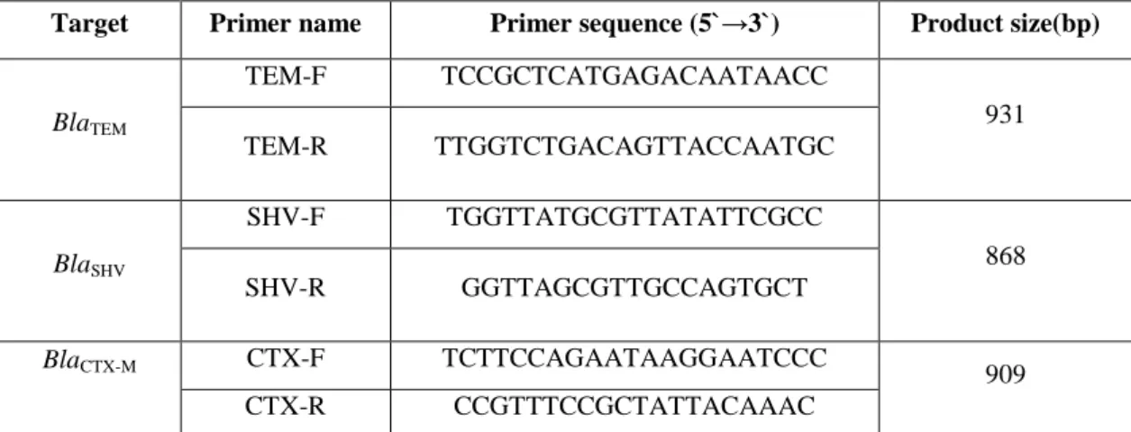
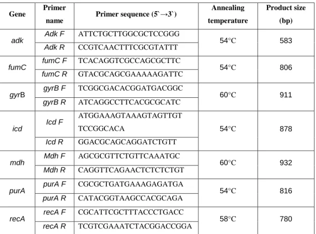
2.6.1. Primers………...……...……..…. 40
2.6.2. Enzymes………..……….…... 43
2.6.3. DNA Markers………..…….... 43
2.7. Susceptibility test discs……….... 44
2. 8. Kits……….… 44
2.8.1. QIA quick PCR Purification Kit 250 (Qiagen, Germany)….….... 44
2.8.2. DIG High Prime DNA Labeling and Detection Starter Kit II…… 44
2. 9. Computer soft wares, programs and sites……….. 45
2.10. Bacterial growth condition………...…. 46
2. 11. Bacterial growth measurement……….…….………..…. 46
2.12. Bacterial storage……….…… 46
2.13. Polymerase chain reaction (PCR)……… ……….. 46
2.14. Agarose gel electrophoresis……….…… 47
2.15. Measurement of DNA concentration with Nano- Drop ND 1000 Spectrophotometer, Thermo Technologies……… 47
2.16. Testing of antibiotic susceptibility and ESBL production……….……….. 48
Table of Contents
2.18. Sequence based allele typing for CTX-M……….…… 49
2.19. Pulsed-Field Gel Electrophoresis (PFGE)-based genotyping……...…….. 49
2.20. Phylogenetic group typing……….….. 49
2.21. Multi-Locus sequence typing………..….….. 50
2. 22. PCR-based replicon typing (PBRT) of plasmid………..…..…. 50
2. 23. Transfer of antibiotic resistance genes by conjugation………..… 50
2.24. Detection and sizing of large plasmids……… 51
2.25. Southern blot………. 52
2.25.1.Depurination………...…..…….… 53
2.25.2.Denaturation………...…..…. 53
2.25.3. Neutralization………..……..…… 53
2.25.4. Setting-up of transfer system……….…… 53
2.25.5. Preparation of nylon membrane for hybridization……… 54
2.25.6. Purification and labeling of the probe………..…….. 54
2.25.7. Prehybridization………...……… 54
2.25.8. Hybridization………..… 54
2.25.9. Detection of hybridization……….……….… 55
3.0. Results………..…… 56
3.1ESBL production and PCR detection of ß-lactamase-encoding genes among enterobacterial isolates derived from Egyptian University Hospitals……….. 56
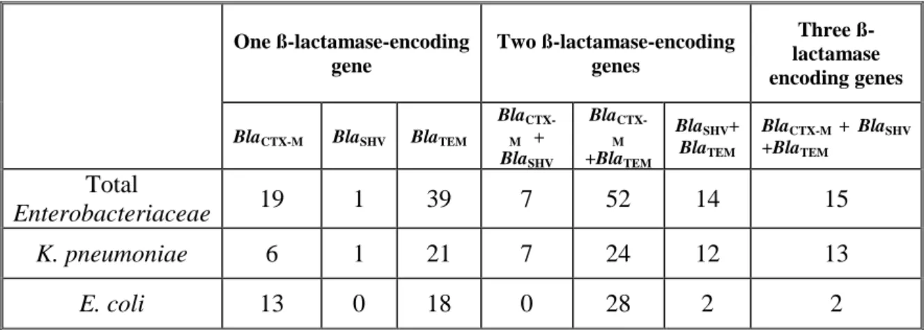
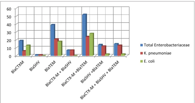
3.1.1. Multiplicity of ß-lactamase-encoding genes among enterobacterial isolates derived from Egyptian University Hospitals.………… …….… 57
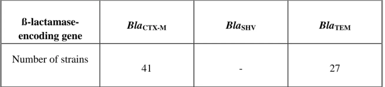
3.2. PCR detection of ß-lactamase encoding genes among enterobacterial isolates derived from Giessen University Hospital, Germany………...….. 59
3.3. Characterization of CTX-M-producing E. coli clinical isolates derived from Egyptian and German University Hospitals………...…. 59
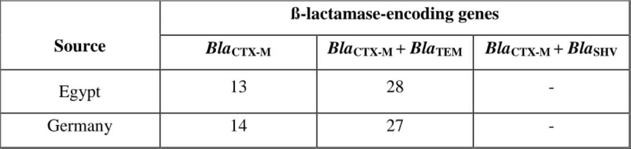
3.3.1. Distribution of ß-lactamase-encoding genes among the clinical E. coli isolates derived from Egyptian and German University Hospitals…….. 59
3.3.2. Sequence based allele typing for blaCTX-M……….….… 60
3.3.3. Antibiotic susceptibility ………...…… 61
3.3. 4. Pulsed-field gel electrophoresis-based genotyping…………..… 63
3.3. 5 Phylogenetic group typing………..…..… 66
3.3.5.1. Distribution of the different blaCTX-M allele types among E. coli phylogenetictype……….………….67
3.3.6. Multi -Locus Sequence Typing (MLST)………..….. 68
Table of Contents
3.3.7.1 Association of the different plasmid replicon types with the different CTX-M alleles in E. coli strains derived from German University Hospital……….. 70 3.3.8. Transfer of antibiotic resistance genes by conjugation………... 71 3.3.9. Transfer of non ß-lactam resistance-encoding genes……… 73 3.3.10 Plasmids size and number………..……… 74 3.3.11. Southern blot and hybridization………....… 76 3.3.11.1. DNA hybridization results for isolates derived from Egyptian University Hospital……… 76 3.3.11.2. DNA hybridization result for isolates derived from German University Hospital………….…..………. 77 3.3.12. Comparison between CTX-M-producing Escherichia coli isolates derived from University Hospitals of Egypt and Germany 78 4.0 Discussion……….……...………..….…... 79
4.1. Prevalence of ß-lactamase-encoding Enterobacteriaceae among clinical isolates derived from Egyptian University Hospitals………..… 80 4.1.1. Determination of the types of ß-lactamases among clinical enterobacterial isolates derived from Egyptian University Hospitals….. ...81 4.2. Screening for blaCTX-M and determination of the associated ß- lactamase types among clinical E. coli isolates derived from German University Hospital 82 4.3. Characterization of CTX-M-producing clinical E. coli isolates derived from Egyptian and German University Hospitals………..…… 83
4.3.1. Sequence based allele typing for blaCTX-M………..…… 83 4.3.2. Antibiotic susceptibility………..…. 84 4.3.3. Pulsed-field gel electrophoresisbased genotyping ………….… 87 4.3.4. Phylogenetic typing ……….…… 89 4.3.5. Multilocus sequence typing (MLST)………..…… 91 4.3.6. Conjugation and co-transfer of non ß–lactam resistance concomitant with ß-lactam resistance……….... 93 4.3.7. Plasmid analysis and location of blaCTX-M. ………. 95 4.4. Concluding remarks……….…. 97 4.5. Limitations of this study and recommendations for further studies in this field. ……… 99
Table of Contents
Summary……….………...……… I Zusammenfassung………..……...… III List of Abbreviations………... V List of Tables………..………….. VIII List of Figures……….. X References……….………..……….……… XII Appendix……….……….………. XXV List of publications………... XLIV Erklärung zur Dissertation……… XLV Acknowledgements………. XLVI
Introduction
1.0. Introduction
The continuous emergence of resistance to antimicrobial agents among the prevalent pathogens is the most dangerous threat for the treatment of infectious disease. The production of ß-lactamases is the major mechanism of bacterial resistance to ß-lactam antibiotics which considered the most widely used class of antibiotic. Curiously, the detection of the first ß-lactamase was reported before the use of penicillin in the medical field. Extended-spectrum-ß-lactams have been introduced in the medical practice in the 1980s for the treatment of serious gram negative bacteria but the resistance to this class of antibiotic has emerged rapidly due to production of a new class of ß-lactamase later termed extended-spectrum ß-lactamase (ESBL) (Al-Jasser, 2006).
In 1983, plasmid born extended-spectrum ß-lactamase, SHV-2, produced by Klebsiella ozaenae isolate was discovered in Germany. TEM and SHV types were the predominant ESBL types until the 1990s. In this duration Klebsiella pneumoniae was the main ESBL-producer. Later-on, prevalence of TEM and SHV and a new ESBL family, CTX-M which is produced mainly by E. coli, has emerged. During the next few years, CTX-M has become the predominant ESBL family and CTX-CTX-M-producing E. coli has spread globally and has been involved in nosocomial outbreaks and community acquired infections (Canton and Coque, 2006; Marcade et al., 2009).
The first blaCTX-M was detected in clinical E. coli isolate in Germany 1990 (Bauernfeind
et al., 1990) then CTX-M-producing Enterobacteriaceae has globally been detected. CTX-M is named after their higher hydrolytic activity against cefotaxime than ceftazidime and the place of first isolation (Munich, Germany). BlaCTX-M is a 291 amino acids encoding enzyme and the change in any one of them result in a new CTX-M variant (Naseer and Sundsfjord, 2011).
Currently, a total of 130 different blaCTX-M allele types have been added to Lahey data base for classification and aminoacids sequences of TEM, SHV, CTX-M, OXA and inhibitor resistant ß-lactamase (http://www.lahey.org /Studies accessed for last time in 10th May 2012).
The members of CTX-M family are grouped into five evolutionary groups (clusters) on the basis of their genetic relatedness and their amino acid homology with each group named after their first described member. These groups include CTX-M group 1, 2, 8, 9 and 25 (Naseer and Sundsfjord, 2011).
Introduction
2
Currently, CTX-M-15 is the most widely distributed ESBL type. It has been detected almost everywhere in the world among clinical enterobacterial isolates (Bonnet, 2004; Bradford, 2001).
In Egypt, CTX-M-15 is the dominant CTX-M-ESBL type in addition to other less frequent types like CTX-M-14 and CTX-M-27 have been also reported (Mohamed Al-Agamy et al., 2006; Fam et al., 2011).
In Germany as well as many other part of Europe, CTX-M-1 and CTX-M-3 in addition to CTX-M-15 are the most prevalent CTX-M variants (Mshana et al., 2009; Cullik et al., 2010).
CTX-M-15 was identified for the first time in 1999 from an isolate from India (Karim et al., 2001) and reported for the first time in the African continent 2005 in Tanzania (Blomberg et al., 2005).
Among the frequently detected CTX-M-types in Europe are CTX-M-1 and CTX-M-3. The spread of CTX-M-3, the precursor of CTX-M-15, is seemed to be restricted to Eastern Europe but it is sporadically detected in other parts of Europe. CTX-M-15 differs from CTX-M-3 only in single amino acid at the position 240 (Asp240Gly) resulting in enhanced activity against ceftazidime (Poirel et al., 2002).
CTX-M-encoding gene has been located on plasmids ranging from 7 to 430 kb in size. It has frequently been located on large conjugative plasmids encoding also genes of resistance to non ß-lactam antimicrobials like tetracycline, aminoglycosides sulphonamide and trimethoprim (Naseer and Sundsfjord, 2011).
BlaCTX-M-types have been associated with certain plasmid replicons like the association of CTX-M-15 with FII either in single or muti-replicons form with FIA and/or FIB. Presence of blaCTX-M-15 on Inc FI plasmids mainly FIA and FIB either in a single or multi-replicon form have recently been reported (Gonullu et al., 2008; Mshana et al.,
2009; Mshana et al., 2011).
Inc F plasmids are narrow host range plasmids highly adapted to Enterobacteriaceae
and frequently carry more than one replicon. These plasmids are a heterogeneous group of plasmids which are variable in size, resistance determinants content and replicon combination (Bergquist et al., 1986; Nordstrom, 2006). The other CTX-M-types like CTX-M-1 and CTX-M-3 are frequently associated with broad host range replicon type plasmids as Inc N, Inc I 1 and Inc L/M enabling them to be transferred to a new host of a distantly related or even unrelated species (Novais et al., 2007). Although A/C replicon type plasmid is considered to be rare in Enterobacteriaceae, it has been
Introduction
detected in CTX-M-producing E. coli (Marcade et al., 2009). Frequent association of
blaCTX-M-15 with blaTEM-1 has been reported previously in 80% of CTX-M-producing E. coli (Marcade et al., 2009). Co-existence of blaCTX-M-15 and blaTEM-1 on the same plasmid has previously been reported (Karisik et al., 2006; Marcade et al., 2009).
The horizontal transfer of ESBL-encoding plasmids among clonally related and unrelated, local endemic and international epidemic clones is responsible for the current high prevalence of ESBLs in the different European regions (Coque et al., 2008 B). The phylogenetic grouping and multi-locus sequence typing are among the most beneficial genotyping way for E. coli which can be used for their comparison on the global level. Recently, the simple PCR based method (Clermont method) has been extensively used for determination of the phylogenetic relationship among E. coli
isolates. CTX-M-15 has been frequently linked to the phylogenetic group B2 and D while CTX-M-14 has been linked to B1, A and D(Naseer and Sundsfjord, 2011). CTX-M family has been associated with many different MLST some of them displaying a global spread feature while the others have emerged and persisted locally. ST131 is the best representative of the first class being responsible for the global dissemination of CTX-M-15. Another examples are ST405 and ST38, those have been also involved in the global spread of CTX-M-15 (Coque et al., 2008 B; Naseer et al., 2009; Oteo et al., 2009 A). ST648 has been reported in many different countries. Birds are suspected to be the disseminator of this sequence type (Guenther et al., 2010). The remaining sequence types which are detected among the ESBL-producing E. coli have emerged and proliferated locally.
1.1.
Enterobacteriaceae
Members of this family are gram-negative, rod-shaped, facultative anaerobes, most of them are motile (with peri-trichous flagella) grow at 37oC on peptone, meat extract without addition of sodium chloride or other supplements, grow well on MacConkey agar, ferment glucose aerobically and anaerobically, reduce nitrate to nitrite and their G + C DNA content range from 39% to 59%. They are found in soil, water, plants and human and animal intestines. The common genera of this family include: Escherichia, Klebsiella, Enterobacter, Salmonella, Proteus, Shigella, Citrobacter, Yersinia, Serratia, Morganella, Providencia, and Hafnia. The most clinically relevant species to ESBL issue are Escherichia coli and Klebsiella pneumoniae (Irving et al., 2005).
Introduction
4
1.1.1.
Escherichia coli
Escherichia coli: is a member of the genus Escherichia which contain mostly motile gram-negative bacilli including also E. blattae, E. fergusonii, E. hermanii and E. vulneris and belong to the family Enterobacteriaceae (Nataro and Kaper, 1998). E. coli
was identified in 1885 by the German pediatrician Theodor Escherich (Feng et al., 2002). E. coli, facultative aerobes, is isolated from clinical samples either on general or selective media (such as MacConkey and eosin methylene-blue agar) at 37oC under aerobic conditions (Nataro and Kaper, 1998).
E. coli is considered to be of fecal origin and is regarded as indicator of fecal contamination but sometimes is found temporarily in the other environments such as raw meat and vegetables (Österblad et al., 1999). E. coli is widely distributed in the intestine of humans and animals maintaining the physiology of their host and are considered as one of the first bacterial genera (in addition to Streptococcus) that colonize the intestine of both human and animal-newborns protecting them against enterotoxigenic E. coli and Salmonella spp. (Feng et al., 2002; Hudault et al., 2001). Based on their clinical and genetic characters, E. coli strains are classified into commensal strains and pathogenic strains. The pathogenic strains are subdivided into intestinal pathogenic (also termed enteric or diarrheagenic) strains and extraintestinal pathogenic strains (Russo and Johnson, 2000). The great majority of E. coli is commensals. But they act as an opportunistic organism causing infections when other factor is implicated like foreign body indwelled in immunocompromised hosts. Most of them belong to phylogenetic group A and are normally devoid of the specialized virulence features that found among pathogenic strains (Feng and Weagant, 2011; Russo and Johnson, 2000).
Intestinal pathogenic strains of E. coli are rarely found among the intestinal flora of healthy hosts. Six different pathogenic classes of intestinal pathogenic groups were identified according to their virulence factors namely, enterotoxigenic E. coli (ETEC), enteropathogenic E. coli (EPEC), enterohemorrhagic E. coli (EHEC), enteroinvasive E. coli (EIEC), enteroaggregative E. coli (EAEC) and diffusely adherent E. coli (DAEC) (Russo and Johnson, 2000).
Introduction
ETEC is the causative agent of travelers’ diarrhea (watery diarrhea without fever) that occurs in developing countries due to consumption of soft foods which contain any of their several enterotoxins that are produced by ETEC.
EIEC is the causative agent of invasive, dysenteric form of diarrhea in humans due to the ability to invade the colonic mucosa.
EHEC is the causative agent of hemorrhagic colitis and bloody diarrhea due to the production of Vero or Shiga toxins (Feng and Weagant, 2011).
Extraintestinal pathogenic E. coli (ExPEC) strains are not able to cause intestinal disease. However, they can colonize the intestinal tract and predominate to the other strains in 20% of healthy hosts. Entry to extraintestinal anatomical site like urinary tract is an essential prerequisite for infection by such group. In contrary to commensal E. coli
strains, ExPEC strains are derived either from the phylogenetic group B2 or D and encode various combinations of genes collectively called extraintestinal pathogenicity virulence factors (Russo and Johnson, 2000).
1.2.2.
Klebsiella spp.
Klebsiella spp.: Gram-negative, non-motile, encapsulated, rod-shaped bacteria belong to the family Enterobacteriaceae. According to their medical importance, the genus
Klebsiella are divided into the following species: K. pneumoniae, K. ozaenae and K. rhinoscleromatis and K. oxytoca (Podschun and Ullmann, 1998).
Klebsiella spp. are found in two habitats. The first environment is the surface water, sewage, soil and on the plants, while the other environment is mucosal surfaces of mammals (Bagley et al., 1978; Brown and Seidler, 1973). In humans, Klebsiella pneumoniae are found as saprophyte in the nasopharynx and in the intestinal tract of human (Davis and Matsen, 1974).
Klebsiella are opportunistic bacteria that cause nosocomial infection in immunocompromised, hospitalized patients. Klebsiella are the causative agent of pneumonia, septicemia and urinary tract infection. Klebsiella pneumoniae and K. oxytoca regarded as the only pathogenic members of the genus Klebsiella (Bennett et al., 1995).
Introduction
6
Klebsiella usually develop a prominent capsule of complex acidic polysaccharide (K antigen) which is an essential determinant of the virulence of Klebsiella. This capsule protects Klebsiella from phagocytosis and from the bactericidal effect of serum factors. Presence of pili, responsible for the adhesive properties, is another contributor of
Klebsiella virulence (Podschun and Ullmann, 1998).
1.2. Antimicrobial agents
The great majority of antimicrobial agents can be classified on the basis of their mechanism of action into four groups:
- Cell wall synthesis inhibitors. - Protein synthesis inhibitors.
- Inhibitors of certain metabolic pathway - Nucleic acid synthesis inhibitors (Neu, 1992).
ß-Lactams like penicillins, cephalosporins, monobactam and carbapenem belong to the first group which inhibits the cell wall synthesis by interference with the enzymes that are involved in the peptidoglycan cross-linking. Tetracycline, aminoglycosides and macrolides selectively inhibit the bacterial protein synthesis but do not affect that of eukaryotes due to their ribosomal difference. Fluroquinolones interfere with DNA replication, while sulphonamides and trimethoprim block the synthetic pathway for folic acid (Tenover, 2006).
1.2.1. ß-Lactam antibiotics
This group of antibiotic includes penicillins, cephalosporins, carbapenems and monobactams. ß-lactam ring is considered as Achilles heel in this group which is easily broken by ß-lactamase enzymes, which are produced by frequent bacterial species, resulting in loss of their anti-bacterial activity.
Penicillins: in these members the ß-lactam ring is fused with thiazolidine ring
Introduction
Cephalosporins: In this group ß-lactam ring is fused with dihydrothiazine (six-membered) ring. Cephalosporins are classified into first, second, third and fourth generations.
Basic cephalosporins structure (source: Wikipedia)
The first generation includes the compounds which were available before 1975 as cefalexin and cefalothin, the second generation includes the compounds which are stable to ß-lactamase as cefuroxime and cefoxitin, the third generation includes the compounds which have ß-lactamase (but not ESBLs) stability, higher intrinsic activity and extended spectrum like cefotaxime, ceftazidime, the fourth generation is a group of new compounds like cefepime (Greenwood et al., 2006).
Monobactams: This class of ß-lactam antibiotics contains non-fused ß-lactam ring. Aztreonam is the only commercially available member of this class. Aztreonam is active against aerobic, Gram negative organisms including Pseudomonas aeruginosae
but inactive against anaerobic bacilli and the Gram positive organisms. The lack of cross-allergy with the other ß-lactam antibiotics enables it be used as an alternative for penicillin and cephalosporin in case of patient allergy to these agents (Bodey, 1990).
Aztreonam structure
Carbapenems: This group include: imipenem, meropenem and ertapenem. Carbapenems have the widest spectrum of activity compared to any other group of antibiotics which provide broad-spectrum umbrella when multiple and/or unknown organisms are expected otherwise it should be reserved for the treatment of infection caused by ESBL– producing organisms (Bodey, 1990).
Introduction
8
ß-Lactamase inhibitors: Are the agents that inhibit ß-lactamase enzymes by irreversible binding to its active site rendering it permanently inactive. The first clinically used ß-lactamase inhibitor was clavulanic acid (isolated from Streptomyces clavuligaris). It has a weak antimicrobial activity. But if combined with amoxicillin, it significantly increases the antimicrobial activity of the later. The other ß-Lactamase inhibitors such as sulbactam and tazobactam are combined with ampicillin and piperacillin respectively. lactamase inhibitors are effective against class-A ß-lactamase including CTX-M, TEM and SHV-ESBLs (Drawz and Bonomo, 2010).
1.3. Antimicrobials resistance
The microbiologists and the clinicians regard an organism to be resistant to an antimicrobial agent when it is inhibited in vitro by a concentration greater than the highest achievable concentration in the human body (Hawkey, 1998).
1.3.1. Types of resistance
I-Intrinsic resistance: The resistance of all members of a bacterial species without any genetic extra-modifications. This type of resistance is due to either the lack of the target for the action of drug or inability of drug to enter the bacterial cell (Normark and Normark, 2002; Greenwood et al., 2006).
II- Acquired resistance: This type of resistance includes:
- Mutational resistance: Occur either by point mutation, deletion, inversion or insertion in the bacterial genome resulting in a very few individuals, among the huge bacterial populations, exhibiting spontaneous resistance. These resistant mutants proliferate under the action of antibiotic selective pressure to constitute the majority or even the whole population (Normark and Normark, 2002; Greenwood et al., 2006).
- Transferable resistance: in which a resistance gene (or genes) transfer from resistant to susceptible bacterial cell. Among the different DNA elements those transfer antibiotic resistances are plasmids, phages, transposons and integrons (Normark and Normark, 2002; Greenwood et al., 2006).
Introduction
1.3.2. Mechanisms of resistance
The ability of an antimicrobial agent to inhibit a bacterial cell requires the following conditions:
- Presence of susceptible vital target in the bacterial cell (frequently enzymes or essential proteins)
- Sufficient and metabolically active concentration reaches to the target site.
The antimicrobial agents enter the bacterial cell either across the cell wall and the outer membrane or are carried by an active transport mechanism. Consequently, the differences in the susceptibilities of the different bacterial species are largely due to the difference in their cell wall structure. Therefore, the complex structure of gram negative bacteria offers a comparatively greater barrier to many antimicrobials than gram positive bacteria(Greenwood et al., 2006).
The mechanisms of drug resistance are divided into the following: - Inactivation or destruction of the antimicrobial agent.
- Alteration or protection of the target site.
- Blocking the active transport mechanism, decreasing the cell surface permeability or (efflux) removal from the cell.
- Creation of alternative metabolic pathway instead of that was inhibited by antimicrobial agent (McManus, 1997; Tenover, 2006).
1. 3.3. Genetics of resistance
Knowledgeof the basic genetics of the microbial resistance enables us to understand the evolution and spread of resistance and hence, it is suitable to start with the recognition of the different DNA elements that play a role in the evolution and spread of resistance.
Plasmids: Extrachromosomal, self-replicating double stranded DNA, present in bacterial host cell in a number of copies ranges from one to hundreds of copies, many of them encode a toxin-antitoxin based system called addiction system that eliminates the daughter cell which did not receive the relevant plasmid during cell division ensuring the survival of the plasmid regardless of the antimicrobial selective pressure. On the other hand, most of the other plasmids are devoid of such system. And hence, their survival depends on the selection of their hosts which encode an antimicrobial
Introduction
10
resistance gene under the presence of the relevant antimicrobial selective pressure (Carattoli, 2009). Conjugative plasmids contain tra genes which encode all necessary requirements for conjugation. There are also mobilisable (non self-transmissible) plasmids which can transfer in association with the conjugative (self-transmissible) plasmid (Carattoli, 2001).
Bacteriophages: These are viruses infecting only small number of strains of related bacteria (specific host range) and are divided into virulent (lytic) phage and temperate (lysogenic) phage. During transduction, the phage particle can carry exogenous bacterial DNA and transfer it to other recipient bacteria. The narrow host range of the phage greatly limits their role as a vector transferring the resistance genes(Carattoli, 2001).
Transposons: are DNA sequences that have the ability of transposition from one replicon (either plasmid or chromosome) to another and consist of either individual or a group of resistance genes flanked by short (often 40 bp) DNA sequences called direct or inverted repeats. These DNA sequences serve as recognition sites for transposase enzymes that catalyze the transfer of the transposon from one replicon to another. Transposition is an extremely important mechanism for the natural transfer of antibiotic resistance genes from one bacterial replicon and recombination into another. Theoretically, any two similar insertion sequences can bracket any gene and convert it into a transposon. All replicons, at least from the theoretical point of view, are liable to transposition and all genes are transposable and hence transposons and insertion sequences play an important role in the evolution of the resistance explaining how an antibiotic resistance gene can emerge and disseminate over a wide range of non-related replicons (Greenwood et al., 2006).
Integrons: These specialized DNA elements have been frequently observed in multi-drug resistant isolates either located on the chromosome or broad host range plasmids. Integrons are composed of two conserved regions flanking a variable region containing one or more of resistance genes. The essential components of any integron include the integrase gene (intI), the attachment site (attI) and the promoter (Carattoli, 2001).
Introduction
1. 3.3.1. Modes of transfer of resistance
Bacterial resistance may develop vertically as a result of chromosomal mutation followed selection under antibiotic selective pressure. Bacterial resistance may also develop horizontally when a susceptible bacterial cell acquires determinants of resistance from a resistant strain through one the following modes of the genetic exchange (Tenover, 2006).
Conjugation: The process in which DNA passes on one direction from a bacterial cell (the donor) to another bacterial cell when the two cells contact one another through an elongated proteinaceous structure called pillus (McManus, 1997).
Transformation: The process in which the bacterial cell acquires naked DNA from the surrounding medium. This process is of a rare occurrence in vivo and depends largely on the competence of the recipient cell to uptake the naked DNA(McManus, 1997).
Transduction: The process in which the phage particle serves as a vector transferring the bacterial DNA that is incorporated into the bacteriophage particle to the next infected cell (McManus, 1997).
The evolution of resistance via mutation and selection and the horizontal transfer of the resistance determinant enable the bacteria to accommodate the introduction of antimicrobial agents quickly and to increase the spectrum of resistance of the bacteria. Single mutation may develop initially to reduce the sensitivity of the host to the present antimicrobial until additional determinants of resistance or further mutations are acquired. In very rare occasion, a single mutation may result in high level of resistance (Tenover, 2006).
1.4. ESBL Definition and classification
Extended-spectrum ß-lactamase (ESBL): This term was used initially to refer to TEM and SHV enzymes that have the ability to hydrolyze oxyimino-cephalosporins. Later on, this term has been widened to include:
- Enzymes derived from other source and have resistance spectra similar to that of TEM and SHV mutants e.g. CTX-M and VEB types.
Introduction
12
- Enzymes exhibit wider resistance than their parents but do not belong to 2be group e.g. OXA and Amp C mutants with increased activity against cefepime (Livermore, 2008).
Although there is no consensus on the exact ESBL definition, the currently used definition for ESBL is ß-lactamase that is able to render the bacteria resistant to the penicillins, first, second, and third-generation cephalosporins and aztreonam (but not cephamycins or carbapenems) by hydrolysis (which could be inhibited by ß- lactamase inhibitors) of these antibiotics (Paterson and Bonomo, 2005).
There are two general schemes for classification of ß-lactamases:
- Ambler molecular classification scheme (see Table 1) which is based on the protein sequence similarity. Accordingly, ß-lactamase enzymes are classified into four classes A, B, C and D based on conserved and variable amino acid motifs. Class A, C, and D include the enzymes that hydrolyze their substrates by forming acyl enzymes via the active site serine, while class B (metalloenzymes) utilizes active site zinc to facilitate ß-lactam hydrolysis(Bush and Jacoby, 2010).
- Bush-Jacoby-Medeiros functional classification scheme (see Table 1) which classify these enzymes according to the similarity in their functional (substrates and inhibitors profile) characteristics. Although the molecular classification is the easiest scheme to group these diverse enzymes, the functional classification enables the clinicians and laboratory microbiologists to correlate these enzymes with their clinical roles (Bush et al., 1995; Bush and Jacoby, 2010).
Introduction
Table 1: Classification of ß-lactamases (Bush et al., 1995).
B us h -J a co by g ro up M o lecula r cla ss substrate Inhibitor Characteristic Example CA o r TZB E DT A 1 C Cephalosporins No No
Greater hydrolysis of cephalosporins than benzylbenicillin; hydrolyzes cephamycin.
AmpC, ACT-1, CMY-2, FOX-1,MIR-1
1e C Cephalosporins No No Increased hydrolysis of ceftazidime and
often oxyimino-ß-lactams CMY-37
2a A Penicillins Yes No Greater hydrolysis of benzylbenicillin
than cephalosporins. PC1
2b A Penicillins, early
cephalosporins Yes No
Similar hydrolysis of benzyl penicillin and cephalosporins.
TEM-1, TEM-2, SHV-1
2be A Extended–spectrum
cephalosporins, monobactam Yes No
Increased hydrolysis of oxyimino-ßlactams (cefotaxime, ceftazidme, ceftriaxone ,cefepime, aztreonam)
TEM-3, SHV-2, CTX-M-15, PER-1, VEB-1
2br A Penicillins No No Resistance to clavulanic acid,
sulbactam, tazobactam TEM-30, SHV-10
2ber A Extended–spectrum
cephalosporins, monobactam No No
Increased hydrolysis of oxyimino ß-lactamswith resistance to clavulanic acid, sulbactam, tazobactam
TEM-50
2c A Carbenicillin Yes No Increased hydrolysis of carbinicillin PSE-1, CARB-3
2ce A Carbenicillin, cefepime Yes No Increased hydrolysis of carbenicillin,
cefepime, cefpirome RTG-4
2d D Cloxacillin V No Increased hydrolysis of cloxacillin or
oxacillin OXA-1, OXA-10
2de D Extended–spectrum
cephalosporins V No
Hydrolyzes cloxacillin, oxacillin
oxyimino-ß-lactams OXA-11, OXA-15
2df D Carbapenems V No Hydrolyzes cloxacillin, oxacillin,
carbapenems OXA-23, OXA-48
2e A Extended–spectrum
cephalosporins Yes No
Hydrolyzes cephalosporins inhibited
by clavulanic acid but not aztreonam CepA
2f A Carbapenems V No Increased hydrolysis of carbapenems, oxyimino-ß-lactams,cephamycins
KPC-2, IMI-1, SME-1
3a B (B1) Carbapenems No Yes Broad-spectrum hydrolysis including carbapenems but not monobactams
IMP-1, VIM-1, IND-1
3b B (B2) Carbapenems No Yes Preferential hydrolysis of carbapenems CphA, Sfh-1
CA :Clavulanic acid. TZB: Tazobactam. V : Variable
Introduction
14
1.4.1. ESBL types
1.4.1.1. TEM
TEM type ESBLs are derivatives of TEM-1 and TEM-2.
TEM-1 was detected for the first time in 1965 in Greece in an Escherichia coli isolate recovered from a patient named Temoneira, and hence the designation TEM (Datta and Kontomichalou, 1965).
TEM-1 hydrolyzes ampicillin at a rate higher than that of carbenicillin, oxacillin, and cephalothin but fail to hydrolyze the extended-spectrum cephalosporins.
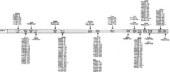
TEM-2 has the same hydrolytic activity of TEM-1 but has more active native promotor and a different isoelectric point (5.6 instead 5.4). The plasmid mediated ß-lactamase TEM-3 (an ESBL member) was detected in 1987 in Klebsiella pneumoniae isolate in France. It was originally named CTX-1 due to its higher activity against cefotaxime. The amino acid substitutions in the different TEM variants in comparison with TEM-1 are illustrated in the following Figure which is adapted from Bradford (2001).
Figure 1: The amino acid substitutions in the different TEM variants in comparison with TEM-1 adapted from Bradford (2001).
1.4.1.2. SHV
SHV-type ESBL was the most frequent ESBL-type that has been found in clinical isolates (Jacoby, 1997). SHV refer to sulfhydryl variable because it was thought that the inhibition of the enzyme activity by p-chloromercuribenzoate was substrate-dependent and variable according to the substrate used in the assay (Sykes and Bush, 1982).
In1983, a new SHV-ß-lactamase (designated SHV-2) efficiently hydrolyzes cefotaxime and to lesser extent ceftazidime has been detected in Klebsiella ozaenae in Germany
Introduction
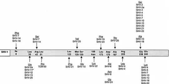
(Knothe et al., 1983). SHV-2 differs from SHV-1 by only one amino acid at the 238th position (glycine replaced by serine). This substitution (Gly238Ser) that resulted from a point mutation accounts for the activity of this enzyme against extended-spectrum cephalosporin. SHV-2 spread globally due to the selection pressure exerted by third-generation cephalosporins being detected in a wide range of Enterobacteriaceae but mainly Klebsiella spp. (Paterson and Bonomo, 2005; Paterson et al., 2003).
The amino acid substitutions in the different SHV variants in comparison with SHV-1 is illustrated in the following Figure
Figure 2: The amino acid substitutions in the different SHV variants in comparison with SHV-1 adapted from Bradford (2001).
1. 4.1.3. CTX-M
The designation CTX refers to the potent hydrolytic activity of these enzymes against cefotaxime. However, some CTX-M-types hydrolyze ceftazidime. CTX-M-types hydrolyze cefepime with high efficiency (Baraniak et al., 2002; Paterson and Bonomo, 2005; Yu et al., 2002). The hydrolytic activity of CTX-M is inhibited by ß-lactamase inhibitors. Tazobactam exhibits 10-fold greater inhibitory activity than clavulanic acid (Bush et al., 1993). CTX-M-type lactamases are related to the chromosomal ß-lactamase of Kluyvera spp. (Decousser et al., 2001). The number of CTX-M-ESBL-types is rapidly increasing and worldwide dissemination can be observed (Paterson and Bonomo, 2005).
Introduction
16
1. 4.1.4. OXA-ß-lactamase
OXA name refers to the oxacillin-hydrolyzing ability of these ß-lactamases. They hydrolyze oxacillin and cloxacillin at a rate greater than 50% that of benzylpenicillin
(Bush et al., 1995). OXA-ß-lactamases are mainly found in Pseudomonas aeruginosae (Weldhagen et al., 2003). The most common OXA-type ß-lactamase is OXA-1. It has been detected in up to 10% of E. coli isolates (Livermore, 1995). Most OXA-type ß-lactamases do not hydrolyze the extended- spectrum cephalosporin and hence are not regarded as ESBLs. OXA ESBLs includes, OXA10 (weak), 11, 14, 16, 17. 19, -15, -18, -28, -31, -32, -35 and -45 (Paterson and Bonomo, 2005).
1.4.1.5. Other ESBL-types
:
includes PER-1, PER-2, VEB-1, VEB-2, GES andSFO which share only 25% and 27% homology with TEM and SHV types. PER-1 is widely spread in Turkey and share 86% amino acid homology with PER-2 which is found almost exclusively in South America. VEB-1 discovered in an E. coli isolate from Vietnamese patient hospitalized in France (Bradford, 2001). TLA-1 was detected in a clinical E. coli isolate in Mexico. PER-1, PER-2, VEB-1 and TLA-1 are related and have some homology to the chromosomal ß-lactamase in Bacteroides spp. Therefore, it is likely to be originated from this genus (Bradford, 2001).
1.5. ESBL Epidemiology
ESBLs currently are a universal problem in hospitalized patients and community settings. Prevalence of ESBLs among clinical isolates is variable from institution to institution and from country to country and from continent to continent (Al-Jasser, 2006). Determination of the prevalence of ESBL-producing organisms at a wider geographical level is difficult and very likely to be under/overestimated. This is attributed to the different methods used for detection, sometimes the difficulty of the detection itself, the difference in the minimum inhibitory concentration (MIC) breakpoints, the variation of the incidence of ESBL among the different hospitals or medical centers located in the same country in addition to discontinuous monitoring and reporting (Sturenburg and Mack, 2003; Al -Jasser, 2006). All these factors complicate the task. Nevertheless, recent studies refer to a significant global increase in the ESBL rate. In North America the ESBL rate in Klebsiella spp., E. coli and Proteus mirabilis ranges from 4.2% - 44%, 3.3 - 4.7% and 3.1 - 9.5%, respectively. In Latin America the
Introduction
ESBL rate of Klebsiella spp., E. coli and Proteus mirabilis lie in the ranges 40% - 47.3%, 6.7 -25.4% and 9.5- 35.5%, respectively. In the Far East-Western Pacific area, the ESBL rate in Klebsiella spp., E. coli, Salmonella spp. and Proteus mirabilis ranges between 11.3% - 51%, 7.9% - 23.6%, 3.4% and 1.4 -1.8%, respectively (Sturenburg and Mack, 2003). On the national and global levels the overall ESBL production rate for the combined Enterobacteriaceae was 10.5%, the highest rate was detected in Egypt (38.5%) and Greece (27.4%) and the lowest rate was in Netherlands (2%) and Germany (2.6%) according to the Pan European Antimicrobial Resistance Local Surveillance (PEARLS) study (2001-2002) (Bouchillon et al., 2004). In the United States the prevalence of ESBL-encoding Enterobacteriaceae was around 3% (Bradford, 2001). In Europe, the incidence of ESBL-producing Enterobacteriaceae is greatly variable from geographical region to another and from country to country the published studies reflect that lower ESBL prevalence in Northern European countries compared to Southern and Eastern European countries (Bradford, 2001 and Coque et al ., 2008 A). In Spain, only 1.5% among1962 invasive E. coli isolates in 2001 were found to produce ESBL (Oteo
et al., 2002). In France, 11.4% of 6121 K. pneumoniae isolates and 47.7% of 2353 E. aerogenes were found to produce ESBL in a surveillance covering many medical centers in the period from 1996 to 2000 (Albertini et al., (2002). Northern European countries still have the lowest prevalence of ESBL-producing Enterobacteriaceae
ranging from <1% in the Netherlands to 3% in Sweden. In 2001, a study accomplished by Paul-Ehrlich-Gesellschaft (PEG) covering many medical centers in Germany revealed that the prevalence of ESBL-producing K. pneumoniae 8.2%, E. coli 0.8% and
K. oxytoca 1.3% (Sturenburg and Mack, 2003). Another study carried out by Gröbner et al at the University Hospital of Tübingen during the period from 2003 to 2007 reported that the overall ESBL prevalence still low (1.6%), however, the study recorded slight but continuous increase in the percentage of ESBL-producing Enterobacter spp. from 0.8% in 2003 to 6.4% in 2007 and in E. coli from 0.5% in 2003 to 3.8% in 2007. Percentage of ESBL-producing Klebsiella spp. ranged from 1.3% in 2003 to 2.9% in 2007 (Grobner et al., 2009). A previous study carried out in Giessen, Germany by Mshana et al. on 63 E. coli isolates, collected in the period from August 2006 to April 2007 recorded high prevalence (77.7%) of blaCTX-M encoding isolates (Mshana et al.,
2009). In an earlier study in Dresden, Germany carried out by Schmitt et al. on 39 ESBL positive enterobacterial isolates (including 10 E. coli) collected in the period
Introduction
18
from January to September 2003, 80% of ESBL positive E. coli isolates were encoding for blaCTX-M, 50% of the isolates were encoding blaTEM and 20% were encoding blaSHV.
One half of the CTX-M-encoding E. coli isolates were also encoding blaTEM gene, while the rest were harboring CTX-M gene alone. None of the enterobacterial isolates were found to harbor blaCTX-M together with blaSHV (Schmitt et al., 2007). In a relatively recent study carried out byCullik et al. on 22 ESBL-producing E. coli isolates collected during 2006 from a German University Hospital, blaCTX-M was the most frequent ß-lactamase-encoding gene being detected in 95.5% of the tested isolates followed by
blaTEM (63.6%), while only one isolate (4.5%) was positive for blaSHV. Co-existence of both blaCTX-M and blaTEMwas detected in (59.1%), while the presence of blaCTX-M alone was detected in (36.4%) of the tested isolates (Cullik et al., 2010).
In a study carried out by Fang et al. on 87 ESBL-producing E. coli isolates in Stockholm, Sweden collected during the period from 2001 to 2006, 92% of isolates were encoding blaCTX-M, 63% were encoding blaTEM and 6% were encoding blaSHV
(Fang et al., 2008).
In the United Arab Emirates, five (11.3%) multidrug-resistant enteroaggregative E. coli
strains demonstrated ESBL production (Sonnevend et al., 2006). The first detection of CTX-M-ESBL-type in Saudi Arabia was documented in 2009 by Al-Agamy et al. in a study targeting the estimation of the prevalence of ESBL-producers among 400 K. pneumoniae isolates. In which, high ESBL rate (55%) has been detected. Among those 97.3%, 84.1% and 34.1% were positive for blaSHV, blaTEM and blaCTX-M-ß-lactamase
genes, respectively. Sixty percent of the blaCTX-M belonged to blaCTX-M-1 group and the remainders belonged to the blaCTX-M-9 group(Al-Agamy et al., 2009).
In Egypt, the first study that referred to the potential high rate of ESBL production that was reflected by the reduced rate of susceptibility of E. coli, Klebsiella and
Enterobacter to ceftazidime recording 62%, 40% and 46%, respectively (El-Kholy et al., 2003). The first study that aimed at the determination of the molecular basis of ESBL resistance in clinical E. coli isolates derived from Egyptian University Hospitals was carried out by Mohamed Al-Agamy et al. who reported a very high ESBL rate (60.9%). All isolates were positive for blaTEM and blaCTX-M genes. BlaCTX-M was further
differentiated into blaCTX-M-15, blaCTX-M-14 and blaCTX-M-27 (Mohamed Al-Agamy et al.,
2006). Another study investigating 85 gram-negative bacterial isolates collected during 2002 from the Intensive Care Unit at Theodor Bilharz Research Institute, Cairo, Egypt
Introduction
revealed very high overall ESBL rate (65.8%) including Klebsiella pneumoniae
(55.3%), E. coli (35.7%), Proteus mirabilis (5.3%), Enterobacteraerogenes (1.7%) and
Citrobacter freundii (1.7%). Furthermore, ten of these ESBL-producing isolates (5 E. coli and 5 K. pneumoniae) were subjected to molecular analysis revealing that all strains were positive for blaCTX-M (all of them were molecularly identified as blaCTX-M-15) but were negative for both of blaSHV and blaTEM (Fam and El-Damarawy, 2008). Twenty-nine percent of the organisms that caused nosocomial blood stream infection in Assuit University Hospital in Upper Egypt were gram-negative bacilli of which 10.3% were K. pneumoniae, 8.6% were E. coli. Moreover, another 135 gram-negative isolates obtained from ICU environment were tested for ESBL production revealing that 18.4% were ESBL-producers. The molecular typing of the K. pneumoniae strains (by RAPD method) revealed that two isolates derived from ICU environment and one isolate derived from a patient manifested blood stream infection were identical emphasizing the role of hospital in the dissemination of ESBL-producers (Ahmed et al., 2009). In the same year, another study targeting molecular characterization of CTX-M-ESBLs harbored by five clinical isolates (3 isolates of K. pneumoniae, an E. coli isolate and an
Enterobacter cloacae isolate) demonstrated the presence of blaCTX-M-14 in Klebsiella pneumoniae and Enterobacter cloacae for the first time in Egypt as well as the presence of blaCTX-M-15 in E. coli. This study also reported the transferability of blaCTX-M-15 but
not blaCTX-M-14 and reported also the association of both types with ISEcp1element
(Khalaf et al., 2009).
Recently, a study investigating 520 enterobacterial isolates collected during the period from May 2007 till August 2008 at Theodor Bilharz Research Institute, Cairo, Egypt reported that a total of 16% of all isolates, 19% of E. coli and 14% of K. pneumonae
were ESBL-producers (Fam et al., 2011). Sometimes certain ESBL allele may be restricted to certain country or a certain geographical region. For instance blaTEM-10 has been detected in the United States in several outbreaks for many years before the detection of this allele in Europe(Bradford et al., 1994).
Another example demonstrated by blaTEM-3 which has not been detected in the United States but it is frequently found in France(Nordmann, 1998; Soilleux et al., 1996). In contrast, there are ESBL alleles which are commonly encountered worldwide like SHV-5 and CTX-M-15 (Bradford, 2001).
Global dissemination of an antibiotic resistance determinant encoded by a bacterial host can be achieved either by human travelers, migrating birds, imported animals or
Introduction
20
imported agricultural and meat products (Okeke and Adelman, 2001). Travel plays an important role in the dissemination of antibiotic resistance (Naseer and Sundsfjord, 2011). It was suggested that, the global dissemination of CTX-M-producing / ST131 E. coli is attributed to colonization or infection of travelers returning from high risk area like Indian subcontinent and the Middle East (Pitout et al., 2009). CTX-M-15-producing isolates were the most common among ESBL-CTX-M-15-producing isolates that were recovered from travelers returning from Indian sub-continent and the Middle East, while CTX-M-14-producers were the predominant ESBL-producing E. coli isolates that were recovered from travelers returning from Asia confirming the notion that returning travelers are most likely to acquire the most predominant ESBL-determinant in the visited country. Such acquisition can be achieved even without hospitalization or contact with the health care system in the visited country (Pitout et al., 2009).
Migration of the wild birds could contribute to the dissemination of resistance over the globe. CTX-M-type has been detected in the water fowls including gull species in a comparatively high level. CTX-M allele types in the wild bird were the same as those were found in the clinical setting and food-producing animals in the same region (Bonnedahl, 2011). Thirty-five different sequence types have been detected among the avian E. coli. The majority of them (ST10, ST90, ST648 and ST69) have been detected among human clinical isolates suggesting interspecies transmission, while the avian ST746 has not been detected among the clinical isolates (Guenther et al., 2011). Other than water fowls, CTX-M-producing E. coli has been isolated from Euro-Asian black birds, rock pigeon and white-fronted goose in Germany. All the recovered isolates were assigned to ST648 (Guenther et al., 2010). Therefore, the bird picks up E. coli strains derived from human origin and act as reservoir and vector posing potential to re-infect human populations (Bonnedahl et al., 2009). CTX-M-producing E. coli are likely to be globally present in chickens. CTX-M variants that have previously been isolated from chickens include CTX-M-1 (the most prevalent variant), CTX-M-2, CTX-M-14 and CTX-M-15. But the pandemic CTX-M-15-producing/ST131 E. coli clone has not been isolated from poultry (Randall et al., 2010). In addition to their presence in chicken, CTX-M has been recovered from other food- producing animals and has been found in high prevalence in retailed meat representing potential threat to human health (Simões
et al., 2010). Imported zoo animals, especially those from developing countries, represent a potential hazard to the native animals and the public health as a source of multidrug resistant bacteria (Sato et al., 2009).
Introduction
ESBL variants are selected by de novo selection then spread by different means such as clonal dissemination of the host strain or by horizontal transmission of the ESBL-gene carrying plasmid either to related strains, in case of narrow–host range plasmids, or to non-related strains in wide host range plasmids (Hibbert-Rogers et al., 1994; Gniadkowski, 2001; Palucha et al., 1999; Villa et al., 2000). Epidemic plasmids have been implicated in the dissemination and the high prevalence of blaESBL in the European region and have been detected among local or international epidemic clones (Canton et al., 2008).
Several outbreaks have been reported, the great majority of which occurred in tertiary hospitals where the transfer of a colonized patient provide a chance for dissemination of the ESBL-producing organism (Patterson, 2001; Palucha et al., 1999). Frequently, the exact source of outbreaks has never been detected but many of these resistant bacteria were characterized epidemiologically as illustrated by the following examples:
- In a French Hospital, SHV-5 expressing K. pneumoniae were isolated from six peripartum women and two neonates. PFGE profiles of these strains indicated that all of the strains have PFGE-patterns identical to that of a strain isolated from contaminated ultrasonography coupling gel (Gaillot et al., 1998).
- In South Africa, an ESBL-producing K. pneumoniae carried by cockroaches infesting the neonatal ICU has the same PFGE-type of the strain that were implicated in an outbreak caused high mortality rate among neonates in that hospital (Cotton et al., 2000).
- In another outbreak, ESBL-producing E. coli and K. pneumoniae having different PFGE-types, but carrying identical plasmid encoding TEM-10 have been isolated from many patients in different hospitals in Chicago. The occurrence of the same plasmid in the strains of different PFGE-genotypes is a clue for plasmid transfer (Bradford et al., 1994; Wiener et al., 1999).
- More interestingly, blaTEM-24 encoding, 180 kb, conjugative plasmid was detected in four different enterobacterial species E. coli, K. pneumoniae, E. aerogenes and P. rettgeri isolated from the same patient suggesting horizontal transfer between the normal flora of the gut (Marchandin et al., 1999).
The first two examples can be regarded as an example of clonal dissemination while the latter two could be considered as an example of horizontal transfer of epidemic plasmid.
Introduction
22
During 1990s, infection and colonization with ESBL-producers was mainly hospital-acquired principally in intensive care units (ICU) in addition to surgical, pediatrics, neonatology and oncology wards. Also community clinics and nursing homes are regarded as potential reservoir (Babini and Livermore, 2000; Bermudes at al., 1997; Wiener et al., 1999). Currently, ESBL-producing bacteria are no longer restricted to hospital environment, but they are circulating in the community setting which plays an essential role in the persistence of ESBL-producing organisms (Andriatahina et al., 2010). In general, TEM and SHV type ß-lactamases, produced commonly by K. pneumoniae, have spread throughout the hospital setting, while CTX-M enzymes, produced mainly by E. coli, has become predominant ESBL-family in the community (Pitout et al., 2008; Mirelis et al., 2003).
Recent studies demonstrated significant increase in ESBL-producers in the community setting in many part of the world. Asymptomatic colonization with ESBL-producing organisms has also been described (Hollander et al., 2001). Intestinal carriage is the main reservoir for ESB-producing organism in the hospital setting (Andriatahina et al., 2010). The detected rates of ESBL carriage among hospitalized patients was 11.7% in Spain, 16% in Lebanon and 26% in Saudi Arabia (Valverde et al., 2004; Moubareck et al., 2005; Kader et al., 2007). The existence of ESBL-producing organisms in the intestinal tract increases the risk of transmission to other individuals through human to human transmission or through the environment (Andriatahina et al., 2010). Emergence of producers in the community could be attributed to acquisition of an ESBL-producer by a patient during hospitalization or due to antibiotic overuse by community patients (Kader and Kamath, 2009). Extensive administration of expanded-spectrum ß-lactam antibiotics along with one or more of specific risk factors including the prolonged hospital stay, sever illness, admission to ICU, intubation, mechanical ventilation, urinary or arterial catheterization, undergoing hemodialysis, abdominal surgery, gut colonization, low birth weight and patient transfer between the different units in the hospital are the most common aspects among the hospitals that were infected by ESBL-producing organisms (Pena et al., 1997 ; Rice, 1999).
Introduction
1.6. ESBL detection methods
The detection methods are divided into - Phenotypic methods.
- Molecular methods.
Phenotypic methods are based on the resistance of ESBL-producers to oxyimino-lactams such as cefotaxime, ceftriaxone, ceftazidime and aztreonam and the ability of ß-lactamase inhibitors to inhibit this resistance. Several tests are recommended including:
1.6.1. Double disk diffusion (double disk approximation/ double disk
synergy DDS)
The test was earlier performed by swabbing the organism onto a Muller-Hinton agar plate then a disk containing amoxicillin-clavulanates (20µg/10µg) was placed in the center of the plate and disks containing 30 µg of ceftazidime, ceftriaxone, cefotaxime and aztreonam were placed at a distance of 30 mm (center to center). Enhancement of the zone of inhibition of the oxyimino-ß-lactam caused by the synergy of the clavulanate in the amoxicillin-clavulanate disk was interpreted as positive test indicating the production of ESBL. The use of cefpodoxime as the oxyimino-cephalosporin of choice is recommended. It gives test sensitivity up to 97% and test specificity up to 100%. If the test is negative with an isolate that is highly suspected to be an ESBL-producer the test should be repeated with closer distance (20 mm) but generally the test is reliable, convenient and non-expensive method for screening of ESBL production (Drieux et al., 2008).
1.6.2. Combination disk method
This method is based on measuring the inhibition zone around the disks of cephalosporin (ceftazidime 30µg, cefotaxime 30µg and/or cefpodoxime 30µg) and around the disk of the same cephalosporin plus clavulanate (30 µg/10 µg). A difference of ≥ 5 mm between the two diameters or 50% expansion of the inhibition zone is considered a positive indication for ESBL production (Carter et al., 2000; M'Zali et al.,
2000). Sensitivity and specificity of this method is up to 96% and 100%, respectively (Linscott and Brown, 2005).
Introduction
24
1.6.3. ESBL E-test
The purpose of this test is the assessment of the synergism between extended-spectrum cephalosporin and ß-lactamase inhibitors. E-test is a two-sided strip containing gradient concentrations of cefotaxime (CT) or ceftazidime (TZ) or cefepime (PM) alone at one side of the strip and combined with clavulanate 4 mg/L at the other end giving the designation CT / CTL, TZ / TZL and PM / PML. The reduction of MIC of the tested cephalosporin by more than three doubling dilution i.e. if MIC ratio ≥ 8 and / or if phantom zone appeared just below the lowest concentration of CTL, TZL or PML and /or deformation of the CT, TZ or PM inhibition ellipse at the tapering end the test is interpreted as positive indicating ESBL production (Cormican et al., 1996; Leverstein-van Hall et al., 2002).
1.6.4. Agar supplemented with clavulanate
In this method, two Müller-Hinton agar plates one of which freshly supplemented with 4µg/ml clavulanate are swabbed with the test bacteria and antibiotic disk of ceftazidime (30 µg), cefotaxime (30 µg), ceftriaxone (30 µg) and aztreonam (30 µg) are placed on clavulanate-supplemented and clavulanate-free plates. If the difference in inhibition zone diameter around ß-lactam on the two media is ≥10 mm the test is positive indicating ESBL production. The sensitivity of this test is up to 96% and the specificity is 100% for ceftazidime (Al- Jasser., 2006; Ho, 1998; Vercauteren et al., 1997).
1.6.5. Automated method
The automated antimicrobial susceptibility test systems include VITEK test, Phoenix ESBL test.
1.6.5.1. VITEK 2 ESBL test (BioMerieux, France)
This method relies on simultaneous quantification of the antimicrobial activity of cefotaxime, ceftazidime and cefepime with and without clavulanate. A card wells contains 1 mg/L of cefepime or 0.5 mg/L cefotaxime or ceftazidime either alone or with clavulanate 4mg/L. After inoculation with the suspected ESBL- producing organisms, the cards are introduced into the VITEK 2 machine and the turbidity for each antibiotic tested is measured at regular intervals. The growth in the wells containing cephalosporin
Introduction
with clavulanate compared with that of the cephalosporin alone and the result is interpreted by a computerized system (Drieux et al., 2008).
1.6.5.2. The automated Phoenix ESBL test (Becton Dickinson, USA)
This method depends also on the growth response to certain expanded-spectrum cephalosporins and the result also interpreted via computerized system (Drieux et al., 2008).
1.6.6. Molecular detection methods
A number of methods can be used for characterization of ESBL. In the past, the determination of the isoelectric point was enough for the identification of ESBL that was present in a clinical isolate. But after the emergence of many ESBL-types that have identical isoelectric points the determination of ESBL type by isoelectric point is no longer feasible(Bradford, 2001).
Detection of earlier ß-lactamase genes was achieved by using specific DNA probes for TEM and SHV-encoding genes but this method is labour intensive(Arlet and Philippon, 1991).
PCR with oligonucleotide primers that are specific for an ESBL-encoding gene is the most convenient and the most widely used method to detect the ESBL family of the variant but will not differentiate among the different variants (Bradford, 2001).
For determination of a specific ß-lactamase gene that is present in a strain, nucleotide sequencing is the gold standard that can detect all variants. But it is labour intensive, technically challenging (Bradford, 1999). So, several molecular methods for detection and differentiation of ESBL without sequencing have been suggested like the oligotyping method that has been used to differentiate between blaTEM-1 and blaTEM-2.
This method is based on the use of oligonucleotide probes that are designed to detect point mutation under strict hybridization conditions. Several blaTEM variants were identified by this method (Bradford, 2001).
Sometimes mutation result in creation or disappearance of restriction sites and hence if this part of the gene is amplified by PCR and the resulting amplicon digested with restriction endonuclease, the analysis of the restriction profile may lead to identification of the new ESBL variant. This lead to another approach combines restriction fragment length polymorphism analysis with PCR (PCR-RFLP). The created pattern by each