3
Evaluation of specimen preparation techniques for
elemental localisation in hyperaccumulating tissues
*A
BSTRACT
Hybanthus floribundus subsp. floribundus and Pityrogramma calomelanos var. austroamericana are promising species for use in phytoremediation of contaminated sites. Micro-proton-induced X-ray emission (µ-PIXE) spectroscopy was used to map the elemental distribution of the accumulated metal(loid)s, Ca and K in leaf or pinnule tissues prepared by two contrasting specimen preparation techniques: freeze-substitution in tetrahydrofuran (THF) and freeze-drying. The specimens were analysed to compare the suitability of each technique in preserving (i) the spatial elemental distribution and (ii) the tissue structure of the specimens. Further, the µ-PIXE results were compared with concentration of elements in the bulk tissue obtained by inductively coupled plasma-atomic emission spectrometer (ICP-AES) analysis. In H. floribundus subsp. floribundus, µ-PIXE analysis revealed Ni, Ca and K concentrations in freeze-dried leaf tissues were at par with bulk tissue concentrations. Elemental distribution maps illustrated that Ni was preferentially localised in the adaxial epidermal tissues (1% DW) and least concentration was found in spongy mesophyll tissues (0.53% DW; as determined by region selection analysis). Conversely, elemental distribution maps of THF substituted tissues indicated significantly lower Ni, Ca and K concentrations than freeze-dried specimens and bulk tissue concentrations. Moreover, Ni concentrations were uniform across the whole specimen and no localisation was observed. In P. calomelanos var. austroamericana freeze-dried pinnule tissues, µ-PIXE revealed statistically similar As, Ca and K concentrations as compared to bulk tissue concentrations. Elemental distribution maps showed that As localisation was relatively uniform across the whole specimen. Tetrahydrofuran freeze-substituted tissues revealed a significant loss of As compared to freeze-dried specimens and the concentrations obtained by bulk tissue analysis. The results demonstrate that freeze-drying is a suitable sample preparation technique to study elemental distribution of ions in H. floribundus and P. calomelanos plant tissues using µ-PIXE spectroscopy. Furthermore, cellular structure was preserved in samples prepared using this technique.
3.1 I
NTRODUCTION
The phenomenon of metal(loid) hyperaccumulation is expressed in ca. 450 species worldwide
that exhibit the unique ability of elevated metal(loid) accumulation (> 0.1 % nickel (Ni) or arsenic (As) dry weight; DW) in aboveground tissues without adverse effects on plant growth (Reeves and Baker 2000; Ma et al. 2001; Reeves 2003). These species may be utilised in
phytoextraction of contaminated soils, whereby roots absorb the contaminants from soil into aboveground biomass which is harvested and safely processed by drying, ashing or composting (Garbisu and Alkorta 2001). To comprehend the ecology and physiology of these potentially very useful plant resources, quantitative spatial distribution of the hyperaccumulated metal(loid) is required. Micro-proton induced X-ray emission spectrometry (µ-PIXE) analysis is a versatile micro-analytical technique that is frequently used to study spatial distribution and localisation of elements in metal(loid) hyperaccumulating species (Table 1.3). To date, studies have examined concentrations to the mg kg–1 level in leaf, stem and seed hyperaccumulating tissues (Krämer et al. 1997; Bhatia et al. 2003; Fernando et al.
2006a). Typically, the acquired µ-PIXE spectra are extracted using the dynamic analysis (DA) matrix transform method in order to construct two-dimensional elemental maps. Quantitative concentration data is then extracted and interpreted from arbitrarily selected regions within the two-dimensional maps representing, for example a specific cellular structure (Przybyłowicz et al. 2001).
To proceed with µ-PIXE analysis of hyperaccumulating tissues, sample preparation is the most crucial step in order to obtain reliable data and provide an accurate representation of the spatial localisation of elements in vivo. Owing to the high sensitivity of µ-PIXE spectroscopy,
purity at all stages of sample preparation is of paramount importance (Mesjasz-Przybyłowicz and Przybyłowicz 2002). Biological materials are highly complex and heterogeneous, and therefore sample preparation techniques allowing i) preservation of tissue structure and ii) prevention of elemental redistribution must be followed, however these two requirements are often in conflict (Przybyłowicz et al. 1999). For example, conventional chemical fixation
protocols for electron microscopy ensure preservation of cellular structures, however, are unsuitable for investigating spatial localisation as they often result in a dramatic loss and redistribution of mobile ions. Such protocols are carried out at ambient conditions with abrasive fixatives such as glutaraldehyde and solvents such as acetone or ethanol which can all potentially introduce artifacts during sample preparation. For example, Davies et al. (1991)
using energy dispersive X-ray micro-analysis reported a 78% loss of Zn in root meristematic cells of three Festuca rubra cultivars prepared using a glutaraldehyde-osmium tetroxide
protocol. Cryofixation of hand-sectioned tissue into a liquid coolant (such as liquid nitrogen [LN2] or propane) followed by freeze-drying may be employed in order to overcome
limitations of conventional chemical fixation protocols. This technique is relatively simple and easy to handle and has been successfully demonstrated by Mesjasz-Przybyłowicz et al.
(1997) and Bhatia et al. (2004) in preserving elemental composition in hyperaccumulating
plant material.
Alternatively, freeze-substitution is another technique where water molecules in tissues are substituted with a suitable medium (or solvent) at ultra low temperatures (–90 °C). Of paramount importance is the duration of water substitution as determined by the chemical properties of the freeze-substitution medium. For example, tetrahydrofuran (THF) is a fast acting solvent, used with success in the preservation of elements in animal tissues (Pålsgård et al. 1994; Pålsgård et al. 1996), however, there is little information pertaining to the use of
THF in hyperaccumulating plant tissues. Recently, Budka et al. (2005) suggested that THF
was unsuitable in localisation of Ni in leaves of Berkheya coddii, however, it is not known if
this is consistent with other hyperaccumulating species, particularly species other than Ni hyperaccumulators. Moreover, published protocols are often lacking pertinent information regarding methodological execution such as ensuring anhydrous conditions of solvents prior and during freeze-substitution and similarly, resin/solvent mixtures during infiltration and embedding (e.g. Budka et al. 2005).
Therefore, in this study, freeze-substitution using THF as the freeze-substitution medium and freeze-drying protocols were investigated using foliar tissues from two model hyperaccumulating species, Hybanthus floribundus subsp. floribundus (Violaceae) and Pityrogramma calomelanos var. austroamericana (Pteridaceae). Hybanthus floribundus is a
native Australian perennial shrub and has been reported to hyperaccumulate up to 13,500 mg Ni kg–1 DW in leaf tissues (Farago et al. 1977). Three subspecies of H. floribundus namely floribundus, adpressus and curvifolius have been reported to hyperaccumulate Ni (Bennett
1972; Severne 1974; Bidwell et al. 2001). Subspecies adpressus and curvifolius are restricted
to the ultramafic outcrops and nickeliferous soils in the Eastern Goldfields region of Western Australia. Subspecies floribundus has a rather disjunctive distribution and is a facultative
(pseudo-) nickelophyte – inhabiting nickeliferous, ultramafic and non-ultramafic soils across south-eastern regions of Australia (Bennett 1972; Severne and Brooks 1972; Severne 1974).
Pityrogramma calomelanos var. austroamericana (Domin) Farw.(nomenclatural synonym - Pityrogramma austroamericana Domin), or gold dust fern is a perennial and rhizomatous fern
native to South America. This weedy species has naturalized across many tropical and subtropical regions across the world and is frequently found thriving in disturbed areas such as road cuttings, mine overburden and tailings (McCarthy 1998; Ashley et al. 2003). In the
present study, µ-PIXE spectroscopy was employed to ascertain the efficiency of elemental and morphological preservation of leaf (H. floribundus subsp. floribundus) or pinnule (P. calomelanos var. austroamericana) tissues prepared using both protocols.
3.2 M
ATERIALS AND METHODS
3.2.1 Selection of plant material
3.2.1.1 Hybanthus floribundus subsp. floribundus
Tube stock of young H. floribundus (Lindl.) F.Muell. subsp. floribundus plants were
purchased from a commercial nursery at Bendigo (Victoria, Australia). Prior to transplanting, approximately 800 g of air-dried potting mix (Debco® general container mix with 10% coarse
perlite) was weighed into eight plastic pots (∅ 14 cm; 2 L capacity). To half the pots, 500 mL of 1,000 mg Ni kg–1 (as nickel sulphate; NiSO
4·6H2O) solution was applied equating to a total
of 1,500 mg Ni kg–1. The remaining four pots were maintained as untreated controls. A plastic saucer was placed under each pot to collect leachate, hence maintaining a closed system. Pots were allowed to incubate for two weeks prior to planting, and each pot contained a single tube stock. The pots were arranged in a completely randomized design in a greenhouse with 11 h daily photoperiod with photon flux > 370 µmol m–2 s–1, temperature range between 19 ºC (night) to 32 ºC (day) and relative humidity of ~65%. Pots were watered with deionised water to maintain approximately 60% water holding capacity. Four grams of Osmocote® native sustained release fertiliser (N:P:K of 17:1.6:8.7) was applied two weeks after transplanting.
3.2.1.2 Pityrogramma calomelanos var. austroamericana
Pityrogramma calomelanos var. austroamericana ferns growing in mine tailings at Mt
Morgan, Queensland, Australia, were exhumed and transplanted into plastic pots (∅ 14 cm; 2 L capacity) that contained approximately 800 g of air dried potting mix (Debco® general
container mix with 10% coarse perlite). Prior to transplanting, roots were rinsed to remove native soil and debris, and hardened off for 4 weeks. Ferns wereexposed to 50 mg As kg–1 (as sodium arsenate; Na2HAsO4·7H2O) and grown for a period of 20 weeks. The pot experiment
was conducted in a greenhouse with 11 h daily photoperiod with photon flux > 370 µmol m–2
s–1, temperature range between 19 ºC (night) to 32 ºC (day) and relative humidity of ~65%.
3.2.2 Sample preparation
3.2.2.1 Freeze-drying
During the course of the experiment, phytotoxicities were absent from all metal(loid) treated plants. Specimens were prepared following the procedure outlined by Bhatia et al. (2004).
Briefly, fresh pinnule and stipe samples were carefully excised; hand-sectioned without delay using a stainless steel razor blade and immediately plunged into LN2 (time lapse in the whole
procedure being < 5 s). Sections were then freeze-dried for up to 48 h and individually mounted onto Formvar (in 2% ethylene dichloride) support films on aluminium strips (25 mm × 75 mm × 0.3 mm). Each strip had been prepared by floating a Formvar film across a 5 mm diameter hole punched through the strip. A second film was placed over the section, air dried for 10–15 min, glued to an aluminium target holder using double-sided carbon tape at the Australian Nuclear Science and Technology Organisation (ANSTO) microprobe facility. Prior to µ-PIXE analysis mounted specimens were photographed to assist with the location of anatomical features and target holders were stored in a desiccator at all other times. Representative leaf and pinnule samples from the same plants were also excised and processed for ICP-AES analysis.
3.2.2.2 Chemical analysis
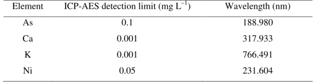
Tissue samples were digested in a mixture of nitric and perchloric acids as described in section 2.2.3. Digests were analysed for various elements using Vista CCD Varian® inductively coupled plasma-atomic emission spectrometer (ICP-AES). Analytical accuracy was determined by including NIST-SRM #1547 (peach leaves) and NIST-SRM #1575 (pine needles). Recovery of analysed elements was within ± 10% of the recommended values and analytical precision was below 10% RSD for all samples. The detection limit and wavelengths of each element are shown in Table 3.1.
Table 3.1 Detection limits and wavelengths used for inductively coupled plasma-atomic emission spectrometer (ICP-AES) analysis.
Element ICP-AES detection limit (mg L–1) Wavelength (nm)
As 0.1 188.980
Ca 0.001 317.933
K 0.001 766.491
Ni 0.05 231.604
3.2.2.3 Freeze-substitution using tetrahydrofuran
A hole punch was used to cut 1.7 mm discs of leaf/pinnule tissue from three replicates of both species and eight discs per replicate were obtained, each disk from a different leaf/pinnule. The discs were immediately transferred into the cavity (∅ 1.2 mm × 200 µm in depth)of gold-plated specimen carriers pre-filled with a cryoprotectant, 1-hexadecene. The specimen carriers were then rapidly frozen using a high pressure freezer (Leica EM Pact; Leica Microsystems, Australia) and stored under liquid nitrogen prior to freeze-substitution.
For the preparation of specimens, a modified freeze-substitution protocol used by Hyde et al.
(2003) was followed. Briefly, frozen leaf specimens were freeze-substituted in tetrahydrofuran with freshly activated molecular sieve (4 Å) in a Leica automatic freeze-substitution unit (Leica Microsystems, Australia) for three days at –80 °C. Samples were gradually warmed to –30 °C (@ 5 °C h–1), held for 24 h; further warmed to 6 °C and held for 1 h, before allowing the specimens to warm up to room temperature. Specimens were then transferred into a glove box at room temperature flushed with dry nitrogen gas and infiltrated with Spurr’s resin (Spurr 1968) in one day steps of 30, 60, 90 and 100% with relative humidity kept below 12%. Resin mixes were prepared with freshly activated molecular sieve and stored at –80 °C prior to use. Infiltrated leaf samples were then flat embedded into fresh Spurr’s resin and polymerized overnight at 60 °C. Sections were cut at 10 µm using dry glass knives on a Leica Ultracut S microtome (Leica Microsystems, Australia), mounted onto target holder as described above and stored in a dry box prior to µ-PIXE analysis.
3.2.3 Micro-PIXE setup
Nuclear microprobe analyses were performed using the 10 MV Tandem accelerator at the Australian Nuclear Science and Technology Organization (Siegele et al. 1999). Samples were
analysed using a 3 MeV proton beam with a typical spot size of between 5 and 8 µm. Aluminium target holders were introduced through a vacuum lock into the target chamber for proton beam irradiation (Figure 3.1). X-ray data was collected using a high purity Ge detector (Canberra Industries, USA). The detector was located 33 mm from the sample and had a 100 mm2 active area. The µ-PIXE set up was calibrated using an obsidian rock standard
(NIST-SRM #278) which contained 3.6 ± 0.3 µg Ni g–1. The proton beam was scanned over samples
up to a maximum of 2.5 × 2.5 mm2 and samples were typically irradiated with a total charge between 2 and 5 µC. A 100 µm Mylar foil was used to attenuate X-rays from the light elements and thus minimise pile-up in the µ-PIXE spectrum. The density and composition of plant sections analysed were not homogeneous and consequently the composition of the sample matrix was assumed to be comparable to cellulose (following the procedure of Pineda and Peisach 1988).
Figure 3.1 (a) Nuclear microprobe end station. The target chamber is indicated by the red arrow. (b) Inside the target chamber. The Ge detector is indicated by the yellow
arrow.
The analysis of PIXE spectra was performed using GeoPIXE II software (Ryan et al. 2002).
Region selection analysis (RSA) using the Dynamic Analysis (DA) matrix transform method of GeoPIXE II (Ryan et al. 2005) was used to extract representative concentration data and
minimum detection limits from select regions within pinnule and stipe sections. Regions were selected from on-screen quantitative distribution maps reproduced from data accumulated by Oxford Microprobe Data Acquisition System (Bhatia et al. 2004). The quantitative images
obtained using the DA method were free of artefacts due to overlapping elements, detector effects (such as escape peaks and tailing) and were background subtracted (Ryan et al. 2005).
In addition, quantitative point analysis (QPA) based on DA method was performed by
(a) (a) (a)
drawing area transects across leaf and stem elemental maps. Transects were located on the basis of visual inspection of the elemental distribution maps in order to provide a typical elemental profile across leaf and pinnule sections (Bhatia et al. 2004).
3.2.4 Statistical analysis
.The results of RSA obtained were subject to ANOVA using GenStat version 8.1.0.152 (Payne
et al. 2005) and significant differences among the treatment means were separated by least
significant differences (LSD) at P≤0.05.
3.3 R
ESULTS
3.3.1 Micro-PIXE analysis
3.2.3.1 Hybanthus floribundus subsp. floribundus
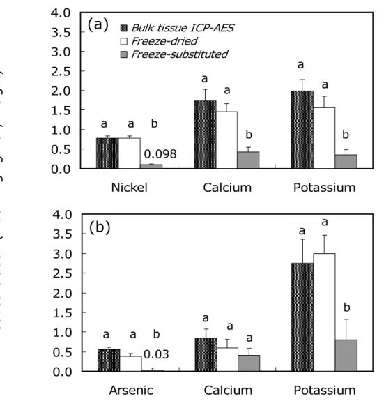
The concentrations of Ni, Ca and K determined by RSA in H. floribundus subsp. floribundus
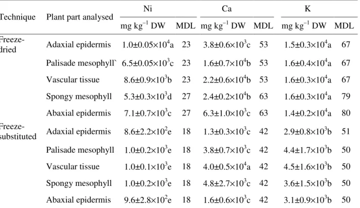
freeze-dried leaf cross-sections were in agreement with bulk tissue concentrations and followed the order K > Ca > Ni (Figure 3.2a). Nickel concentration and distribution in freeze-dried leaves were significantly higher than freeze-substituted tissues, and showed a distinctive distribution pattern that was not uniform (Figure 3.3d). The highest Ni concentration was found in adaxial epidermal tissues (1.0×10⁴ mg kg–1 DW) and the lowest in spongy mesophyll tissues (5.3×10³ mg kg–1 DW; Table 3.2). The concentration of Ni, Ca and K in freeze-substituted leaf cross-sections as determined by RSA was 4–5 times lower than their corresponding bulk tissue concentrations and µ-PIXE concentrations of freeze-dried tissues (Figure 3.2a). Nickel concentrations in freeze-substituted sections showed little variation (Figure 3.3b) and were statistically similar amongst tissue types (Table 3.2). Quantitative point analysis (QPA) revealed a peak Ni concentration of 1.45×10⁴ mg kg–1 DW in the adaxial epidermis of freeze-dried samples, whereas in the freeze-substituted samples, a peak Ni concentration of only 0.215×10⁴ mg kg–1 DW occurred in the vascular bundle (Figure 3.3).
0.0 0.5 1.0 1.5 2.0 2.5 3.0 3.5 4.0
Nickel Calcium Potassium
Bulk tissue ICP-AES Freeze-dried Freeze-substituted C o n c e n tr a ti o n ( × 1 0 4 m g k g – 1 d ry w e ig h t) 0.0 0.5 1.0 1.5 2.0 2.5 3.0 3.5 4.0
Arsenic Calcium Potassium
Figure 3.2 Average concentration (± standard error) of elements in Hybanthus floribundus subsp. floribundus leaf (a) and Pityrogramma calomelanos var. austroamericana pinnule (b) after ICP-AES analysis of bulk tissue and µ-PIXE analysis of freeze-dried and freeze-substituted (tetrahydrofuran) cross-sections.
Micro-PIXE concentrations were obtained following dynamic analysis across whole cross-sections (n=3).
Means followed by a different letter indicate a significant difference between the treatments for each element (P≤0.05) (a–b denotes significance between the concentrations for each element).
0.098 0.03 a a b a a b b a a a a b a a a b a a
(a)
(b)
F ig u re 3 .3 E le m en ta l m a p s o f N i in a h ig h p re ss u re f ro ze n , te tr a h y d ro fu ra n f re ez e-su b st it u te d ( b ) a n d h a n d se ct io n ed , cr y o -f ix ed fr e ez e-d ri ed (d ) le a f c ro ss -s e ct io n o f H yb a n th u s fl o ri b u n d u s su b sp . fl o ri b u n d u s sa n d w ic h ed b et w ee n F o rm v a r fi lm s a ft er 2 0 w ee k s o f N i ex p o su re . A ty p ic a l o p ti ca l m ic ro g ra p h o f a f re e ze -s u b st it u te d ( a ) a n d f re e ze -d ri ed ( c) H . fl o ri b u n d u s su b sp . fl o ri b u n d u s W he re E (a d) , a da xi al e pi de rm is ; P, p al is ad e m es op hy ll; S M , s po ng y m es op hy ll; V B , v as cu la r bu nd le a nd E (a b) , a ba xi al e pi de rm is . T he g ra ph ic al o ut pu t ad jo in in g ea ch e le m en ta l m ap r ep re se nt s qu an tit at iv e po in t an al ys is ( m ic ro -P IX E ). S ta tis tic al e rr or s of e ac h po in t a re in di ca te d on th e Q PA p ro fi le b y er ro r ba rs . T he c on ce nt ra tio n le ge nd p ro vi de s a qu al ita tiv e m ea su re o f th e re la tiv e in te ns ity of th e N i m ap pe d.
Table 3.2 Average concentration of elements (mg kg–1 DW) in Hybanthus floribundus subsp. floribundus leaf cross-sections after freeze-drying and freeze-substitution with tetrahydrofuran.
Concentrations were obtained following dynamic analysis of individually selected regions. The results are presented as means ± SE of three replicates. Different letters within the same column indicate a significant difference (P≤0.05).
Minimum detection limit (MDL; mg kg–1 DW) of each measurement are also presented.
Ni Ca K
Technique Plant part analysed
mg kg–1 DW MDL mg kg–1 DW MDL mg kg–1 DW MDL Freeze-
dried Adaxial epidermis 1.0±0.05×10⁴a 23 3.8±0.6×10³c 53 1.5±0.3×10⁴a 67 Palisade mesophyll` 6.5±0.05×10³c 23 1.6±0.7×10⁴b 53 1.6±0.4×10⁴a 67 Vascular tissue 8.6±0.9×10³b 23 2.2±0.6×10⁴b 53 1.6±0.3×10⁴a 67 Spongy mesophyll 5.3±0.3×10³d 27 2.4±0.2×10⁴b 63 1.6±0.3×10⁴a 79 Abaxial epidermis 7.1±0.7×10³c 27 6.3±1.0×10³c 63 1.4±0.2×10⁴a 80 Freeze-
substituted Adaxial epidermis 8.6±2.2×10²e 18 1.3±0.3×10³c 42 2.9±0.8×10³b 51 Palisade mesophyll 1.0±0.2×10³e 18 3.8±0.7×10³c 42 4.4±1.7×10³b 50 Vascular tissue 1.0±0.1×10³e 18 4.0±0.5×10⁴a 42 4.5±1.6×10³b 50 Spongy mesophyll 1.0±0.2×10³e 18 4.8±2.7×10³c 42 3.6±1.5×10³b 50 Abaxial epidermis 9.6±2.8×10²e 18 1.6±0.6×10³c 42 3.1±0.9×10³b 50
3.2.3.2 Pityrogramma calomelanos var. austroamericana
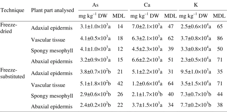
An examination of elemental maps generated from freeze-dried and freeze-substituted pinnule cross-sections of P. calomelanos var. austroamericana revealed that the concentrations of As,
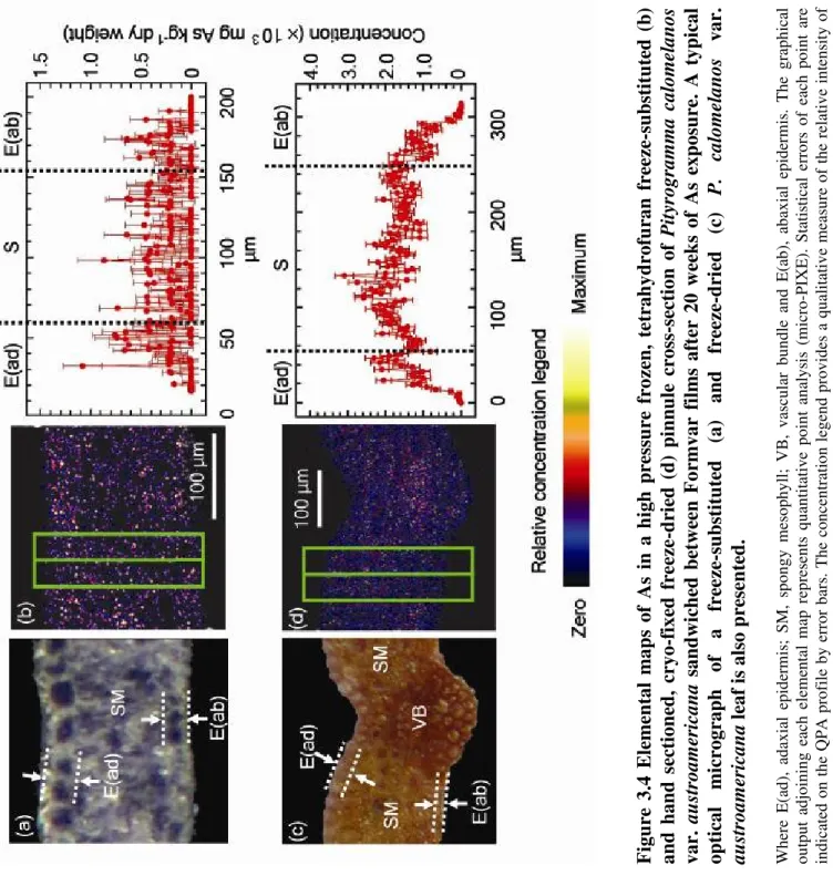
Ca and K determined by RSA were in agreement with bulk tissue concentrations, and followed the order K > Ca > As (Figure 3.2b). The concentration of As in freeze-substituted sections was almost 20-times lower than the bulk tissue concentrations, and K and Ca concentrations were 2–3 times lower than their corresponding bulk tissue concentrations (Figure 3.2b). In the freeze-dried samples, QPA revealed a peak As concentration of 3.1×10³ mg kg–1 DW in spongy mesophyll tissues; however in the freeze-substituted samples, a peak of 1.1×10³ mg kg–1 DW was observed in the adaxial epidermis (Figure 3.4). Arsenic concentration was significantly higher in the freeze-dried tissues than in the freeze-substituted tissues; however, the differences were non-significant between anatomical regions (Table 3.3).
F ig u re 3 .4 E le m en ta l m a p s o f A s in a h ig h p re ss u re f ro ze n , te tr a h y d ro fu ra n f re ez e-su b st it u te d ( b ) a n d h a n d s ec ti o n ed , cr y o -f ix ed f re ez e -d ri ed ( d ) p in n u le c ro ss -s ec ti o n o f P it yr o g ra m m a c a lo m el a n o s v a r. a u st ro a m er ic a n a sa n d w ic h ed b et w ee n F o rm v a r fi lm s a ft er 2 0 w ee k s o f A s ex p o su re . A t y p ic a l o p ti ca l m ic ro g ra p h o f a fr ee ze -s u b st it u te d (a ) a n d fr ee ze -d ri ed (c ) P . ca lo m el a n o s v a r. a u st ro a m er ic a n a le a f is a ls o p re se n te d . W he re E (a d) , ad ax ia l ep id er m is ; SM , sp on gy m es op hy ll; V B , va sc ul ar b un dl e an d E (a b) , ab ax ia l ep id er m is . T he g ra ph ic al ou tp ut a dj oi ni ng e ac h el em en ta l m ap r ep re se nt s qu an tit at iv e po in t an al ys is ( m ic ro -P IX E ). S ta tis tic al e rr or s of e ac h po in t ar e in di ca te d on t he Q PA p ro fi le b y er ro r ba rs . T he c on ce nt ra tio n le ge nd p ro vi de s a qu al ita tiv e m ea su re o f th e re la tiv e in te ns ity o f th e A s m ap pe d.
Table 3.3 Average concentration of elements (mg kg–1 DW) in Pityrogramma calomelanos var. austroamericana pinnule sections after freeze-drying and freeze-substitution with tetrahydrofuran.
Concentrations were obtained following dynamic analysis of individually selected regions. The results are presented as means ± SE of three replicates. Different letters within the same column indicate a significant difference (P≤0.05).
Minimum detection limit (MDL; mg kg–1 DW) of each measurement are also presented.
As Ca K
Technique Plant part analysed
mg kg–1 DW MDL mg kg–1 DW MDL mg kg–1 DW MDL Freeze-
dried Adaxial epidermis 3.1±1.0×10
3 a 14 7.0±2.1×10³a 47 2.5±0.6×10⁴a 65 Vascular tissue 4.1±0.5×10³a 18 6.3±2.1×10³a 62 3.7±0.8×10⁴a 86 Spongy mesophyll 4.1±1.0×10³a 12 4.5±2.3×10³a 39 3.3±0.8×10⁴a 50 Abaxial epidermis 3.2±0.9×10³a 15 6.6±2.2×10³a 51 2.3±0.5×10⁴a 71 Freeze-
substituted Adaxial epidermis 3.8±0.7×10
2 b 21 5.1±2.2×10³a 31 9.5±1.0×10³a 35 Vascular tissue 5.1±1.8×10²b 42 1.2±0.6×10⁴a 64 3.5±1.5×10⁴a 71 Spongy mesophyll 2.9±0.6×10²b 26 2.1±1.7×10³b 40 7.3±0.7×10³b 44 Abaxial epidermis 2.4±0.2×10²b 22 3.7±1.5×10³a 34 7.7±0.2×10³b 38
3.4 D
ISCUSSION
The µ-PIXE results of this comparative study clearly demonstrate that choice of sample preparation procedure is crucial for successful localisation and quantification of elements in the tissue of metal(loid) hyperaccumulating plants. For in vivo localisation and quantification
of elements, it is important to prevent or minimise the migration of ions from and within a tissue. The high sensitivity of µ-PIXE spectroscopy requires purity of samples and therefore incorrect sample preparation may produce artefacts and in turn erroneous results. Of the two sample preparation procedures investigated, plunge freezing specimens into a liquid cryogen (i.e. LN2) followed by freeze-drying produced results consistent with bulk tissue analysis; and
also prevented the redistribution of Ni, As and other elements in both H. floribundus subsp. floribundus and P. calomelanos var. austroamericana plant species. Moreover, cryofixation
of tissues once excised from plants was executed without delay, which further reduced the introduction of potential artefacts. In both species, the concentration of elements measured using the freeze-drying technique were statistically similar to the chemical analysis of the
bulk tissue using ICP-AES. The freeze-drying procedure is relatively simple, requires minimum sample preparation and the elemental distribution patterns obtained using this technique are consistent with those reported in studies of other Ni and As hyperaccumulating species. For example, Bhatia et al. (2004) observed highest Ni concentration in
hyperaccumulator Stackhousia tryonii epidermal and vascular tissues (5,400 mg Ni kg–1 DW)
that were more than 2-fold higher than concentrations recorded in palisade cells (2,500 mg Ni kg–1 DW). Similarly, As enrichment has been reported in hyperaccumulators Pteris cretica
var. nervosa and Pteris vittata mesophyll tissues (Chen et al. 2003; Chen et al. 2005). A
comprehensive discussion of elemental localisation and physiology of hyperaccumulation in
P. calomelanos var. austroamericana and H. floribundus subsp. floribundus freeze-dried
tissues is provided in Chapters 4 and 5, respectively.
A drawback of freeze-drying biological tissues is the low lateral resolution due to the thickness of the sections (ca. 50 µm) and the lack of spatial resolution at a subcellular level.
The removal of ice by sublimation during the freeze-drying process may result in the migration of soluble elements to the nearest solid surface, such as from a vacuole to the cell wall. However, it is unlikely to result in redistribution between cells and therefore mapping of tissues prepared using this technique is limited only to the cellular level.
Recently, Budka et al. (2005) investigated THF, diethyl ether, acetone and methanol as
possible freeze-substituting solvents for the localisation and quantification of Ni in leaves of hyperaccumulator Berkheya coddii following an extended freeze-substitution protocol
spanning 29 days. These authors demonstrated redistribution and loss of Ni from leaf tissue prepared using THF and indicated movement of Ni on the solvent front from the inner to the outer tissues by a quantitative Ni map. The duration of freeze-substitution is critical in order to reduce elemental redistribution and is inversely related to the solubility of water in the solvent at the substitution temperature (Pålsgård et al. 1994). As THF is highly soluble in
water, a shorter protocol was adopted which has been successfully used elsewhere (Pålsgård
et al. 1994; Pålsgård et al. 1996). In the present investigation, a shorter freeze-substitution
program did not appear to reduce elemental redistribution. These results are consistent with the observations made by Budka et al. (2005). Presumably, the leaching of elements may have
occurred during thawing of tissues and resin infiltration processes. Alternative sample preparation procedures, such as cryomicrotomy, may be followed in order to minimise
physiological disruption, thus preservation of elemental localisation. A review of suitable biological sample preparation protocols has been presented elsewhere (Kirby and Legge 1993).
3.3 C
ONCLUSION
The present study highlights the importance of specimen preparation for localisation and quantification of elements in metal(loid) hyperaccumulating plants using µ-PIXE spectroscopy. Concentration of elements in freeze-dried leaves of H. floribundus subsp. floribundus and pinnules of P. calomelanos var. austroamericana were comparable to the
results obtained following ICP-AES analyses. The freeze-drying technique ensured good preservation of the cellular structure and satisfactory lateral and spatial resolution, which enabled true elemental imaging and quantification of elements at a cellular level. Conversely, using THF as a freeze-substitution solvent resulted in the loss and migration of elements and misleading elemental images.