The perception of body vertically (subjective
postural vertical) in peripheral and central
vestibular disorders
A. R. Bisdorff, C. J. Wolsley, D. Anastasopoulos, A. M. Bronstein and M. A. Gresty
MRC Human Movement and Balance Unit, NationalHospital for Neurology and Neurosurgery, London, UK
Correspondence to: Dr Adolfo Bronstein, MRC Human Movement and Balance Unit, National Hospital for Neurology and Neurosurgery, 8-11 Queen Square, London WC1N 3BG, UK
Summary
The perception of body verticality (subjective postural vertical, SPV) was assessed in normal subjects and in patients with peripheral and central vestibular lesions and the data were compared with conventional neuro-otological assessments. Subjects were seated with eyes closed in a motorized gimbal which executed cycles of tilt at low constant speed (1.5° s~'), both in the frontal (roll) and sagittal (pitch) planes. Subjects indicated with a joystick when they entered
and left verticality, thus defining a sector of subjective uprightness in each plane. The mean angle of tilt (identifying a bias of the SPV) and the width of the sector (defining sensitivity of the SPV) were then determined. In normal subjects, the angle of the 'verticality' sector was 5.9° for pitch and roll. Patients with bilateral absence of vestibular function, patients with vertigo, i.e. acute unilateral lesions, benign paroxysmal positional vertigo (BPPV) and Meniere's disease, and patients with positionally modulated up-/ downbeat nystagmus all had enlarged sectors (i.e. loss in sensitivity). Mean sector angle in these groups ranged from 7.8 to 11° and the abnormality was present both in pitch and roll, regardless of the direction of nystagmus or body sway. Patients with chronic unilateral peripheral vestibular lesions
and those with position-independent vertical nystagmus had normal sensitivities. No significant bias of the SPV was found in any patient group, not even those with acute unilateral vestibular lesions who had marked tilts of the subjective visual vertical (SW). Complementary experiments in normal subjects tested under galvanic vestibular or roll-plane optokinetic stimulation also failed to show biases of the SPV. In contrast, a significant bias in the SPV could be induced in normal subjects by asymmetric cycles of gimbals tilt, presumably by proprioceptive adaptation. The following conclusions can be drawn, (i) The perception of body verticality whilst seated is mainly dependent on proprioceptive/contact cues but these are susceptible to tilt-mediated adaptation, (ii) Vestibular input improves the sensitivity of the SPV, even in vestibular disorders, as long as the abnormality is stable. (Hi) There can be marked dissociation between vestibulo-motor (ocular and postural) phenomena and the perception of body verticality, and between the SPV and SW. (iv) The postural sway asymmetries in patients with peripheral and central vestibular lesions, like those induced by galvanic or optokinetic stimulation in normal subjects, are not consequences of changes of the SPV. Keywords: spatial orientation; vestibular disorders; proprioception; visual vertical; postural vertical
Abbreviations: B = backward; BPPV = benign paroxysmal positional vertigo; F = forward; L = left; R = right; SPV = subjective postural vertical; S W = subjective visual vertical
Introduction
The functional neuro-anatomy of the vestibular system can be inferred from the symptoms arising from an acute unilateral peripheral vestibular lesion. These comprise the following: nystagmus due to asymmetrical function in the vestibulo-ocular reflex pathways; unsteadiness caused, in part, by © Oxford University Press 1996
vestibulo-spinal dysfunction; the turning sensation 'vertigo' mediated by vestibulo-cortical sensory projections; nausea due to autonomic involvement. Although these sensations are tightly clustered in the acute stage, it is a common clinical observation that the motor and perceptual sequelae
1524 A. R. Bisdorffet al.
of vestibular disease rapidly become dissociated in the recovery or chronic phase. Examples of the dissociation include the numerous patients who, having sustained a peripheral insult such as a vestibular neuritis, report enduring symptoms in spite of normal neuro-otological examination (Rudge and Bronstein, 1995). Conversely, although more frequent in central than in peripheral vestibular lesions, some patients with little or no vertigo may still have significant nystagmus.
Clinical tests of vestibular dysfunction focus largely on motor phenomena, despite reports that their specificity and sensitivity to disease is less than for perceptual tests of vestibular disorder (Brookes et al., 1993; Kanayama et al., 1995). This prompted us to study spatial orientation in patients with vestibular disease, in particular the SPV, and how this function compared with motor phenomena. The estimation of uprightness, which one could expect to be pivotal to both orientation in space and motor coordination, was the specific orientation task selected for study.
A priori, one would expect accurate perception of postural vertical to be important in the maintenance of upright stance and gait and, conceivably, that disordered perception may lead to clinical unsteadiness. Patients with bilateral absence of vestibular function have been reported to have a reduced ability to perceive uprightness of their body and this finding may well underlie the non-specific unsteadiness they experience (Guedry, 1974). However, larger groups of patients such as those with unilateral labyrinthine lesions or CNS disorders with vertical (up- or downbeating) nystagmus have never been examined and these would be particularly important since it has been suggested that the abnormal sway present in these patients may be related to their perception of body orientation in space (Biichele et al., 1983). Patients with acute unilateral vestibular lesions tend to sway more in the frontal plane and to veer to the side of their lesion on walking (Dichgans et al., 1976), particularly in the absence of vision. Similarly, patients with up- or downbeat nystagmus are reported to have increased sway in the sagittal plane (Biichele et al., 1983). Thus, the deviation of the body may correspond to an adjustment of body posture to a putative altered subjective vertical.
Patients and normal subjects
Normal subjects
Fifty-two normal subjects (26 male and 26 female, mean age 40.4 years, range 21-80) gave their informed consent to the study according to the guidelines of the local ethics committee. In addition to providing control data, the normal subjects also took part in experiments on somatosensory adaptation during optokinetic and galvanic stimulation; their details are given in the section concerning these experiments.
Patients
All patients gave their written informed consent to the study according to the guidelines of the local ethics committee. They were divided into the following seven groups.
Bilateral loss of vestibular function. These patients
(four males and four females, mean age 47 years, range 30-64) had bilateral loss of vestibular function defined by absent responses to caloric testing with 20°C and with rotational testing using 80° s~' velocity-steps in the absence of visual fixation. The aetiologies were meningitis (three), idiopathic (three), ototoxic drugs (one) and bilateral VIII-nerve section due to neurofibromatosis II (one). The duration of vestibular loss was s=2 years. On clinical examination, they had no additional neurological abnormalities.
Acute unilateral peripheral vestibular lesions. Eight
patients (four male and four female, mean age 44.1 years, range 26-62) with acute unilateral vestibular lesions were investigated. Six patients had undergone vestibular neurec-tomy for unilateral Meniere's disease or secondary hydrops. One patient with vestibular neuritis, and one with a labyrin-thine infarct during surgical VH-nerve decompression for hemifacial spasm, were also tested. The time between the acute event and testing was 3-7 days. The clinical signs on the day of testing were spontaneous nystagmus towards the healthy side, sway towards the lesion on Romberg testing and deviation to the side of the lesion on walking with eyes closed.
Chronic stable unilateral peripheral vestibular
lesions. Six asymptomatic patients (four male and two female, mean age 37.5 years, range 26-45) with long-standing complete unilateral lesions after VHI-nerve sections were tested. The indications for the operation had been acoustic neuroma (four), Meniere's disease (one) and VHI-nerve astrocytoma (one).
Benign paroxysmal positional vertigo (BPPV).
These nine patients (three male and six female, mean age 54.3 years, range 41-72) had typical signs and symptoms on Hallpike's manoeuvre and had been symptomatic for at least several weeks before testing. In eight cases the BPPV was idiopathic and in one it appeared after an episode of labyrinthitis. Two of the patients had mild asymmetries of vestibular responses on rotational testing.
Meniere's disease. These 12 patients (nine male and three female, mean age 44.3 years, range 26-59) had frequent and/ or severe attacks of vertigo and were evaluated for surgical treatment. Eleven patients had the diagnosis of Meniere's disease and one of delayed hydrops after acquiring deafness at the age of 18 months. The tests were performed between attacks, in a symptom-free interval. All patients underwent rotational testing with a velocity-step paradigm of 60° s"1. In 11 cases the directional preponderance of the nystagmus was <15% and in one case 37%. Nine patients underwent caloric testing: two had canal pareses of 44% and 100% and seven had percentage asymmetries of =£12%.
eight female, mean age 47.1 years, range 19-77), six had upbeat and nine had downbeat nystagmus. The aetiologies were idiopathic (six), cerebellar degeneration (three), Arnold-Chiari malformation (two), post encephalitic (one), Vitamin B) deficiency (one), postoperative shunt for aqueduct stenosis (one) and brainstem cavernoma (one). On neurological examination six patients were ataxic on stance and gait, the other nine had no neurological abnormality except for their eye-movement disorder.
Five patients had no position-dependent modulation of their nystagmus. Three patients showed persistent changes in the intensity of their nystagmus when adopting different head positions (positional modulation). Seven had a transitory increase of their nystagmus on positional testing (positioning modulation), usually accompanied by vertigo. These modulations were exclusively dependent on head position or movement because these findings were reproducible when neck movement was avoided during the position change. Positional effects are likely to be otolith-mediated whereas positioning effects are more likely to be semicircular canal-mediated. However, a dynamic otolithic contribution is also possible.
Longitudinal study of peripheral vestibular disease.
Three male patients (aged 36, 37 and 47 years) and one female patient (aged 25 years) with severe and/or frequent vertiginous attacks because of Meniere's disease were tested before, and during the first week after unilateral vestibular nerve section. Patients were in a symptom-free interval preoperatively. At the time of testing postoperatively they had vertigo, nystagmus, a directional bias of sway to the side of their lesion on Romberg testing and a veering in that direction on walking with their eyes closed. The preoperative SPV and SVV tests were done on the same day, as were the postoperative tests.
Methods
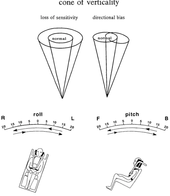
The subjects were seated in a padded chair in a motorized gimbal with the head and torso restrained and their eyes closed (Fig. 1). The gimbal was externally controlled and it executed 7-10 cycles of 15° tilt to either side around the vertical at 1.5° s"1 constant velocity in pitch and, after a few minutes rest, the same in roll. This slow velocity does not provide significant semicircular canal input (Benson et al., 1989). The (machine) vertical was defined when the plane of the seat was orthogonal to the gravitational vertical. Subjects were simply asked to indicate when they entered and left the upright position, rather than a single position of uprightness. (The actual instruction invited subjects to use all available body sensations: 'indicate as soon as you start to feel upright and as soon as you start to feel tilted again'). This protocol would therefore identify 'uprightness' as perceived within a zone of tilt ('cone of verticality', Fig. 1). Subjects started in a 15° tilt position to the right or back
and indicated when they began to feel upright, and again when they began to feel tilted, with an analogue, three-position joystick. The subjects were not informed about their starting position, or that the cycles would be equal during the whole procedure and they did not get any feedback about their performance. With this paradigm a sector of subjective verticality was determined during motion from forwards to backwards (F-»B) and from backwards to forwards (B->F) in pitch, and during motion from the left to right (L—>R) and right to left (R—>L) in roll. If a subject signalled that they felt they were tilted at an angle of > 10° the gimbal continued in that direction for an additional 5° before the direction was changed. In this case, the same amount of tilt was performed in the opposite direction in this particular cycle to keep the time spent on either side of vertical identical, i.e. to avoid the possibility of inducing a bias by asymmetrical exposure to tilt. Adaptation (see below) was avoided by employing cyclic symmetric tilts about upright and rejecting the data of the first cycle.
It might have been theoretically possible for the subjects to derive verticality perception through rational pondering (e.g. detecting the turning points in the cycles and assuming that they were moving equal lengths of time in each direction and finally calculating the half-time in the cycle). However, there was no evidence that this might be the case: subjects in our experiments were not told they would stay for equal lengths of time in each direction Pilot experiments randomizing angles of tilt achieved similar results to those with our standard protocol. Finally, subjective reports from normal subjects and patients indicated that estimates of verticality during these slow movements were based on compelling bodily sensations rather than on rational estimates.
Tilt-induced adaptation of the SPV
To test this effect, eight normal subjects (two male and six female, aged 16-40 years, mean 25.8, SD = 7.8) were tested in roll with the centre of the cycles of movement offset by 0°, 5° and 10° and the amplitude of the cycles preserved. Four subjects were allocated to an offset to the left and four to an offset to the right; the sequence of the different offset angles was allocated according to a latin square. To induce an offset of say 10° to the right, subjects started from upright and then they were tilted 15° to the right; they were then tilted 5° to the left, 25° right and from then on oscillated between these two points. The first cycle was rejected and the following eight measured. For presentation the data were normalized as if for an offset to the right.
Effect of full field optokinetic stimulation
A motor driven cone, 56 cm in diameter and 23.5 cm depth, with fluorescent stripes spaced every 45° on black background was mounted in front of the subject at a distance of 30 cm from the nasion. The cone subtended -160° of the visual
1526 A. R. Bisdorffet al.
cone of verticality
loss of sensitivity directional bias
roll
pitch
10
Fig. 1 Schematic drawing illustrating the method used to investigate the SPV in roll and pitch planes
(bottom). The SPV sector was determined for each half cycle. The underlying assumption for this
procedure is that subjective verticality is confined to a 'cone' in space (top). The abnormalities expected are a loss of sensitivity, a directional bias or combinations of both.
field. Subjects were tested in dark surroundings, first while fixating the centre of the static disc and then while the disc was spinning counterclockwise at 60° s~' during 10 cycles of tilt for an approximate total time of 6 min. Preliminary trials showed that this velocity of rotation gave the strongest sensation of circularvection and the largest clinically observable sway to the left in most subjects whilst standing, and it is similar to the speed used by previous investigators (Dichgans et al., 1972). Ten normal subjects (seven male and three female aged 21-48 years, mean 36.4 years, SD = 10.2 years) were tested and subsequently questioned about their subjective impressions.
Effect of galvanic stimulation
A constant 1 mA current was passed between two surface electrodes placed on the mastoids. During the tilt cycles the current was switched on automatically during the half cycle from R—>L motion starting at a 6° right tilt and lasting for 10 s. This timing was chosen so that subjects would perform both judgements (entering and leaving the vertical) during galvanic stimulation. About 20 cycles were executed and the stimuli were randomly applied: anodal stimulation on the right, anode on the left and no current. Seven subjects were tested (four male and three female, mean age 35.6 years, range 23-48).
On a separate occasion five of these subjects also undertook estimates of the SVV during galvanic stimulation. Whilst in the gimbal, in complete darkness, they had to align a luminous rod of 18.5 cm length placed at 40 cm from the nasion, with the gravitational vertical. The galvanic stimulus was switched on when the subjects were asked to open their eyes and reset the rod, and switched off when the subjects signalled that they had finished. No time limit was given but most judgements were performed well within 10 s. The two directions of polarization and the 'no stimulus' condition were pseudo-randomized with equal frequency of presentation. A total of 15 SVV measurements were obtained.
Before either of these tests was performed, the effect of the electrical stimulus was checked on each subject while they were standing with eyes closed; it consistently induced sway in the direction of the anode. The subjects were questioned about subjective impressions of sway.
Visual vertical measurements
The patients of the longitudinal study viewed a randomly tilted luminous straight line in an otherwise dark room and adjusted its orientation by remote control until it appeared earth vertical (SVV). Twelve settings were made for random displacements in right and left tilts. There were no time constrains and, in between settings, subjects closed their eyes whilst the line was arbitrarily positioned away from vertical by the experimenter. The head was held upright with a chin rest while seated upright.
Data analysis
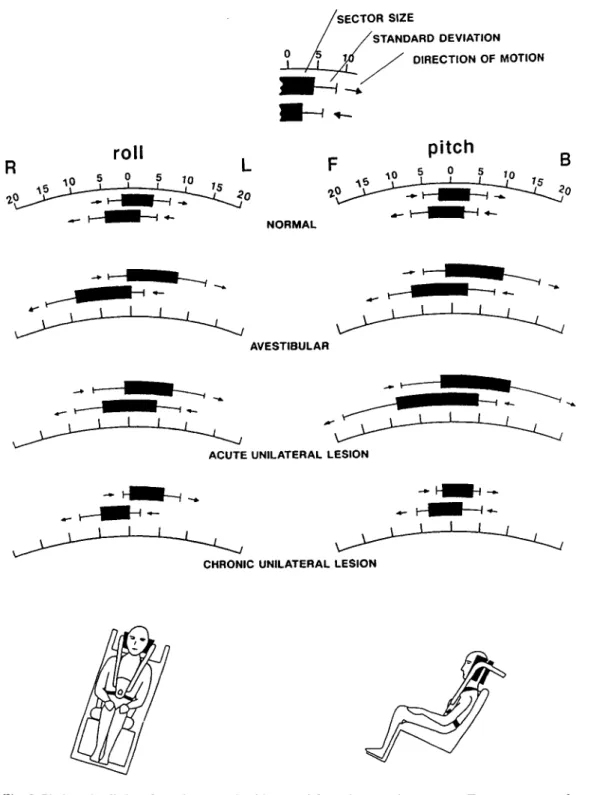
After rejection of the first cycle of tilt, the angles of entering and exiting vertical for each subsequent cycle were averaged to give a mean position for the SPV (a negative value indicates a backwards or leftwards mean SPV, i.e. a negative bias or 'lean' to the SPV). The width of the sector is a parameter for the sensitivity of perception of uprightness (see inset in Fig. 2). This is defined as the absolute difference between the angle of entering upright and the angle of exit, and was similarly averaged over cycles of tilt. Since sector angle was similar for F—>B and B—>F tilts and R—>L and L—»R tilts the overall means are reported here.
In groups of patients with unilateral lesions, the roll data were normalized for left lesions. Statistical comparisons were carried out with both the two-tailed / test and non-parametric statistics (Mann-Whitney and Wilcoxon) in all cases. There were no differences between the outcome of these two analyses and significance values given are those of the t test.
Results
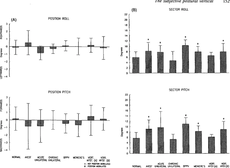
Figure 2 shows the results from normal subjects and three patient groups, plotted separately to show different planes and direction of motion. Figure 3A shows mean results of position of the SPV ('bias' or 'lean' of the SPV) and
Fig. 3B, mean sector angles ('sensitivity'). Statistics are presented in Table 1.
Normal controls
In normal subjects, the width of the sector of the SPV in pitch was 5.8±2.P and in roll 5.9±2.1°. It can be seen in Fig. 2 that the verticality sectors were shifted in the direction of gimbal's motion, i.e. they were not positioned symmetrically around vertical. This aspect of the postural vertical will be discussed in a future publication. For the purpose of this study, we have combined both directions of motion, thus reporting results as mean position of the SPV. For normal subjects, the mean SPV was 0.16± 1.55° in pitch and 0.12±0.95° in roll.
The magnitudes of the sectors in roll and pitch correlated (r = 0.796, P < 0.0001). There was a slight increase of the sectors with age (for roll r = 0.347, P = 0.02 and for pitch r = 0.473, P < 0.0001), i.e. there is a loss of sensitivity with age but this effect was small compared with the inter-individual variability.
Patients
Bilateral vestibular loss
These patients had significantly larger sectors in roll and pitch, i.e. a non-specific loss of sensitivity. The angle at which they entered subjective vertical was not different from normal but the tilt angle at which they left subjective vertical was larger (compare the top two data sets, 'normal' and 'avestibular', in Fig. 2). The mean position of the SPV was not different from that in normals.
Acute unilateral peripheral lesions
The patients with acute unilateral vestibular lesions had enlarged sectors in both planes. In contrast to those of avestibular patients, their sectors of verticality widened in both directions, i.e. they entered subjective verticality earlier and left it later than the normals. There was no bias in the mean position of the SPV in pitch but for roll there was a tendency for a bias in the direction of the lesion (with left normalization, mean position in normal subjects = -O.12±O.95° and in patients, -0.84±0.91°; P = 0.051 in the two-tailed t test).
On preliminary testing assessment, before the test, these patients swayed or veered in the direction of their lesion. When questioned, the subjective perception of sway direction matched the actual direction of sway.
Chronic unilateral peripheral vestibular lesions
The patients with complete long-standing unilateral lesions after VHI-nerve section had normal sectors in pitch and in both roll, and they had no directional bias.
1528 A. R. Bisdorffet al. SECTOR SIZE STANDARD DEVIATION DIRECTION OF MOTION
pitch
NORMAL AVESTIBULARACUTE UNILATERAL LESION
J L
CHRONIC UNILATERAL LESION
Fig. 2 Pitch and roll data from the normal subjects and from three patient groups. Two sectors, one for each hemicycle, are shown in each example. Since there were no significant differences overall between the data from the two hemicycles, pooled data were used in the subsequent analyses, normalized for movement direction and asymmetrical pathology when appropriate.
Vertical nystagmus
The patients with vertical nystagmus had enlarged sectors in both planes but no directional bias. Separate evaluation of up- and downbeat patients did not reveal a directional bias of the mean SPV, in particular, not in the pitch plane.
The presence of positional modulation of the nystagmus had an effect on the SPV estimates (Table 1). The patients
without modulation were normal. The group of patients with positioning or positional modulation had enlarged sectors in both planes.
Benign paroxysmal positional vertigo (BPPV)
These patients did not experience their vertigo during this experimental procedure. They had a loss of sensitivity, i.e.
(A)
- 2
POSITION ROLL
(B) SECTOR ROLL
1529
POSITION PITCH SECTOR PITCH
3 8 1 2 1 o £-1 - 2
NORMAL AVEST ACUTE CHRONIC BPPV MENIERE'S VERT. VERT UNILATERAL UNILATERAL NYST. (A) NYST. ( B )
» : NOT POSITION MODULATED B : POSTCW UOOULATED
ACUTE CHRONIC VERT. VERT. NORMAL AVEST UNILATERAL UNILATERAL 8PPV MENIERE'S NYST.(A) NYST.(B)
Fig. 3 Mean sector positions (A) and mean sector angles (B) for normal subjects and the different patient groups in pitch (sagittal) and roll (frontal) planes, AVEST = bilateral loss of vestibular function; ACUTE/CHRONIC UNILATERAL = acute/chronic unilateral loss of vestibular function; VERT. NYST. = vertical nystagmus. *Different from normal subjects at P < 0.05 (see Table 1 for details).
Table 1
Subjects
Summary of results (n)
in normals and patients Sector angle Pitch (°) Roll (°) Position Pitch (°) Roll (°) Normals (52) Patients Avestibular (8) Acute unilateral (8)* Chronic unilateral (6)* BPPV (9)* Meniere's disease (12)* Vertical nystagmus A (5) Vertical nystagmus B (10) 5.8+2.0 9.2±3.3 (P < 0.001) 9.7±5.9 (P = 0.001) 5.3±1.9 (n.s.) 11.0+2.3 (P < 0.001) 8.3±2.O (P = 0.001) 6.2+0.9 (n.s.) 9.0±2.9 (P < 0.001) 5.9±2.1 8.2 + 3.2 (P = 0.01) 7.8+2.5 (P = 0.027) 4.7±3.4 (n.s.) 10.3+2.0 (P < 0.001) 7.9+2.0 (P = 0.004) 6.5+1.2 (n.s.) 7.9+3.0 (P = 0.016) 0.2±1.5 -0.8±3.0 (n.s.) -0.8±2.2 (n.s.) 0.0±1.2 (n.s.) -0.4±1.3 (n.s.) -0.6+1.5 (n.s.) 0.5+1.5 (n.s.) 0.2±1.5 (n.s.) -0.1 ±1.0 0.5+1.6 (n.s.) -0.8±0.9 P = 0.051 -0.3±0.6 (n.s.) 0.1 ±1.3 (n.s.) 0.0±l.l (n.s.) 0.2±0.9 (n.s.) -0.2±1.5 (n.s.) A = nystagmus was not position-sensitive; B = nystagmus was position sensitive; n.s. = not satistically significant. *Data normalized for left lesions.
1530 A. R. Bisdorff et al.
L
preop postop
Fig. 4 Pre-operative and (1 week) postoperative positions of the subjective postural vertical (SPV) and visual vertical (SVV) in four patients who underwent vestibular neurectomy (courtesy of Mr G. Brookes and Mr P. Riordan Eva). Data normalized as for a left-sided operation.
enlarged sectors in both planes, but no directional bias in either plane.
Meniere's disease
These patients had enlarged sectors in both planes and no directional biases. There was no relationship between the degree of canal pareses or directional preponderance of rotational nystagmus responses, and the mean SPV or sector width.
Longitudinal, post-vestibular neurectomy study
These five patients were tested before, and during the first week after, neurectomy. Their tilt estimates had not changed post-operatively. However, their SVV changed significantly with an average tilt of 8.5 ± 1.4° in the direction of the lesion (Fig. 4).
Complementary experiments
Adaptation to asymmetric tilt and the effects of optokinetic and galvanic stimulation was studied in the normal subjects {see summary of results in Table 2)
Adaptation induced by cycle offset
After offsetting the centre of the cycles, a significant bias in the same direction was observed (2.1° for 5° offset and 5° for 10° offset, see Fig. 5). The bias induced in the mean position of the SPV was larger than any seen in any patients.
Table 2 Summary of the results of the experiments
manipulating visual, vestibular and somatosensory input to the perception of body vertical
Mean sector width (°) Optokinetic stimulation
Static cone Spinning cone Effect of cycle offset
0° 5° 10° Galvanic stimulation* No stimulation Anode right Anode left 5.1 + 1.4 5.8+1.6 (n.s.) 6.2±2.9 4.9±1.8(n.s.) 7.2±2.9 (n.s.) 4.3±0.9-1.6±1.4 5.0±l.l (n.s.) 4.8±l.°(n.s.) Mean sector position (°) 0.3±0.8 -0.1 + 1.8 (n.s.) -0.4±0.9 2.1±1.7 (P = 0.003) 5.0±2.6 (P = 0.02) -1.6±1.4 (n.s.) -2.2±1.2(n.s.) n.s. = not satistically significant. *Values from the hemicycle during which stimulation was applied, i.e. during movement from right to left. o — 7 « 6 -o | 5 : — 4
1 3
-o a. 2 -0)1 1
.5 0 -CO 1 -o /Y
- /
/ • i 1 1 -5 10 15 offset (°)Fig. 5 Data from eight normal subjects showing the effect of offseting tilt cycles, namely a bias of the SPV in the direction of the offset.
The first cycle was rejected and monitoring the SPV over the next eight consecutive cycles did not show a time-dependent effect. The fluctuation of the SPV in one individual over testing time was smaller than the variation across the group. This suggests that the main adaptation to tilt about an offset centre occurs rapidly, within the first minute. There was no significant change in the sector angle (Table 2).
Optokinetic stimulus
The mean position of the SPV with the cone static for all subjects was 0.3 ±0.8° and not significantly different from that when the cone was rotated counterclockwise (-0.1 +1.8°).
On further anlysis, there was a non-significant tendency to report entry into 'uprightness' at a higher angle when moving from left to right than when coming from right to left. There was a non-significant increase in mean sector widths with cone rotation (static 5.1 ±1.4°; dynamic 5.8±1.6°). Even though the SPV estimates were normal, subjects reported circularvection and disorientation during the test.
Galvanic stimulation
Subjective visual vertical. Without galvanic stimulation the judgements of the SVV were accurate (0.27±0.40°). The galvanic stimulus tended to induce a tilt of the SVV in the direction of the anode (anode right, 0.57±0.31°; anode left, 0.04±1.43°) but this effect was small and not statistically significant.
Subjective postural vertical. Sector widths increased (a
decrease in sensitivity) when the subjects had to make their judgement during galvanic stimulation compared with the control situation. This increase was small but reached statistical significance (no stimulus, 4.27±0.89°; anode right, 5.03± 1.07°, P < 0.05). The effects on the mean SPV during the hemicycle when the galvanic stimulation was applied were not significant. During the pre-test assessment, subjects correctly reported sway in the direction of the anode when stimulated while standing.
Discussion
The main findings of this study can be summarized as follows. (i) The abnormality found in patients was an increase in sector width of the SPV, i.e. a loss of sensitivity for the perception of body vertically. This was observed in patients with vertigo (acute unilateral lesions, BPPV, Meniere's disease), patients with positionally modulated up-/downbeat nystagmus and patients with absent vestibular function. The degree of abnormality was similar across these groups. Patients with chronic unilateral lesions and patients with position-insensitive vertical nystagmus showed normal results.
(ii) The loss of sensitivity, when present, was observed in both sagittal and frontal planes, regardless of the plane or direction of veering or nystagmus.
(iii) Directional sway, veering or nystagmus was not reflected in a directionally biased SPV in any of the patient groups.
(iv) The SVV and SPV were profoundly dissociated after vestibular neurectomy.
(v) Stimuli which induced directional sway and orientation illusions in normals (galvanic and optokinetic stimulation) did not induce consistent changes in the SPV.
(vi) The only significant bias of the SPV observed was the one induced in normal subjects by offsetting the cycles of tilt asymmetrically around the real vertical.
Sensory inputs controlling the perception of
body verticality
The afferent information upon which estimates for uprightness are based in this test consists primarily of somatosensory and otolithic 'graviceptive' signals. We found that subjects without vestibular function showed a mild loss of sensitivity, as shown by increased sector width, but they had a normal mean SPV. This suggests that proprioception alone may be sufficient for a reasonable estimate of uprightness but that 'reliable' vestibular input increases sensitivity. This is in line with previous findings showing that estimates of uprightness in vestibular defective subjects have higher variance than those in normal subjects (Clark and Graybiel, 1963) and that reduction of proprioceptive cues by whole body compression or immersion in water impairs estimates, particularly in avestibular subjects (Padden, 1959; Brown, 1961; Graybiel et al., 1968; Nelson, 1968).
Adaptation of the SPV is shown by the study in which the envelope of tilts was vertically asymmetrical; the SPV deviates in the direction of the greater tilt. In fact, our experiments show that a more marked bias of the SPV can be induced by asymmetric tilt than with galvanic or optokinetic stimulation or vestibular disease. Adaptation of otolith effects has not been shown even after prolonged centrifugation, as judged by the subjective visual horizontal (Clark and Graybiel, 1962). Adaptation of body vertical is also present in avestibular subjects and tends to be even more pronounced than in normals (Clark and Graybiel, 1964) suggesting that it is a property of the proprioceptive input to SPV estimates. It is perhaps surprising that otolithic information is not able to counteract proprioceptive-mediated adaptation effects fully. This may be partly due to the fact that subjects were tested restrained and immobile, as it has been observed in studies underwater that estimates of upright improve if subjects are allowed to move actively (Brown 1961).
It should be noted that the effect induced by asymmetrical tilt was restricted to the mean SPV without changing sector angle (i.e. creating a bias without reducing sensitivity). This is just the opposite to the abnormality found in vestibular patients. It can be concluded that a healthy or, at least, stable vestibular input {see below) is required for normal sensitivity in the perception of body verticality. However, detection of body tilt seems to be heavily dependent on proprioceptive-contact cues. The mechanisms by which adaptation-induced biases are counteracted in everyday life are not well established but it is possible that active movements play an important part.
Postural and visual vertical
Our patients with VHI-nerve section had large ipsilateral deviations of the SVV immediately after operation and yet no concomitant tilt of their SPV. Other investigators in unilateral vestibular disease have reported that, although a luminous rod was set tilted in the dark, none of the patients
1532 A. R. Bisdorff et al.
reported to feel tilted while sitting upright (Dai et al., 1989; Curthoys et al., 1991). In more dramatic reports of 90° tilts (Hagstrom et al., 1969) or even inversions of the visual world (Charles et al., 1992; Stracciari et al, 1993) in cerebellar and cerebral disease a simultaneous feeling of body tilt was not described even on specific questioning (Solms et al., 1988). Similarly during optokinetic stimulation, which is known to induce large tilts of the SVV (Dichgans et al., 1972, 1975), our subjects did not experience a consistent tilt of the SPV, in spite of the intense concurrent circularvection. However, Dichgans et al. (1972) found a bias of the SPV in the direction of an optokinetic stimulus when pilots were required to defend their upright orientation against random perturbations. In this study, however, the effects noticed on the SPV were smaller than on the SVV and the testing conditions were different from those in our study. Specifically, subjects in the Dichgans et al. (1972) study had active control of the flight simulator whereas our subjects were seated immobile, engaged in a more purely perceptual task.
Therefore, from results in patients and normal subjects under vestibular or optokinetic stimulation, one concludes that the SPV remains largely veridical despite concurrent perceptions of vertigo, circularvection and SVV tilts. In the light of dissociations between the SVV and SPV, we must assume different mechanisms and pathways subserving these two perceptions. This agrees with Mittelstaedt's observations (e.g. Mittelstaedt, 1988) that normal subjects can adjust their body to the horizontal accurately but fail to set the SVV veridically in that body position. The SVV can be critically influenced by vestibulo-ocular (Curthoys et al., 1991; Dieterich and Brandt 1992), ocular (Balliet and Nakayama, 1978) and somatosensory mechanisms, e.g. by tilt-mediated effects which are smaller in deafferented patients (Yardley, 1990) and larger in patients without vestibular function (Miller et al., 1968; Bronstein et al., 1996). Thus, tilts of the SVV cannot be solely ascribed to tonic vestibular asymmetry. The mean SPV, on the other hand, is more refractory to visuo-vestibular stimulation and dysfunction, even in the acute stage {see below). In addition, the emerging role of visceral sensing of gravitational forces acting on the body will have to be considered in future studies (Mittelstaedt, 1992).
Postural vertical and sway
Patients with Meniere's disease tested with a stepping test with eyes closed deviate to the side of the lesion (Okubo et al., 1986) as do patients with acute unilateral lesions (Dichgans et al., 1976). In this study, no formal posturography was necessary, since the lateral veering in patients with acute unilateral lesions and normals during galvanic/optokinetic stimuli was a marked clinical feature. Our patients with acute unilateral lesions also swayed away from the intact side and when questioned, reported their veering or sway direction correctly, as did normal subjects during galvanic or
optokinetic stimulation. If their sway were a postural adjustment to abnormally tilted perception of verticality, they should have felt offset from vertical while standing upright. This was not reported, in agreement with the lack of a significant bias of the the SPV.
Galvanic stimulation induces sway and nystagmus with slow phase in the direction of the anode (Pfaltz, 1970; Coats, 1972, 1973). Our subjects did not report that they had adjusted their posture in response to galvanic stimulation, e.g. because of perception of tilt induced by the onset of the stimulus. The effect of galvanic stimulation is brought upon by short latency (40-100 ms) vestibulo-spinal reflexes which can be modulated by the initial postural set, e.g. by different head-on-trunk positions (Nashner et al., 1974; Britton et al., 1993). It cannot readily be explained how such short latency responses in the limbs could result from, and be modulated by, a higher order perceptual change. These findings suggest that the sway induced by galvanic stimulation and by acute unilateral peripheral vestibular disease is a primary vestibulo-spinal motor response, the consequence of which is subsequently perceived. The perception of body verticality itself is undisturbed and cannot be held responsible for the abnormal sway.
The SPV in vestibular disease
Patients with chronic unilateral peripheral lesions and patients with constant amplitude vertical nystagmus during the tilt procedure showed no abnormality of the SPV. They have in common that their vestibular input, albeit abnormal, does not fluctuate. In contrast, patients with continuous or recurrent vertigo (recent vestibular nerve sections, BPPV, Meniere's disease) or positionally modulated up-/downbeat nystagmus, had an increase of sector angle. This abnormality was, however, not plane-specific. The significance of this finding lies in the fact that their abnormalities were similar to those of subjects with bilateral vestibular loss, which suggests that they tended not to utilize vestibular input in making estimates of the SPV. This could represent a strategy to protect the spatial orientation system against fluctuating or unreliable vestibular signals. It is interesting, however, that in patients with vertigo, complaints of continuous feelings of off-balance can be present, to varying degrees, in between attacks. It can be speculated that such symptoms partly depend on the loss of an accurate sense of verticality in the postural system, as identified in this study.
The dissociations shown experimentally and observed in our patients between vestibular and proprioceptive perceptions of orientation are typical experiences of patients with vestibular disease and they may lead to such bizarre sounding symptoms that in the differential diagnosis a functional disorder is often considered (Gresty et al., 1992). Patients typically report sensations of continuous self-motion or of floating of the support surface and yet they have no clinical signs. This apparent paradox becomes understandable when one considers that conflicting perceptual estimates of
orientation may coexist. A scheme of 'orientation in space' can be mediated relatively independently by visual, vestibular or proprioceptive channels. In the various experimental situations described above, the subject can experience the co-existence of contradicting perceptions. When pushed to make a realistic appraisal of his orientation, the sensory channel(s) which, from experience, appear more veridical, are selected for the task. With the type of patients reported in this study, namely patients with vestibular problems, or in normal subjects under disorienting visuo-vestibular stimulation, proprioception predominates. However, patients with somatosensory loss may give more weight to vestibular cues. A similar 're-weighting' process also operates in the postural system, where postural responses to unstabilizing sensory stimuli, such as slowly moving visual scenes, can be suppressed if subjects have reliable proprioceptive information from the lower limbs (Bronstein, 1986; Bronstein
et al., 1990). Indeed, such re-weighting may be a requisite
underlying compensation for the postural and perceptual effects of acute balance disorders (Bles etai, 1983; Bronstein, 1995). This scheme, however, may not apply to acute, focal brainstem lesions with marked lateropulsion, e.g. lateral medullary syndrome. In this case a tighter association between motor and perceptual aspects may exist, but only the SVV, not the SPV, has been measured in such patients (Friedman, 1970; Dieterich and Brandt, 1992). However, as shown in this paper, studies of the visual vertical are not necessarily representative of other aspects of spatial orientation.
Acknowledgements
Financial support from the CEC Human Capital and Mobility Program (Network of Fellows and Access to Large Scale Facilities) and from the Ministry of Cultural Affairs (Luxembourg) is gratefully acknowledged.
References
Balliet R, Nakayama K. Egocentric orientation is influenced by trained voluntary cyclorotary eye movements. Nature 1978; 275: 214-6.
Benson AJ, Brown SF. Visual display lowers detection threshold of angular, but not linear, whole-body motion stimuli. Aviat Space Environ Med 1989; 60: 629-33.
Bles W, Vianney de Jong JM, de Wit G. Compensation of labyrinthine defects examined by the use of a tilting room. Acta Otolaryngol (Stock) 1983; 95: 576-9.
Britton TC, Day BL, Brown P, Rothwell JC, Thompson PD, Marsden CD. Postural electromyographic responses in the arm and leg following galvanic vesiibular stimulation in man. Exp Brain Res
1993; 94: 143-51.
Bronstein AM. Suppression of visually evoked postural responses. Exp Brain Res 1986; 63: 655-8.
Bronstein AM. Visual vertigo syndrome: clinical and postural findings. J Neurol Neurosurg Psychiatry 1995; 59: 472-6.
Bronstein AM, Hood JD, Gresty MA, Panagi C. Visual control of balance in cerebellar and parkinsonian syndromes. Brain 1990; 113: 767-79.
Bronstein AM, Yardley L, Moore AP, Cleeves L. Visually and posturally mediated tilt illusion in Parkinson's disease and in labyrinthine defective subjects. Neurology 1996. In press. Brookes GB, Gresty MA, Nakamura T, Metcalfe T. Sensing and controlling rotational orientation in normal subjects and patients with loss of labyrinthine function. Am J Otol 1993; 14: 349-51. Brown JL. Orientation to the vertical during water immersion. Aerospace Med 1961; 32: 209-17.
Biichele W, Brandt T, Degner D. Ataxia and oscillopsia in downbeat-nystagmus vertigo syndrome. Adv Otorhinolaryngol 1983; 30: 291-7.
Charles N, Froment C, Rode G, Vighetto A, Turjman F, Trillet M, et al. Vertigo and upside down vision due to an infarct in the territory of the medial branch of the posterior inferior cerebellar artery caused by dissection of a vertebral artery. J Neurol Neurosurg Psychiatry 1992; 55: 188-9.
Clark B, Graybiel A. Visual perception of the horizontal during prolonged exposure to radial acceleration on a centrifuge. J Exp Psychol 1962; 63: 294-301.
Clark B, Graybiel A. Perception of the postural vertical in normals and subjects with labyrinthine defects. J Exp Psychol 1963; 65: 490-4.
Clark B, Graybiel A. Perception of the postural vertical following prolonged bodily tilt in normals and subjects with labyrinthine defects. Acta Otolaryng (Stockh) 1964; 58: 143-8.
Coats AC. The sinusoidal galvanic body-sway response. Acta Otolaryngol (Stockh) 1972; 74: 155-62.
Coats AC. Effect of varying stimulus parameters on the galvanic body-sway response. Ann Otol Rhinol Laryngol 1973; 82: 86—102. Curthoys IS, Dai MJ, Halmagyi GM. Human ocular torsional position before and after unilateral vestibular neurectomy. Exp Brain Res 1991; 85: 218-25.
Dai MJ, Curthoys IS, Halmagyi GM. Linear acceleration perception in the roll plane before and after unilateral vestibular neurectomy. Exp Brain Res 1989; 77: 315-28.
Dichgans J, Held R, Young LR, Brandt Th. Moving visual scenes influence the apparent direction of gravity. Science 1972; 178:
1217-9.
Dichgans J, Brandt Th, Held R. The role of vision in gravitational orientation. Fortschr Zool 1975; 23: 255-63.
Dichgans J, Mauritz KH, Allum JHJ, Brandt Th. Postural sway in normals and atactic patients: analysis of the stabilising and destabilising effects of vision. Agressologie 1976; 17 (c spec No):
15-24.
Dieterich M, Brandt Th. Wallenberg's syndrome: lateropulsion, cyclorotation, and subjective visual vertical in thirty-six patients. Ann Neurol 1992; 31: 399^108.
1534 A. R. Bisdorffet al.
with peripheral and central vestibular lesions. Brain 1970; 93: 313-28.
Graybiel A, Miller EF, Newsom BD, Kennedy RS. The effect of water immersion on perception of the oculogravic illusion in normal and labyrinthine defective subjects. Acta Otolaryngol (Stockh) 1968; 65: 599-610.
Gresty MA, Bronstein AM, Brandt Th, Dieterich M. Neurology of otolith function, peripheral and central disorders. [Review]. Brain
1992; 115: 647-73.
Guedry FE. Psychophysics of vestibular sensation. In: Komhuber HH, editor. Handbook of sensory physiology, Vol. VI/2. Berlin: Springer-Verlag 1974: 3-154.
Hagstrbm L, Hornsten G, Silfverskiold BP. Oculostatic and visual phenomena occurring in association with Wallenberg's syndrome. Acta Neurol Scand 1969; 45: 568-82.
Kanayama R, Bronstein AM, Gresty MA, Brookes GB, Faldon ME, Nakamura T. Perceptual studies in patients with vestibular neurectomy. Acta Otolaryngol (Stockh) 1995; Suppl 520: 408-11. Miller EF, Fregly AR, Graybiel A. Visual horizontal perception in relation to otolith function. Amer J Psychol 1968; 81: 488-96. Mittelstaedt H. The information processing structure of the subjective vertical. A cybernetic bridge between its psychophysics and its neurobiology. In: Marko H, Hauske G, Struppler A, editors. Processing structures for perception and action. Weinheim: VCH-Verlagsgesellschaft, 1988: 217-63.
Mittelstaedt H. Somatic versus vestibular gravity reception in man. Ann NY Acad Sci 1992; 656: 124-39.
Nashner LM, Wolfson P. Influence of head position and
proprioceptive cues on short latency postural reflexes evoked by galvanic stimulation of the human labyrinth. Brain Res 1974 67: 255-68.
Nelson JG. Effect of water immersion and body position upon perception of the gravitational vertical. Aerosp Med 1968; 39: 806-11.
Okubo J, Watanabe I, Ishida H, Kotaka S. Posture and gait in Meniere's disease. In: Bles W, Brandt Th, editors. Disorders of posture and gait. Amsterdam: Elsevier, 1986: 113-26.
Padden DA. Ability of deaf swimmers to orient themselves when submerged in water. Res Q Am Ass Hlth Phys Educ 1959; 30: 214-26.
Pfaltz CR. The diagnostic importance of nystagmography in the galvanic test. In: Stahle J, editor. Vestibular function on earth and in space. Oxford: Pergamon 1970: 187-99.
Rudge P , Bronstein AM. Investigations of disorders of balance. [Review]. J Neurol Neurosurg Psychiatry 1995; 59: 568-78. Solms M, Kaplan-Solms K, Saling M, Miller P. Inverted vision after frontal lobe disease. [Review]. Cortex 1988; 24: 499-509. Stracciari A, Guarino M, Ciucci G, Pazzaglia P. Acute upside down reversal of vision in vertebrobasilar ischaemia [letter]. J Neurol Neurosurg Psychiatry 1993; 56: 423.
Yardley L. Contribution of somatosensory information to perception of the visual vertical with body tilt and rotating visual field. Percept Psychophys 1990; 48: 131-4.
Received February 2, 1996. Revised May 9, 1996. Accepted June 13, 1996