The following full text is a publisher's version.
For additional information about this publication click this link.
http://hdl.handle.net/2066/142565
Please be advised that this information was generated on 2017-12-05 and may be subject to
change.
CHEST RADIOGRAPHY:
IMPACT ON OBSERVER PERFORMANCE
Steven Schalekamp
Design/lay-out
Promotie In Zicht, Arnhem
Ipskamp Drukkers, Enschede
© 2015, Steven Schalekamp
All rights are reserved. No part of this book may be reproduced, distributed or transmitted in any form or by any means, without prior written permission of the author.
Proefschrift
ter verkrijging van de graad van doctor aan de Radboud Universiteit Nijmegen
op gezag van de rector magnificus prof. dr. Th.L.M. Engelen, volgens besluit van het college van decanen
in het openbaar te verdedigen op donderdag 10 september 2015 om 14:30 uur precies door Steven Schalekamp geboren op 19 juni 1984 te Gorinchem
CHEST RADIOGRAPHY:
Copromotor
Dr. C.M. Schaefer-Prokop Manuscriptcommissie
Prof. dr. M.M. Rovers
Prof. dr. H.J.M. Groen (UMCG)
Chapter 1 Chest Radiography: New Technological Developments and their Applications
Seminars of Respiratory and Critical Care Medicine (2014)
15
Chapter 2 Bone Suppressed Images for the Detection of Pulmonary Nodules
European Journal of Radiology (2014)
43
Chapter 3 Bone Suppression for the Detection of Invasive Pulmonary Aspergillosis
PLOS ONE (2014)
59
Chapter 4 Effect of Bone Suppressed Images on the Detection of Common Thoracic Diseases
submitted
75
Chapter 5 Effect of Computer-Aided Detection on the Detection of Pulmonary Nodules
Radiology (2013)
89
Chapter 6 New Methods for Using Computer-Aided Detection British Journal of Radiology (2014)
107 Chapter 7 Independent Combination of Reader and Computer-Aided
Detection submitted
127
Chapter 8 Retrospective Evaluation of Computer-Aided Detection on a Large Series of Radiographs
to be submitted
143
Chapter 9 Influence of Study Design in ROC Studies: Sequential versus Independent Reading
Journal of Medical Imaging (2014)
159
Summary and Discussion 179
Nederlandse Samenvatting 197
Dankwoord 203
List of Publications 205
Introduction
On a New Kind of Rays (1895), was the title of the manuscript submitted to the Würzburg Physical Medical Institute by Wilhelm Conrad Röntgen, the discoverer of X-rays. X-rays are electromagnetic radiation that can penetrate through the body. The energy absorption of the radiation is dependent on the type of tissue, allowing for visualization of objects with different energy absorption levels, for instance bones and soft tissue. In the beginning, the new X-ray technology was mainly used in the battlefields for the localization of bullets and other foreign objects in soldiers. Radiography immediately proved its value and was quickly used for diagnosis, but also treatment, of many kinds of diseases. Almost 120 years later radiography is still the backbone of the radiology department, and is very important hospital-wide. Radiography of the chest is currently the most commonly acquired radiological
exam1. Chest radiography (CXR) is being used to rule out cardiopulmonary disease,
to study the effect of treatment, and to follow-up patients, but also represents an important tool for the diagnosis of lung cancer.
Lung Cancer and Chest Radiography
Lung cancer is the most common and most deadly cancer worldwide2. Patient survival is
strongly dependent on the disease stage upon discovery, with a five-year survival ranging
from 54% in an early stage to less than 5% in an advanced stage3. Unfortunately, lung
cancer is often diagnosed when the cancer has already spread to other body parts. Early detection of lung cancer is therefore crucial. However, a significant amount of lung cancers is being missed on chest radiographs, although being visible in the
image in retrospect4,5. Literature that looked at the actual frequency of lung cancers
missed at chest radiography report numbers that differ widely ranging from 19% to 90%4,6-9. Those numbers are based on an outdated screen film radiographic technique
suffering from a lower image quality. But also recent publications using a modern digital radiographic technique reported comparable results, and thus confirm that the
problem of misdiagnosis of early lung cancer on radiographs is still present today10,11.
Even though chest radiography is not as good for the detection of lung nodules as 3D imaging with computed tomography (CT), lung nodules can be present and should be reported. Especially, given the fact that lung nodules, in the size range detectable
by CXR, harbor a high risk to represent early stage lung cancer4,7.
Why are lung cancers being missed on chest radiographs?
Lung cancers on chest radiographs can be missed for several reasons. Observer errors can be made since chest radiographs are complex radiological examinations to review. Multiple anatomical structures contribute to a complex 2D image of a 3D volume. Factors that contribute to detection errors include image quality, lesion characteristics
as size and conspicuity12-14, overprojection of multiple anatomical structures (e.g.
bones)5,9, the presence of accompanying abnormalities12 (satisfaction of search), and
finally the radiologists’ experience and human perception. Observer errors are in principle avoidable when the image quality is optimal. Three types of observer errors can be distinguished: scanning errors, recognition errors and interpretation errors. In scanning errors, the radiologist does not look at the correct location in the image, and hereby misses the lung lesion. In recognition errors, the radiologist does look at the correct lesion location but the lesion is not identified as abnormal. In interpretation errors, the radiologist does look at the correct location and identifies a lesion, but decides not to report this lesion. Results of studies that used eye-tracking in radiologists that search for lung nodules in chest radiographs showed that interpretation errors by
the radiologist is the most important factor for missing lung cancer15,16. In this situation
the radiologist does correctly identifies the location of the lesion, but no appropriate action is triggered.
Advanced Processing
To reduce miss rates of lung cancer in chest radiography, advanced processing methods have been developed. Advanced processing refers to techniques that are aimed to aid detection performance. They produce processed images that differ from the original images in such a way that they are used as an adjunct to the original images with the idea to enhance image information or draw the observer’s attention to a certain area or structure. All of these techniques are designed to reduce overlapping potentially distracting structures (e.g., bones) or highlight areas of suspicion (e.g., circles around nodules produced by a computer aided detection system) with the ultimate goal to support the detection performance of the observer. These post- processing methods, and other developments of the chest radiography technique, are elaborated in Chapter 1. In this thesis mainly two advanced processing techniques are discussed, which refer to software analysis tools for CXR that can aid the radio - logists in the detection of lung cancer. These two techniques include digital bone suppression software and computer-aided detection of lung nodules.
Thesis Outline
This thesis handles the effect of both digitally bone suppressed images and computer- aided detection on the detection of lung nodules in chest radiography. We wanted to know if lung nodule detection could be improved with the use of bone suppressed images, or a combination of bone suppressed images and computer- aided detection (CAD), if bone suppressed images could be used outside lung nodule detection, and if so, what the effect of BSI would be in a clinical chest x-ray evaluation task. Further, we tried to find optimal ways to use CAD information, either to optimize the detection performance or to optimize the clinical applicability.
Chapter 1 handles recent developments in chest radiography. Different hardware techniques are explained, but also previous work on image processing techniques including bone suppression and computer-aided detection are discussed in this chapter.
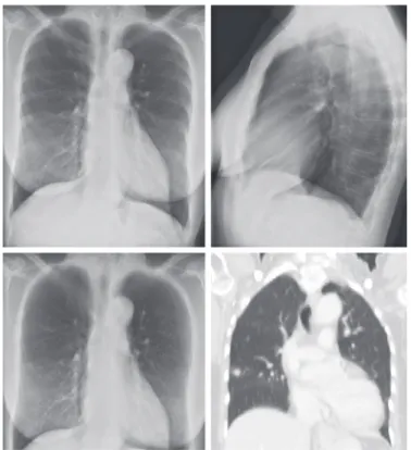
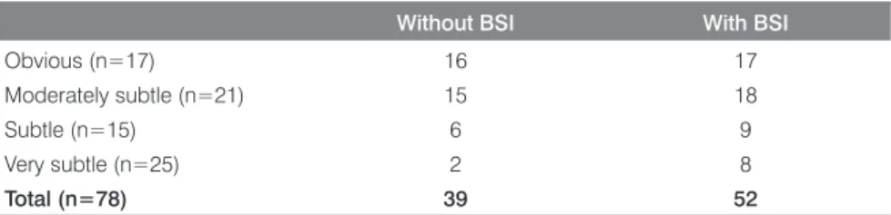
Chapter 2 focuses on the effect of BSI on lung nodule detection. In this study eight radiologists assessed 300 cases with and without BSI in search for lung nodules. Cases included both posteroanterior and lateral digital radiographs, and analysis Figure 1 PA radiograph with a solitary nodule in the right lung. The nodule is overlapped by a projection of 7th posterior rib. Digital bone suppression (Riverain Technologies) produces a bone suppressed image (b), where the rib is perfectly suppressed without suppressing the lung nodule.
Figure 2 The PA radiograph of a 48 years old female shows a 17 mm adenocarcinoma in the left upper lobe. On the right side a CAD output image is shown, in which the suspicious region in the left upper lobe is highlighted.
focused on performance in a high specificity range. In chapter 3 the effect of BSI on the detection of aspergillus infections in critically ill patients was tested. This critically ill patient group did not only contain upright radiographs but also bedside images with lower image quality, making the CXR evaluation more difficult. In order to be used in clinical practice, not only the effect of BSI on focal lesions is important. In chapter 4 the effect of BSI on multiple types of common chest diseases, including disease with a diffuse distribution, was investigated. In this study 261 cases were evaluated by six radiologists.
In the next chapters we investigated the effect of CAD on the detection of pulmonary nodules. In chapter 5 we evaluated the effect of CAD beyond the support of bone suppressed images. In this study CAD was used as a second reader as it is currently recommended by the manufacturers. In chapter 6 and 7 new ways to apply CAD in chest radiography were investigated. The effect of an interactive CAD system in which CAD marks remain hidden unless their location was queried by the radiologist was tested. Further, a computerized combination of reported findings by the radiologist and the CAD system was evaluated. Results were compared with a similar study of the effect of CAD for breast cancer detection in mammography. In chapter 8 the CAD system processed more than 11,000 clinical chest radiographs. The most suspicious CAD marks were evaluated in search for missed cancers. A clinical algorithm for usage of a highly specific CAD system was designed.
Finally, this thesis contains mainly observer studies. In chapter 9 the effect of different observer study designs on observer variability, the effect size and statistical power was investigated.
References
1 Radiation Protection NC, Measurements. Ionizing radiation exposure of the population of the United States. National Council on Radiation Protection report no. 160. National Council on Radiation Protection
and Measurements 2009;.
2 Ferlay J, Shin H, Bray F, et al. GLOBOCAN 2008 v2.0, Cancer Incidence and Mortality Worldwide: IARC CancerBase no. 10. internet 2010.
3 Howlader N, Noone A, Krapcho M, et al. SEER Cancer Statistics Review, 1975-2010, National Cancer Institute. Bethesda, MD, http://seer.cancer.gov/csr/1975_2010/ based on November 2012 SEER data submission, posted to the SEER web site, April 2013;.
4 Quekel LG, Kessels AG, Goei R, et al. Miss rate of lung cancer on the chest radiograph in clinical practice.
Chest 1999;115:720–724.
5 Shah PK, Austin JHM, White CS, et al. Missed non-small cell lung cancer: Radiographic findings of potentially resectable lesions evident only in retrospect. Radiology 2003;226:235–241.
6 Austin JH, Romney BM, Goldsmith LS. Missed bronchogenic carcinoma: radiographic findings in 27 patients with a potentially resectable lesion evident in retrospect. Radiology 1992;182:115–122. 7 Muhm JR, Miller WE, Fontana RS, et al. Lung cancer detected during a screening program using
four-month chest radiographs. Radiology 1983;148:609–615.
8 Heelan RT, Flehinger BJ, Melamed MR, et al. Non-small-cell lung cancer: results of the new york screening program. Radiology 1984;151:289–293.
9 Monnier-Cholley L, Arrivé L, Porcel A, et al. Characteristics of missed lung cancer on chest radiographs: a French experience. Eur Radiol 2001;11:597–605.
10 de Hoop B, Schaefer-Prokop CM, Gietema HA, et al. Screening for lung cancer with digital chest radiography: sensitivity and number of secondary work-up CT examinations. Radiology 2010;255:629– 637.
11 Wu M, Gotway MB, Lee TJ, et al. Features of non-small cell lung carcinomas overlooked at digital chest radiography. Clin Radiol 2008;63:518–28.
12 Samuel S, Kundel HL, Nodine CF, et al. Mechanism of satisfaction of search: eye position recordings in the reading of chest radiographs. Radiology 1995;194:895–902.
13 Kundel HL. Predictive value and threshold detectability of lung tumors. Radiology 1981;139(1):25–29. 14 Sorenson JA, Mitchell CR, Armstrong J 2nd, et al. Effects of improved contrast on lung-nodule detection.
a clinical roc study. Invest Radiol 1987;22(10):772–780.
15 Manning DJ, Ethell SC, Donovan T. Detection or decision errors? Missed lung cancer from the post-eroanterior chest radiograph. Br J Radiol 2004;77:231–5.
16 Kundel HL, Nodine CF, Carmody D. Visual scanning, pattern recognition and decision-making in pulmonary nodule detection. Invest Radiol 1978;13:175–181.
Chest Radiography:
New Technological Developments
and their Applications
S. Schalekamp; B. van Ginneken; N. Karssemeijer; C. Schaefer-Prokop Seminars of Respiratory and Critical Care Medicine
Abstract
Digital chest radiography is still the most common radiological examination. With the upcoming 3D acquisition techniques the value of radiography seems to diminish. But because radiography is cheap, readily available, and requires very little dose, it is still being used for the first line detection of many cardiothoracic diseases. In the last decades major technical developments of this 2D technique are being achieved. First, hardware developments of digital radiography have improved the contrast to noise, dose efficacy, throughput and workflow. Dual energy acquisition techniques reduce anatomical noise by splitting a chest radiograph into a soft tissue image and a bone image. Second, advanced processing methods are developed to enable and improve detection of many kinds of disease. Digital bone subtraction by a software algorithm mimics the soft tissue image normally acquired with dedicated hardware. Temporal subtraction aims to rule out anatomical structures clotting the image, by subtracting a current radiograph with a prior radiograph. Finally, computer aided detection systems help radiologists for the detection of various kinds of disease such as pulmonary nodules or tuberculosis.
Introduction
The conversion from conventional screen film to digital technique took place in the eighties and became clinically feasible after introduction of storage phosphor detector technique. Organizational advantages as instant availability of the images in multiple locations eased the acceptance by clinicians. From the radiologists’ viewpoint options for image processing and lower vulnerability to over/underexposure represented major progress. Additionally, digitization of radiographic examinations represented the last step towards a complete digital imaging department since other cross-sectional imaging techniques such as computed tomography (CT) or magnetic resonance tomography had been from beginning in a digital format.
Today, the process of optimization of processing seems more or less finalized. Differences between techniques and manufacturers have become very small and there appears to be a common sense for what is considered an ‘optimal radiographic image’. Recently, within the area of processing, the focus has dramatically changed. Elaborate processing schemes including dual energy subtraction or digital bone suppression, temporal subtraction or tomosynthesis (chapter 2) are increasingly available. They all have the goals to decrease distracting anatomic noise in the image, to improve perception of pathology and decrease inter- and intra-reader variability. Computer aided detection (CAD) schemes analyze morphologic image features in the background to provide the radiologists with candidate lesions in areas that need increased attention.
The following article will provide an overview over currently available detector systems with special emphasis on their capabilities with respect to dose efficiency. Principles of processing will be summarized including most modern elaborate processing options that go beyond optimization of image quality but are designed for detection support and diagnostic aid.
Detector Technology
A variety of detector technologies has been exploited in modern digital radiography; each coming with different characteristics in terms of physical performance and thus image quality, different levels of dose efficiency and processing options, and last but not least various organizational and financial aspects.
Basically three types of detector systems can be differentiated that are in use for radiographic applications today: storage phosphor radiography (CR), flat panel direct radiography (DR) and slot scanning CCD technology, the latter being the least applied one.
Computed Radiography (Storage phosphor radiography)
CR technology was the first detector technique for digital radiography to be introduced in the early eighties. It was immediately accepted by the radiological and clinical
community due to its organizational advantages and the fact that this cassette based system could be used with already existing hardware components.
The name “storage phosphor” refers to the fact that the image information (relief of absorbed radiation) is stored within the detector layer of photostimulable phosphor material (BaFX: Eu2+): Exposure of the image plate to x-ray photons releases electrons that are trapped within the crystalline structure; only illumination of the image plate to a thinly focused laser light during a dedicated read-out process causes the trapped electrons to relax to their ground state while emitting light. The stimulated light emission is collected via an optically efficient light guide, converted to electronic current by a photomultiplier, logarithmically amplified, filtered and finally digitized using a 12bit analogue-to-digital converter.
In its first version after introduction CR offered limited options for reducing the radiation dose per exposure compared with film-screen. In fact image quality was characterized by relatively high noise especially in high absorption areas. Detector material and read-out technology, however, were continuously improved over the
following years1,2. Most recent more radical innovations such as dual read-out
technology or needle crystalline detector technology have further attributed to substantial improvement of dose efficiency that is now approaching that of flat panel technology and by far exceeding that of conventional film/screen combination with a
speed of 4003-5.
Storage phosphor needle-like detectors use a more efficient X-ray absorption
material (CsBr:Eu2+) and are structured in needle-like columns instead of in small crystals. The result is a marked reduction of lateral scattering of the emitted fluorescent light allowing for increasing detector thickness and thus improving dose efficiency
without losing geometric resolution3.
‘Dual-sided read-out’ captures the laser-stimulated phosphorescence light from both sides. This dual-sided approach can access a larger proportion of the trapped electrons and as a result achieve a more complete read-out of the latent image. The result is an improved signal and signal-to-noise ratio for a given absorption of radiation within the detector. The dual-sided read-out does not affect the spatial resolution since the amount of scattering of light remains unaffected. Literature reports an increase of fractional x-ray absorption efficiency of the image plate by 50% through the dual-read out. Physical and clinical evaluations confirmed a substantial increase
in image quality4,5.
CR used to be a cassette based system and is as such still in use e.g., on intensive care wards. It is also available in dedicated chest stands or bucky units that offer automatic read out and require no manual interaction with cassettes. Also the needle- type detector is available as cassette based system as well as system integrated in bucky and fluoroscopy units.
When using CR systems or reading comparison studies that include CR technique it is very important to pay attention to the type of detector (needle versus powder storage phosphor, which generation of powder storage phosphor plate) and which read out system has been used. The pure description of CR is too nonspecific and
refers to various systems with a broad range of performance6.
Direct Radiography (Flat panel detector systems)
Flat-panel detector systems - also referred to as direct radiography (DR) - are characterized by a direct readout-matrix of electronic elements that are made of thin layers of amorphous silicon thin-film transistors (a Si-TFT elements) that are deposited on a piece of glass. The TFT layer is coupled with an x-ray absorption medium
responsible for capturing the radiographic image information7,8.
Depending on the material used, there are two types of DR detectors:
a) Detectors using a scintillator (Cesium Iodide = CsI or Gadolinim Oxysulphide = GOS) and light sensitive photodiodes are called indirect conversion TFT detectors
or opto-direct systems. Similar to CR technology, the absorbed radiation is
transferred into light signals in the CsI layer. CsI-TFT systems are widely applied for chest and skeletal radiography and are also amenable to real-time display. More recently also mobile DR units became available, suited to be used at the bedside.
b) In direct conversion systems, the detector elements consist of condensator elements made up of amorphous selenium (or other semiconducting material) that is deposited on the TFT array. Absorbed x-ray energy is directly converted into charge, obviating the intermediate step of a scintillator to provide conversion to visible light. These systems are not amenable to real-time imaging due to the tendency to produce persistent latent images. They are mostly applied in mammography units because they provide a high dose efficiency for the high frequency ranges needed in mammography.
DR has a markedly higher dose efficiency as compared to first generations standard CR (Figure 1). As compared to most modern CR units, however, differences with respect to image quality and work flow organization become much less prominent. It is therefore important to consider which type of storage phosphor system (detector generation, read-out) is used. DR (CsI/TFT) systems are amenable to high frame rates (up to 30/sec) making them also suited for fluoroscopy applications. By integrating acquisition, read-out and processing into one system, throughput and workflow can be optimized. Integrated systems are available for both CR and DR. When CR is used as cassette based system, e.g., at the bedside, these advantages (e.g., immediate availability of pre read, no cassette handling) perish.
CCD and CMOS-based Detectors
Charge coupled devices (CCD) and complementary metal oxide semiconductor devices (CMOS) were initially suffering from lower dose efficiency that could not compete with DR or CR systems. They also use scintillating phosphor as an absorption medium; emitted light is directed to multiple CCD or CMOS cameras that form the radiograph. Various optical arrangements including lenses or fiber-optic tapers are used for the coupling between the phosphor layer and the mostly relatively small cameras.
Because only a fraction of the light could be captured by the cameras, image formation was less efficient with respect to signal-to-noise. This limitation was even more obvious for clinical applications that require a large area (e.g., chest). Recent developments markedly improved the coupling efficiency by using larger sensors and improved phosphor efficiency, making these systems also suitable for high quality chest and large area skeletal radiography. CCD and CMOS systems have a wider distribution in the US, but are only scarcely applied in Europe.
Irradiating the body by a sliding slit beam instead of irradiating the whole body at once is called slot-scan technology and provides excellent scatter rejection. The increased signal-to-noise yielded by scatter reduction effectively compensates for the 2.5 times lower intrinsic detective quantum efficiency (DQE) of CCD technology allowing for a successful combination of slot-scan and CCD technology. Advantages of this CCD slot-scan technology are especially prominent for the mediastinum: both CsI-DR at 75% dose reduction and CCD slot scanning at 50% reduction outperformed
a standard CR system9. Though these systems provide excellent image quality at
reasonable doses, they did not find a broad international application, most likely to specific hardware requirements and the fact that they are exclusively amenable to chest imaging.
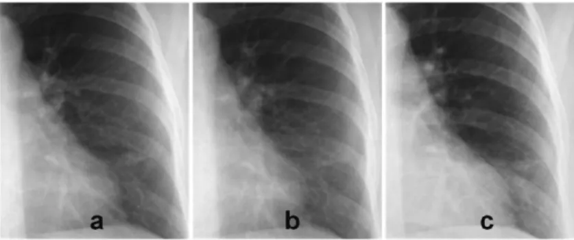
Figure 1 Images of the left lower lung demonstrate the potential of dose reduction of DR as compared to (powder) CR: image b (DR) was obtained with 50% of the dose compared to a (DR) and c (CR).
Physical parameters of detector systems that determine
image quality
Dynamic range is defined as the of absorption differences that can be accommodated
by the detector system and thus contribute information to the recorded image1. In thoracic
imaging the absorption difference between normally ventilated lung and mediastinal soft tissue is 1:80. In screen-film radiography the dynamic range was defined by the radiographic response of the film and was rather limited (1:10) as illustrated by the somewhat too white mediastinum or too black lung. In digital radiography (true for CR and DR), the maximum signal capability is set by the detector medium itself and its read-out mechanism, the minimum signal capability is set by the image noise and the greyscale discrimination capability of the system. The dynamic range of digital radiography (CR and DR) is about 400 times wider as conventional film meaning that digital systems can obtain image information over a much wider range of entrance doses. The spatial resolution of an image detector refers to its ability resolve two (or more)
small high-contrast image features as independent entities1. The spatial resolution is
influenced by many factors such as the detector medium itself (e.g., needle channeled CR versus powder crystals CR), the detector thickness, the size of the laser beam, the pre- and post-processing and finally the pixel size. CR plates offer a pixel sampling interval between 0.5µm to 200µm, DR systems between 140 and 200µm. More important for visual discernability of e.g., parenchymal details, however, is the relationship between detail size and detail contrast, described in the modulation transfer function (MTF): the higher the MTF the smaller the details an imaging system displays with visually discernable contrast. Especially processing has a vast influence on the MTF, and is optimized to improve the rendition of fine and small detail structures on one side and increase visualization of ill-defined low contrast lesions on the other side. It is therefore inadequate to focus on the pixels sampling interval alone. Multiple studies could show that there is no diagnostically relevant difference between 2K and 4K CR systems (pixel sampling difference 100 and 200 µm), for chest abnormalities, given an appropriate processing.
The term ‘detective quantum efficiency’ (DQE) is regarded as the best single
indicator to describe the performance of digital radiographic systems10. The DQE of
an imaging system refers to the ratio between the SNR at the entrance to the image detector (flux of x-ray photons incident upon the image detector) and the SNR recorded by the image detector (the value which is computed from the output data). Ideally that value would be 100% (what goes in also comes out) but dependant on the amount of extraneous noise sources in the image detector itself the DQE will be less than 100%. The greater the value of DQE, the more efficiently the detector records x-ray image information. The magnitude of DQE is influenced by the effective beam energy, the detector entrance dose level, the detector system itself and the targeted image frequencies.
Image Processing
One of the most important advantages of all digital radiographic detectors is their wide dynamic range making them less vulnerable towards under- or over exposure. Together with adequate image processing, these systems produce over a wide range of exposures adequate image quality (Figure 2). A process called signal normalization – automatically running in the background – yields images with adequate density and contrast independent of acquisition dose (Figure 3). However, this harbors advantages and disadvantages: visual control of acquisition dose is more or less lost and there is
a certain risk for over-exposure (e.g., in pediatric units11 which will even be awarded
by excellent image quality. A too strong dose reduction on the other hand leads to loss of fine detail because of increased image noise (Figure 4). Thus - though less critical as compared to traditional film/screen radiography - there are limitations for how much the dose should be increased or decreased.
Image processing critically influences image quality and thus diagnostic performance: while adjustments of gradation curves influence overall image contrast and density, frequency processing enhances local contrast or even selectively enhances Figure 2 The wide dynamic range of digital imaging technique allows for optimized display of structures in high and low absorption areas: e.g., the tip of the central venous catheter, signs of vascular congestion in the left lung and the right sided pathologic subcapital humerus fracture.
structures of a certain size or contrast12. Unsharp mask filtering is the simplest type of
frequency processing but has the disadvantage that stronger filter settings lead to (edge) artifacts.
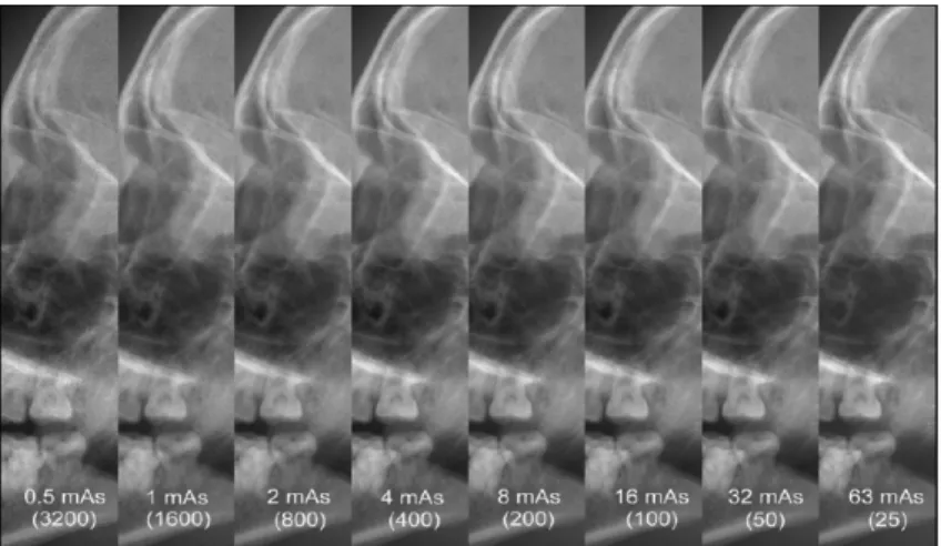
Figure 3 Images of a scull phantom: Over a dose range between 0.5 mAs and 63 mAs (factor > 100) digital technique is able to yield images with comparable and diagnostically useful contrast and density. Though images on the left side (low dose) have a higher noise and therefore demonstrate a less sharp delineation of the linear details compared to images on the right side.
Figure 4 Image b was obtained with one third of the dose used for image a. Image contrast and density appear to be comparable. However, the ratio between signal and noise became too low in image b to capture the linear density of the intravenous catheter. Thus a too strong dose reduction will lead to loss of detail and thus potentially diagnostic information.
Most vendors have made the transition to multi-frequency processing algorithms
(e.g., MUSICA, UNIQUE, MFP)13,14. Multifrequency processing makes it possible to
separately enhance and suppress image structures dependent on their contrast (amplitude), their size (spatial frequency bands) and their background density (mediastinum versus lung).
The desired result is an ‘image harmonization’ with a more transparent mediastinum and improved visualization of low contrast structures throughout the chest with simultaneous display of soft tissue and osseous structures and preserved high resolution for low contrast differences in the lung (Figure 5). Today, there is general agreement that a too strong enhancement of edges along anatomic structures (e.g., vascular shadows) produce diagnostic misinterpretation (e.g., of cardiogenic congestion or interstitial lung disease), and may in fact lead to loss of diagnostic detail (Figure 6).
Advanced Processing
Advanced processing refers to techniques that are aimed to aid detection performance. They produce processed images that differ from the original images in such a way that they are used as an adjunct to the original images with the idea to enhance image formation or draw the observer’s attention to a certain area or structure. All of these techniques are designed to reduce overlapping potentially distracting structures (e.g., bones) or highlight areas of suspicion (e.g., circles around nodules produced by CAD) with the ultimate goal to support the detection performance of the observer.
Techniques such as digital bone suppression, dual energy acquisition or tomosynthesis are designed to reduce overlying structures, e.g. bones, that way Figure 5 Desired effects of processing are compression of the dynamic range with increased transparency of the mediastinum and simultaneously increased detail contrast in the lung parenchyma (visible in b as compared to a).
reducing the “anatomic noise” and providing the reader with an unobscured display of the lung parenchyma. Tomosynthesis goes a step further, displaying a subvolume of the lung similarly to CT that way reducing projection effects. This technique will be discussed in chapter 2. Temporal subtraction and CAD highlight areas of suspicion that subsequently need to be accepted or dismissed by the radiologist as real pathology.
Bone Subtraction
Chest radiographs are complex radiological examinations to review. Multiple anatomical structures contribute to a complex 2D image of a 3D volume. This high level of so called “anatomical noise” is one important factor contributing to the difficulty of interpretation of chest radiographs. Bones have shown to be a major
contributor to this noise, and are an important cause of missed lung nodules15,16.
Therefore several techniques have been proposed to suppress overlying bone structures; this can be achieved either digitally or using a dedicated dual energy- acquisition technique.
Dual Energy Radiography
Dual energy radiography is based on the principle that radiation absorption by tissues is dependent on the energy of roentgen photons. At high energy level (>100 kVp) absorption of photons differs not much for bone and soft tissue. At lower energy levels, however, photoelectric absorption is much more effective for tissue containing Figure 6 Unwanted effects of image processing: suboptimal processing parameters lead to too strong enhancement of vascular structures and noise but obscuration of the faint density (seen in b) caused by an interlobular effusion.
calcium and thus bones. This effect is used to produce images mainly displaying soft tissue information and images mainly displaying calcified structures as ribs, spine or calcified nodules.
Two techniques of energy subtraction (ES) can be distinguished: single-shot ES radiography and dual-shot ES radiography.
Dual shot ES radiography uses two exposures: one at a high and one at a low energy level. Subtraction of these two radiographs using specific weighting factors generates a “soft tissue image” and a “bone image”. The images have usually high quality with high contrast and low image noise. But the short interval of 150-200 ms between the two acquisitions can cause motion artifacts, mainly caused by movement of the heart, diaphragm and hilar vessels. Also the need for two acquisitions slightly increases the total dose of the examination.
Single-shot ES radiography requires only one exposure. In one cassette are two detector plates separated by a copper filter. While the first detector plate is radiated by the full radiation spectrum producing a “normal” radiograph, the second detector plate is only radiated by a high energy radiation spectrum since the low energy photons were absorbed by the copper filter. The image detected by the second plate has low bone contrast, and is nosier than the radiograph detected by the first plate. Subtraction of the two acquired images using specific weighting factors produces again a soft tissue and a bone image. Since the image detected by the second plate is noisier, also the resulting images suffer from higher image noise and thus lower overall image quality. In opposite to the dual shot technique they do not suffer from subtraction artifacts.
Several studies testing these dual energy techniques have been performed. ROC analysis in multiple observer studies showed an increased performance for the detection of lung nodules with help of dual energy images (soft tissue image and
bone image)17-23 (Table 1). Studies found an increase of sensitivity that varied between
5 and 19%. Also few studies failed to prove added value of dual energy images,
mainly because a decrease in specificity24,25. Besides solid lung nodules also
part-solid nodules and ground glass opacities could be more easily detected with
dual energy radiographs18,20,23. When calcification of nodules can be identified on
the bone images, further CT examinations might not be needed26. However, small
amounts of calcium may not be seen with dual energy radiography. The bone image can also be used to clarify other calcified disease like pleural plaques and mediastinal
calcified nodules27. Also some studies found beneficial effects for the detection of
cardiac calcification28,29. Besides, radiopaque foreign objects, like medical devices,
catheters, drains, silicone breast implants, are more easily seen (Figure 7). No improvement
was found for the detection of rib fractures based on the bone image30.
There is no clear preference for single or dual shot energy subtraction techniques for the detection of lung nodules. One study explicitly compared dual and single shot
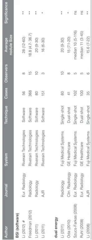
Ta b le 1 O ve rv ie w o f s tu di es u si ng d ig ita l b on e s up pr es si on o r d ua l e ne rg y s ub tra ct io n i m ag es fo r t he d et ec tio n o f l un g n od ul es . Au th o r Jo ur na l S ys te m Te chni qu e C ase s O b ser ver s Av er ag e no d ul e S ize S ig ni fic an ce B SI ( so ft w are ) Li ( 20 12 ) # Eu r. R ad io lo g y R iv er ai n T ec hn ol og ies S of tw are 56 8 28 (12 -6 0) ** Fr ee dm an (2 01 2) R ad io lo g y R iv er ai n T ec hn ol og ies S of tw are 36 8 15 18 .8 (4 .7 - 3 6. 7) ** Li (20 11 ) R ad io lo g y R iv er ai n T ec hn ol og ies S of tw are 80 10 20 (9 -3 0) ** Li (20 11 ) A JR R iv er ai n T ec hn ol og ies S of tw are 151 3 16 (6 -3 0) * d ua l en er g y Li (20 11 ) R ad io lo g y Fu ji M edi ca l S ys te m s S in gl e-sh ot 80 10 20 (9 -3 0) ** O da (2 010 ) Cl in . R adi ol og y G E H eal th ca re D ua l-sh ot 41 8 15 (7. 1-20 ) * S zu cs -Fa rk as (2 00 8) Eu r. R ad io lo g y Fu ji M edi ca l S ys te m s S in gl e-sh ot 10 2 5 me di an 1 0 ( 5-11 6) ns R uh l (2 00 8) Eu r. R ad io lo g y G E H eal th ca re D ua l-sh ot 10 0 5 me di an 1 1 ( 3-45 ) ns Li (20 08 ) A JR Fu ji M edi ca l S ys te m s S in gl e-sh ot 35 6 15 .6 (7-22 ) ** # s tu d y u se d s ub je ct s w ith f oc al p ne um on ia i ns te ad o f l un g n od ul es . A JR = A m er ic an J ou rn al o f R ad io lo g y. * p < 0 .0 5; * * P < 0 .0 1. n s = n on -s ig ni fic an t.
techniques and could not demonstrate a significant difference in the detection of lung
nodules between the two techniques26.
Despite the mainly positive results, both techniques (dual shot and single shot) have never found their way into broad clinical application. Both techniques require specific hardware and software facilities, meaning that this technique cannot be applied using already existing radiographic equipment. For a long time, the image quality of the soft tissue and bone images was considered insufficient. The quality of these images, however, could be substantially improved over the time making them a useful adjunct, especially for the detection of focal disease. It is most likely a combination of all – the need for specific hardware, the increasing availability of CT and the need to integrate an additional soft tissue image into the reading process – that has contributed to the still somewhat limited use of this technique.
Bone Suppression
The need for special hardware is an important drawback for dual-energy radiography. Therefore investigators focused on digital removal of bony structures in the chest radiograph using dedicated software applicable to already existing radiographic equipment (Figure 8). Advantages of such a solution are that image quality is not suffering from subtraction artifacts (by motion in dual-shot energy subtraction), no increase in dose for the patient and no special hardware is required. The software product can be integrated in the picture archiving and communication system. Disadvantage is that the software algorithm is designed to detect solely bone structures and not all structures with high density. Therefore other high calcium structures, like calcified nodules or old rib fractures, are not recognized and not suppressed, as in energy subtracted images.
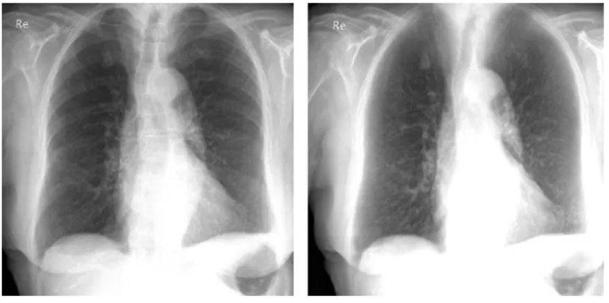
Figure 7 PA radiograph (a) with circular opacification in the right upper lobe suspicious for a cavitation. Soft tissue (b) and bone image (c) produced by dual energy subtraction technique demonstrate a linear scarring (b) and a radio-opaque vascular stent (c) as explanation for the circular opacity (courtesy of Peter Vock, Bern, Switzerland).
First papers about a bone suppression technique by a computer algorithm have
been published in 200631,32. Studies using the bone suppression technique found an
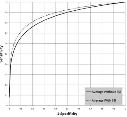
increase in performance for the detection of lung nodules17,33,34 (Table 1). The largest
observer study which incorporated 15 observers and 368 cases found an average
increase in sensitivity of 17%, but also a loss in specificity of 4%33. The software
solution might also be used for the detection of other abnormalities. Li et al.35 found
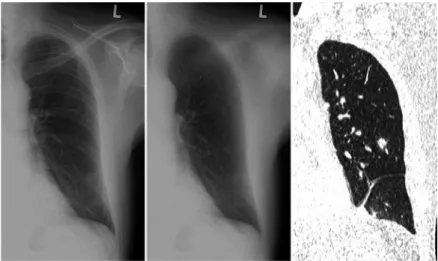
a significant increase for the detection of focal pneumonias, and improved detection of other disease is conceivable. Artifacts of the suppression techniques can cause pseudolesions, which result in a somewhat lower specificity. Most artifacts are caused by overprojection of multiple structures, like in the apical regions where both the clavicle and the first rib obscure the lung field (Figure 9).
One of the papers also compared bone suppression imaging software with dual
energy bone suppressed images17. In an observer study 10 radiologists searched 80
CXRs for lung nodules first unaided, followed by and interpretation of the digitally bone suppressed image, and finally with dual energy image. Biggest increase in performance was seen with help of the dual energy bone suppressed images, although both techniques showed significant improvement over original chest radiographs.
Considering that digital bone suppression is still a relatively young technique, more beneficial effects might be demonstrated in the future. Artifacts could be minimized and with further experience with this technique artifacts may be more easily denied. Clinical studies will have to show this effect.
Computer Aided Detection
The approach of computer aided detection (CAD) techniques is to decrease the intrinsic limitations of human perception, by alerting the observer to suspicious areas in a chest x-ray.
Figure 8 PA radiograph with a solitary nodule in the right lung. The nodule is overlapped by a projection of 7th posterior rib. Digital bone suppression (Riverain Technologies) produces a bone suppressed image, where the rib is perfectly suppressed without suppressing the lung nodule.
First computer analysis techniques of chest radiographs were already published
in the 1960s36,37. A number of generations of CAD software have been introduced
and most of them focusing on the detection of nodules, meaning focal densities smaller than 3cm. Other indications less well advanced computerized detection systems so far refer to the detection of spine fractures, heart size, interstitial lung disease, emphysema and tuberculosis.
Development of techniques for the automatic detection of lung nodules in chest
radiographs started in the 1970s38,39. Matsumoto et al.40 was one of the first to apply
a computer aided detection scheme on a series of radiographs with lung cancer. CAD was able to reach a sensitivity of 62%, which was comparable to the radiologists. On the other hand, the system produced an average of 15 false positive (FP) finding per image leading to an overall loss of performance of radiologists because of
acceptance of too many FPs41. In a study with a simulated improved performance of
the same CAD with a sensitivity of 80% at 1 FP/image, radiologists did benefit from
the CAD system41. Already these early studies pointed out to the main challenge for
radiologists when using CAD, namely to take advantage of CAD by detecting more focal lesions that would have been otherwise missed without losing specificity by accepting too many false positive candidate lesions.
Since then CAD has been applied on multiple sets of radiographs, showing the
potential for detection of lesions that were missed by human observers42-44. Sensitivities
ranged from 35% at 5.9 FP per image to 49% at 1.8 FP per image for these missed lesions.
Still the various CAD systems are difficult to compare, since training and testing of the system is often done with different data and studies use different sets of lesions. But multiple studies have reported their CAD performance on a publicly available
database from the Japanese Society of Radiological Technology45. Looking at the
Figure 9 Bone suppressed image (b) shows a pseudolesion in the right apex, due to incomplete suppression of calcification of the first rib.
CAD performances on this database, CAD evolved quickly from a sensitivity of 35%
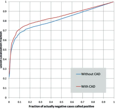
with 6 FP / image in 1999 to a sensitivity of 75% with 0.5 FP / image in 2012 (Figure 10) 46-53.
Compared to an average sensitivity of 70% at a specificity of 81% for radiologists for this database, CAD is approaching the performance of an average radiologist.
As mentioned above, the effect of CAD on observer performance highly depends on its ratio between true and false positive candidates. Although significant improvements are made over the last ten years, CAD is still not able to achieve performance of a human observer. Therefore, CAD is designed to be used as a second reader. This means that the human observer, after his first evaluation, can ask CAD to show suspicious regions. The human observer than has to accept or neglect the suggested findings of the computer. Several studies with different CAD systems have been performed to investigate the added value of CAD as a second reader. These studies reported variable results. While some studies were able to demonstrate an increased
accuracy for the detection of potential lung cancer when aided by the computer44,54-59,
Figure 10 Published performances of CAD systems applied on a publicly available database from the Japanese Society of Radiological Technology (JSRT). Over the last 15 years performance of CAD systems dramatically increased from a sensitivity of 35% at 6 FP/image to a sensitivity of 75% at 0.5 FP/image. Riverain 2012 Chen 2011 Snoeren 2010 Hardie 2008 Campadelli 2006 Schilham 2006 Coppini 2003 Wei 2002 Carreira 1998 0 10 20 30 40 50 60 70 80 90 0 1 2 3 4 5 6 7 S en siti vit y
False Positives per image
Riverain 2012 Chen 2011 Snoeren 2010 Hardie 2008 Campadelli 2006 Schilham 2006 Coppini 2003 Wei 2002 Carreira 1998
also several studies reported no increase in performance60-63 (Table 2). The main issue
that is highlighted in these papers is that it is very difficult to discriminate between true positive and false positive CAD marks. When a system produces many FP CAD marks, it is likely that a substantial number of these false positives gets accepted, potentially resulting in over-diagnosis and overtreatment to the patient. With a decline in FP CAD marks in the latest studies, improvement is more easily achieved.
To improve performance of the CAD system, bone suppressed images or dual energy acquired images can be used. Since many false positives are generated by overprojections of bony structures, inclusion of these images could improve CAD
performance64,65. Usage of either digital bone suppression or dual energy images
does not seem to make a lot of difference66. However, indifferent results were seen
when radiologists were offered the combination of bone suppression and CAD for the
detection of pulmonary nodules67,68.
Not only for pulmonary nodules, but also for the detection of other disease, CAD in chest radiography starts to play a role. Software algorithms for the detection of tuberculosis (TB) have been developed for application in high burden countries. Expertise of readers in high burden countries is often poor. Since CXR is a powerful TB screening instrument, research has focused on automated detection of TB in CXRs. Background of different TB detection techniques are discussed in Arzhaeva et
al. and Ginneken et al.69,70. TB can have multiple different characteristics on chest
x-rays. Therefore these detection applications do often not only focus only on focal lesions, but also try to qualify an image as normal or abnormal based on multiple factors. The latest developed software achieves performance comparable to clinical officers in Zambia, and therefore could be used as point-of-care decision tool to
select subjects that should undergo further tests71.
For interstitial lung disease (ILD) computerized detection started more than
20 years ago, with many approaches72-74. Automated detection of ILD was found to
be extremely challenging. Since interstitial disease presents itself with diffuse patterns, software algorithms focus on specific texture analysis. The main approach is to detect the various patterns, in which ILD can be present. Abnormality scores generated by the system can be based on local areas or a more global approach where the whole image is given one score. Another complicating factor is the large amount of anatomical structures occluding the interstitial patterns. Therefore CAD approaches for texture analysis could benefit from inclusion of digitally bone
suppressed images or dual energy acquired images75,76.
Other research focuses on the computerized detection of emphysema in chest
radiographs77,78, detection of pneumothorax79 and the automatic detection of catheters
and tubes80-82. Also CAD software for the detection of vertebral compression fractures
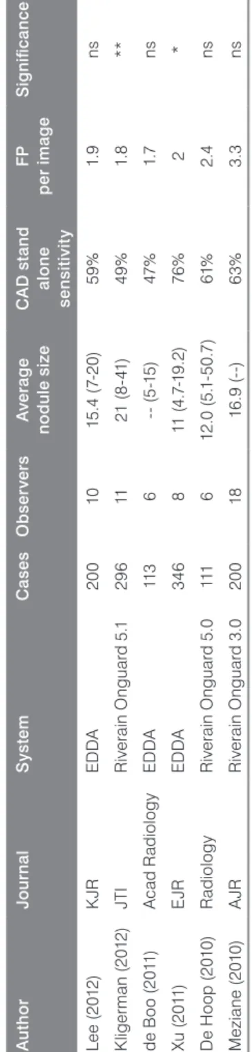
Ta b le 2 O ve rv ie w o f t he m os t r ec en t s tu di es e va lu at in g c om pu te r a id ed d et ec tio n fo r l un g n od ul es . Au th o r Jo ur na l S ys te m C ase s O b ser ver s Av er ag e no d ul e s ize C A D s ta nd al one se nsi tiv ity F P per im ag e S ig ni fic an ce Le e ( 20 12 ) K JR E D DA 20 0 10 15 .4 (7-20 ) 59 % 1. 9 ns K lige rm an (2 01 2) JTI R iv er ai n O ng ua rd 5 .1 29 6 11 21 (8 -4 1) 49% 1. 8 ** de B oo ( 20 11 ) A cad R ad io lo g y E D DA 11 3 6 (5 -1 5) 47 % 1. 7 ns Xu ( 20 11 ) E JR E D DA 34 6 8 11 (4 .7 -1 9. 2) 76 % 2 * D e H oo p ( 20 10 ) R ad io lo g y R iv er ai n O ng ua rd 5 .0 111 6 12 .0 ( 5. 1-50 .7 ) 61% 2.4 ns M ez ia ne (2 010 ) A JR R iv er ai n O ng ua rd 3 .0 20 0 18 16 .9 (--) 63% 3. 3 ns K JR = K or ea n J ou rn al o f R ad io lo g y. J TI = J ou rn al o f T ho ra ci c I m ag in g. E JR = E ur op ea n J ou rn al o f R ad io lo g y. A JR = A m er ic an J ou rn al o f R ad io lo g y. C A D = C om pu te r a id ed d et ec tio n. F P = f al se p os iti ve. * p < 0 .0 5; * * P < 0 .0 1. n s = n on -s ig ni fic an t.
Currently there are two U.S. Food and Drug Administration (FDA) approved, computer aided detection systems (IQQA-chest, EDDA Technology, Inc., Princeton Junction, NJ, USA; ClearRead + Detect, Riverain Technologies, Miamisburg, Ohio) for the detection of lung nodules on the market. Besides these commercial systems there are also multiple prototypes used for research purposes in various hospitals around the world. Other developed CAD systems for the detection of tuberculosis, ILD, emphysema, pneumothorax, catheters and tubes or vertebral fractures are not yet being used in clinical practice.
Temporal Subtraction
An important part of the evaluation of a chest radiograph is based on the comparison with previous radiographs. Since nowadays all the images are digitally stored in a digital archive system, temporal subtraction has become technically feasible and attractive. Temporal subtraction is aimed to enhance the changes over time. This refers to new lesions as well as to the assessment of progression or regression of pathology over time.
With temporal subtraction a prior image is registered to the current image, using registration algorithms to warp the position of the prior radiograph to the current radiograph. If the registration is successful and the image is subtracted, an interval change stands out as a dark or bright area in the radiograph. One major benefit of temporal subtraction is that it is able to totally rule out the background anatomical noise. If the registration is perfect, solely interval changes thus potential pathology are highlighted.
Although temporal subtraction was already discussed in 197683, reasonable
attempts to automatically subtract radiographs started in 1994 by the University of
Chicago84. Registration mismatch is responsible for most of the artifacts and represent
the crucial challenge. Depth of inspiration, positioning and rotation of the patient, makes registration of chest radiographs very difficult. In one of the early studies, where a consecutive set of cases was analyzed, 81% of the subtraction images were
of clinically acceptable quality85. Because ribs are very radiopaque, mismatch of
the ribs causes the most pronounced artifacts. Usage of dual energy subtracted or
bone suppressed images, could reduce this effect86. That is why developers are now
using a combination of bone suppression and temporal subtraction (Figure 11). Several studies tested the effect of temporal subtraction images on the detection
performance of various abnormalities. Difazio et al.87 found significant improvement
in a study with 50 chest radiographs, including 29 lungs with abnormalities like new opacities and lung nodules. Eleven radiologists significantly improved their performance when using temporal subtraction images in addition to the current and previous radiograph. Later studies confirmed these results in studies with radiographs
disease patterns89,90, and lung nodules91-93. Not only detection performance could be
improved, also reading time of CXRs reduced significantly87, and was more pronounced
in cases without registration mismatch artifacts94. The latest available commercial
software package includes bone suppressed images in the temporal subtraction process. In their FDA approval study they found a significant increase in the detection
of lung nodules in a study with 422 pairs of radiographs and 15 radiologists95. Also,
recently temporal subtraction images are being used to improve CAD systems96.
Up to now, only in Japan temporal subtraction is fully integrated in daily clinical practice. Commercial available systems are Truedia/XR (Mitsubishi space software) and Compare (Riverain Technologies). Mismatch artifacts is the biggest cause why temporal subtraction has not been adapted into clinical practice.
Summary
The two major detector systems widely applied for digital radiography are computed radiography (CR) based on storage phosphor plates and solid-state (flat panel) direct radiography (DR) systems.
CR represents the older system, matured over decades with some important recent improvements with respect to dose efficiency and work-flow efficiency that strengthened its position. It represents a very versatile medium, economically attractive system that is equally suited for integrated systems as well as for cassette based imaging at the bedside.
DR systems offer superb image quality and realistic options for dose reduction based on their high dose efficiency. While for a long time only integrated systems were available suited for a large patient throughput, also mobile DR systems became recently available.
Figure 11 Prior radiograph (a) is registered to the current radiograph (b) and subtracted to generate a subtraction image (c) (Riverain Technologies). Bone suppressed images are used to improve the subtraction image. The subtraction image clearly reveals newly formed focal lesions in the left and right lung, while only minor misregistration artifacts of ribs can be seen.
While for the next years, it is likely that DR and CR systems will coexist, the long term perspective of CR will depend on innovations with respect to dose efficiency and signal – to – noise characteristics while for DR economical aspects and broader availability of mobile systems will play a role.
Advanced processing techniques have become available for clinical use in the last decade. Dual energy radiography, digital bone subtraction, tomosynthesis, temporal subtraction and CAD have been shown to considerably increase detectability of focal lesions in radiography, that way strengthening the role of radiography compared to 3D acquisition techniques like CT. Although many of the above mentioned techniques are not yet commonly used in clinical practice, many of them show an improved discernability of pathology in chest radiographs. It seems to be a matter of time until certain advanced processing techniques are being adapted to standard clinical care. Digital software solutions seem to have advantages over hardware solutions, since those can easily being incorporated in the radiology department, and are often much cheaper.
References
1. Cowen AR, Davies AG, Kengyelics SM. Advances in computed radiography systems and their physical imaging characteristics. Clin Radiol 2007;62:1132–1141.
2. Veldkamp WJH, Kroft LJM, Geleijns J. Dose and perceived image quality in chest radiography. Eur J
Radiol 2009;72:209–217.
3. Berger-Kulemann V, Pötter-Lang S, Gruber M, et al. Needle image plates compared to conventional CR in chest radiography: is dose reduction possible? Eur J Radiol 2012;81:4156–4160.
4. Körner M, Treitl M, Schaetzing R, et al. Depiction of low-contrast detail in digital radiography: comparison of powder- and needle-structured storage phosphor systems. Invest Radiol 2006;41:593–599. 5. Gruber M, Uffmann M, Weber M, et al. Direct detector radiography versus dual reading computed
radiography: feasibility of dose reduction in chest radiography. Eur Radiol 2006;16:1544–1550. 6. Schaefer-Prokop C, Neitzel U, Venema HW, et al. Digital chest radiography: an update on modern
technology, dose containment and control of image quality. Eur Radiol 2008;18:1818–30.
7. Neitzel U. Status and prospects of digital detector technology for CR and DR. Radiat Prot Dosimetry 2005;114:32–38.
8. Schaefer-Prokop CM, Boo DWD, Uffmann M, et al. DR and CR: Recent advances in technology. Eur J
Radiol 2009;72:194–201.
9. Veldkamp WJH, Kroft LJM, Boot MV, et al. Contrast-detail evaluation and dose assessment of eight digital chest radiography systems in clinical practice. Eur Radiol 2006;16:333–41.
10. Bertolini M, Nitrosi A, Rivetti S, et al. A comparison of digital radiography systems in terms of effective detective quantum efficiency. Med Phys 2012;39:2617–2627.
11. Willis CE. Optimizing digital radiography of children. Eur J Radiol 2009;72:266–273.
12. Prokop M, Neitzel U, Schaefer-Prokop C. Principles of image processing in digital chest radiography. J
Thorac Imaging 2003;18:148–164.
13. Stahl M, Aach T, Dippel S. Digital radiography enhancement by nonlinear multiscale processing. Med Phys 2000;27:56–65.
14. Vuylsteke P, Schoeters EP. Multiscale image contrast amplification (MUSICA). In Medical Imaging 1994. International Society for Optics and Photonics 1994; pages 551–560.
15. Monnier-Cholley L, Arrivé L, Porcel A, et al. Characteristics of missed lung cancer on chest radiographs: a French experience. Eur Radiol 2001;11:597–605.
16. Shah PK, Austin JHM, White CS, et al. Missed non-small cell lung cancer: Radiographic findings of potentially resectable lesions evident only in retrospect. Radiology 2003;226:235–241.
17. Li F, Engelmann R, Pesce LL, et al. Small lung cancers: improved detection by use of bone suppression imaging–comparison with dual-energy subtraction chest radiography. Radiology 2011;261:937–949. 18. Oda S, Awai K, Murao K, et al. Computer-aided volumetry of pulmonary nodules exhibiting ground-glass
opacity at MDCT. AJR Am J Roentgenol 2010;194, February 2010.
19. Li F, Engelmann R, Doi K, et al. Improved detection of small lung cancers with dual-energy subtraction chest radiography. AJR Am J Roentgenol 2008;190:886–891.
20. Ide K, Mogami H, Murakami T, et al. Detection of lung cancer using single-exposure dual-energy subtraction chest radiography. Radiat Med 2007;25:195–201.
21. Ricke J, Fischbach F, Freund T, et al. Clinical results of CsI-detector-based dual-exposure dual energy in chest radiography. Eur Radiol 2003;13:2577–2582.
22. Kido S, Ikezoe J, Naito H, et al. Clinical evaluation of pulmonary nodules with single-exposure dual-energy subtraction chest radiography with an iterative noise-reduction algorithm. Radiology 1995;194:407–412. 23. Kelcz F, Zink FE, Peppler WW, et al. Conventional chest radiography vs dual-energy computed
radiography in the detection and characterization of pulmonary nodules. AJR Am J Roentgenol 1994;162:271–278.
24. Szucs-Farkas Z, Patak MA, Yuksel-Hatz S, et al. Single-exposure dual-energy subtraction chest radiography: detection of pulmonary nodules and masses in clinical practice. Eur Radiol 2008;18:24–31. 25. Rühl R, Wozniak MM, Werk M, et al. CsI-detector-based dual-exposure dual energy in chest radiography
26. Ho JT, Kruger RA. Comparison of dual-energy and conventional chest radiography for nodule detection.
Invest Radiol 1989;24:861–868.
27. Fischbach F, Freund T, Röttgen R, et al. Dual-energy chest radiography with a flat-panel digital detector: revealing calcified chest abnormalities. AJR Am J Roentgenol 2003;181:1519–1524.
28. Gilkeson RC, Novak RD, Sachs P. Digital radiography with dual-energy subtraction: improved evaluation of cardiac calcification. AJR Am J Roentgenol 2004;183:1233–1238.
29. Mafi JN, Fei B, Roble S, et al. Assessment of coronary artery calcium using dual-energy subtraction digital radiography. J Digit Imaging 2012;25:129–136.
30. Szucs-Farkas Z, Lautenschlager K, Flach PM, et al. Bone images from dual-energy subtraction chest radiography in the detection of rib fractures. Eur J Radiol 2011;79:e28–e32.
31. Suzuki K, Abe H, MacMahon H, et al. Image-processing technique for suppressing ribs in chest radiographs by means of massive training artificial neural network (MTANN). IEEE Trans Med Imaging 2006;25:406–416.
32. Loog M, van Ginneken B. Bony Structure Suppression in Chest Radiographs. In Computer Vision
Approaches to Medical Image Analysis, volume 4241 of Lect Notes Comput Sci 2006; pages 166–177.
33. Freedman MT, Lo SCB, Seibel JC, et al. Lung nodules: improved detection with software that suppresses the rib and clavicle on chest radiographs. Radiology 2011;260:265–273.
34. Oda S, Awai K, Suzuki K, et al. Performance of radiologists in detection of small pulmonary nodules on chest radiographs: effect of rib suppression with a massive-training artificial neural network. AJR Am J
Roentgenol 2009;193:W397–W402.
35. Li F, Engelmann R, Pesce L, et al. Improved detection of focal pneumonia by chest radiography with bone suppression imaging. Eur Radiol 2012;22:2729–2735.
36. Becker HC, Nettleton WJ, Meyers PH, et al. Digital computer determination of a medical diagnostic index directly from chest X-ray images. IEEE Trans Biomed Eng 1964;BME-11:67–72.
37. Meyers PH, Nice Jr CM, Becker HC, et al. Automated computer analysis of radiographic images.
Radiology 1964;83:1029–1034.
38. Toriwaki J, Suenaga Y, Negoro T, et al. Pattern recognition of chest X-ray images. Comput Graph Image
Process 1973;2:252–271.
39. Ballard DH. Hierarchic recognition of tumors in chest radiographs. Birkhauser Verlag, New York 1976. 40. Matsumoto T, Yoshimura H, Doi K, et al. Image feature analysis of false-positive diagnoses produced by
automated detection of lung nodules. Invest Radiol 1992;27:587–579.
41. Matsumoto T, Yoshimura H, Giger ML, et al. Potential usefulness of computerized nodule detection in screening programs for lung cancer. Invest Radiol 1992;27:471–475.
42. Li F, Engelmann R, Metz CE, et al. Lung cancers missed on chest radiographs: results obtained with a commercial computer-aided detection program. Radiology 2008;246:273–80.
43. White CS, Flukinger T, Jeudy J, et al. Use of a computer-aided detection system to detect missed lung cancer at chest radiography. Radiology 2009;252:273–281.
44. Kligerman S, Cai L, White CS. The effect of computer-aided detection on radiologist performance in the detection of lung cancers previously missed on a chest radiograph. J Thorac Imaging 2013;28:244–252. 45. Shiraishi J, Katsuragawa S, Ikezoe J, et al. Development of a digital image database for chest radiographs with and without a lung nodule: receiver operating characteristic analysis of radiologists’ detection of pulmonary nodules. AJR Am J Roentgenol 2000;174:71–74.
46. Carreira MJ, Cabello D, Penedo MG, et al. Computer-aided diagnoses: automatic detection of lung nodules. Med Phys 1998;25:1998–2006.
47. Wei J, Hagihara Y, Shimizu A, et al. Optimal image feature set for detecting lung nodules on chest x-ray images. In Computer Assisted Radiology and Surgery (CARS 2002). Springer, Berlin 2002; pages 706–711. 48. Coppini G, Diciotti S, Falchini N Massimo andVillari, et al. Neural networks for computer-aided diagnosis:
Detection of lung nodules in chest radiograms. IEEE Trans Inf Technol Biomed 2003;7:344–357. 49. Schilham AMR, van Ginneken B. Computer-aided diagnosis as a second reader for nodule detection in
chest radiographs versus single and double reading. In European Congress of Radiology 2005; page 205. 50. Campadelli P, Casiraghi E, Artioli D. A fully automated method for lung nodule detection from postero-