Received: June 25, 2018. Accepted: May 2, 2019. Pre-published: May 9, 2019.
©2020 Ferrata Storti Foundation
Material published in Haematologica is covered by copyright. All rights are reserved to the Ferrata Storti Foundation. Use of published material is allowed under the following terms and conditions:
https://creativecommons.org/licenses/by-nc/4.0/legalcode. Copies of published material are allowed for personal or inter-nal use. Sharing published material for non-commercial pur-poses is subject to the following conditions:
https://creativecommons.org/licenses/by-nc/4.0/legalcode, sect. 3. Reproducing and sharing published material for com-mercial purposes is not allowed without permission in writing from the publisher.
Correspondence:
LÜDER HINRICH MEYERHaematologica
2020
Volume 105(1):170-181
doi:10.3324/haematol.2018.199364
Check the online version for the most updated information on this article, online supplements, and information on authorship & disclosures: www.haematologica.org/content/105/1/170
Ferrata Storti Foundation
A
lterations of the tumor suppressor gene
TP53
are found in different
cancers, in particular in carcinomas of adults. In pediatric acute
lym-phoblastic leukemia (ALL),
TP53
mutations are infrequent but
enriched at relapse. As in most cancers, mainly DNA-binding domain
mis-sense mutations are found, resulting in accumulation of mutant p53, poor
therapy response, and inferior outcome. Different strategies to target
mutant p53 have been developed including reactivation of p53’s wildtype
function by the small molecule APR-246. We investigated
TP53
mutations
in cell lines and 62 B-cell precursor ALL samples and evaluated the activity
of APR-246 in
TP53
-mutated or wildtype ALL. We identified cases with
TP53
missense mutations, high (mutant) p53 expression and insensitivity to
the DNA-damaging agent doxorubicin. In
TP53
-mutated ALL, APR-246
induced apoptosis showing strong anti-leukemia activity. APR-246 restored
mutant p53 to its wildtype conformation, leading to pathway activation
with induction of transcriptional targets and re-sensitization to genotoxic
therapy
in vitro
and
in vivo
. In addition, induction of oxidative stress
con-tributed to APR-246-mediated cell death. In a preclinical model of
patient-derived
TP53-
mutant ALL, APR-246 reduced leukemia burden and
syner-gized strongly with the genotoxic agent doxorubicin, leading to superior
leukemia-free survival
in vivo
. Thus, targeting mutant p53 by APR-246,
restoring its tumor suppressive function, seems to be an effective
therapeu-tic strategy for this high-risk group of
TP53
-mutant ALL.
Introduction
Although most pediatric patients diagnosed with acute lymphoblastic leukemia (ALL) have a favorable prognosis, achievement of long-term survival remains a major clinical challenge, particularly at relapse.1Alterations of cell death programs
cause treatment failure and resistance in many cancers including leukemia. The nuclear phosphoprotein p53 is a transcription factor that controls cellular respons-es to strrespons-ess, including DNA damage. Originally identified more than three decadrespons-es ago,2,3p53 was characterized as a tumor suppressor negatively regulating cell cycle
and growth, inhibiting the cancer cell’s oncogenic potential.4,5The gene coding for
p53 (TP53) is localized on the short arm of chromosome 17 (17p13) and it is the most frequently mutated gene across different cancers.6,7Both deletions and point
mutations have been described and mutations often co-occur with loss of the cor-responding wildtype allele.8,9 The majority are TP53 missense mutations found
within the DNA-binding domain coding region (codons 100-300, exons 5-8) and
Therapeutic targeting of mutant p53
in pediatric acute lymphoblastic leukemia
Salih Demir,1,2Elena Boldrin,1,2,3Qian Sun,1Stephanie Hampp,4Eugen Tausch,5 Cornelia Eckert,6Martin Ebinger,7Rupert Handgretinger,7Geertruy te Kronnie,8 Lisa Wiesmüller,4Stephan Stilgenbauer,5Galina Selivanova,9Klaus-Michael Debatin1and Lüder Hinrich Meyer11Department of Pediatrics and Adolescent Medicine, Ulm University Medical Center, Ulm,
Germany; 2International Graduate School of Molecular Medicine, Ulm University, Ulm,
Germany; 3PhD Program in Biosciences, University of Padova, Padova, Italy; 4Department
of Obstetrics and Gynecology, Ulm University Medical Center, Ulm, Germany; 5Department
of Internal Medicine III, Ulm University Medical Center, Ulm, Germany; 6Department of
Pediatrics, Charité Center Gynecology, Perinatal, Pediatric and Adolescent Medicine, Berlin, Germany; 7Department of General Pediatrics, Hematology and Oncology, Children's
University Hospital Tübingen, Tübingen, Germany; 8Department of Women’s and
Children’s Health, University of Padova, Padova, Italy and 9Department of Microbiology,
Tumor and Cell Biology, Karolinska Institute, Stockholm, Sweden
affect the structural integrity and DNA-binding ability of p53, leading to accumulation of dysfunctional p53 protein and increased oncogenic potential.10-13
TP53mutations are found frequently, in up to 95% of carcinomas, typically in older patients.7,8 In ALL, recent
studies identified alterations of TP53in subsets of up to 16%, with higher rates in T-ALL, at relapse, and in elderly patients.14-18Moreover, more than 90% of ALL cases with
a low hypodiploid karyotype (including loss of chromo-some 17) carry somatic TP53 alterations19,20 and TP53
germline mutations confer a high risk for hypodiploid ALL.21 In pediatric ALL, TP53 alterations are associated
with poor response to chemotherapy and an inferior out-come, particularly at relapse, identifying TP53-mutant B-cell precursor (BCP)-ALL patients as a high-risk subgroup with a particular need for alternative therapies.14,16-18,22
Different strategies to interfere with the p53 pathway have been evaluated. Inhibition of the interaction of p53 and its negative regulator, mouse double minute 2 (MDM2), leads to sustained p53 transcriptional activity, but requires the presence of wildtype p53.23 Therefore,
direct targeting of mutant p53 has been investigated, identifying small molecules that reactivate p53 function.24
In line, anti-tumor activity has been observed in murine lymphoma and liver cancer models upon genetic restora-tion of p53, supporting the principle of p53 reactivarestora-tion as a therapeutic strategy.25,26 APR-246 (PRIMA-1Met), the
structural analog of PRIMA-1 (p53 reactivation and induc-tion of massive apoptosis) is a small molecule, identified in a screen for mutant p53-dependent growth suppression in sarcoma cells, showing activity on both structural and DNA-binding mutants.27APR-246 is a prodrug that is
con-verted into methylene quinuclidinone, which binds cova-lently to the core domain of mutant p53 interacting with thiol groups of cysteines, restoring p53 wildtype confor-mation and function.28,29In addition, induction of
oxida-tive stress has been reported as a second activity of APR-246, deriving from glutathione depletion, thioredoxin reductase inhibition and other effects.30-33
APR-246 demonstrated preclinical antitumor activity and synergism with DNA-damaging drugs in different cancers32,34-39 and showed very moderate side effect
pro-files in a first-in-human phase I/IIa clinical trial in patients with refractory prostate cancer, acute myeloid leukemia, chronic lymphocytic leukemia, multiple myeloma and lymphoma.40 Accordingly, APR-246 is currently being
investigated in ovarian and esophageal cancer, myeloid neoplasms and melanoma in phase II clinical trials (ClinicalTrials.gov).41However, mutant p53 has so far not
been addressed as a target for therapeutic intervention in ALL.
In this study, we investigated a large cohort of patient-derived pediatric BCP-ALL primograft samples identify-ing TP53-mutated cases and analyzed the effects of APR-246 in TP53-mutated (TP53mut) and TP53-wildtype (TP53wt) BCP-ALL. We identified strong and selective antileukemia activity of APR-246 in TP53mut ALL pro-viding the basis to develop personalized therapy regi-mens for this high-risk subgroup of ALL.
Methods
Additional detailed information is provided in the Online Supplementary Data.
Sixty-two patient-derived xenograft samples established by transplantation of patients’ ALL cells onto NOD.CB17-Prkdcscid/J mice42and six BCP-ALL cell lines were studied. Leukemia samples
were obtained from pediatric BCP-ALL patients at diagnosis or relapse upon informed consent from the children and/or their legal guardians in accordance with the institution’s ethical review boards. All animal experiments were approved by the appropriate authority (Regierungspräsidium Tübingen) and carried out follow-ing the national animal welfare guidelines. TP53mutations were analyzed by denaturing high-performance liquid chromatography and confirmed by Sanger sequencing, 17p deletions were assessed by fluorescence in situ hybridization. Mutation information was matched to the IARC-TP53database.43The sensitivity of leukemia
samples to doxorubicin, APR-246 (kindly provided by Aprea Therapeutics, Stockholm, Sweden) or the combination was assessed after incubation of ALL cells with increasing drug concen-trations, analyzing cell death by flow-cytometry according to for-ward- and side-scatter criteria. Data from three independent eximents performed in triplicate (cell lines) or of one experiment per-formed in triplicate (primografts) were analyzed by t-test, and dif-ferences of half maximal inhibitory concentrations (IC50) titrations
by F-test. Pvalues ≤0.05 were considered statistically significant. Synergies of drug combinations were assessed calculating combi-nation indices (CI), indicating strong synergism (CI 0.1-0.3), syner-gism (CI <1), an additive effect (CI=1) or antagonism (CI>1). Apoptosis was analyzed assessing annexin-V-FLUOS positivity and caspase-3 activity. Proteins (p53, PUMA, p21, NOXA, GAPDH) were detected by western blot analysis using the respec-tive antibodies. The wildtype conformation of p53 was detected by immunoprecipitation using a conformation-specific anti-p53 wildtype antibody (PAb1620) followed by western blot analysis with an anti-p53 (total) antibody (DO-7). An immunoglobulin light chain-specific peroxidase conjugated binding protein was used for western blot analyses carried out following immuno-pre-cipitation. Depletion of p53 was achieved by lentiviral shRNA-mediated knockdown or siRNA-shRNA-mediated downregulation in
TP53mut or TP53wt ALL cells. Forin vivotreatment, transplanted recipients showing >5% human ALL cells in peripheral blood were randomized and treated (for 3 weeks) with solvent, APR-246 (days 1-5), doxorubicin (day 1), or the combination (APR-246 days 1-5, doxorubicin day 5) and sacrificed at the end of treatment for analy-sis of leukemia loads. For survival analyses, recipients were fol-lowed up after treatment until onset of leukemia-related morbidity and sacrificed. High loads of human ALL cells were detected in bone marrow and spleen in all cases, confirming reoccurrence of manifest leukemia.
Results
Identification of
TP53
mutations in B-cell precursor
acute lymphoblastic leukemia
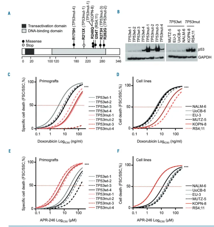
We investigated 62 patient-derived pediatric BCP-ALL samples, which were established in our NOD/SCID/huALL xenograft model from patients at diagnosis (n=53) or relapse (n=9). TP53mut cases were identified by denaturing high-performance liquid chromatography and confirmed by Sanger sequencing (exons 4-10). Four TP53mut cases were found, one derived from a patient at second relapse (TP53mut-1) and three at diagnosis (TP53mut-2, -3, -4) (Online Supplementary Table S1). In parallel, we character-ized six BCP-ALL cell lines and identified two TP53mut (RS4;11, KOPN-8) and four TP53wt (MUTZ-5, EU-3, UoCB-6 and NALM-6) lines. All samples carried missense mutations previously described (p53.iarc.fr),15,43 localized
within the region encoding the DNA-binding domain, sug-gesting loss of p53’s tumor suppressive function (Figure 1A, Table 1). In the TP53mut samples, the second allele carried a nonsense mutation (TP53mut-1), was absent (loss of 17p, TP53mut-2, -3), or carried the same missense mutation (TP53mut-4) (Table 1). Somatic and germline TP53mut are
associated with (low) hypodiploid ALL.15,19-21 One
primo-graft sample (TP53mut-3) showed a hypodiploid karyotype with 44 chromosomes (Table 1). In line with disrupted degradation and accumulation of mutant p53 protein, TP53mut cases showed higher p53 protein levels compared to TP53wt leukemias (Figure 1B).
Figure 1. TP53-mutated acute lymphoblastic leukemias are DNA-damage resistant but sensitive to APR-246.(A) All TP53mut B-cell precursor (BCP) acute lym-phoblastic leukemia (ALL) primograft and cell lines harbor missense mutations (filled circles) localized in the DNA-binding domain of TP53. The primograft sample
TP53mut-1 carries an additional stop mutation (R213X, open circle). See also Table 1. (B) Increased p53 protein expression in TP53mut compared to TP53wt ALL in primograft (left) and cell line (right) leukemia samples. Western blot, anti-p53 antibody (total, clone DO-7) with GAPDH as a loading control. (C-F) Significantly high-er half maximal inhibitory concentrations (IC50) for doxorubicin in TP53mut (red curves) primograft (C) and cell line (D) BCP-ALL, and significantly lower IC50values
for APR-246 in TP53mut primograft (E) and cell line (F) samples, indicating insensitivity to the DNA-damaging agent doxorubicin but sensitivity to APR-246 in
TP53mut BCP-ALL. Dose-response curves reflect cell death induction in response to increasing concentrations summarizing one (primografts, 24 h; C, E) or three (cell lines, 48 h; D, F) independent experiments, each performed in triplicate. Comparison of sensitivities of TP53wt and TP53mut leukemias, F-test, ***P<0.001. See also Online Supplementary Table S2.
A B
D C
TP53-
mutated leukemias are sensitive to APR-246 but
not to genotoxic therapy
In response to genotoxic agents and stress, wildtype p53 suppresses cellular viability and proliferation. However, dysfunctional, mutant p53 fails to mediate tumor-suppres-sive functions such as induction of cell death. Therefore, we analyzed cell death in TP53mut and TP53wt ALL pri-mografts (TP53mut n=4, TP53wt n=4) and cell lines (TP53mut n=2, TP53wt n=4) in response to increasing concentrations of the DNA-damaging agent doxorubicin, a standard genotoxic drug regularly used in ALL treatment protocols, and to APR-246. All TP53mut primografts and cell lines showed, as expected, insensitivity to doxorubicin indicated by significantly higher IC50values, in contrast to
doxorubicin-sensitive TP53wt leukemias (Figure 1C, D; Online Supplementary Table S2A, B). An opposite effect was observed upon exposure to APR-246 with high sensitivity and cell death induction in all TP53mut leukemias, but low APR-246 sensitivity in TP53wt ALL (Figure 1E, F; Online Supplementary Table S2C, D). Interestingly, diagno-sis- (TP53mut-2, -3, -4) or relapse-derived (TP53mut-1) pri-mograft samples did not show differences in APR-246 or doxorubicin sensitivity.
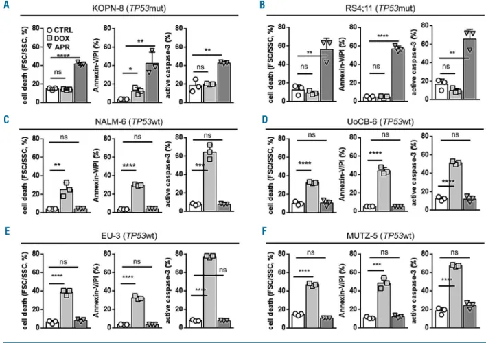
Activation of the p53 pathway results in apoptosis induction. Along with cell death, APR-246 led to annexin-V/propidium iodide positivity and caspase-3 activation indicating apoptosis induction in TP53mut ALL. In con-trast, apoptosis was induced by doxorubicin but not APR-246 in TP53wt cells (Figure 2 and Online Supplementary Figure S1).
Thus, all identified TP53mut leukemias carried missense mutations in the DNA-binding domain, showed accumu-lation of p53 indicative of dysfunctional mutant p53, resistance to the genotoxic agent doxorubicin, and were highly sensitive to APR-246-induced apoptosis.
APR-246 restores p53’s wildtype conformation
reactivating tumor suppressive functions
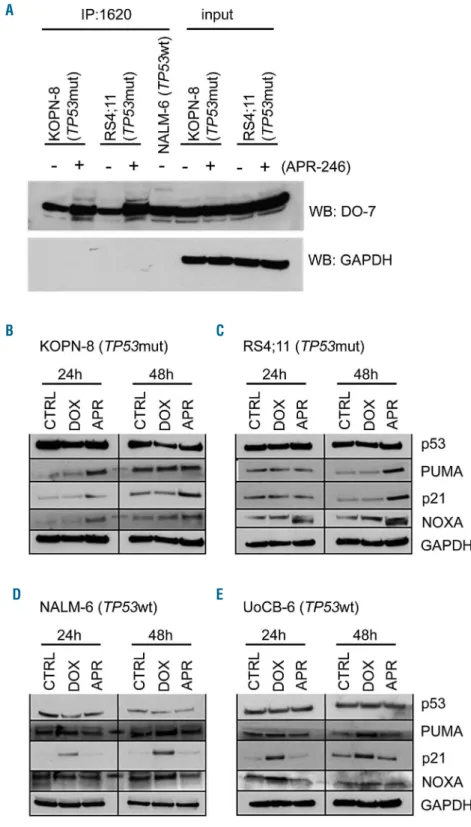
We further addressed the mode of action of APR-246 in
TP53mut ALL and examined the conformation of p53 and activation of the pathway in response to APR-246. Using a p53 wildtype conformation-specific antibody (PAb1620), larger amounts of p53 with wildtype confor-mation were immunoprecipitated from lysates of TP53mut ALL cells exposed to APR-246, indicating recon-stitution of p53 wildtype conformation in TP53mut ALL by APR-246 (Figure 3A). However, this effect was not observed in TP53wt leukemia cells (Online Supplementary Figure S2). Next, we assessed expression of the p53 tran-scriptional targets PUMA (P53-Upregulated Modulator of Apoptosis), p21 (Cyclin Dependent Kinase Inhibitor 1A, CDKN1), and NOXA upon APR-246 or doxorubicin treat-ment in TP53mut (KOPN-8, RS4;11) and TP53wt (NALM-6, UoCB-6) ALL lines. In TP53mut ALL, APR-246 led to induction of all p53 targets (Figure 3B, C and Online Supplementary Figure S3). In contrast, an opposite picture of activation of p53 transcriptional targets in TP53wt but not in TP53mut leukemias was observed upon incubation with doxorubicin (Figure 3D, E and Online Supplementary Figure S3). Thus, APR-246 induces restoration of mutant p53 to wildtype conformation, transcriptional target expression, and apoptosis in TP53mut ALL.
Induction of oxidative stress contributes
to APR-246-mediated cellular death
Induction of oxidative stress has been described as a sec-ond activity of APR-246 in different cancers.30-32APR-246
was reported to interfere with different regulators of the cellular redox system, such as thioredoxin reductase, thioredoxin and glutathione, and with the transcription factor NRF2, leading to induction of reactive oxygen species (ROS).28,30-33,44-47 Given the activity of APR-246 in TP53mut ALL, we addressed whether TP53mut and TP53wt ALL display distinct sensitivities in response to ROS generation. Upon treatment with 3-morpholinosyd-nonimine, a spontaneous generator of reactive oxygen and nitrogen species, and the oxidant tert-butyl hydroxyper-oxide, increased ROS levels were observed in both
Table 1. TP53mutations in acute lymphoblastic leukemia cell lines and primograft samples.
Sample Exon Mut. (bp) Mut. (aa) Region Mut. type del (17p) Genotype Number of chr.s.
TP53mut-1 exon 6 c.637C>T p.R213X DBD stop - heterozygous 47
exon 7 c.743G>A p.R248Q missense TP53mut-2 exon 8 c.844C>G p.R282G DBD missense del hemizygous 47
TP53mut-3 exon 8 c.818G>A p.R273H DBD missense del hemizygous 44
TP53mut-4 exon 5 c.524G>A p.R175H DBD missense - homozygous 46
RS4;11 exon 7 c.761T>C p.I254T DBD missense - heterozygous 47
KOPN-8 exon 7 c.743G>A p.R248Q DBD missense - heterozygous 46 Mut: mutation; bp: base pair; aa: amino acid; del: deletion; DBD,: DNA-binding domain; Chr.s: chromosomes.
Table 2. TP53mutations in primary samples from patients with acute lymphoblastic leukemia.
Sample Exon Mut. (bp) Mut. (aa) Region Mut. type del (17p) Genotype Diploidy
Patient-1 exon 4 c.375G>A p.T125T DBD splice site del hemizygous diploid Patient-2 exon 7 c.743G>A p.R248Q DBD missense - heterozygous diploid Patient-3 exon 7 c.743G>A p.R248Q DBD missense - heterozygous diploid Patient-4 exon 7 c.733G>C p.G245R DBD missense - heterozygous diploid Mut: mutation; bp: base pair; aa,: amino acid; del: deletion; DBD: DNA-binding domain.
TP53mut and TP53wt ALL cells, leading to similar cell death rates (Online Supplementary Figure S4).
Next, we investigated whether induction of oxidative stress is involved in APR-246-mediated cell death in TP53mut ALL. Importantly, methyl quinuclidinone, the active drug spontaneously formed from APR-246, binds covalently to cysteine residues in the core domain of p53, but also to cysteines in the widely used antioxidant and ROS inhibitor N-acetylcysteine (NAC).28,44 Thus, NAC
directly blocks APR-246 activity and cannot be used to investigate the role of ROS in APR-246-mediated cell death. Therefore, the synthetic antioxidant compound and ROS inhibitor superoxide dismutase mimetic Mn (III) tetrakis (5, 10, 15, 20-benzoic acid) porphyrin (MnTBAP) was used. Cell death was analyzed together with ROS lev-els in TP53mut (KOPN-8 and RS4;11) and TP53wt (NALM-6 and UoCB(NALM-6) leukemia cells exposed to APR-24(NALM-6 with or without NAC or MnTBAP. Similar ROS levels were observed upon APR-246 treatment in bothTP53mut and TP53wt ALL (Online Supplementary Figure S5A, C, E, G), however induction of cell death was only seen in TP53mut cells (Online Supplementary Figure S5F, H) but not in TP53wt cells (Online Supplementary Figure S5B, D). Interestingly, ROS inhibition by MnTBAP partially inhibited APR-246-induced cell death in TP53mut ALL, indicating that ROS
contribute to APR-246- induced cell death. It was also interesting that, even in the presence of MnTBAP, i.e. in the absence of ROS, APR-246 retained a statistically significant cytotoxic effect (Online Supplementary Figure S5F, H). In line with previous reports,44the activity of APR-246 was
com-pletely blocked by NAC.
Taken together, these data show that induction of oxidative stress might contribute to APR-246-mediated cell death in ALL, in line with previously reported data of a dual mode of action of APR-246 in other malignan-cies.28,30-33,44-46
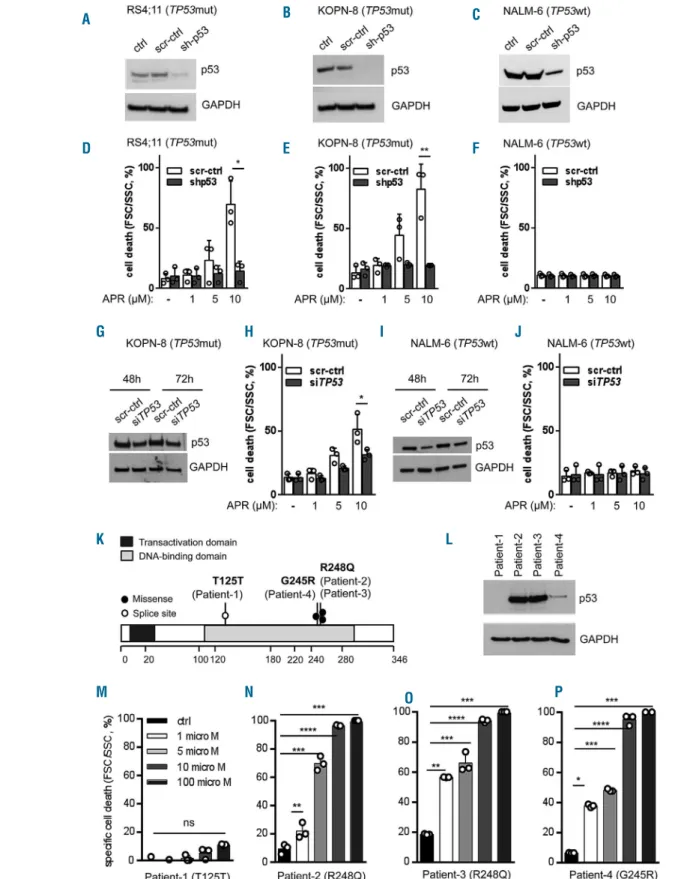
APR-246 activity depends on mutant p53
Activity of APR-246 was observed in TP53mut but not TP53wt ALL. Therefore, we analyzed the effect of APR-246 in TP53mut and TP53wt cell lines upon lentiviral shRNA-mediated knockdown of p53 (Figure 4A-C). In both TP53mut lines (KOPN-8 and RS4;11) depletion of p53 led to APR-246 insensitivity and cell death resistance, in contrast to dose-dependent cell death induction in con-trol-transduced cells (Figure 4D, E). However, TP53wt cells with p53 depletion were unresponsive to APR-246, like the corresponding control transduced cells (Figure 4F). A similar result was observed upon siRNA-mediated p53 downregulation with clearly lower cell death induction in
Figure 2. APR-246 induces apoptosis in TP53-mutated acute lymphoblastic leukemia.(A-F) Induction of cell death (left diagrams, forward/side scatter criteria, flow cytometry), annexin-V/propidium iodide (PI) positivity (middle diagrams) and caspase-3 activation (right diagrams) by APR-246 in TP53mut cell lines KOPN-8 (A) and RS4;11 (B), in contrast to cell death and apoptosis induction by doxorubicin in TP53wt lines NALM-6 (C), UoCB-6 (D), EU-3 (E), and MUTZ-5 (F). Proportions of cells after 48 h exposure to solvent (CTRL), APR-246 (APR, 5 mM), or doxorubicin (DOX, 15 ng/mL). Mean values ± standard deviation of three independent experiments, each performed in triplicate. Student t-test, ****P<0.0001; ***P<0.001; **P<0.01; *P<0.05; n.s., not significant.
A B
D C
TP53mut ALL but no effect in TP53wt cells (Figure 4G-J and Online Supplementary Figure S6). Together with our observations on APR-246 insensitivity in TP53wt ALL (Figure 1E, F) and the absence of p53 transcriptional target expression upon APR-246 treatment in TP53wt ALL (Figure 3D, E), these findings indicate that the activity of APR-246 is associated with the presence of mutant p53. Accordingly, distinct sensitivities to APR-246 were found in four primary ALL samples obtained from patients with therapy-resistant disease or relapse carrying different TP53mutations (Figure 4L, Table 2). Robust
dose-depen-dent cell death induction was observed in leukemia cells from patients 2, 3, and 4 carrying missense mutations resulting in expression of mutant p53 (Figure 4L, N-P), whereas APR-246 did not induce cell death in ALL cells of patient 1 carrying a hemizygous splice site mutation with-out detectable expression of p53 protein (Figure 4L, M).
APR-246 re-sensitizes
TP53
-mutated acute
lymphoblastic leukemia to doxorubicin
TP53mut cancer cells show resistance to DNA damage. Therefore, we analyzed whether reactivation of mutant
Figure 3. Conformational and functional restoration of mutant p53 by APR-246.
(A) Increased levels of p53 with wildtype conformation in TP53mut ALL (KOPN-8, mutation R248Q; RS4;11, mutation I254T) upon exposure to APR-246 (5 mM, 24 h). Immunoprecipitation (IP, anti-wt p53 specific antibody PAb1620) and western blot analysis (WB, p53 body DO-7, light chain-specific goat anti-mouse peroxidase conjugated binding protein), GAPDH expression in input lysates and absence in precipitates, NALM-6 serves as a wildtype p53 positive control. (B-E) Expression of p53 transcrip-tional targets PUMA, p21 and NOXA in (B, C) TP53mut ALL upon APR-246 treatment and in (D, E) TP53wt ALL upon doxoru-bicin treatment. Western blot, exposure to solvent (CTRL), doxorubicin (DOX, 15 ng/mL), or APR-246 (APR, 5 mM) for the indicated times, with GAPDH as a loading control. The results of one representative out of two independent experiments are shown. See also Online Supplementary Figures S2 and S3. A B D C E
Figure 4. APR-246 activity depends on mutant p53. (A-C) Stable lentiviral shRNA-mediated p53 knockdown in TP53mut (RS4;11 and KOPN-8) and TP53wt (NALM-6) cell lines. Western blot, anti-p53 antibody DO-7, with GAPDH as a loading control, non-transduced cells (ctrl), cells transduced with scrambled control (scr-ctrl) and TP53-specific shRNA (sh-p53). (D-F) Increasing cell death (forward/side scatter criteria, flow cytometry) in control transduced cells and abrogated cell death induction upon p53 knockdown at increasing concentrations of APR-246 (APR, 48 h) in TP53mut but not TP53wt cells. Mean values ± standard deviation (SD) of three independent experiments, each performed in triplicate. Student t-test, *P<0.05; **P<0.01. (G-J) si-RNA-mediated p53 downregulation in TP53mut (KOPN-8), (G) and TP53wt (NALM-6), (I) cells leading to clearly lower cell death induction upon p53 downregulation as compared to higher cell death in control cells (H), while p53 downregulation in TP53wt cells did not affect cell death induction upon APR-246 treatment (J). Mean values ± SD of three independent experiments. Student
t-test, *P<0.05; **P<0.01. (K) TP53mutations identified in primary samples from patients with acute lymphoblastic leukemia (ALL): the mutations were localized in the DNA-binding domain with one splice site mutation (open circle, Patient-1) and three missense mutations (filled circles, Patients -2, -3, -4). (L) No detectable p53 protein in ALL cells from Patient-1 (western blot, anti-p53 antibody DO-7, GAPDH as a loading control), and (M) no APR-246 activity in these cells (Patient-1), in contrast to cell death induction in cases carrying missense hot spot TP53mutations (N, O, P; Patients-2, -3, -4). Mean values ± SD, measurements performed in triplicate. Student t-test, ****P<0.0001; ***P<0.001; **P<0.01; *P<0.05; n.s., not significant.
A B D C E F G H I J K L M N O P
p53 re-sensitizes TP53mut ALL to the DNA-damaging agent doxorubicin, which is also used in treatment of pediatric ALL. TP53mut and TP53wt ALL cell lines and primograft samples were exposed to APR-246, doxoru-bicin, or to combinations of both at increasing concentra-tions. Strongly increased cell death rates were observed in all four TP53mut primografts and two cell lines upon
com-bination treatment with 246, as compared to APR-246 or doxorubicin alone, indicating synergistic activity for APR-246 and doxorubicin in TP53mut ALL (Figure 5A-F,Online Supplementary Table S3A). In TP53wt leukemias however, only doxorubicin showed cell death-inducing activity, which was not increased by adding APR-246 (Figure 5G-L, Online Supplementary Table S3B).
Figure 5. APR-246 synergizes with doxorubicin. Synergistic activity of APR-246 in combination with the DNA-damage-inducing agent doxorubicin in TP53mut B-cell precursor (BCP)-acute lymphoblastic leukemia (ALL) cell lines (A, B) and TP53mut primograft leukemias (C-F) leading to doxorubicin re-sensitization, in contrast to no increased activity compared to treatment with doxorubicin alone in TP53wt cell lines (G, H) and TP53wt primograft ALL (I-L). Cell death (forward/side scatter cri-teria, flow cytometry) after exposure (primografts 24 h, cell lines 48 h) at indicated concentrations of APR-246 (APR), doxorubicin (DOX) or the combination (COMBI, 3 h APR-246 pre-incubation). Mean values ± standard deviation (SD) of three independent experiments, each performed in triplicate (cell lines: A, B, G, H). Mean values ± SD, three measurements (primografts: C-F, I-L). Combination indices (CI) indicating a strong synergistic (CI 0.1-0.3), a synergistic (CI <1), an additive (CI=1) or an antagonistic effect (CI>1) upon combination.
A B D C E F G H I J K L
We also addressed whether induction of oxidative stress would increase the antileukemia activity of APR-246. In contrast to clearly increased cell death upon treatment with APR-246 together with the DNA-damaging agent doxorubicin, combining APR-246 with the ROS inducers 3-morpholinosydnonimine and tert-butyl hydroxyperox-ide did not lead to clearly increased cell death (Online Supplementary Figure S7). Thus, APR-246 effectively syner-gizes with doxorubicin and re-sensitizes TP53mut ALL to DNA-damage-induced cell death, while additional ROS induction did not increase APR-246-mediated leukemia cell death.
Preclinical antileukemia activity of APR-246
and
in vivo
synergy with genotoxic therapy
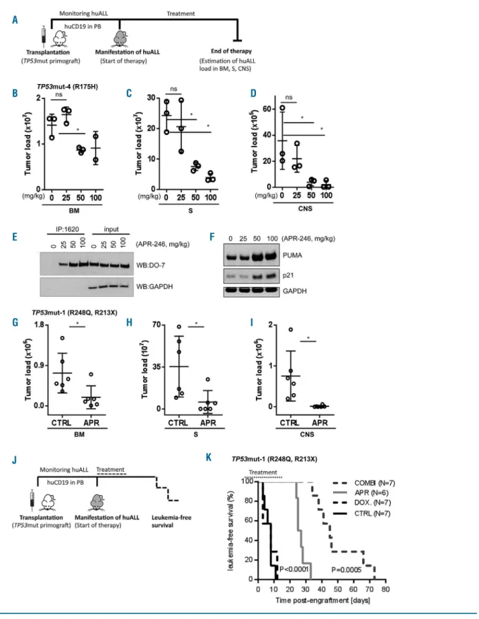
Based on our findings, we investigated the antileukemia activity of APR-246 in a preclinical setting in vivo. Mice were transplanted with the TP53mut primograft (TP53mut-4; R175H). Upon manifestation of leukemia, as indicated by 5% or more human CD19+ALL cells in the
recipients’ peripheral blood, mice were treated with APR-246 (25, 50 or 100 mg/kg) or vehicle until control-treated animals showed signs of leukemia-related morbidity (3 weeks, days 1-5) (Figure 6A). Upon APR-246 treatment, a clear dose-dependent reduction of leukemia loads was observed in all three organ compartments: spleen, bone marrow and central nervous system (Figure 6B-D). Moreover, in leukemia cells isolated from these APR-246-treated animals, dose-dependent increases in mutant p53 with wildtype conformation and expression of PUMA and p21 were detected (Figure 6E, F), indicating restoration of wildtype p53 conformation and function in vivo.
Furthermore, APR-246 demonstrated strong in vivo antileukemia activity in another TP53mut ALL primograft sample (TP53mut-1; R248Q/R213X) leading to significant-ly reduced leukemia loads in the spleen, bone marrow and central nervous system upon therapy of leukemia-bearing recipients (Figure 6G-I).
We also addressed the effects of APR-246 in combina-tion with doxorubicin in vivo. Recipients with manifest ALL (TP53mut-1; R248Q/R213X; 5% or more human ALL cells in the peripheral blood) were treated with APR-246, doxorubicin, or the combination of both for 3 weeks. After treatment, the animals were followed up and the time until onset of ALL-related morbidity was analyzed for each animal (Figure 6J). Upon sacrifice, high loads of human ALL were detected in the spleen and bone marrow of all recipients, confirming recurrence of manifest leukemia at clinical onset. Importantly, in addition to clear antileukemia activity as a single agent, leading to increased post-treatment survival (P<0.0001), APR-246 synergized strongly with doxorubicin and re-sensitized TP53mut ALL to genotoxic therapyin vivo, resulting in sig-nificantly prolonged survival as compared to APR-246 alone (P=0.0005) (Figure 6K). In all treatment experiments, application of APR-246 was well tolerated and no side effects were observed in the recipients.
Taken together, our findings in ALL carrying TP53 mis-sense mutations in the DNA-binding domain, which lead to accumulation of dysfunctional p53, indicate that target-ing mutant p53 with APR-246 results in refoldtarget-ing of mutant p53 into its native wildtype conformation, induc-tion of p53 transcripinduc-tional targets, involvement of oxida-tive stress, induction of apoptosis, sensitization to DNA damage and, most importantly, preclinical antileukemia
activity with significant reduction of leukemia loads, re-sensitization to genotoxic therapy and clearly prolonged survivalin vivo. Thus, application of APR-246 can provide an effective strategy for directed therapeutic intervention in the high-risk subtype of TP53mut BCP-ALL.
Discussion
Investigating a large cohort of 62 patient-derived BCP-ALL samples, all identified TP53mut cases showed mis-sense mutations leading to alterations in the DNA-binding domain of p53, high levels of p53 protein and insensitivity to doxorubicin. Interestingly, APR-246 demonstrated robust antileukemia activity in these cases, including induction of apoptosis, effective reduction of leukemia loads, and sensitization to doxorubicin in an in vivomodel of TP53mut ALL. Both in vitro and in vivo experiments showed that treatment with APR-246 led to restored con-formation and activation of mutant p53, and induction of transcriptional targets.
Alterations in TP53have been described in diverse can-cers at high frequencies of up to 95%.7In our cohort, TP53
mutations were identified in four out of 62 cases (6.5%), in line with reported rates in ALL of 6-16%.14-16,18All
muta-tions identified in the primograft and cell line samples were missense mutations localized in the DNA-binding domain, with additional loss of the second allele in some of the cases, consistent with mutational patterns reported throughout different cancer types.10,43OneTP53mut
sam-ple showed hypodiploidy, in line with reported associa-tions of hypodiploidy with germline and somatic TP53 mutations.19-21
Mutated dysfunctional p53 results in resistance to ther-apy-induced DNA damage48and poor patient outcome.
14,16-18,22 Correspondingly, increased numbers of TP53
alter-ations are seen at ALL relapse16and all TP53mut leukemias
were insensitive to the DNA-damaging agent doxoru-bicin. Importantly, these TP53mut leukemias were sensi-tive to APR-246, likely by reactivation of high levels of dysfunctional p53 accumulated in the cells. Most impor-tantly, in line with reports in ovarian cancer,32,39APR-246
clearly synergized with doxorubicin in vitro, ex vivoand in vivo, re-sensitizing initially resistant TP53mut ALL to DNA damage. Therefore, combining functional p53 restoration with genotoxic therapies triggering the p53-mediated DNA-damage response would be the rationale to apply APR-246 together with doxorubicin, a classical DNA-damage-inducing agent used in ALL treatment regimens. Importantly, a favorable pharmacological profile and anti-tumor effects were observed upon first clinical use of APR-246 in patients with refractory cancers40and APR-246
is being tested in combination with anticancer agents, including doxorubicin, in ongoing phase II trials (ClinicalTrials.gov).41
We addressed the molecular mechanism of action of APR-246 and demonstrated restoration of p53 wildtype conformation, p53 pathway activation with induction of downstream transcriptional targets, and a contribution of oxidative stress leading to apoptosis of TP53mut BCP-ALL cells. Importantly, this antileukemia effect was also observed in vivoin TP53mut ALL, but not in TP53wt ALL, upon p53 knockdown or in a patient’s sample with a splice site mutation and loss of p53 protein expression. High levels of misfolded mutant p53 were described to be
Figure 6. Anti-leukemia activity of APR-246 and synergy with genotoxic therapy in TP53-mutated acute lymphoblastic leukemia in vivo.(A) Schematic representa-tion of the experimental procedure: endpoint analysis assessing leukemia loads in differently treated recipients. (B-D) Dose-dependent reducrepresenta-tion of leukemia load in bone marrow (BM) (B), spleen (S) (C) and central nervous system (CNS) (D) upon treatment of mice bearing TP53mut-4 (mutation R175H) acute lymphoblastic leukemia (ALL) with solvent or increasing doses of APR-246 for 3 weeks as indicated (n=3 recipients per group, except n=2 for BM 100 mg/kg). Student t-test, *P<0.05; n.s., not significant. (E) Restoration of p53 wildtype conformation upon in vivo APR-246 therapy, immunoprecipitation (IP: anti-wt p53 specific antibody PAb1620, western blot: anti-p53 antibody DO-7, light chain-specific goat anti-mouse peroxidase conjugated binding protein, GAPDH as a loading control), and (F) dose-dependent induction of p53 transcriptional targets PUMA and p21 (western blot, GAPDH as a loading control). (G-I) Significant reduction of leukemia load in bone marrow (BM) (G), spleen (S) (H) and central nervous system (CNS) (I) upon treatment of TP53mut-3 (mutations R248Q, R213X) ALL-bearing mice with APR-246 (APR, 100 mg/kg) or solvent (CTRL) for 3 weeks, n=6 mice per group, Student t-test, *P<0.05. (J) Schematic representation of the experimental procedure: survival analysis. (K) Superior survival of animals treated with APR-246 (APR, 50 mg/kg, 3 weeks, days 1-5; n=6) as compared to doxorubicin (DOX, 2 mg/kg, 3 weeks, day 1; n=7) or vehicle (CTRL, 3 weeks, days 1-5; n=7) (P<0.0001); and synergy of the combination of APR-246 and doxorubicin (COMBI, APR-246, 50 mg/kg, days 1-5, and doxorubicin, 2 mg/kg, day 5; n=7) leading to increased survival as compared to APR-246 treatment alone (P=0.0005). Kaplan-Meier analysis, log-rank test.
A B C D G H I K J E F
associated with APR-246 sensitivity in cancer cell lines,38,49,50 consistent with our observation of high
APR-246 activity in TP53mut ALL with high p53 expression. However, evaluation of larger cohorts of patients together with outcome data would be required to explore the value of the level of expression of p53 as an indicator of APR-246 responsiveness in TP53mut ALL.
APR-246 activity has been reported to be mediated independently of p53 by induction of oxidative stress in other types of cancer, including acute myeloid leukemia and multiple myeloma.30-33 However, we only observed
cell death in TP53mut ALL, although TP53mut and TP53wt ALL showed no differences in induction of and sensitivity to oxidative stress. Interestingly, APR-246 activity in TP53mut ALL was partially inhibited by ROS neutralization. This suggests that induction of oxidative stress contributes to APR-246-mediated cell death in ALL, in line with reports on a dual mode of action of APR-246, 30-32which might vary between tumor types and cellular
con-text.
The presence of p53 in a mutated, dysfunctional form, as typically is the case for missense mutations in the DNA-binding core domain, enables binding of the active moiety of APR-246, leading to activity in BCP-ALL.11,28
This is of clinical relevance, since the majority of TP53 mutations in BCP-ALL are missense hot spot mutations in the DNA-binding domain15,43resulting in accumulation of
misfolded p53 protein, which is targeted by APR-246. However, the precise mechanism of activity on DNA con-tact mutations is not yet known. Importantly, we and oth-ers 27have demonstrated antitumor activity on structural
and contact mutants including clear preclinical
antileukemia activity on TP53mut ALL carrying either a structure (R175H) or contact (R248Q) mutation.
Taken together, our study shows that the small molecule APR-246 exhibits profound antileukemia activity in TP53mut BCP-ALL, targeting non-functional mutant p53 resulting from missense mutations in the DNA-binding domain of TP53, the most frequent mutation type reported throughout different malignancies. Mechanistically, we showed that APR-246 led to restoration of p53’s wildtype conformation, pathway activation with expression of tran-scriptional targets and induction of apoptosis in TP53mut ALL. Moreover, we found a clear synergism between APR-246 and doxorubicin treatment, strongly suggesting that the combination of p53 reactivation and DNA-damage induction could be an effective antileukemia strategy for BCP-ALL patients with TP53missense mutations. Hence, targeting mutant p53 appears to be a promising, directed treatment for this high-risk subgroup of TP53mut ALL.
Acknowledgments
The authors would like to thank Aprea Therapeutics (Stockholm, Sweden) for kindly providing APR-246 for the study, S. Volk and S. Essig for excellent technical assistance, the Ulm University Sorting and Animal Facilities and Pharmacy of the Ulm University Medical Center, and the INFORM study group. The authors would also like to thank the International Graduate School in Molecular Medicine Ulm (SD, EB), Madeleine-Schickedanz-Stiftung and “Förderverein für Krebskranke Kinder Tübingen” (ME, RH), Swedish Research Council and Swedish Childhood Cancer Society (GS), EU COST Action CA16223 (GtK), the German Research Foundation, SFB 1074 B6 (LHM, KMD) and B1 (SS) for supporting the work.
References
1. Hunger SP, Mullighan CG. Acute lym-phoblastic leukemia in children. N Engl J Med. 2015;373(16):1541-1552.
2. Lane DP, Crawford LV. T antigen is bound to a host protein in SV40-transformed cells. Nature. 1979;278(5701):261-263.
3. Linzer DI, Levine AJ. Characterization of a 54K dalton cellular SV40 tumor antigen pres-ent in SV40-transformed cells and uninfect-ed embryonal carcinoma cells. Cell. 1979;17(1):43-52.
4. Finlay CA, Hinds PW, Levine AJ. The p53 proto-oncogene can act as a suppressor of transformation. Cell. 1989;57(7):1083-1093. 5. Chen PL, Chen YM, Bookstein R, Lee WH.
Genetic mechanisms of tumor suppression by the human p53 gene. Science. 1990;250 (4987):1576-1580.
6. Isobe M, Emanuel BS, Givol D, Oren M, Croce CM. Localization of gene for human p53 tumour antigen to band 17p13. Nature. 1986;320(6057):84-85.
7. Kandoth C, McLellan MD, Vandin F, et al. Mutational landscape and significance across 12 major cancer types. Nature. 2013;502(7471):333-339.
8. Nigro JM, Baker SJ, Preisinger AC, et al. Mutations in the p53 gene occur in diverse human tumour types. Nature. 1989;342 (6250):705-708.
9. Baker SJ, Fearon ER, Nigro JM, et al.
Chromosome 17 deletions and p53 gene mutations in colorectal carcinomas. Science. 1989;244(4901):217-221.
10. Hollstein M, Sidransky D, Vogelstein B, Harris CC. p53 mutations in human cancers. Science. 1991;253(5015):49-53.
11. Levine AJ. p53, the cellular gatekeeper for growth and division. Cell. 1997;88(3):323-331.
12. Cho Y, Gorina S, Jeffrey PD, Pavletich NP. Crystal structure of a p53 tumor suppressor-DNA complex: understanding tumorigenic mutations. Science. 1994;265(5170):346-355. 13. Kim MP, Zhang Y, Lozano G. Mutant p53: multiple mechanisms define biologic activi-ty in cancer. Front Oncol. 2015;5:249. 14. Chiaretti S, Brugnoletti F, Tavolaro S, et al.
TP53 mutations are frequent in adult acute lymphoblastic leukemia cases negative for recurrent fusion genes and correlate with poor response to induction therapy. Haematologica. 2013;98(5):e59-61. 15. Stengel A, Schnittger S, Weissmann S, et al.
TP53 mutations occur in 15.7% of ALL and are associated with MYC-rearrangement, low hypodiploidy, and a poor prognosis. Blood. 2014;124(2):251-258.
16. Hof J, Krentz S, van Schewick C, et al. Mutations and deletions of the TP53 gene predict nonresponse to treatment and poor outcome in first relapse of childhood acute lymphoblastic leukemia. J Clin Oncol. 2011;29(23):3185-3193.
17. Richter-Pechanska P, Kunz JB, Hof J, et al.
Identification of a genetically defined ultra-high-risk group in relapsed pediatric T-lym-phoblastic leukemia. Blood Cancer J. 2017;7(2):e523.
18. Stengel A, Kern W, Haferlach T, Meggendorfer M, Fasan A, Haferlach C. The impact of TP53 mutations and TP53 dele-tions on survival varies between AML, ALL, MDS and CLL: an analysis of 3307 cases. Leukemia. 2017;31(3):705-711.
19. Muhlbacher V, Zenger M, Schnittger S, et al. Acute lymphoblastic leukemia with low hypodiploid/near triploid karyotype is a specific clinical entity and exhibits a very high TP53 mutation frequency of 93%. Genes Chromosomes Cancer. 2014;53(6): 524-536.
20. Holmfeldt L, Wei L, Diaz-Flores E, et al. The genomic landscape of hypodiploid acute lymphoblastic leukemia. Nat Genet. 2013;45(3):242-252.
21. Qian M, Cao X, Devidas M, et al. TP53 germline variations influence the predisposi-tion and prognosis of B-cell acute lym-phoblastic leukemia in children. J Clin Oncol. 2018;36(6):591-599.
22. Krentz S, Hof J, Mendioroz A, et al. Prognostic value of genetic alterations in children with first bone marrow relapse of childhood B-cell precursor acute lym-phoblastic leukemia. Leukemia. 2013;27(2): 295-304.
23. Gu L, Zhu N, Findley HW, Zhou M. MDM2 antagonist nutlin-3 is a potent inducer of
apoptosis in pediatric acute lymphoblastic leukemia cells with wild-type p53 and over-expression of MDM2. Leukemia. 2008;22 (4):730-739.
24. Bykov VJ, Wiman KG. Mutant p53 reactiva-tion by small molecules makes its way to the clinic. FEBS Lett. 2014;588(16):2622-2627. 25. Martins CP, Brown-Swigart L, Evan GI.
Modeling the therapeutic efficacy of p53 restoration in tumors. Cell. 2006;127(7): 1323-1334.
26. Xue W, Zender L, Miething C, et al. Senescence and tumour clearance is trig-gered by p53 restoration in murine liver car-cinomas. Nature. 2007;445(7128):656-660. 27. Bykov VJ, Issaeva N, Shilov A, et al.
Restoration of the tumor suppressor func-tion to mutant p53 by a low-molecular-weight compound. Nat Med. 2002;8(3):282-288.
28. Lambert JM, Gorzov P, Veprintsev DB, et al. PRIMA-1 reactivates mutant p53 by cova-lent binding to the core domain. Cancer Cell. 2009;15(5):376-388.
29. Zhang Q, Bykov VJN, Wiman KG, Zawacka-Pankau J. APR-246 reactivates mutant p53 by targeting cysteines 124 and 277. Cell Death Dis. 2018;9(5):439. 30. Tessoulin B, Descamps G, Moreau P, et al.
PRIMA-1Met induces myeloma cell death independent of p53 by impairing the GSH/ROS balance. Blood. 2014;124(10): 1626-1636.
31. Ali D, Mohammad DK, Mujahed H, et al. Anti-leukaemic effects induced by APR-246 are dependent on induction of oxidative stress and the NFE2L2/HMOX1 axis that can be targeted by PI3K and mTOR inhibitors in acute myeloid leukaemia cells. Br J Haematol. 2016;174(1):117-126. 32. Mohell N, Alfredsson J, Fransson A, et al.
APR-246 overcomes resistance to cisplatin and doxorubicin in ovarian cancer cells. Cell Death Dis. 2015;6:e1794.
33. Peng X, Zhang MQ, Conserva F, et al. APR-246/PRIMA-1MET inhibits thioredoxin reductase 1 and converts the enzyme to a dedicated NADPH oxidase. Cell Death Dis. 2013;4:e881.
34. Zandi R, Selivanova G, Christensen CL, Gerds TA, Willumsen BM, Poulsen HS. PRIMA-1Met/APR-246 induces apoptosis and tumor growth delay in small cell lung cancer expressing mutant p53. Clin Cancer Res. 2011;17(9):2830-2841.
35. Nahi H, Merup M, Lehmann S, et al. PRIMA-1 induces apoptosis in acute myeloid leukaemia cells with p53 gene dele-tion. Br J Haematol. 2006;132(2):230-236. 36. Bykov VJ, Zache N, Stridh H, et al.
PRIMA-1(MET) synergizes with cisplatin to induce tumor cell apoptosis. Oncogene. 2005;24 (21):3484-3491.
37. Izetti P, Hautefeuille A, Abujamra AL, et al. PRIMA-1, a mutant p53 reactivator, induces apoptosis and enhances chemotherapeutic cytotoxicity in pancreatic cancer cell lines. Invest New Drugs. 2014;32(5):783-794. 38. Liu DS, Read M, Cullinane C, et al. APR-246
potently inhibits tumour growth and over-comes chemoresistance in preclinical mod-els of oesophageal adenocarcinoma. Gut. 2015;64(10):1506-1516.
39. Fransson A, Glaessgen D, Alfredsson J, Wiman KG, Bajalica-Lagercrantz S, Mohell N. Strong synergy with APR-246 and DNA-damaging drugs in primary cancer cells from patients with TP53 mutant high-grade serous ovarian cancer. J Ovarian Res. 2016;9(1):27.
40. Lehmann S, Bykov VJ, Ali D, et al. Targeting p53 in vivo: a first-in-human study with p53-targeting compound APR-246 in refrac-tory hematologic malignancies and prostate cancer. J Clin Oncol. 2012;30(29):3633-3639. 41. Zarin DA, Tse T, Williams RJ, Rajakannan T. Update on trial registration 11 years after the ICMJE policy was established. N Engl J Med.
2017;376(4):383-391.
42. Meyer LH, Eckhoff SM, Queudeville M, et al. Early relapse in ALL is identified by time to leukemia in NOD/SCID mice and is char-acterized by a gene signature involving sur-vival pathways. Cancer Cell. 2011;19(2): 206-217.
43. Bouaoun L, Sonkin D, Ardin M, et al. TP53 variations in human cancers: new lessons from the IARC TP53 database and genomics data. Human Mutat. 2016;37(9):865-876. 44. Bykov VJ, Zhang Q, Zhang M, Ceder S,
Abrahmsen L, Wiman KG. Targeting of mutant p53 and the cellular redox balance by APR-246 as a strategy for efficient cancer therapy. Front Oncol. 2016;6:21.
45. Lisek K, Walerych D, Del Sal G. Mutant p53-Nrf2 axis regulates the proteasome machin-ery in cancer. Mol Cell Oncol. 2017;4(1): e1217967.
46. Liu DS, Duong CP, Haupt S, et al. Inhibiting the system xC(-)/glutathione axis selectively targets cancers with mutant-p53 accumula-tion. Nat Commun. 2017;8:14844. 47. Haffo L, Lu J, Bykov VJN, et al. Inhibition of
the glutaredoxin and thioredoxin systems and ribonucleotide reductase by mutant p53-targeting compound APR-246. Sci Rep. 2018;8(1):12671.
48. Hientz K, Mohr A, Bhakta-Guha D, Efferth T. The role of p53 in cancer drug resistance and targeted chemotherapy. Oncotarget. 2017;8(5):8921-8946.
49. Bykov VJ, Issaeva N, Selivanova G, Wiman KG. Mutant p53-dependent growth sup-pression distinguishes PRIMA-1 from known anticancer drugs: a statistical analy-sis of information in the National Cancer Institute database. Carcinogenesis. 2002;23 (12):2011-2018.
50. Synnott NC, Murray A, McGowan PM, et al. Mutant p53: a novel target for the treat-ment of patients with triple-negative breast cancer? Int J Cancer. 2017;140(1):234-246.