Abstract
Glioblastoma (GBM) is the most common and lethal malignant brain tumor. Current treatment involves surgical resection, radiation, and chemotherapy. Temozolomide (TMZ) is the only FDA approved systemic chemotherapeutic available and many tumors are known to have both extrinsic and intrinsic resistance to the TMZ. Resistance to TMZ has generated a pressing need for novel therapies. ONC201, an Akt/ERK dual inhibitor has shown promising preliminary results in phase II clinical trials and its potent analog, ONC212 is believed to be more clinically efficacious. However, in vitro studies with ONC212 are necessary prior to clinical application.
Normal human astrocytes (NHA) and normal human astrocytes with mutant Ras (NHA Ras), along with 6 established glioblastoma human cell lines (U251, U87, U373, LN18, LN229, and D54MG) were each evaluated for toxicity with 3 individual drugs (TMZ, ONC201, and ONC212) and IC50 values were calculated from toxicity curves. TMZ was found to be less effective in cell lines with high MGMT expression (LN18). ONC212 was found to be more potent than ONC201 and has the potential to be a possible treatment option for GBM. Short-term goals involve repeating the experiment with glioblastoma stem cells which better mimic tumor structure with the potential of beginning in vivo
animal studies and evaluating efficacy and blood brain barrier penetrance of ONC212, followed by clinical trials. The ultimate goal remains to develop effective novel therapies for patients with GBM.
Acknowledgements
Table of Contents
Abstract ... 1
Acknowledgements ... 1
Introduction ... 3
Materials and Methods ... 5
Results... 7
Discussion ... 12
Conclusion and Future Directions ... 14
Introduction
The majority of malignant brain tumors are gliomas, and most gliomas are
astrocytomas. Glioblastoma (GBM), also known as grade 4 astrocytoma, is the most common and lethal malignant brain tumor.1 The current median survival rate of patients with GBM is approximately 15 months after diagnosis. Standard of care involves
surgical resection, radiation, and chemotherapy. However,GBM is diffusely invasive and tumors are molecularly heterogenous, making recurrence inevitable and
development of effective therapeutics difficult.2-4 Additionally, difficulty with central nervous system (CNS) penetrance can make developing adequate drug therapies for GBM difficult.4 Despite the heterogenous nature of GBM, the current standard of care is the same across patients.2, 3 Temozolomide (TMZ), a mono-alkylating agent that
methylates nitrogenous bases, mainly guanine, is the only FDA approved systemic chemotherapeutic available. However, TMZ treatment often leads to harsh off-target effects including hemotoxicity and cytotoxicity.2, 5, 6
Additionally, many tumors also have intrinsic resistance to TMZ or acquire resistance over time. A common form of intrinsic resistance in GBM is obtained by overexpression of DNA repair protein O6-methylguanine methyltransferase (MGMT), which is capable of removing the lesion conferred by TMZ.2, 7, 8 MGMT expression is often governed by the methylation of the promoter region of the MGMT gene.9 Furthermore, while the
mechanisms are not well elucidated, many tumors acquire resistance to TMZ over the course of treatment. GBM cells have been shown to induce the expression of MGMT protein, decrease Tumor Necrosis Factor-Alpha-Induced Protein 3 (TNFAIP3)
effects associated with TMZ as well as intrinsic resistance in certain tumor cells, there is a pressing need to develop novel and potent therapies that provide targeted treatment with little to no detrimental secondary response.
To this end, ONC201 is a small molecule inhibitor within the imipridone class that is currently being tested in phase II clinical trials of patients with GBM.14, 15 ONC201 acts by crossing the blood brain barrier and inhibiting the phosphorylation of the
communicating Akt and ERK pathways, as well as inducing the dephosphorylation of Foxo3a. Dephosphorylated Foxo3a translocates into the nucleus and induces
transcription of the TNF-related apoptosis inducing ligand (TRAIL). ONC201 also activates EIF2α which stimulates downstream transcription factors ATF4 and CHOP and ultimately upregulates death receptor, DR5. The upregulation of both TRAIL and DR5 leads to apoptosis, regardless of p53 status.14-16
Recent studies utilizing ONC201 in breast cancer cell lines point to the direct targeting of mitochondria through suppression of multiple mtDNA genes, independent of TRAIL. ONC201 has been shown to target mitochondrial cellular respiration, making cells reliant on anaerobic respiration resistant to ONC201.17 However, GBM has specifically been shown to heavily rely on glycolysis for energy production.18 This is a potential roadblock which may occur when utilizing ONC201 to treat GBM.
investigation regarding the CNS penetrance, potential molecular targets, and toxicity of ONC212 is necessary prior to clinical trials.
Materials and Methods Tissue culture
Normal human astrocytes (NHA) and normal human astrocytes with mutant Ras (NHA Ras) cell lines were established and immortalized by expressing HPV oncogenes E6 and E7 to inhibit the TP53 and Retinoblastoma pathways and hTERT to maintain telomere length.22 In addition to NHA and NHA Ras, six established GBM cell lines (ECLs) were used: U251, U87, U373, LN18, LN229, and D54MG. Cell lines were maintained at 37°C and 5% CO2 in Dulbecco’s Modified Eagle’s Medium (DMEM) with 10% Fetal Bovine Serum (FBS) and 1% Penicillin-Streptomycin.
Proliferation and cell number optimization
colored product. MTS was added to cells and plates were incubated for 2 hours at 37°C. Absorbance was recorded at a wavelength of 490 nm in order to quantify cell counts.23
Drug treatment
Once cell number was optimized, adherent cells were plated onto a 96-well tissue culture treated plate at the optimized densities to have the same cell counts at time of treatment. Cells were treated with one of nine different concentrations of drug (serially diluted) or DMSO control the following day. Dimethyl sulfoxide (DMSO) was used as a solvent for the drug. Since DMSO is known to be toxic to cells at concentrations greater than 1%, it was also incorporated into the serial dilution.24 An equivalent concentration of DMSO was used with all cells in order to eliminate its presence as a confounding cause of cellular toxicity. MTS Cell Proliferation Assay Kit (Colorimetric) (197010) was, again, utilized to quantify the number of viable cells five days following treatment. The same procedure involving the MTS Cell Proliferation Assay Kit as conducted in
proliferation and cell number optimization experiments was used to quantify cell data.23
Statistics
A triplicate blank was averaged in each plate and subtracted from each well’s absorbance to eliminate the absorbance due to the background media color. To
calculations. Error bars on dot plots (panel B in each figure) are 95% confidence intervals.
Results
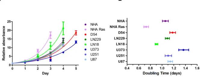
NHA was used as an approximation of a normal human astrocyte, similar to a negative control, while NHA Ras was used as an aggressively oncogenic astrocyte, similar to a positive control. NHA and NHA Ras were compared to the 6 ECLs.
A B
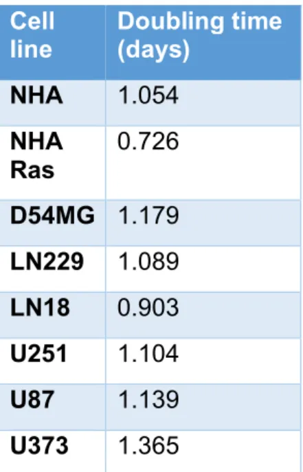
These doubling times were used to determine the plating cell densities 24 hours prior to drug treatment. Doubling times for NHA, NHA Ras, and ECLs are summarized in the table below.
Cell line
Doubling time (days)
NHA 1.054
NHA
Ras 0.726
D54MG 1.179 LN229 1.089 LN18 0.903 U251 1.104
U87 1.139
U373 1.365
Table 1 Proliferation curves were developed after plotting relative absorbance at 490 nm against time for each of the 8 cell lines (NHA, NHA Ras, U251, U87, U373, LN18, LN229, D54MG). Doubling times were calculated from these plots.
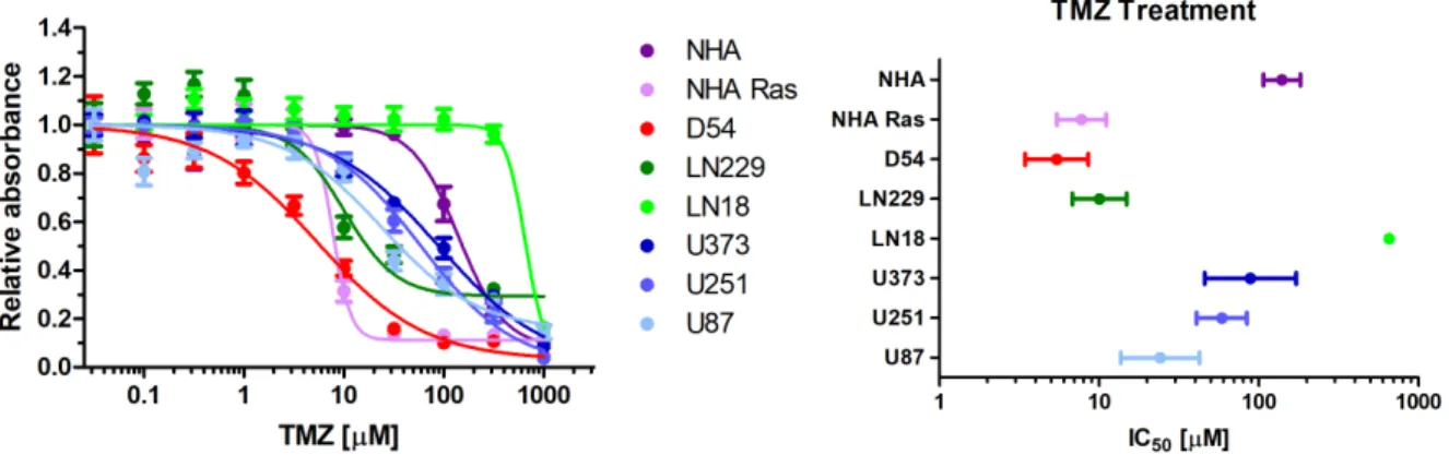
Figure 2 (A) Each cell line was treated with varying concentrations of TMZ and was plotted with relative absorbance of light at 490 nm to obtain toxicity graphs. Bars represent standard error. (B) IC50 values were calculated from the TMZ toxicity curves. IC50 values were calculated as follows in: NHA (139 µM), NHA Ras (7.77 µM), D54MG (5.43 µM), LN229 (10.1 µM), LN18 (654 µM), U251 (58.7 µM), U87 (24.2 µM), U373 (88.7 µM). Bars represent 95% confidence intervals.
IC50 values for TMZ ranged from minimum of 5.43 µM in D54MG to a maximum of 654
µM in LN18. NHA had an IC50 of 139 µM and NHA Ras had an IC50 of 7.77 µM.
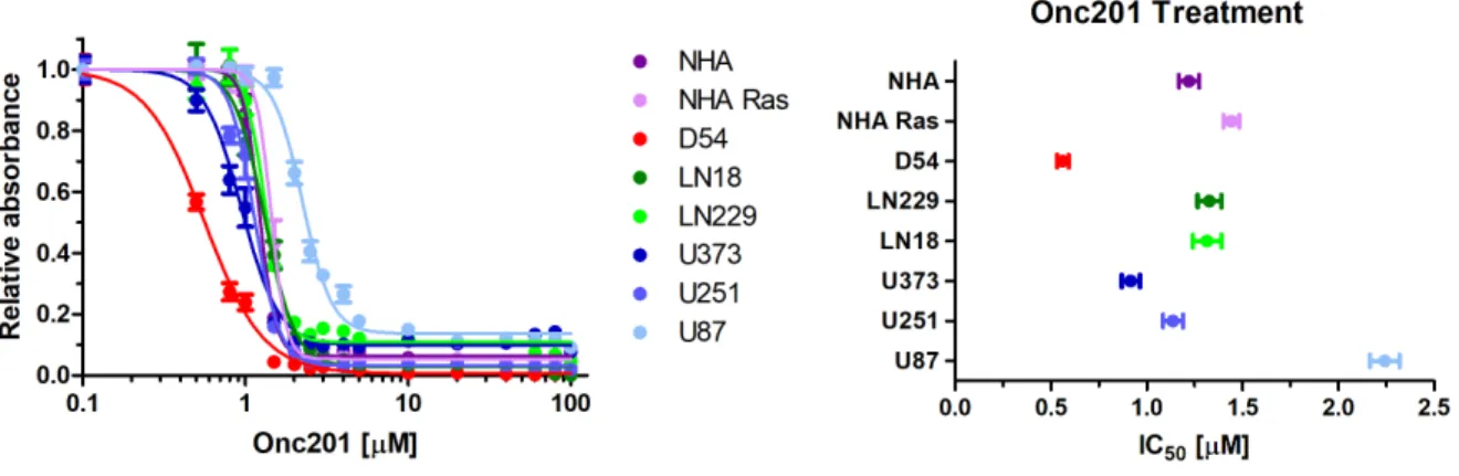
Figure 3 (A) Each cell line was treated with varying concentrations of ONC201 and was plotted with relative absorbance of light at 490 nm to obtain toxicity graphs. Bars
represent standard error. (B) IC50 values were calculated from the ONC201 toxicity curves. IC50 values were calculated as follows in: NHA (1.22 µM), NHA Ras (1.44 µM), D54MG (0.56 µM), LN229 (1.33 µM), LN18 (1.31 µM), U251 (1.14 µM), U87 (2.24 µM), U373 (0.944 µM). Bars represent 95% confidence intervals.
IC50 values for ONC201 ranged from minimum of 0.056 µM in D54MG to a maximum of 2.24 µM in U87. NHA had an IC50 of 1.22 µM and NHA Ras had an IC50 of 1.44 µM.
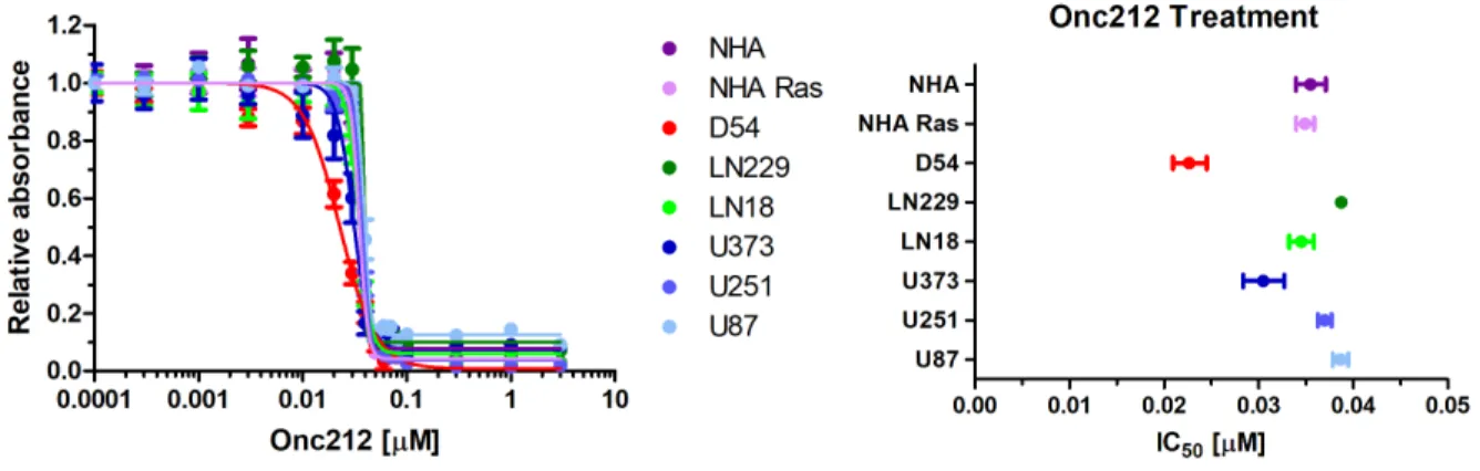
Figure 4 (A) Each cell line was treated with varying concentrations of ONC212 and was plotted with relative absorbance of light at 490 nm to obtain toxicity graphs. Bars
represent standard error. (B) IC50 values were calculated from the ONC212 toxicity curves. IC50 values were calculated as follows in: NHA (35.5 nM), NHA Ras (34.9 nM), D54MG (22.7 nM), LN229 (38.7 nM), LN18 (34.5 nM), U251 (37.0 nM), U87 (38.6 nM), U373 (30.5 nM). Bars represent 95% confidence intervals.
IC50 values for ONC212 ranged from minimum of 22.7 nM in D54MG to a maximum of 38.7 nM in LN229. U87 had the second highest IC50 of 38.6 nM. NHA had an IC50 of 35.5 µM and NHA Ras had an IC50 of 34.9 µM.
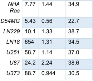
IC50 values for NHA, NHA Ras, and ECLs following treatment with TMZ, ONC201, and ONC212 are summarized in the table below.
Cell line
TMZ (µM)
ONC201 (µM)
NHA Ras
7.77 1.44 34.9
D54MG 5.43 0.56 22.7
LN229 10.1 1.33 38.7
LN18 654 1.31 34.5
U251 58.7 1.14 37.0
U87 24.2 2.24 38.6
U373 88.7 0.944 30.5
Table 2 Toxicity curves were developed after plotting relative absorbance at 490 nm against drug concentration for each of the 8 cell lines (NHA, NHA Ras, U251, U87, U373, LN18, LN229, D54MG). IC50 values were calculated from these plots. IC50 values for each cell lines were calculated in response to TMZ, ONC201, and ONC212.
Discussion
Doubling times vary from 0.726 days to 1.365 days in U373. NHA Ras had a
2A, 2B, 3A, 3B). These relatively lower IC50 values of ONC201 are clinically promising due to the harsh off-target effects associated with TMZ therapy, such as neutropenia, thrombocytopenia, nausea, and fatigue.2, 5, 6, 30 A recent phase II clinical trial with
ONC201 displayed promising results, reporting a median overall survival of 41.6 weeks. Two patients continue to receive ONC201 treatment for greater than 12 months. One of these patients exhibited regression by 85% in one lesion and 76% in another. The second patient remains disease-free following re-resection. The study also reported excellent drug tolerance. 31 Due to the high tolerance of ONC201 at 625 mg every 3 weeks in clinical trials, other studies have begun to explore increased dosage
frequencies.32 It may be worthwhile to consider ONC212, a potent analog of ONC201, as a more effective treatment option.15, 19, 20
Certain cell lines were more sensitive to treatments than others. D54MG was the most sensitive cell line in all treatments. U87 was the least sensitive cell line in ONC201 treatments and was the second least sensitive cell lines in ONC212 treatment.
Conclusion and Future Directions
ONC201 and ONC212 having a narrow range of IC50 concentrations for all 8 cell lines can indicate applicability across a wide variety of molecularly variable tumors and the capacity to act as improved therapeutics for GBM in comparison to TMZ. Furthermore, utilizing a combined multi-drug treatment plan can aid in accounting for both intrinsic and extrinsic drug resistance in molecularly heterogenous tumors. Since ONC212 and ONC201 operate as dual Akt/ERK inhibitor, it can be particularly efficacious in tumors with high Akt or ERK expression. Inhibiting both Akt and ERK can help quench potential response that may occur if only one is inhibited due to Akt and ERK being
communicating pathways.15, 19, 20 Future directions include performing similar
experiments with glioblastoma stem cells to better emulate tumor structure due to their 3-dimensional shape compared to the adherent cell lines which were used. In vivo
References
[1] Ostrom QT, Gittleman H, Liao P, Rouse C, Chen Y, Dowling J, Wolinsky Y, Kruchko C, Barnholtz-Sloan J: CBTRUS statistical report: primary brain and central nervous system tumors diagnosed in the United States in 2007-2011. Neuro Oncol 2014, 16 Suppl 4:iv1-63.
[2] Fernandes C, Costa A, Osorio L, Lago RC, Linhares P, Carvalho B, Caeiro C: Current Standards of Care in Glioblastoma Therapy. Glioblastoma. Edited by De Vleeschouwer S. Brisbane (AU): Codon Publications Copyright: The Authors., 2017.
[3] Inda MM, Bonavia R, Seoane J: Glioblastoma multiforme: a look inside its heterogeneous nature. Cancers 2014, 6:226-39.
[4] Harder BG, Blomquist MR, Wang J, Kim AJ, Woodworth GF, Winkles JA, Loftus JC, Tran NL: Developments in Blood-Brain Barrier Penetrance and Drug Repurposing for Improved Treatment of Glioblastoma. Frontiers in oncology 2018, 8:462.
[5] Niewald M, Berdel C, Fleckenstein J, Licht N, Ketter R, Rübe C: Toxicity after radiochemotherapy for glioblastoma using temozolomide--a retrospective evaluation. Radiat Oncol 2011, 6:141.
[6] Stupp R, Mason WP, van den Bent MJ, Weller M, Fisher B, Taphoorn MJ, Belanger K, Brandes AA, Marosi C, Bogdahn U, Curschmann J, Janzer RC, Ludwin SK, Gorlia T, Allgeier A, Lacombe D, Cairncross JG, Eisenhauer E, Mirimanoff RO, Groups
EOfRaToCBTaR, Group NCIoCCT: Radiotherapy plus concomitant and adjuvant temozolomide for glioblastoma. N Engl J Med 2005, 352:987-96.
[8] Ramirez YP, Weatherbee JL, Wheelhouse RT, Ross AH: Glioblastoma multiforme therapy and mechanisms of resistance. Pharmaceuticals (Basel, Switzerland) 2013, 6:1475-506.
[9] Uno M, Oba-Shinjo SM, Camargo AA, Moura RP, Aguiar PH, Cabrera HN, Begnami M, Rosemberg S, Teixeira MJ, Marie SK: Correlation of MGMT promoter methylation status with gene and protein expression levels in glioblastoma. Clinics (Sao Paulo, Brazil) 2011, 66:1747-55.
[10] Bredel M, Bredel C, Juric D, Duran GE, Yu RX, Harsh GR, Vogel H, Recht LD, Scheck AC, Sikic BI: Tumor necrosis factor-alpha-induced protein 3 as a putative regulator of nuclear factor-kappaB-mediated resistance to O6-alkylating agents in human glioblastomas. Journal of clinical oncology : official journal of the American Society of Clinical Oncology 2006, 24:274-87.
[11] Happold C, Roth P, Wick W, Schmidt N, Florea AM, Silginer M, Reifenberger G, Weller M: Distinct molecular mechanisms of acquired resistance to temozolomide in glioblastoma cells. Journal of neurochemistry 2012, 122:444-55.
[12] Kohsaka S, Wang L, Yachi K, Mahabir R, Narita T, Itoh T, Tanino M, Kimura T, Nishihara H, Tanaka S: STAT3 inhibition overcomes temozolomide resistance in glioblastoma by downregulating MGMT expression. Molecular cancer therapeutics 2012, 11:1289-99.
[14] Allen JE, Krigsfeld G, Patel L, Mayes PA, Dicker DT, Wu GS, El-Deiry WS:
Identification of TRAIL-inducing compounds highlights small molecule ONC201/TIC10 as a unique anti-cancer agent that activates the TRAIL pathway. Molecular cancer 2015, 14:99.
[15] Ralff MD, Lulla AR, Wagner J, El-Deiry WS: ONC201: a new treatment option being tested clinically for recurrent glioblastoma. Translational cancer research 2017,
6:S1239-s43.
[16] Allen JE, Krigsfeld G, Mayes PA, Patel L, Dicker DT, Patel AS, Dolloff NG,
Messaris E, Scata KA, Wang W, Zhou JY, Wu GS, El-Deiry WS: Dual inactivation of Akt and ERK by TIC10 signals Foxo3a nuclear translocation, TRAIL gene induction, and potent antitumor effects. Science translational medicine 2013, 5:171ra17.
[17] Greer YE, Porat-Shliom N, Nagashima K, Stuelten C, Crooks D, Koparde VN, Gilbert SF, Islam C, Ubaldini A, Ji Y, Gattinoni L, Soheilian F, Wang X, Hafner M, Shetty J, Tran B, Jailwala P, Cam M, Lang M, Voeller D, Reinhold WC, Rajapakse V, Pommier Y, Weigert R, Linehan WM, Lipkowitz S: ONC201 kills breast cancer cells in vitro by targeting mitochondria. Oncotarget 2018, 9:18454-79.
[18] Zhou Y, Zhou Y, Shingu T, Feng L, Chen Z, Ogasawara M, Keating MJ, Kondo S, Huang P: Metabolic alterations in highly tumorigenic glioblastoma cells: preference for hypoxia and high dependency on glycolysis. The Journal of biological chemistry 2011, 286:32843-53.
[20] Wagner J, Kline CL, Ralff MD, Lev A, Lulla A, Zhou L, Olson GL, Nallaganchu BR, Benes CH, Allen JE, Prabhu VV, Stogniew M, Oster W, El-Deiry WS: Preclinical
evaluation of the imipridone family, analogs of clinical stage anti-cancer small molecule ONC201, reveals potent anti-cancer effects of ONC212. Cell cycle (Georgetown, Tex) 2017, 16:1790-9.
[21] Ishida CT, Zhang Y, Bianchetti E, Shu C, Nguyen TTT, Kleiner G, Sanchez-Quintero MJ, Quinzii CM, Westhoff MA, Karpel-Massler G, Prabhu VV, Allen JE, Siegelin MD: Metabolic Reprogramming by Dual AKT/ERK Inhibition through
Imipridones Elicits Unique Vulnerabilities in Glioblastoma. Clinical cancer research : an official journal of the American Association for Cancer Research 2018, 24:5392-406. [22] Sonoda Y, Ozawa T, Hirose Y, Aldape KD, McMahon M, Berger MS, Pieper RO: Formation of intracranial tumors by genetically modified human astrocytes defines four pathways critical in the development of human anaplastic astrocytoma. Cancer research 2001, 61:4956-60.
[23] Assay Guidance Manual. Edited by Sittampalam GS, Coussens NP, Brimacombe K, Grossman A, Arkin M, Auld D, Austin C, Baell J, Bejcek B, Caaveiro JMM, Chung TDY, Dahlin JL, Devanaryan V, Foley TL, Glicksman M, Hall MD, Haas JV, Inglese J, Iversen PW, Kahl SD, Kales SC, Lal-Nag M, Li Z, McGee J, McManus O, Riss T, Trask OJ, Jr., Weidner JR, Wildey MJ, Xia M, Xu X. Bethesda (MD): Eli Lilly & Company and the National Center for Advancing Translational Sciences, 2004.
3-(4,5-dimethylthiazol-2-yl)-2,5-diphenyl-tetrazolium bromide. Agricultural and biological chemistry 1990, 54:2961-6.
[25] Aasland D, Reich TR, Tomicic MT, Switzeny OJ, Kaina B, Christmann M: Repair gene O(6) -methylguanine-DNA methyltransferase is controlled by SP1 and up-regulated by glucocorticoids, but not by temozolomide and radiation. Journal of neurochemistry 2018, 144:139-51.
[26] Cui B, Johnson SP, Bullock N, Ali-Osman F, Bigner DD, Friedman HS: Decoupling of DNA damage response signaling from DNA damages underlies temozolomide resistance in glioblastoma cells. Journal of biomedical research 2010, 24:424-35. [27] Sasai K, Akagi T, Aoyanagi E, Tabu K, Kaneko S, Tanaka S: O6-methylguanine-DNA methyltransferase is downregulated in transformed astrocyte cells: implications for anti-glioma therapies. Molecular cancer 2007, 6:36.
[28] Wang HH, Chang TY, Lin WC, Wei KC, Shin JW: GADD45A plays a protective role against temozolomide treatment in glioblastoma cells. Scientific reports 2017, 7:8814. [29] Zhu Z, Du S, Du Y, Ren J, Ying G, Yan Z: Glutathione reductase mediates drug resistance in glioblastoma cells by regulating redox homeostasis. Journal of
neurochemistry 2018, 144:93-104.
[30] Bae SH, Park MJ, Lee MM, Kim TM, Lee SH, Cho SY, Kim YH, Kim YJ, Park CK, Kim CY: Toxicity profile of temozolomide in the treatment of 300 malignant glioma patients in Korea. Journal of Korean medical science 2014, 29:980-4.
administered every three weeks in recurrent glioblastoma. Oncotarget 2017, 8:79298-304.