497
© 2018 by the Serbian Biological Society How to cite this article: Zhao X, Jiang L, Liu K, Ma M, Zhou Y, Tian Y, Li M. Tomato aspermy virus elimination improves medicinal quality of chrysanthemum. Arch Biol Sci. 2018;70(3):497-502.
Tomato aspermy virus elimination improves medicinal quality of chrysanthemum
Xiting Zhao1,2,3,*, Liwei Jiang1, Ke Liu1, Mengdan Ma1, Yingyuan Zhou1, Ying Tian1 and Mingjun Li1,2,3
1College of Life Science, Henan Normal University, Xinxiang 453007, Henan, China
2Engineering Technology Research Center of Nursing and Utilization of Genuine Chinese Crude Drugs in Henan Province,
Xinxiang 453007, Henan, China
3Engineering Laboratory of Biotechnology for Green Medicinal Plant of Henan Province, Xinxiang 453007, Henan, China
*Corresponding author: [email protected]
Received: December 26, 2017; Revised: February 19, 2018; Accepted: March 15, 2018; Published online: March 22, 2018
Abstract: Chrysanthemum morifolium cv. ‘Huaihuang’, a medicinal chrysanthemum of China, undergoes long-term asexual reproduction and virus infection that change its quality characteristics. Our previous studies have shown that tomato aspermy virus (TAV) is the main virus infecting ‘Huaihuang’. Many studies indicate that plant virus elimination can improve plant growth, but only a few studies have focused on the effects of detoxification on the medicinal compo-nents of medicinal plants. In this paper, the content of medicinal compocompo-nents, including chlorogenic acid, luteoloside and 3,5-O-dicaffeoylquinic acid, was compared between the TAV-free and TAV-infected (control) chrysanthemum. In addition, the reason why TAV elimination improves the medicinal components of chrysanthemum is explored. Our results suggest that TAV elimination significantly improves plant growth, enhances the enzyme activities of phenylalanine ammonia-lyase, cinnamate-4-hydroxylase and 4-coumarate:CoA ligase, and increases the levels of CmHCT and CmCHS expression, thereby greatly improving the medicinal quality of chrysanthemum.
Key words: chrysanthemum; tomato aspermy virus (TAV) elimination; chlorogenic acid; luteoloside; 3, 5-O-dicaffeoylquinic acid
INTRODUCTION
Chrysanthemum morifolium cv. ‘Huaihuang’ is a well-known medicinal plant produced mainly in the ancient Huaiqing area of Henan in China, and it has been used for medicinal and ornamental purposes, as well as food and tea. ‘Huaihuang’ contains abundant chlorogenic acid (CA), luteoloside (LO) and 3,5-O-dicaffeoylquinic acid (DCQA, an isomer of CA). Thus, CA, LO and DCQA have been chosen as standard compounds for evaluating the medicinal quality of chrysanthemum [1]. Asexual reproduction, such as cuttings and plant division propagation, is the main cultivation method for chrysanthemum, but virus contaminations greatly reduce chrysanthemum yield and quality [2-4]. Our previous studies showed that the main virus infect-ing ‘Huaihuang’ is tomato aspermy virus (TAV) [5], which causes severe mottling, dwarfing and twisting [6]. Long-term accumulation of TAV leads to slow growth, fewer flowers and substantial reduction in the amount of CA, LO and DCQA. These negative effects
limit the large-scale cultivation and human consump-tion of ‘Huaihuang’, rendering it urgent to remove the virus and improve the quality of ‘Huaihuang’.
Many studies have shown that the agronomic traits of virus-free plants, such as plant height, crown width, leaf number and yield, are significantly higher than those in infected plants [7,8]. For medicinal plants, virus elimination improves not only the agro-nomic traits, but also the medicinal quality. However, the effect of detoxification on medicinal quality has received little scientific attention.
levels of CmHCT and CmCHS in the biosynthesis of medicinal components in chrysanthemum.
MATERIALS AND METHODS
Plant material and growth conditions
Naturally TAV-infected plants of ‘Huaihuang’ grow-ing in the ancient Huaiqgrow-ing area of Henan in China were used in the present study. Stems with a young axillary bud (about 1 cm in length) were excised and used as explants for the establishment of stock cultures
in vitro. After surface disinfection with 75% alcohol for 5 min and 0.1% HgCl2 for 40 s, and then wash-ing three times in sterile distilled water, the explants were grown on solid Murashige and Skoog (MS) me-dium supplemented with 2 mg/L kinetin and 0.5 mg/L α-naphthylacetic. Stock cultures were maintained at a temperature of 25±2°C under a 14 h light and a 10 h dark photoperiod with a light intensity of 36.0 μmol/ (m2 s) at the Engineering Technology Research Center of Nursing and Utilization of Genuine Chinese Crude Drugs in Henan Province (Henan Normal University, China). Subculturing was performed every four weeks. Shoot tip culture and TAV detection
Apical and lateral shoots (about 0.3 mm) from virus-infected in vitro stock cultures were excised using a microscope under sterile conditions and were cultured in MS containing 0.03 mg/L kinetin; the medium was replaced every two weeks. After four weeks of culture, the leaves from the shoot-tip seedlings were sampled for total RNA extraction. Total RNA was used as a template for the synthesis of cDNA. The specific prim-ers TAV-F (5’ ATG GCC CAA AAC GGT ACG 3’) and TAV-R (5’ TCA CAC CGG GAG CGT TGA AG 3’) were designed to amplify the TAV coat protein gene sequence (KF432415) that was obtained in a pre-vious study [5]. The presence of TAV was determined using RT-PCR according to Zhao et al. [9].
Field growth
After RT-PCR, TAV-free plants of shoot-tip seedlings and TAV-infected plants (control) of stock cultures were transplanted into the field after subculturing and
rapid propagation. In this experiment, a randomized block design including three blocks was adopted. Five hundred plants of the TAV-free group and 500 of the control group were transplanted in each block with a planting density of 60000 plants/hm2. At the pre-har-vest time (early November), the plant height and crown width of the TAV-free and control group plants were measured. Random sampling was performed according to the five-point sampling method [10], with 10 plants per point. At harvest time (mid-November), the plants were randomly harvested according to the five-point sampling method, with 20 plants per point. Flowers were harvested and dried at 55°C to a constant weight. The content of main medicinal components
The concentrations of CA, LO and DCQA in dry flow-ers were determined using high-performance liquid chromatography (HPLC). The sample was ground to a powder and 0.25 g of the powder was suspended in 25 mL 70% methanol (v/v), ultrasonically processed for 40 min, and filtered through a 0.22-μm membrane prior to HPLC analysis.
An Agilent 1260 HPLC system (Agilent Technolo-gies, Palo Alto, USA) was used to obtain HPLC chro-matograms for each sample. Chromatographic separa-tion was performed using solvents A (acetonitrile) and B (0.1% H3PO4) in the following separation program: 0-11 min (10-18% A), 11-30 min (18-20% A), and 30-40 min (20% A). The number of theoretical plates was ≥8000. The detection wavelength was set to 348 nm. The identification of CA, LO and DCQA was based on comparison between the retention times obtained for the standards and those in the samples run under the same experimental conditions. Each sample was in-jected three times with 5 μL each time. Preparation of the standard substances and calibration curves was as follows: three replicates of each standard CA, LO and DCQA (obtained from Wei Ke Qi Biological Technology in China, Sichuan) were injected (5 μL) and analyzed. The calibration curves were constructed by plotting the peak area and the total amount (μg) of each sample. Enzyme activities and gene expression
the biosynthesis of the main medicinal components, were determined. All enzyme extraction procedures were conducted at 4°C. Fresh petals (0.1 g) obtained during the full-blossom period were homogenized in 1 mL 50 mM Tris-HCl (pH 8.9) containing 15 mM β-mercaptoethanol, 5 mM EDTA, 5 mM ascorbic acid, 10 μM leupeptin, and 0.15% (w/v) polyvinyl pyrrol-idone. The homogenate was then centrifuged at 14650
xg for 20 min at 4°C. The supernatant was collected and used as a crude enzyme extract.
The activity of PAL was determined and expressed as the conversion rate of L-phenylalanine to trans-Cinnamic acid following Solecka and Kacperska [11] with slight modifications. The reaction mixture con-taining 0.5 mL crude enzyme extract and 2.5 mL 50 mM Tris-HCl (pH 8.9) buffer with 16 mM L-PAL, 3.6 mM NaCl was incubated for 1 h at 30°C. The reaction was stopped with 6 M HCl and the absorbance was measured at 290 nm. One unit of PAL was defined as the amount of enzyme that caused an increase in A290 of 0.001 units/min.
The activity of C4H was determined according to Lamb and Rubery [12]. The reaction mixture was composed of 0.8 mL crude enzyme extract and 2.2 mL 50 mM Tris-HCl (pH 8.9) buffer containing 2 µM trans-cinnamic acid2 µM NADPNa2 and 5 µM glucose-6-PNa2 and was incubated for 30 min at 25°C. The reaction was stopped with 6 M HCl and the ab-sorbance was measured at 340 nm. One unit of C4H was defined as the amount of enzyme that caused an increase in A340 of 0.001 units/min.
The activity of 4CL was measured as the increase in A333 with p-coumarate as the substrate following Knobloch and Hahlbrock [13]. The reaction mixture was composed of 0.8 mL crude enzyme extract and 2.2 mL 50 mM Tris-HCl (pH 8.9) buffer containing 5 µM p-coumarate, 50 µM ATP, l µM CoA-SH, and 15 µM MgSO4·7H2O. One unit of 4CL was defined as the amount of enzyme that caused a decrease in A333 of 0.01 units/min.
Quantitative RT-PCR (qRT-PCR) [14] was used to determine the expression levels of CmHCT and
CmCHS at full-blossom period. Primers were de-signed based on the CmHCT and CmCHS sequences in the chrysanthemum transcriptome database
(Prim-ers F-5ʹ CAC CAG GTT ACT TTG GGA ATG 3ʹ and R-5ʹ CAG GCT GAA GTT CCA AGT AAT CGA 3ʹ for CmHCT; F-5ʹ GGC AGC CCA AGT CAA AGA 3ʹ and R-5ʹ CAG AGC AGA CGA CAA GAA CG 3ʹ for CmCHS). The housekeeping gene UBI of chrysan-themum was used as the internal reference (Primers F-5ʹ AGC TGA GCA GAC TCC CGA TG 3ʹ and R-5ʹ AGG CGA ATC ATC AGT ACC AAG T 3ʹ for UBI). The reaction program included initial denaturation at 95°C for 5 min, and 40 cycles at 95°C for 15 s and at 60°C for 45 s. The relative expression levels were calculated using the 2-∆∆CT method [14].
Statistical analysis
For all experiments, three biological replicates were used, and each was repeated three times. Student’s t -test was carried out to determine significant differenc-es in plant height, crown width, the main medicinal components, enzyme activities and gene expression in TAV-free and control plants. The differences between the two groups of data in all the experiments were evaluated as statistically significant (*p<0.05) or as extremely significant (**p<0.01).
RESULTS
Shoot-tip seedling acquisition and TAV detection
‘Huaihuang’ is mainly infected with TAV [5], so in this study we examined the effects of TAV elimina-tion. After 6 days, the shoot tips began to swell and sprout. After 25 days, the shoot tips grew into robust plantlets. RT-PCR showed that for 35% of shoot-tip seedlings there was no specific amplification, and that a fragment of 657 bp was amplified in control plants (Fig. 1). Thus, TAV-free plantlets were obtained from the shoot-tip culture.
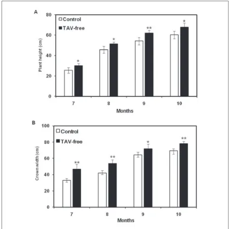
Effect of TAV elimination on ‘Huaihuang’ growth
results of various agronomic traits showed that the growth of TAV-free plants was better than the control. Effect of TAV elimination on the main medicinal components of ‘Huaihuang’
We measured the content of CA, LO and DCQA in ‘Huaihuang’ dry flowers using HPLC with gradient elution (Fig. 4). The regression equations are as fol-lows: y=7.358x1-5.529 (r2=0.9999), y=13.39x
2+2.293 (r2=0.9999) and y=6.8578x
3-1.3964 (r2=0.9998). Y represents the peak area, x1, x2, and x3 are the concen-trations of CA, LO and DCQA, respectively. Com-pared with control plants, the contents of CA, LO and DCQA in TAV-free plants increased by 3.92%, 33.33%, 10.27%, respectively (Table 1). Our results show that TAV elimination greatly improved the content of the main medicinal components of ‘Huaihuang’.
Fig. 3. Effect of TAV elimination on ‘Huaihuang’ plant height (A) and crown width (B). Plant height and crown width in TAV-free plants was significantly higher than in control plants (July to October). The data are the mean±SD of ten biological replicates. The asterisks indicate statistically significant differences between the plants from which TAV has been eliminated and control plants (*P<0.05; **P<0.01).
Fig.1. TAV detection in plantlets derived from control (lanes 1, 2, 3) and shoot-tip seedlings (lanes 4, 5, 6) of ‘Huaihuang’.
Fig. 2. The appearance of control (A, B) and TAV-free (C, D) ‘Huaihuang’ plants in the field. Disease symptoms were observed when the ‘Huaihuang’ seedlings were transplanted into the field in the fifth month. Compared with the control, TAV-free plants grow well and do not present leaf yellowing, mottling and other disease symptoms. A and C – long-range shoots; B and D – close-up shoots. Bar=5cm.
The effect of TAV elimination on enzyme activities and gene expression
As shown in Fig. 5, the activities of PAL, 4CL and C4H, which are the key enzymes involved in the bio-synthesis of the principal medicinal components of ‘Huaihuang’, are significantly higher in TAV-free plants than in control plants. The relative expression levels of
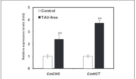
CmHCT and CmCHS, which are genes encoding for the major enzymes involved in the biosynthesis of the main medicinal components, were 2.39- and 3.71-fold higher, respectively, in TAV-free plants when compared to control plants (Fig. 6). These results show that the elimination of TAV increases the activities of the major enzymes and the relative expression levels of genes en-coding the key enzymes related to the biosynthesis of the main medicinal components, thus improving the principal medicinal component content of ‘Huaihuang’.
DISCUSSION
For most crops, research is focused on establishing a suitable detoxification method and obtaining virus-free plants [15-18] while little is known about the ef-fect of detoxification on the contents of the plants’ medicinal ingredients. ‘Huaihuang’ is a traditional Chinese herbal medicine, and the content of its main medicinal components (CA, LO and DCQA) is one of the main criteria for evaluating its value. Whether virus elimination can maintain or improve its medici-nal value is the key factor determining whether virus elimination technology should be popularized and further applied. In this study, we increased the content of three medicinal components of ‘Huaihuang’ by TAV elimination, which indicated that detoxification can improve the medicinal value of ‘Huaihuang’.
CA, LO and DCQA are all secondary metabolites generated by the phenylalanine metabolic pathway. In this pathway, coumaroyl-CoA is an important in-termediate product that can be further transformed into CA and LO, and CA can be isomerized to DCQA. Phenylalanine can be converted to coumaroyl-CoA through sequential catalysis by PAL, C4H and 4CL. Hydroxycinnamoyl-CoA transferase (HCT) and chal-cone synthase (CHS) are the key enzymes for CA and LO synthesis, respectively. Coumaroyl-CoA and shi-kimic acid are catalyzed by HCT to synthesize CA,
Fig. 6. Real-time PCR quantification of CmHCT and CmCHS in
control and TAV-free ‘Huaihuang’ plants. CmHCT and CmCHS
are the principal genes involved in the biosynthesis of the main medicinal components in ‘Huaihuang’. TAV elimination signifi-cantly increased the expression of CmHCT and CmCHS.The data are the mean±SD of three biological replicates. The asterisks indi-cate statistically significant differences between the TAV-free and control plants (**P<0.01).
Table 1. The effect of TAV elimination on the content of the main medicinal components of ‘Huaihuang’ (%).
Plant Chlorogenic acid Luteoloside 3,5-O-dicaffeoylquinic acid
Control 0.485±0.027 0.087±0.005 1.714±0.066
TAV-free 0.504±0.006* 0.116±0.035* 1.890±0.234*
The data of all samples are presented as the mean±SD of three biological replicates. Student’s t-test was carried out to determine significant differences in TAV-free and control plants. The asterisks indicate statistically significant differences (*P<0.05; **P<0.01).
and coumaroyl-CoA and 3 × malonyl CoA are cata-lyzed by CHS to synthesize LO [19]. It is speculated that the levels of the three in ‘Huaihuang’ are closely related to the activities of PAL, C4H, 4CL, HCT and CHS. Therefore, in this study we measured the en-zyme activities of PAL, C4H and 4CL and the expres-sion levels of CmHCT and CmCHS as the reasons for the improvement of the main medicinal components of TAV-free chrysanthemum.
Our results showed that TAV elimination sig-nificantly improved plant growth and enhanced the activities and gene expression levels of the principal enzymes involved in the biosynthesis of the main medicinal components of the plant, thereby greatly improving its medicinal quality. Moreover, this study also proves that detoxification is an effective method to improve the medicinal quality of chrysanthemum.
Funding: This work was supported by grants from National Natural Science Foundation of China (No. 31372105), the China Postdoctoral Science Foundation (No. 2011M500457), the Pro-gram for Innovative Research Team (in Science and Technology) of the University of Henan Province (No. 15IRTSTHN020), and the Graduate Innovation Foundation of Henan Normal University (No. YL201621).
Author contributions: Xiting Zhao and Liwei Jiang conceived and designed the study; Yingyuan Zhou and Ying Tian performed the experiments; Ke Liu and Mengdan Ma performed the data analysis; Xiting Zhao and Liwei Jiang wrote the paper; Mingjun Li revised the paper. All authors read and approved the final manuscript.
Conflict of interest disclosure: The authors declare that they have no competing interests.
REFERENCES
1. China Pharmacopeia Commission. Pharmacopoeia of the People’s Republic of China. 10th ed. Beijing: Chemistry Industry Publishing House; 2015. 1749 p.
2. Kondo T, Yamashita K, Sugiyama S. First report of
impa-tiens necrotic spot virus infecting chrysanthemum (
Chry-santhemum morifolium) in Japan. J Gen Plant Pathol. 2011;77(4):263-5
3. Miller AJ, Cramer MD. Root nitrogen acquisition and assimi-lation. Plant Soil. 2005;274(1):1-36
4. Verma N, Sharma A, Ram R, Hallan V, Zaidi AA, Garg ID. Detection, identification and incidence of
chrysanthe-mum B carlavirus in chrysanthechrysanthe-mum in India. Crop Prot. 2003;22(2):425-9
5. Zhao X, Liu X, Ge B, Li M, Hong B. A multiplex RT-PCR for simultaneous detection and identification of five viruses and two viroids infecting chrysanthemum. Arch Virol. 2015a;160(5):1145-52
6. Kong BH, Li DB. Tomalo aspermy virus causing chry-santhemum disease. J Zhejiang Univ Agricult Life Sci. 1990;16(1):55-60. Chinese
7. Komar V, Vigne E, Demangeat G, Fuchs M. Beneficial effect of selective virus elimination on the performance of Vitis vinifera cv. Chardonnay. Am J Enol Vitic. 2007;58(2):202-10 8. Deepthi DC, Makeshkumar T. Elimination of cassava mosaic
disease through meristem culture and field evaluation for yield loss assessment in cassava genotypes. J Root Crops. 2016;42(1):45-52
9. Zhao XT, Liu XX, Hong B. Characterization of tomato aspermy virus isolated from chrysanthemum and eluci-dation of its complete nucleotide sequence. Acta Virol. 2015b;59(2):204-6
10. Bailey TJ. Statistics method in biology. 2nd ed. London: Hod-der and Stoughton; 1981. p.
11. Solecka D, Kacperska A. Phenylpropanoid deficiency affects the course of plant acclimation to cold. Physiol Plant. 2003;119(2):253-62
12. Lamb CJ, Rubery PH. A spectrophotometric assay for trans-cinnamic acid 4-hydroxylase activity. Anal Biochem. 1975;68(2):554-61
13. Knobloch KH, Hahlbrock K. Isoenzymes of p-coumarate:
CoA ligase from cell suspension cultures of glycine max. Eur J Biochem. 1975;52(2):311-20
14. Livak KJ, Schmittgen TD. Analysis of relative gene expression data using real-time quantitative PCR and the 2(-Delta Delta C(T)) Method. Methods. 2001;25(4):402-8
15. Yi JY, Gian L, Jongwook J, Sokyoung L, Younggyu L. Elimi-nating potato virus Y (PVY) and potato leaf roll virus (PLRV) using cryotherapy of in vitro-qrown potato shoot tips. Han-guk Jakmul Hakhoe Chi. 2014;59(4):498-504
16. Li BQ, Feng CH, Hu LY, Wang MR, Wang QC. Shoot tip culture and cryopreservation for eradication of Apple stem pitting virus (ASPV) and Apple stem grooving virus (ASGV) from apple rootstocks ‘M9’ and ‘M26’. Ann Appl Biol. 2016;168(1):142-50
17. Vieira RL, Silva AL, Zaffari GR, Steinmacher DA, Freitas Fraga HP, Guerra MP. Efficient elimination of virus complex from garlic (Allium sativum L.) by cryotherapy of shoot tips. Acta Physiol Plant. 2015;37(1):1733