© 2020 by the Serbian Biological Society How to cite this article: Todorović L, Mandušić V, Vučetić Tadić B, Živaljević V, 31 Paunović I, Stanojević B.Altered expression of mi-croRNA-30a-3p in papillary
thyroid cancer and its association with clinicopathological characteristics. Arch Biol Sci. 2020;72(1):31-6.
Altered expression of microRNA-30a-3p in papillary thyroid cancer and its association
with clinicopathological characteristics
Lidija Todorović1,*, Vesna Mandušić1, Biljana Vučetić Tadić2, Vladan Živaljević3,4, Ivan Paunović3,4 and Boban
Stanojević1
1Laboratory for Radiobiology and Molecular Genetics, “Vinča” Institute of Nuclear Sciences, University of Belgrade, Mike
Petrovića Alasa 12-14, 11351 Belgrade, Serbia;
2Institute for Mother and Child Healthcare of Serbia, Radoja Dakića 6, 11000 Belgrade, Serbia
3Center for Endocrine Surgery, Clinical Center of Serbia, Dr Koste Todorovića 8, 11000 Belgrade, Serbia; 4School of Medicine, University of Belgrade, Doktora Subotića 8, 11000 Belgrade, Serbia.
*Corresponding author: [email protected]
Received: October 4, 2019; Revised: October 14, 2019; Accepted: October 15, 2019; Published online: October 25, 2019
Abstract: A growing number of studies suggest a tumor suppressive role and potential prognostic signif-icance of miR-30a-3p in different types of cancer. However, relatively few studies have focused on this microRNA in neoplastic thyroid lesions, including papillary thyroid cancer (PTC). The aim of our study was to shed more light on the potential involvement and clinical relevance of miR-30a-3p in this type of cancer. We examined the expression levels of this microRNA in 42 pairs of PTCs and matched non-tumor thyroid tissues using quantitative RT-PCR. We analyzed their association with clinical and histopathological parameters. The results revealed that miR-30a-3p was significant-ly downregulated in the majority of PTC tissues compared to corresponding non-tumor tissues. Moreover, decreased expression of miR-30a-3p was associated with advanced clinical stage, pres-ence of multiple tumor foci and capsular invasion, suggesting a role in aggressive disease. Although the role of this microRNA and its prognostic utility remain to be elucidated, the presented data sug-gest that downregulated expression of miR-30a-3p indicates poorer prognosis in PTC patients, war-ranting further investigations.
Keywords: miR-30a-3p; papillary thyroid cancer (PTC); thyroid cancer; expression, advanced stage
INTRODUCTION
Papillary thyroid cancer (PTC) is the most prevalent type of thyroid malignancies, accounting for 75-85% of all thyroid cancer cases. Over the last decades, the incidence of thyroid cancer has been increasing all over the world, and this increase is mainly due to an increase in PTC incidence [1,2]. Risk stratification of PTC patients is currently based on clinicopathological characteristics such as age, gender, tumor size, extra-thyroidal spread, nodal metastases, distant metastases and clinical stage. Although this approach is reliable in predicting the mortality risk, it has proved to be uncertain in predicting the risk of recurrence, which occurs more often than death from PTC, so it is more clinically relevant [3,4]. Therefore, the stratification of PTC patients into low- and high-risk prognostic
groups still needs to be improved in order to accurately tailor management. Better understanding of molecular mechanisms of PTC occurrence and progression may be helpful in the identification of novel diagnostic and prognostic biomarkers as well as potential therapeutic targets for the treatment of more aggressive and/or advanced PTC cases.
been associated with processes such as tumor growth, invasion, angiogenesis, metastasis and immune eva-sion. Depending on their target genes, microRNAs can function as oncogenes or tumor suppressors [5,6].
One of the microRNAs with potential tumor sup-pressor activity that is frequently found to be deregu-lated and associated with aggressive tumor features and progression in various human cancers is miR-30a-3p. So far, downregulated expression of this member of the miR-30 family has been reported in lung cancer [7-10], breast cancer [11-13], colorectal cancer [14], bladder cancer [15], ovarian cancer [16], endometrial cancer [17], hepatocellular carcinoma [18], hematologic malignancies [19] and others.
The aim of this study was to investigate the expres-sion levels of miR-30a-3p in papillary thyroid cancer and corresponding non-tumor thyroid tissue, and to evaluate the association with clinical and pathological parameters in order to determine the potential sig-nificance of miR-30a-3p in PTC malignancy. To our knowledge, this is one of only a few studies of miR-30a-3p expression in this type of cancer.
MATERIALS AND METHODS
Patients and clinicopathological characteristics
Tissue samples (paired tumor and corresponding non-tumor thyroid tissues) were obtained from 42 patients with PTC, following surgical excision at the Center for Endocrine Surgery, Clinical Center of Serbia, between 2012 and 2015. Samples were snap-frozen in liquid ni-trogen and stored at -70°C until RNA extraction. Clinical and histopathological parameters are shown in Supple-mentary Table S1. All patients included in the study gave their informed consent. The study was approved by the Ethics Committee of Clinical Center of Serbia.
RNA extraction and reverse transcription
Total RNA extraction from tumor and non-tumor thy-roid tissues was performed using TRI Reagent Solution (Ambion, Foster City, CA) according to manufacturer’s instructions, following homogenization in liquid ni-trogen. RNA quantification was done on NanoDrop™ 1000 (Thermo Fisher Scientific, USA). Next, 10 ng of
total RNA was reverse transcribed using a TaqMan Micro-RNA Reverse Transcription Kit and specific stem-loop RT primer from TaqMan miR-30a-3p Assay (ID 000416), according to the manufacturer’s protocol (Applied Biosystems, Foster City, CA). Small nuclear RNA (nRNA) RNU6B (Assay ID 001093) was used as an endogenous control.
Quantitative PCR (qPCR)
RT-qPCR was performed on a 7500 Real Time PCR System (Applied Biosystems, Foster City, CA). PCR reactions were carried out in a 20-µL reaction volume using the TaqMan Universal Master Mix, No Amperase UNG (Applied Biosystems, Warrington, UK), specific TaqMan miR-30a-3p assay (ID 000416) or small nRNA RNU6B assay (ID 001093), which was used for nor-malization. The cycling conditions were as follows: 10 min at 95°C, 40 cycles of 15 s at 95°C and 60 s at 60°C.
Ct values were calculated using Applied Biosys-tems 7500 system SDS software. The relative quantity of target microRNA in each tumor sample (T) was expressed as the fold change relative to the correspond-ing non-tumor tissue sample (N) and normalized to the internal reference RNU6B according to the 2-ΔΔCt
method, following the equation:
T/N=2-ΔΔCt, ΔΔCt=ΔCt(T)- ΔCt(N),
ΔCt=Cttarget-Ctendogenous control.
In order to compare the relative expression level between tumor and non-tumor tissue, the relative quantity of target in each sample was expressed as fold change relative to the calibrator (1x sample, a sample with the lowest expression) following the equation:
RQsample=2-ΔΔCt, ΔΔCt=ΔCt (sample)
-ΔCt(calibrator),ΔCt=Cttarget-Ctendogenous control.
Statistical analysis
difference between pairs of tumor and non-tumor tissues was evaluated by the Wilcoxon matched-pairs test. Clas-sification of the fold change values between tumor and corresponding non-tumor thyroid tissues was based on the following criteria: fold change >1 as “increased”; fold change <1 as “decreased”. P≤0.05 represented statistical significance, P<0.1 represented a statistical trend.
RESULTS
The expression level of miR-30a-3p in PTC and non-tumor thyroid tissues
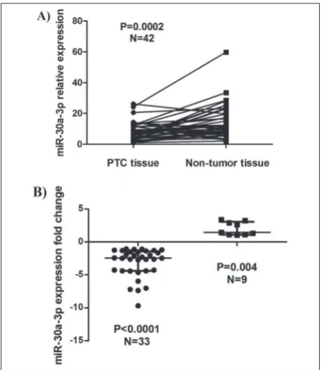
The overall expression level of miR-30a-3p in tumor and non-tumor thyroid samples is shown in Fig. 1A. There was a significant difference in miR-30a-3p ex-pression between tumor and corresponding non-tumor
thyroid tissues (P=0.002). Most of the tumor samples (33/42; 79%) showed a significantly decreased level of miR-30a-3p compared to matched non-tumor samples (P<0.0001), as shown in Fig. 1B.
Relationship between miR-30a-3p expression and clinical and pathological characteristics
The expression levels of miR-30a-3p in PTC with respect to standard clinicopathological parameters are presented in Table 1. A significant association
Fig. 1. The expression level of miR-30a-3p in PTC and non-tumor thyroid tissues. A– miR-30a-3p expression is deregulated in PTC tissues compared to matched non-tumor thyroid tissues. Paired PTC and corresponding non-tumor tissues are connected with lines.
B–78.6% (33/42) of PTCs had significantly decreased expression of miR-30a-3p; 21.4% (9/42) had an increased expression in PTC compared to matched non-tumor thyroid tissue. The dots represent patients with the decreased miR-30a-3p expression in tumor tis-sue while squares represent patients with an increased expression. Statistical analyses were performed by Wilcoxon matched pairs test and P<0.05 was considered statistically significant.
Table 1. The association of miR-30a-3p expression level with clinicopathological parameters in PTC patients.
Clinicopathological
parameters relative expressionmiR-30a-3p P value
Gender
Male (n=10) 0.572 (0.141–0.959)
0.585 Female (n=32) 0.507 (0.270–0.934)
Age Corr.Coef. -0.233 0.142 ≤45 (n=26) 0.660 (0.397–1.043)
0.096* >45 (n=15) 0.378 (0.228–0.726)
Tumor size (cm) Corr.Coef. -0.033 0.834
Histovariant
Classic (n=17) 0.404 (0.225–0.660) 0.099* Follicular+other (n=19) 0.778 (0.234–1.109)
Tumor focality
Multifocal (n=13) 0.376 (0.179–0.610) 0.024 Monofocal (n=28) 0.660 (0.386–1.004)
pT category
pT1+2 (n=21) 0.600 (0.305–1.101) 0.352 pT3+4 (n=21) 0.488 (0.231–0.801)
Lymph node metastases
Yes (n=11) 0.494 (0.378–0.798) 0.841 No (n=31) 0.600 (0.228–1.022)
Capsular invasion
Yes (n=22) 0.392 (0.204–0.736) 0.029 No (n=20) 0.801 (0.408–1.075)
Vascular invasion
Yes (n=15) 0.600 (0.228–0.883)
0.989 No (n=26) 0.504 (0.340–0.968)
Extrathyroidal spread
Yes (n=9) 0.381 (0.188–0.691)
0.191 No (n=32) 0.632 (0.269–1.004)
Clinical stage Corr.Coef. -0.471 0.002
I+II (n=35) 0.657 (0.376–1.022) 0.011 III (n=7) 0.364 (0.139–0.381)
of decreased miR-30a-3p was found with advanced clinical stage (P=0.011), which was also supported by the presence of a significant negative correlation between miR-30a-3p relative expression levels and stage (P=0.002, r=-0.471), obtained by Spearman’s correla-tion test. Lower miR-30a-3p levels were also signifi-cantly associated with the presence of multiple tumor foci (P=0.024) and with capsular invasion (P=0.029). No other significant associations were detected.
DISCUSSION
MicroRNAs have been proposed by numerous studies as promising diagnostic, prognostic, therapeutic and surveillance tools for human cancers because of their critical roles in various biological processes during tumor development and progression [20-25].
In vitro studies have shown that miR-30a-3p is
involved in the regulation of cell proliferation, angio-genesis, cell migration, invasiveness, epithelial-mesen-chymal transition (EMT) and apoptosis [11,18,26,27]. Interestingly, one of the targets experimentally con-firmed to be negatively regulated by miR-30a-3p is C-jun-amino-terminal kinase-interacting protein 4 (SPAG9), a scaffold protein that activates the mitogen-activated protein kinase (MAPK) signaling pathway in tumorigenesis, leading to cell proliferation and inhibition of apoptosis [28]. Constitutive activation of the MAPK signaling pathway is frequently found in PTC [29]. Pathway activation is caused by mutations in proto-oncogene B-Raf (BRAF) or one of the RAS
family genes, or by RET/PTC rearrangements in 70% of cases [30]. Other PTC cases do not harbor any of these genetic alterations suggesting that some other mechanisms, including microRNAs might be involved.
To date, aberrant expression of miR-30a-3p has been detected in various types of cancer, but relatively few studies have investigated the biological role or the clinical relevance of this microRNA in neoplastic thyroid lesions including PTC [31,32].
To shed more light on the involvement of miR-30a-3p and its clinical relevance in PTC, we investigated the level of expression of this microRNA in matched tumor/non-tumor tissue samples and its association with clinical and pathological parameters. The results of the present study show that miR-30a-3p expression
is significantly downregulated in PTC tissues compared to corresponding non-tumor thyroid tissues in the majority of analyzed samples, suggesting a potential involvement of this microRNA in PTC development and/or progression. This is in line with the results of other studies of miR-30a-3p in PTC. miR-30a-3p is significantly downregulated in PTC compared to adjacent non-tumor tissue and to benign goiter tis-sue [31]. Next-generation sequencing and expression analysis of miRNAs in PTC and contralateral normal thyroid tissue specimens revealed miR-30a-3p to be among the most abundantly downregulated miRNAs in PTC [32]. Significantly decreased level of miR-30a-3p in tumor tissue compared to corresponding non-tumor tissue was also found in lung carcinoma [7,8,10,26], esophageal carcinoma [27], gastric cancer [28], colorectal cancer [14] and liver cancer [18].
and more aggressive tumor features such as tumor size, pT category, nodal metastases, TNM stage in lung cancer [8], with higher tumor grade and poorer differentiation in ovarian cancer [16,42], vascular invasion in hepatocellular carcinoma [18], distant metastases in lung and renal carcinoma [43,44] and shorter progression free or overall survival in many of them [8,11-13,28,42].
Although the data presented herein need to be confirmed in larger cohorts, they demonstrate that miR-30a-3p expression is deregulated in PTC and that the decreased expression of this microRNA is associated with more aggressive tumor features and advanced disease stage, which supports its potential usefulness in identifying PTC patients at higher risk for disease progression. The exact role of miR-30a-3p, its potential involvement in the regulation of the MAPK signaling pathway, as well as its utility as a diagnostic and/or prognostic tool, remain to be elucidated.
Funding: This study was supported by the Ministry of Education, Science and Technological Development of the Republic of Serbia, Contract No. ON173049.
Acknowledgments: The authors are grateful to Prof. Shunichi
Yamashita and Dr. Vladimir Saenko from the Atomic Bomb Disease Institute, Nagasaki University, for their generous help and support.
Author contributions: LT – experimental work, statistical analysis, interpretation of the results, preparation of the manuscript; VM – statistical analysis, interpretation of the results, preparation of the manuscript; BVT – critical review of the manuscript; VŽ – sample collection, data collection; IP – sample collection, data collection; BS – design of the study, interpretation of the results, preparation of the manuscript.
Conflict of interest disclosure: The authors declare that they have no conflict of interest.
REFERENCES
1. Sipos JA, Mazzaferri EL. Thyroid cancer epidemiol-ogy and prognostic variables. Clin Oncol (R Coll Radiol). 2010;22(6):395-404.
2. Lim H, Devesa SS, Sosa JA, Check D, Kitahara CM. Trends in Thyroid Cancer Incidence and Mortality in the United States, 1974-2013. JAMA. 2017;317(13):1338-48.
3. Pacini F, Castagna MG, Brilli L, Pentheroudakis G. Thy-roid cancer: ESMO Clinical Practice Guidelines for diag-nosis, treatment and follow-up. Ann Oncol. 2012;23(Suppl 7):vii110-9.
4. Lebastchi AH, Callender GG. Thyroid cancer. Curr Probl Cancer. 2014;38(2):48-74.
5. Hayes J, Peruzzi PP, Lawler S. MicroRNAs in cancer: biomark-ers, functions and therapy. Trends Mol Med. 2014;20(8):460-9. 6. Acunzo M, Romano G, Wernicke D, Croce CM. MicroRNA
and cancer--a brief overview. Adv Biol Regul. 2015;57:1-9. 7. Yu N, Yong S, Kim HK, Choi YL, Jung Y, Kim D, Seo J, Lee
YE, Baek D, Lee J, Lee S, Lee JE, Kim J. Identification of tumor suppressor miRNAs by integrative miRNA and mRNA sequencing of matched tumor-normal samples in lung adeno-carcinoma. Mol Oncol. 2019;13(6):1356-68.
8. Tang R, Liang L, Luo D, Feng Z, Huang Q, He R, Gan T, Yang L, Chen G. Downregulation of MiR-30a is Associ-ated with Poor Prognosis in Lung Cancer. Med Sci Monit. 2015;21:2514-20.
9. Jin X, Chen Y, Chen H, Fei S, Chen D, Cai X, Liu L, Lin B, Su H, Zhao L, Su M, Pan H, Shen L, Xie D, Xie C. Evaluation of Tumor-Derived Exosomal miRNA as Potential Diagnos-tic Biomarkers for Early-Stage Non-Small Cell Lung Can-cer Using Next-Generation Sequencing. Clin CanCan-cer Res. 2017;23(17):5311-9.
10. Zhang Y, Sui J, Shen X, Li C, Yao W, Hong W, Peng H, Pu Y, Yin L, Liang G. Differential expression profiles of microRNAs as potential biomarkers for the early diagnosis of lung cancer. Oncol Rep. 2017;37(6):3543-53.
11. Perez-Rivas LG, Jerez JM, Carmona R, de Luque V, Vicioso L, Claros MG, Viguera E, Pajares B, Sanchez A, Ribelles N, Alba E, Lozano J. A microRNA signature associated with early recurrence in breast cancer. PLoS One. 2014;9(3):e91884. 12. Rodriguez-Gonzalez FG, Sieuwerts AM, Smid M, Look MP,
Meijer-van Gelder ME, de Weerd V, Sleijfer S, Martens JW, Foekens JA. MicroRNA-30c expression level is an indepen-dent predictor of clinical benefit of endocrine therapy in advanced estrogen receptor positive breast cancer. Breast Cancer Res Treat. 2011;127(1):43-51.
13. Turashvili G, Lightbody ED, Tyryshkin K, SenGupta SK, Elliott BE, Madarnas Y, Ghaffari A, Day A, Nicol CJB. Novel prognostic and predictive microRNA targets for triple-nega-tive breast cancer. FASEB J. 2018;32(11):5937-54.
14. Ma Y, Zhang P, Yang J, Liu Z, Yang Z, Qin H. Candidate microRNA biomarkers in human colorectal cancer: system-atic review profiling studies and experimental validation. Int J Cancer. 2012;130(9):2077-87.
15. Ichimi T, Enokida H, Okuno Y, Kunimoto R, Chiyomaru T, Kawamoto K, Kawahara K, Toki K, Kawakami K, Nishiyama K, Tsujimoto G, Nakagawa M, Seki N. Identification of novel microRNA targets based on microRNA signatures in bladder cancer. Int J Cancer. 2009;125(2):345-52.
16. Wang Y, Li L, Qu Z, Li R, Bi T, Jiang J, Zhao H. The expres-sion of miR-30a* and miR-30e* is associated with a dualistic model for grading ovarian papillary serious carcinoma. Int J Oncol. 2014;44(6):1904-14.
17. Tsukamoto O, Miura K, Mishima H, Abe S, Kaneuchi M, Higashijima A, Miura S, Kinoshita A, Yoshiura K, Masuzaki H. Identification of endometrioid endometrial carcinoma-associated microRNAs in tissue and plasma. Gynecol Oncol. 2014;132(3):715-21.
down-regulated in hepatocellular carcinoma. Eur J Surg Oncol. 2014;40(11):1586-94.
19. Ozdogan H, Gur Dedeoglu B, Oztemur Islakoglu Y, Aydos A, Kose S, Atalay A, Yegin ZA, Avcu F, Uckan Cetinkaya D, Ilhan O. DICER1 gene and miRNA dysregulation in mesen-chymal stem cells of patients with myelodysplastic syndrome and acute myeloblastic leukemia. Leuk Res. 2017;63:62-71. 20. Gurbuz N, Ozpolat B. MicroRNA-based Targeted
Therapeu-tics in Pancreatic Cancer. Anticancer Res. 2019;39(2):529-32. 21. Li Y, Sarkar FH. MicroRNA Targeted Therapeutic Approach
for Pancreatic Cancer. Int J Biol Sci. 2016;12(3):326-37. 22. Markopoulos GS, Roupakia E, Tokamani M, Chavdoula E,
Hatziapostolou M, Polytarchou C, Marcu KB, Papavassiliou AG, Sandaltzopoulos R, Kolettas E. A step-by-step microRNA guide to cancer development and metastasis. Cell Oncol (Dordr). 2017;40(4):303-39.
23. Rusek AM, Abba M, Eljaszewicz A, Moniuszko M, Niklinski J, Allgayer H. MicroRNA modulators of epigenetic regulation, the tumor microenvironment and the immune system in lung cancer. Mol Cancer. 2015;14:34.
24. Wang J, Chen J, Sen S. MicroRNA as Biomarkers and Diag-nostics. J Cell Physiol. 2016;231(1):25-30.
25. Xu J, Li J, Zheng TH, Bai L, Liu ZJ. MicroRNAs in the Occur-rence and Development of Primary Hepatocellular Carci-noma. Adv Clin Exp Med. 2016;25(5):971-5.
26. Sui J, Yang RS, Xu SY, Zhang YQ, Li CY, Yang S, Yin LH, Pu YP, Liang GY. Comprehensive analysis of aberrantly expressed microRNA profiles reveals potential biomarkers of human lung adenocarcinoma progression. Oncol Rep. 2017;38(4):2453-63.
27. Fan Y, Bian X, Qian P, Wen J, Yan P, Luo Y, Wu J, Zhang Q. miRNA30a3p inhibits metastasis and enhances radiosensitiv-ity in esophageal carcinoma by targeting insulinlike growth factor 1 receptor. Mol Med Rep. 2019;20(1):81-94.
28. Li D, Yang M, Liao A, Zeng B, Liu D, Yao Y, Hu G, Chen X, Feng Z, Du Y, Zhou Y, He J, Nie Y. Linc00483 as ceRNA regu-lates proliferation and apoptosis through activating MAPKs in gastric cancer. J Cell Mol Med. 2018.
29. Xing M. Molecular pathogenesis and mechanisms of thyroid cancer. Nat Rev Cancer. 2013;13(3):184-99.
30. Nikiforov YE. Thyroid carcinoma: molecular pathways and therapeutic targets. Mod Pathol. 2008;21(Suppl 2):S37-43. 31. Peng Y, Li C, Luo DC, Ding JW, Zhang W, Pan G. Expression
profile and clinical significance of microRNAs in papillary thyroid carcinoma. Molecules. 2014;19(8):11586-99. 32. Riesco-Eizaguirre G, Wert-Lamas L, Perales-Paton J,
Sastre-Perona A, Fernandez LP, Santisteban P. The miR-146b-3p/ PAX8/NIS Regulatory Circuit Modulates the Differentiation Phenotype and Function of Thyroid Cells during Carcino-genesis. Cancer Res. 2015;75(19):4119-30.
33. Lu Z, Sheng J, Zhang Y, Deng J, Li Y, Lu A, Zhang J, Yu H, Zhang M, Xiong Z, Yan H, Diplas BH, Lu Y, Liu B. Clonality analysis of multifocal papillary thyroid carcinoma by using genetic profiles. J Pathol. 2016;239(1):72-83.
34. Kim KJ, Kim SM, Lee YS, Chung WY, Chang HS, Park CS. Prognostic significance of tumor multifocality in papillary thyroid carcinoma and its relationship with primary tumor size: a retrospective study of 2,309 consecutive patients. Ann Surg Oncol. 2015;22(1):125-31.
35. Kim HJ, Sohn SY, Jang HW, Kim SW, Chung JH. Multifocal-ity, but not bilateralMultifocal-ity, is a predictor of disease recurrence/ persistence of papillary thyroid carcinoma. World J Surg. 2013;37(2):376-84.
36. Pacini F, Schlumberger M, Dralle H, Elisei R, Smit JW, Wiers-inga W. European consensus for the management of patients with differentiated thyroid carcinoma of the follicular epithe-lium. Eur J Endocrinol. 2006;154(6):787-803.
37. Cooper DS, Doherty GM, Haugen BR, Kloos RT, Lee SL, Mandel SJ, Mazzaferri EL, McIver B, Pacini F, Schlumberger M, Sherman SI, Steward DL, Tuttle RM. Revised American Thyroid Association management guidelines for patients with thyroid nodules and differentiated thyroid cancer. Thyroid. 2009;19(11):1167-214.
38. Ozdemir D, Ersoy R, Cuhaci N, Arpaci D, Ersoy EP, Koruk-luoglu B, Guler G, Cakir B. Classical and follicular variant papillary thyroid carcinoma: comparison of clinical, ultra-sonographical, cytological, and histopathological features in 444 patients. Endocr Pathol. 2011;22(2):58-65.
39. Shi X, Liu R, Basolo F, Giannini R, Shen X, Teng D, Guan H, Shan Z, Teng W, Musholt TJ, Al-Kuraya K, Fugazzola L, Colombo C, Kebebew E, Jarzab B, Czarniecka A, Bendlova B, Sykorova V, Sobrinho-Simoes M, Soares P, Shong YK, Kim TY, Cheng S, Asa SL, Viola D, Elisei R, Yip L, Mian C, Vianello F, Wang Y, Zhao S, Oler G, Cerutti JM, Puxeddu E, Qu S, Wei Q, Xu H, O’Neill CJ, Sywak MS, Clifton-Bligh R, Lam AK, Riesco-Eizaguirre G, Santisteban P, Yu H, Tallini G, Holt EH, Vasko V, Xing M. Differential Clinicopathological Risk and Prognosis of Major Papillary Thyroid Cancer Vari-ants. J Clin Endocrinol Metab. 2016;101(1):264-74.
40. Agrawal N, Akbani R, Aksoy BA, Ally A, Arachchi H, Asa SL, Auman JT, Balasundaram M, Balu S, S.B. B, Behera M, Bernard B. Integrated genomic characterization of papillary thyroid carcinoma. Cell. 2014;159(3):676-90.
41. Asa SL, Giordano TJ, LiVolsi VA. Implications of the TCGA genomic characterization of papillary thyroid carcinoma for thyroid pathology: does follicular variant papillary thyroid carcinoma exist? Thyroid. 2015;25(1):1-2.
42. Lee H, Park CS, Deftereos G, Morihara J, Stern JE, Hawes SE, Swisher E, Kiviat NB, Feng Q. MicroRNA expression in ovarian carcinoma and its correlation with clinicopathologi-cal features. World J Surg Oncol. 2012;10:174.
43. Daugaard I, Veno MT, Yan Y, Kjeldsen TE, Lamy P, Hager H, Kjems J, Hansen LL. Small RNA sequencing reveals metasta-sis-related microRNAs in lung adenocarcinoma. Oncotarget. 2017;8(16):27047-61.
44. Heinzelmann J, Arndt M, Pleyers R, Fehlmann T, Hoelters S, Zeuschner P, Vogt A, Pryalukhin A, Schaeffeler E, Bohle RM, Gajda M, Janssen M, Stoeckle M, Junker K. 4-miRNA Score Predicts the Individual Metastatic Risk of Renal Cell Carcinoma Patients. Ann Surg Oncol. 2019;26(11):3765-73.
Supplementary Material