TEMPLATE SYNTHESIS, CHARACTERIZATION AND
ANTIMICROBIAL STUDY OF NEW METAL COMPLEXES
FROM 2,6-DIAMINOPYRIDINE AND
1,4-DIHYDROQUINOXALIN-2,3-DIONE
Mahmoud Najim Abid Al-Jibouri
Keywords: template synthesis; 2,6-diaminopyridne; 1,4-dihydroquinoxaline-2,3-dione; zinc(II); cadmium(II); mercury(II) and zirconyl complexes; Schiff base, quinoxalines, benzopyrazine, antimicrobial activity, N2O2 metal complexes
A new series of tetradentate N2O2 acyclic complexes of type [MLCl2] (M: Zn(II), Cd(II), Hg(II); L: tetradentate acyclic ligand) and
[ZrOL]Cl2 have been prepared on the basis of condensation of 2,6-diaminopyridine and 1,4-dihydroquinoxalin-2,3-dione by template
method. The ligand coordinates through the two imine nitrogen atoms formed up on condensation of -NH2 of 2,6diaminopyridine with
-C=O group in 2-position of quinoxalin-2,3-dione, as well as the lactam form of metal complexes confirm the participation of oxygen atoms in coordination with the metal ions. However, the pyridine nitrogen atoms do not take part in coordination with the metal ions. These complexes have been characterized with the help of various spectral techniques 1H and 13C NMR, FT-IR, electronic spectra, elemental
analyses, and measurements of molar conductance in DMF solutions. Furthermore the determination of chloride content in the metal complexes assists in the investigation the concluded structures. The octahedral geometry was proposed for Zn(II), Cd(II) and Hg(II) while, Zr(IV) complex structure was proposed to be square pyramidal. The new metal complexes were investigated for antibacterial and antifungal properties of gram-positive bacteria (Staphylococcus auerus), gram-negative bacteria (Escherichia. Coli) and fungi Aspergillus fumigates
and Candida were used in this study to assess their antimicrobial properties. The results showed that this skeletal framework exhibit marked potency as antimicrobial agents.
Corresponding Author Tel: +964007713460946
E-Mail: [email protected]
[a] Chemistry Department, College of Science, Al-Mustansiriya University, Baghdad, +964, Iraq
Introduction
The chemistry of macrocyclic complexes has attracted the interest of both inorganic and bioinorganic chemists in recent years.1 The field of coordination chemistry of
macrocyclic complexes has undergone remarkable growth during the past few decades and undergone remarkable growth during the past few decades and become a growing class of research. This enormous growth is due to synthesis of great number and variety of synthetic macro cycles which behave as coordinating agents for metal ions.2 Template
reactions lie at the heart of macro cyclic chemistry and are the best aids for the preparation of macro cyclic complexes.3,4 Generally transition metal ions have been used
as templates.5 The importance of macro cyclic complexes in
coordination chemistry is because of its various applications in biological processes such as photosynthesis and dioxide transport catalytic properties, potential applications as metal extract ants and radio therapeutic agents.6 The importance of
macrocyclic complexes is due to their resemblance with in-situ one pot template synthesis is the most widely adopted method for preparation of macrocyclic complexes.7 A
number of nitrogen donor and oxygen macrocyclic and acyclic derivatives have been used for a long time in analytical, industrial and medical applications.8,9 The
macrocyclic complexes have attracted attention because bacterial and fungal growth.10 Some macrocyclic complexes
have been reported to have anti-inflammatory approach.11
Several macrocyclic complexes with tetra aza macrocyclic ligands, such as cyclam, or bicyclam have been reported to exhibit antitumor activity.12 Macrocyclic metal complexes
are of great importance due to their resemblances with many natural systems such as porphyrins andcobalamines.11
Satish and co-workers have investigated the binuclear Co(II), Ni(II), Cu(II) and Zn(II) complexes with tetradentate Shiff bases derived from 2,6-diformyl-4-methylphenol with 2-hydroxy-3-hydrazinoquinoxalin.12 In recently published
papers some Mn(II), Cr(III), Fe(III), V(IV) and Zn(II) complexes with tetradentate N2O2 type quinoxaline organic
moieties were isolated involving template synthesis and investigated them with using GC-MS, 1H NMR and other
analytical techniques.13,14 The template metal(II) complexes
involving quinoxalin-2-one have been found to exhibit potential antibacterial activities and abnormal electronic characterizations.15 The continuous interesting with
coordination chemistry of quinoxaline derivatives, in the present paper, synthesis and characterization of Zn(II), Cd(II), Hg(II) and Zr(IV) complexes derived from 2,6-diaminopyridine and 1,4-dihydroquinoxalin-2,3-dione have been prepared, characterized and biologically tested against some selected bacteria and fungi.
Experimental
The anhydrous metal chlorides ZnCl2, CdCl2, HgCl2 and
ZrOCl2.8H2O were purchased from Sigma-Aldrich company
Synthesis of 2,3-quinoxaline-dione
The 2,3-quinoxaline-dione was prepared according to the method published in literature,14 Scheme 1.
Scheme 1. Preparation of 1,4-dihydroquinoxalin-2,3-dione
Isolation of complexes
Our several attempts to isolate the free macrocyclic or acyclic ligand were unsuccessful. Hence, all the complexes were obtained by template synthesis. To a stirring methanol solution (~60 mL) of 2,6-diaminopyridine (10 mmol) was added divalent ZnCl2, CdCl2, HgCl2 and zirconyl acetate
(that obtained from direct reaction of ZrOCl2.8H2O with
sodium acetate) (5 mmol) dissolved in minimum quantity of methanol (20 mL). The resulting solution was refluxed for 18-20 h, and after overnight cooling a dark colored precipitate formed which was filtered, washed with methanol, acetone, diethyl ether and dried in desiccators. The complexes were found soluble in DMF and DMSO, acetonitrile but were insoluble in water and other organic solvents like ethanol, methanol and chloroform. They were thermally stable up to ~350 °C and then decomposed. The template synthesis of the complexes may be represented by Scheme 2.
Scheme 2. Synthesis of Zn, Cd and Hg(II) complexes, n=2 for M=ZrO, n=0 for M(II)=Zn, Cd and Hg
Instrumentation
The elemental analyses of the new solid complexes were determined using Carlo-Erba 1106 Elemental analyzer. Electronic spectra were recorded for solutions of quinoxalin-2,3-dione and its metal template complexes using Shimadzu spectrometer in the range 200-800 nm in DMF solvent. The
1H and 13C NMR spectra were carried at Al-Bait University,
Amman, Jordan on Bruker 300 MHz spectrometer in DMSO-d6 solvent. The Fourier transform infrared spectra of
the prepared complexes were recorded in KBr and CsI discs on Shimadzu model FT-IR-8400 spectrometer. The molar conductance measurements were made on an ELICO conductivity bridge type CM-82 with a cell having a cell constant of 0.51 cm-1 in DMF solutions. The percents of
metal contents of the complexes were determined by flame atomic absorption on Shimadzu AA-670 spectrometer at instrumental analyses. Finally the estimation of ionic chloride content in the prepared complexes was done by conductometric titrations with standard solution of silver nitrate.15
Antibacterial activity Primary screening
The antibacterial activities of the newly synthesized complexes were evaluated by the Agar Well Diffusion Assay Technique against two gram-positive and negative bacteria, Staphylococcus aureas and, Escherichia coli, two gram-negative and positive fungi, Aspergillus fumigates and
Candida albicans. The bacterial cultures were maintained on the nutrient agar media by sub-culturing them on fresh slants after every 4-6 weeks and incubating them at the appropriate temperature for 24 h. All stock cultures were stored at 5 °C. For the evaluation of antimicrobial activity of the synthesized complexes, a suspension of each test microorganism was prepared. The turbidity of each suspension was adjusted to 0.5 McFarland units by suspending the cultures in sterile distilled water. The size of final inoculums was adjusted to 6×120 CFU mL-1. Volume
of 20 mL of agar media was poured into each Petri plate and the plates were swabbed with broth cultures of the respective micro-organisms and kept for 15 min for adsorption to occur. Using a punch, ≈8 mm diameter wells were bored in the seeded agar plates and a 100 μL volume of each test compound reconstituted in DMSO was added into the wells. DMSO was used as the control for all the test complexes. After holding the plates at room temperature for 2 h to allow diffusion of the compounds into the agar, the plates were incubated at 37 °C for 24 h.16 The antibacterial
activity was determined by measuring the diameter of the inhibition zone. The entire tests were performed in triplicate and the mean of the diameter of inhibition was calculated. The antimicrobial activities of the complexes were compared against standard drugs.
Minimum inhibitory concentration (MIC)
Nutrient broth was adjusted to pH=7.0 for the determination of the MIC of synthesized Zn(II), Cd(II), Hg(II), and ZrO(IV) complexes. The minimum inhibitory concentration (MIC) is the lowest concentration of an antimicrobial agent that prevents the development of visible growth of microorganism after overnight incubation. The inoculum of the test micro organisms were prepared using 20 h-old cultures adjusted by reference to the 0.8 McFarland standards (11 cells mL-1). These cultures were further
diluted up to 5-fold with nutrient broth to obtain an inoculum size of 1.5×10⁶ CFU mL-1. A positive control
(containing inoculum but no complex) and a negative control (containing complex but no inoculums) were also prepared. A stock solution of 5 mg mL-1. of each compound
was prepared in DMSO and further appropriately diluted to obtain final concentrations ranging from 100 to 3.0 μg mL-1.
The requisite quantity of antifungal drug (cyclohexamide) was added to the broth to obtain its desirable final concentration of 250 μg mL-1. Separate flasks were taken for
each test dilution. To each flask was added 100 μL of inoculum. Then the appropriately diluted test sample was added to each flask having broth and microbial inoculum. The contents of the flask were mixed and incubated for 36 h at 37 °C. The test bacterial cultures were spotted in a predefined pattern by aseptically transferring 10 μL of each bacterial culture onto the surface of solidified agar-agar plates and incubated at 37 °C for 24 h for determining the
M.I.C. value.
NH2
NH2
+
C
C
O O
OH
OH . 2H2O
N H
N H
O
O
1, 4 -dihydro - quinoxalin - 2 , 3 - dione oxalic acid
+
N H
N H
O
O 2
N NH2
N H2
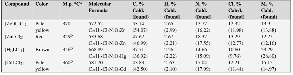
Table 1. The physical properties and elemental analysis of the prepared metal complexes.
Compound Color M.p. °C* Molecular Formula C, % Cald. (found) H, % Cald. (found) N, % Cald. (found) Cl, % Calcd. (found) M, % Cald. (found)
[ZrOL]Cl2 Pale
yellow
370 572.52
C21H15Cl2N7O3Zr
53.14 (54.07) 2.65 (2.99) 15.77 (16.22) 12.32 (11.98) 13.9 (13.88)
[ZnLCl2] Red 329D 533.68
C21H15Cl2N7O2Zn
47.62 (46.99) 2.67 (2.21) 18.37 (17.55) 13.29 (12.77) 12.25 (12.16)
[HgLCl2] Brown 356D 668.89
C21H15Cl2N7O2Hg
37.71 (36.92) 2.26 (2.22) 14.66 (15.09) 10.60 (9.76) 29.29 (28.80) [CdLCl2] Pale
yellow
360D 581.70
C21H15Cl2N7O2Cd
43.83 (42.50) 2. 63 (2.10) 17.04 (17.99) 12.21 (11.44) 15.15 (14.97) *D=decomposed. Antifungal study
Potato dextrose medium (PDA) was prepared in a flask and sterilized. To check the growth of bacterial culture in the medium, the requisite quantity of the standard antibiotic (ampicillin) was added to obtain the desired final concentration of 100 μg mL-1 of the medium. Test samples
were prepared in different concentrations (10, 50 and 100 μg mL-1) in dimethyl sulphoxide and 200 μL of each sample
was spread on the PDA media contained in sterilized Petri plates.
Mycelial discs taken from the standard cultures of fungi (Aspergillus fumigatus and Candida albicans) were grown on the PDA medium for 5-7 days. These cultures were used for the aseptic inoculation in the sterilized Petri dish. Standard cultures inoculated at 28±1 °C were also used as the control.
The efficacy of each sample was determined by measuring the radial mycelial growth. The radial growth of the colony was measured in two directions at right angle to each other and the average of two replicates was recorded in each case. The data are expressed as percent inhibition over the control obtained from the size of colonies. The percent inhibition () was calculated using the formula:
(1)
where
C is the diameter of fungus colony in the control plate after incubation for 96 h and
T is the diameter of the fungus colony in the tested plate after the same incubation period. 16
Results and discussion
The analytical and physical data of template metal complexes are listed in Table 1. The percent’s of C, H, N and M content obtained from elemental and analyse are in good agreement with the general molecular formula proposed for the complexes [MLCl2] (M: Cd(II), Zn(II) and
Hg(II)) and [ZrOL]Cl2, where L = acyclic N2O2 organic
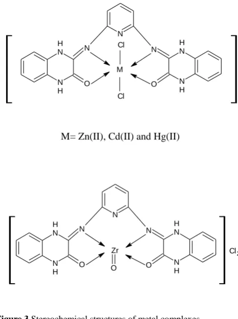
segment formed by template route between 2,6-diaminopyridine and 1,4-dihydroquinoxalin-2,3-dione, as shown in Figure 3.
IR spectra
The IR spectra of acyclic metal template complexes has shown the absence of uncondensed functional groups –NH2
and C=O of 2,6-diaminopyridine and quinoxalin-2,3-dione (i.e., stretching modes of starting materials) which confirm the formation of the proposed acycle organic moiety involving imine –C=N- and –C=O groups13,14 (Table 2.) The
appearance of strong absorption band in the region 1600-1630 cm-1 corresponds to υC=N stretching frequency. A
medium absorption recorded in the regions 3300-3250 cm-1
may be ascribed to υN-H in 1 and 4 positions of pyrazine ring, thus confirms the lactam form corresponding to quinoxaline moiety.14,15 Furthermore, no strong absorption
band was observed near 1675-1660 cm-1 indicating the
absence of >C=O of quinoxalin-2,3-dione at 2-position, this confirms the condensation of carbonyl group of quinoxaline and amino groups of 2,6-diaminopyridine.12-14 These results
provide strong evidence for the formation of acyclic frame.13
A strong absorption band in the region ~1585-1613 cm-1
may be attributed to the C=N group.14-15 The lower values in
the wave numbers of ν(C=N) may be explained on the basis of drift of lone pair density of imine nitrogen towards metal atoms.15-17 The bands present at ~3050-2966 cm-1 may be
assigned due to ν(C-H) vibrations of quinoxaline and pyridine moieties. The bands present in the range ~1150-1130 cm-1 are assigned due to ν(C-N) vibration. The IR
spectra of the complexes do not show any change in the pyridine ring vibrations and interestingly enough, it appears that in these complexes pyridine nitrogen does not take part in coordination.17-18 Thus, in the presence of metal salts, a
quadridentate acyclic system is formed which coordinates through imine nitrogen and two oxygen atoms of –C=O posit ionized in 3 position of lactam quinoxaline of while pyridine nitrogen does not take part in coordination. Moreover, the coordination through pyridine nitrogen does not take place, as it will result in the formation of unstable four member rings.14,15
The far IR spectra show bands in the region ~550-490 and 433-460 cm-1 corresponding to νM-N and νM-O vibrations,
respectively.18 As well as the tautomer’s of in position 2 of
quinoxaline ring between C=O and C=N- supports the coordination of organic moiety with appearance of strong bands at 1680-1640 cm-1. Thus, the acyclic tetradentate
ligand behaves as N2O2 system. Furthermore, the strong
band at 880 cm-1 in the spectrum of zirconium(IV) complex
may be assigned to Zr=O vibration.19 The Zn(II), Cd(II) and
Hg(II) displayed weak absorptions in the regions 260-380 cm-1 which confirms the M-Cl bond.19
100C T
C
Table 2. FT-IR absorptions of the quinoxalin-2,3-dione and its template metal complexes in cm-1.
Complex NH, C=N- C= O Ar-CH, C-N M-N M-O Other bands
Quinoxalin-2,3-dione
3250,1590(s) 1680(s) 3050b,
2962(m)
1150(s) 790-895 b
[ZrOL]Cl2 3180
(m),1533(m)
1657(s) 3060 (w) 2870(m)
1130(m) 450(w) 506(w) 880(Zr=O)
[ZnLCl2] 3180(m), 1580(s) 1677(s)
3080-2970(w)b
1140(m) 433-460(w)
550 265-370a(w),766(m)
[HgLCl2] 3235 , 1610(s) 1666(s) 3100(w),b
2980
1148(s) 421(m), 469(w) 260-380(w)a
2997b(w)
[CdLCl2] 3169, 1530(s) 1640(s) 3050 (w)
2968b
1145(s) 490 (w) 544(m)
270,378(w),780-890(w)b
s=strong,m=medium,br.=broad,w=weak,a =far I.R spectrum of M-Cl and b=vibrational modes of Ar-H.
NMR spectra
The ¹H NMR spectrum of mercury(II) complex shows a multiple signals observed at δ 6.6-8.5 ppm that may be attributed to Ar-H and quinoxaline protons (Fig. 1.) The multiple absorption in the region 9.1-12.5 ppm may be assigned to the spin resonance of the deshielded lactam-NH protons the are effected strongly by the electronic withdrawing of Hg(II) ion (Figure 2). The chemical shifts in the region 4.5-5.3 ppm confirms the tautomerism of –C=N- imine group with C=N- of pyrazine ring, thus support the expected structure of the Hg(II) complex.13,14 Moreover, the 13C NMR spectrum of Hg(II) complex displays resonances
at 40, 110, 130, 150 and 170-200 ppm that are attributed to CH3(DMSO), C=C-(Ar-), C=N, and C=O groups,
respectively.14
The data obtained from 1H and 13C NMR spectra for
Hg(II) complex in DMSO-d6 solvent were shown as below: 13C NMR (300 MHz, DMSO-d6, δ, ppm): 38-40(S, J = 7.2
Hz, DMSO), 155.0 (2C, C=O), 131.0 (1C, Ar-C), 128.1 (2C, N=C), 126.3 (2C, N-C), 128-126 (4C, Ar-C),110-123(4C,C-N-Pyridine) 62.3 (1C, Py-C), 63.2 (1C, C=CH pyridine).
Figure. 1. 1H NMR of [HgLCl
2] complex in DMSO-d6 solution.
Electronic spectra and molar conductivity
The quinoxalin-2,3-dione solution in ethanol shows high intensity peaks at 31446 and 36100 cm-1 that related to
electronic transitions of C=N-, C=O and C=C- groups of the types (π→π* and n→π*).13,15,19 The electronic spectra of the
complexes were recorded in DMF solutions. The pale yellow solution of Zn(II) complex in DMF exhibits two distinct peak in the region 34448 and 27777 cm-1 that are
consistent with ligand field and ligand to metal charge transfer transitions, respectively. The UV-visible spectra of Zn(II), Cd(II) and Hg(II) complexes show high intensity peaks at 22880-34448 due to M→L charge transfer transitions, respectively.22 The electronic spectrum of the
zirconium(IV) complex exhibits bands at 20660 and 30288 cm-1 which are assignable to charge transfer of Zr=O moiety
and ligand field transitions, respectively.20,21 The geometry
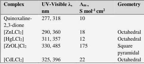
of the zirconium(IV) complex was proved to be square pyramidal based on the charge transfer bands of Zr=O in the UV region besides the 1:2 electrolyte behaviour.
The molar conductance of Zn(II), Cd(II) and Hg(II) complexes in DMF were recorded in the range 10-22 ohm-1
cm2 mol-1 indicating that these complexes were neutral and
agree well with the other spectral and elemental analyses. 23
However, the zirconium(IV) complex solution in DMF shows electrolytic behavior in 1:2 ratio of molar conductance 175 ohm-1 cm2 mol-1 due to the presence of two
chloride ions in the outer sphere of the complex, Table 3.
Table 3. The electronic spectra (cm-1) and molar conductance,
Λm , (S mol-1 cm2) of the prepared complexes
Complex UV-Visible λ, nm
Λm ,
S mol-1 cm2
Geometry
Quinoxaline-2,3-dione
277, 318 10
[ZnLCl2] 290, 360 18 Octahedral
[HgLCl2] 311, 357 12 Octahedral
[ZrOL]Cl2 330, 485 175 Square
pyramidal
[CdLCl2] 325, 396 22 Octahedral
Table 4. Antimicrobial activity of the prepared metal complexes
Compound Inhibition
E. coli S. aurea Fungi
G+ G- Asp. Cand.
[ZnLCl2] 70 66 50 22
[CdLCl2] 49.2 21 18 19
[HgLCl2] 29 17 23 11
[ZrOL]Cl2 22 33 20 44
Control DMSO 10 13 12 9
Streptomycine 25 30 38 33
Antimicrobial studies
In this study, all the chemically synthesized complexes were evaluated against gram-positive and gram-negative bacteria. The MIC values of the synthetic complexes were determined by the method given by Andrews.23,24 Standard
antibiotic, namely streptomycin was used for comparison with the antibacterial activities exhibited by these complexes. All the complexes of the tested series possessed some antibacterial activity against gram-positive bacteria as well as Gram-negative bacteria, Table 4. In the whole series, complexes Zn(II) was found to be most effective against all the tested bacterial strains, showing zone of growth inhibition at 70 mm. The complexes of Cd(II) and Hg(II) exhibited good activity against all the tested bacterial strains with a zone of inhibition in the range from 33.2-49.44 mm and 23.03-29.96 mm, respectively. The ZrO(IV) complex showed activity in the region (22-33 mm) against E. coli and
S. aurea, while it inhibition toward Candida fungus was 44 mm. The complexes of Zn(II) and Cd(II)showed the highest zone of inhibition 60-70 and 21-49.2 mm, respectively 36.70 mm against Staphylloccus aurea and Esherichia coli (Table 4). This would suggest that the chelation could facilitate the ability of a complex to cross a cell membrane and can be explained by Tweedy’s chelation theory.16,25-23 The chelating
will enhance the lipophilic character of the central metal atom, which subsequently favors its permeation through the lipid layers of the cell membraneand blocking the metal binding sites on enzymes of microorganisms.26-29
Conclusion
According to the results obtained from elemental analyses, FT-IR, UV-Visible spectra, magnetic and molar conductivity measurements, the octahedral geometry was proposed for Zn(II), Cd(II) and Hg(II) complexes with formula [MLCl2], while the zirconyl complex was suggested
as square-pyramidal geometry of the formula [ZrOL]Cl2.
The data obtained from infrared spectra revealed that nitrogen atoms of imine groups and two oxygen atoms of carbonyl participate in coordinating with the central metal ions. As well as, this study is a preliminary evaluation of antibacterial activity of acyclic metal complexes against two types of bacteria and fungi. The quinoxaline system is very active biologically. It indicates that the complexes have the potential to generate new antimicrobial metabolites. The acyclic metal complexes demonstrating antibacterial activity could result in the discovery of new chemical classes of antibiotics that could serve as selective agent for the maintenance of animal or human health and provide biochemical tools for the study of infectious diseases.
M= Zn(II), Cd(II) and Hg(II)
Figure 3.Stereochemical structures of metal complexes.
References
1Vigato, P. A., Tamburim, S., Coord. Chem. Rev. 2004, 248,
1717-2118.
2Di Bella, S., Fragala, I., Ledoux, I., Marks, T. J., J. Am. Chem. Soc.
1995, 117, 9481-9485.
3Singh, D. P., Bhantnagar, J. M., Talanta2010, 57, 775-780. 4Patole, J., Singhnapurkar, D., Padhye, S., Ratledge, C., Bioinorg.
Med. Chem. Lett., 2006, 16, 1514-1517.
5Khan, A., Sarkar, S. S. D., Int. J. Antimicrob. Agents 2008, 32,
40-45.
6Daniel, V. P., Murukan, B., Kumari, B. S., Mohanau, K.,
Spectrochim. Acta A. 2008, 70, 403-410.
7Srinivas, Kumar, C. N. S. S. P., Rao, V. J. and Palaniappan, S., J.
Mol. Catal. 2007, 265, 227-230.
8Tarallo, M. B., Costa-Filho, A. J., Vieira, E. D., Monge, A., Leite,
C. Q., Pavan, F. R., Borthagaray, G., Gambino D. and Torre, M. H. J. Arg. Chem. Soc., 2009, 97, 80-89.
9Wang, X. L., Lin, H. Y., Liu, G. C:, Zhao, H-Y., Chen, B. K., J.
Organomet. Chem., 2008, 69, 2767.
10Hu, J., Xie, Z-F., Hui, Y-H., Mo X-X., Sun, N. X., Liu, F.-M.,
Chin. J. Org. Chem., 2007, 279, 1162-1166.
11Zhu, W., Sintic, M., Ou, Z., Sintic, P. J., McDonald, J. A.,
Brotherhood, P. R., Crossley, M. J., Kadish, K. M., Inorg. Chem., 2010, 49, 1027-1031.
12Mohamed, G. G., El-Gamel, N. E. A., Spectrochim. Acta A. 2004,
60, 3141-3151.
13Al-Jibouri, M. N., J. Appl. Chem., 2014, 6(6), 64-73.
14Al-Jibouri, M. N., Emad, A., Al-Mustansiriyah J. Sci.2013, 24(2),
49-64.
N H
N H N
O N
H
N H
O N
N
M
Cl Cl
N H
N H N
O N
H
N
H O
N N
Zr
O
15Satish, M. A., Sathisha, M. P. and Revankar, V. K., Trans. Met.
Chem., 2007, 32, 81-87.
16Atlas, R. M., Brown, A. E., and Parks, L. C., "Laboratory
Manual Experimental Microbiology", Mosby-Year Book Inc.
1995.
17Nakamoto, K., Infrared and Raman spectra of Inorganic and
Coordination Compounds ", 1986, Wiley, New York.
18Siddappa, K., Reddy, T., Mallikarjun, M. and Reddy, C. V., Eur.
J. Chem., 2008, 5(1), 155-162.
19Silverstein, R. M., Bassler, G. C. and Morrill, T. C.,
Spectrometric Identification of Organic Compounds, 4th ed., Wiley, New York, 1981.
20Sutton, D., Electronic Spectra of Transition Metal Complexes,
McGraw-Hill: London, 1968, 388.
21Cotton, F. A., Wilkinson, G., Murillo, C. A., Bochmann, M., Adv.
Inorg. Chem., 6th edition, John Wiley & Sons, 1999.
22Singh, D. P., Malik, V., Kumar, R., Kumar, K., Rasayan J. Chem.,
2009, 2(1), 133-138.
23Geary, W. J., Coord. Chem. Rev., 1971, 7, 81-122.
24Sanmartin, J., Bermejo, M. R., Deibe, A. M. G., Maneiro, M.,
Lage, C., Filho, A. J. C., Polyhedron, 2000, 19, 185-192.
25Rani, D. S., Synthesis and structural studies of transition metal
compounds derived from multidentate disubstituted quinoxalines, Ph.D. Thesis, Osmania University, 1995.
26Raman, N., Raja, J. D., Sakthivel, A., J. Chilean Chem. Soc.,
2008, 53, 3-9.
27Singh, D. P., Kumar, K., Dhiman, S., Sharma, J., J. Enzyme Inhib.
Med. Chem., 2009, 24(3), 795-803.
28Singh, D. P., Eur. J. Med. Chem., 2009, 44(1), 63-69.
29Singh, D. P., Kumar, R., Singh, J., Eur. J. Med. Chem., 2009,
44(4), 1731-1736.