A dissertation submitted to the faculty of the University of North Carolina at Chapel Hill in partial fulfillment of the requirements for the degree of Doctor of Philosophy in the Department of
Biochemistry and Biophysics.
Chapel Hill 2013
modulatory agents that accelerate the slow intrinsic rates of GDP dissociation and GTP hydrolysis. In addition, evidence for the redox regulation of Ras superfamily GTPases is growing, and current data supports regulation by the generation of a thiyl radical. Oxidation and reduction events are critical to physiological and pathological processes and are highly regulated. In Ras GTPases, the redox-sensitive cysteine is in the NKCD motif, which is a motif that is critical for nucleotide binding. However, we show that oxidation that occurs absent of the generation of a thiyl radical does not result in Ras activation.
be in the place I am today without the care and patience of these individuals.
I would also like to acknowledge my mentor, Dr. Sharon Campbell, for always being there to listen to my wild ideas and provide feedback on new data. Her willingness to discuss anything will be greatly missed. While others got me excited to perform research, Sharon taught me how to think like a scientist.
To specifically acknowledge every person that helped along the way would result in an acknowledgements section as long as the dissertation itself. Thus, I say thank you to current and previous lab members, classmates, and friends for asking questions, challenging my thoughts, and providing support when needed. Minh Huynh, who was an undergraduate student under my direction, served with me for two years and deserves a special
encouragement to achieve good grades, instill a solid work ethic, and do what I loved has led me to be who I am today. There are no words to describe the level of gratitude I have for my parents for teaching me right from wrong as well as the importance of never quitting and doing your best.
I also wish the thank Heather. Although she joined me in my last two years of my journey at UNC, they were the most productive two years as well as the most exciting years. She taught me what school could never teach a person, led me out of the lab on some amazing adventures, and let me back into the lab to complete my work. While she has
captured my heart, I will never be able to thank her enough for her help and support through some of the most demanding years in graduate school.
Lastly, I must thank Josh Gough for providing me with one of my favorite sayings, “It sucks to suck.” It truly does suck to suck, and this saying has repeatedly served as motivation and as words of encouragement.
Generation of ROS and RNS in cells and antioxidant defense ... 4
Reactive oxygen and nitrogen species as second messengers ... 9
Free radicals modify and activate Ras through a reactive cysteine in the NKCD motif ... 11
Redox regulation of Rho GTPases by 1e- and 2e- mechanisms through a redox-active cysteine in the phosphoryl-binding loop ... 14
Chapter 2: Glutathiolated Ras: Characterization and Implications for Ras Activation ... 17
Introduction ... 17
Materials and Methods ... 20
Results ... 44
Discussion ... 52
Chapter 4: Oxidation of RhoA at Cys20 regulates nucleotide binding ... 56
Introduction ... 56
Methods ... 60
Results ... 64
Discussion ... 82
Chapter 5: Site-specific monoubiquitination activates Ras by impeding GTPase-activating protein function ... 88
Introduction ... 88
Commentary ... 88
Chapter 6: Biophysical and Proteomic Characterization Strategies for Cysteine Modifications in Ras GTPases ... 101
Introduction ... 101
Materials ... 104
Methods ... 106
3.1: Cysteine pKa determination using 4-fluoro-7-aminosulfonylbenzofurazan (ABD-F) ... 106
3.2: Generation of cysteine-modifying redox-active compounds (CysNO/GSNO generation) ... 109
3.3 NO2• generation and NONOates ... 110
3.4 Quantitative mass spectrometry ... 112
Notes ... 122
Chapter 7: Magnesium coordination of RhoG ... 127
LIST OF FIGURES
Figure 1. The GTPase cycle of Ras family GTPases ... 2
Figure 2. Generation and interconversion of ROS and RNS in cells ... 5
Figure 3. Mass spectrometry of glutathiolated Ras. ... 25
Figure 4. Biochemical characterization of glutathione-modified Ras. ... 27
Figure 5. 2D NMR 1H-15N HSQC comparison of RasWT and RasSSG. ... 29
Figure 6. MANTGDP nucleotide dissociation assays in the presence of the NO-generating agent DEANO. ... 31
Figure 7. Effect of oxidized glutathione on nucleotide binding in Ras. ... 33
Figure 8. Modification of Ras by glutathione can proceed by three different mechanisms. ... 36
Figure 9. CD spectroscopy of K-RasWT and K-RasG12C. ... 46
Figure 11. Nitrosylated K-Ras and nucleotide binding. ... 49
Figure 12. Nucleotide exchange and hydrolysis kinetics of RasG12C and RasG12S. ... 50
Figure 13. NMR analysis of Ras position 12 mutants. ... 51
Figure 14. Reaction of RhoA and RhoA variants with ABD-F. ... 68
Figure 15. Nucleotide binding assays of oxidized RhoA. ... 70
Figure 16. Oxidation mimetic RhoAC20D shows the critical role of Cys20 in nucleotide binding. ... 71
Figure 17. Biochemical characterization of RhoA redox-insensitive mutants. ... 73
Figure 18. GTP hydrolysis of RhoA and RhoA redox-insensitive mutants. ... 75
Figure 19. Thermal denaturation of RhoA and RhoA variants. ... 76
Figure 20. 2D NMR analyses of RhoA and RhoA variants. ... 79
Figure 21. Residues with chemical shifts plotted on pymol-generated structures. ... 80
Figure 30. Generalized ICAT-based relative quantification schematic for
measuring cysteine peptides. ... 116
Figure 31. The oxICAT schematic for measuring cysteine oxidation... 118
Figure 32. Nucleotide dissociation of RhoG and Rac1. ... 131
LIST OF TABLES
Table 1. Tm of K-Ras determined by CD spectroscopy ... 47 Table 2. Thiol accessibility in RhoA crystal structures ... 66 Table 3. Nucleotide dissociation and hydrolysis rates in the presence and
β-ME Mercaptoethanol
CD circular dichroism
CFC Cardio-facio-cutaneous syndrome
CO3•- carbonate radicals
DAB denaturing alkylation buffer
DAF diaminofluorescein
DEANO NONOate diethylammonium
(Z)-1-(N,N-diethylamino)diazen-1-ium-1,2-diolate DETANO-NONOate
(Z)-1-[N-(2-aminoethyl)-N-(2-ammonioethyl)amino]diazen-1-ium-1,2-diolate
DMPO 5,5-dimethyl-1-pyrroline N-oxide
DTBA dithiobutylamine
DTT dithiothreitol
GAPs GTPase activating proteins
GDIs guanine dissociation inhibitors
GEFs guanine exchange factors
GRX-1 glutaredoxin-1
GSSG oxidized glutathione
H2O2 peroxide
HCD-MS/MS higher energy collisional dissociation
Hepes 4-(2-hydroxyethyl)-1-piperazineethanesulfonic acid
HSQC heteronuclear single quantum coherence
spectroscopy
IAA iodoacetamide
ICAT Isotope-coded affinity tag
iNOS inducible NOS
IST immuno-spin trapping
LC-MS liquid chromatography-mass spectrometry MALDI-MS matrix-assisted laser desorption/ionization-MS mantGDP
2'-/3'-O-(N'-methylanthraniloyl)guanosine-5'-O-diphosphate
MES 2-(N-morpholino)ethanesulfonic acid
MS mass spectrometry
mUb monoubiquitination
mUbRas mUb of KRas
NEM n-ethyldmaleimide
N2O3 dintrogen trioxide
NO+ nitrosonium ion
ONOO- peroxynitrite
PAO phenylarsine oxide
PAPA-NONOate
(Z)-1-[N-(3-aminopropyl)-N-(n-propyl)amino]diazen-1-ium-1,2-diolate
PKC Protein Kinase C
p-loop phosphoryl binding loop
PI3K phosphoinositide 3-kinase
PTIO 2-Phenyl-4,4,5,5-tetramethylimidazoline-1-oxyl 3-oxide
PTMs post-translational modifications
RBD Ras-binding domain
REF52 Rat embryonic fibroblasts
ROS reactive oxygen species
RSO2- sulfinic acid
SCX strong cation-exchange
SDS sodium dodecyl sulfate
SIM selected-ion monitoring
SIN-1 5-amino-3-(4-morpholinyl)-1,2,3-oxadiazolium chloride
SOD superoxide dismutase
SOS Son of Sevenless
TCEP Tris(2-carboxyethyl)phosphine
TEV tobacco etch virus
(guanine exchange factors [GEFs]), whereas the GDP-bound form reduces effector binding, turning signaling “off” (GTPase activating proteins [GAPs]). In general, three distinct classes of proteins regulate the level of activated GTPases in the cell (Figure 1).
Ras superfamily GTPases can be modified by a variety of post-translational modifications (PTMs) that drive differences in localization and activity (5). Ras and Rho GTPases associate with the membrane upon lipid modification at the carboxyl-terminus (6, 7). Rac1, RhoA, and Cdc42 are geranylgeranylated and associate with the inner leaflet of the
plasma membrane (8). Rho guanine dissociation inhibitors (GDIs) recognize the lipid moiety and prevent the GTPase from re-associating with the lipid membrane after dissociation (9).
Figure 1. The GTPase cycle of Ras family GTPases
residues in Ras has been observed, which directly regulates its activity (15, 16). Recent evidence indicates that a subset of Rho GTPases can be ubiquitinated (17); however, Rac1 is the only Rho family GTPases that has been shown to be monoubiquitinated (18), whereas RhoA and Cdc42 have only been observed to be polyubiquitinated, which results in deactivation and degradation (17, 19, 20). Further, monoubiquitin can be removed by deubiquitinating enzymes (DUBs); however, Ras- and Rho-specific DUBs have yet to be discovered.
24). A cysteine residue within a protein is generally considered ‘redox-sensitive’ if it is
solvent accessible and contains a depressed pKa, which has been shown to increase the reactivity of the thiol to oxidants (25). If a cysteine forms interactions within the protein that stabilize the thiolate (S-) form, then the residue will have a lower pKa relative to free cysteine (26). In general, a free cysteine has a pKa of approximately 8.5, which leaves it in the thiol (SH) form at physiological pH. The rate of thiol reactivity depends on the thiol ionization state and the oxidant, with the thiolate state being more sensitive to oxidation than the thiol state (27). Furthermore, there is no known consensus sequence that can be used to predict redox-sensitive cysteines (28). While peroxynitrite has been shown to be able to oxidize the thiol form, it is more reactive with the thiolate form, although the radical-mediated
breakdown products are more likely to oxidize cellular thiols (Figure 2) (29). In addition, peroxide will only react with the thiolate (S-) form of cysteines (30), whereas, nitric oxide has been shown to react too slowly with thiols to be physiologically relevant (31). However, an altered pKa is not always a prerequisite for redox sensitivity as Ras Cys118, a known redox-sensitive cysteine, does not have an altered pKa; however, Ras is only regulated through Cys118 by free radical-mediated oxidants.
Thus, as the Ras field is still largely unfamiliar with the redox regulation of GTPases and the redox field as a whole, in this review, we will present some basic information on redox signaling in cells and describe the current understanding of the redox regulation of Rho GTPases.
Generation of ROS and RNS in cells and antioxidant defense
Figure 2. Generation and interconversion of ROS and RNS in cells
detail the role of ROS and RNS in the regulation of Rho GTPases, we will give a brief overview of the production of ROS and RNS in cells, antioxidant defense, and interconversion of cellular ROS and RNS (outlined in Figure 2).
While ROS and RNS are generally grouped into a category of reactive intermediates, it is important to understand that there are two classes of oxidants, one-electron and two-electron oxidants. Two two-electron oxidants have been shown to oxidize thiols to generate sulfenic acid and a predominant cellular two-electron oxidant is hydrogen peroxide (26). One-electron oxidants, such as nitrogen dioxide, generates a thiol radical, which will
predominantly react with other thiols, including cellular glutathione, to form disulfide bonds and produces superoxide (Figure 2) (35, 36). Under normal cellular signaling, only the most reactive thiols will be subject to oxidation and signaling through ROS and RNS, and ROS and RNS have been observed to function as second messengers (23). However, it is important to note that oxidation observed in vitro does not always correlate with oxidation in vivo. This is because in vitro experiments tend to have few substrates in solution, which
leaves the oxidant little competition. Therefore, for a protein to function in oxidative signaling, it needs to be readily oxidized in vivo under normal cellular conditions (27).
A primary source of cellular ROS exists within the mitochondria (37). As all aerobic organisms require oxygen for oxidative phosphorylation, the mitochondria is a central organelle that generates a number of cellular oxidants. One of the most common ROS generated by the mitochondria is superoxide (O2•-) due to incomplete reduction of molecular oxygen (O2). Superoxide does not readily diffuse across membranes, which limits the
p67phox, and Rac1 to form the membrane-associated NADPH oxidase complex, which catalyzes the one-electron reduction of oxygen to superoxide (O2•-) to eliminate invading pathogens by oxidation (40). In addition, superoxide (O2•-) generated in the arteries by Nox2 and Nox4 compete with antihypertensive effects of nitric oxide (NO•) by reacting with nitric oxide to form peroxynitrite (ONOO-; Figure 2) (41). In lung fibroblasts, the presence of different Nox isoforms promotes superoxide (O2•-) production in the cytosol and
extracellular space.
Another source of cellular ROS is in liver and kidney cells. These cells use cellular substructures called peroxisomes, which contain a variety of ROS and RNS generating enzymes and peroxisomal catalase that function in metabolic pathways and have been shown to produce as much as 35% of cellular peroxide (42).
in cells and when activated, can produce low levels of nitric oxide (NO•). Activation of eNOS, like nNOS, is regulated by calcium-bound calmodulin (45), interactions with various effector proteins (46), and phosphorylation (47). Lastly, iNOS expression is induced in macrophages and other cell types and is constitutively active once expressed (48). The function of iNOS is to produce nitric oxide (NO•) to inhibit iron-sulfur proteins, damage tumor cells, and oxidize the DNA of invading parasites.
Discussion regarding the generation of endogenous oxidants would not be complete without considering the lungs. The lungs are constantly exposed to pollutants, cigarette smoke, ozone, and other industrial oxidants. This extra exposure to exogenous ROS and RNS makes the lungs more susceptible to oxidative stress, which can have different effects on protein function than oxidative signaling, which are beyond the scope of this
introduction.
The cell has also evolved a network of antioxidant enzymes and small molecules to protect the cell from oxidative damage. An abundant antioxidant enzyme that deserves consideration is SOD. Three distinct isoforms of SOD: SOD1, a dimeric protein that binds copper and zinc and is localized in the cytosol; SOD2, a homotetrameric protein that binds one manganese ion per subunit and is localized in the mitochondria; and SOD3, a
homotetrameric protein anchored to the extracellular matrix. SODs reduce O2•- to H2O2. Peroxide, in turn, can be reduced to H2O by catalase and glutathione peroxidase (Figure 2). In addition, GSH-Pxs can reduce lipid peroxidases to their respective lipid alcohols. Other antioxidant enzymes include thioredoxin reductases, peroxiredoxins, and glutaredoxins, which all function in the removal of peroxide (49).
(HO•), and examples of RNS are nitric oxide (NO•), nitrogen dioxide (NO2•), peroxynitrite (ONOO-), and dintrogen trioxide (N2O3). However, for these molecules to function in cellular signaling, they must meet the definition of second messengers. As second messengers, RNS and ROS are generated in response to a specific stimulus, short lived, specifically target effectors, and transiently and reversible effect signaling. Superoxide (O2•-) and peroxide (H2O2) are considered second messengers as these reactive species are less reactive than other ROS, such as HO•, which lacks specificity as it reacts at the rate of diffusion with any molecule it contacts. For RNS, nitrogen dioxide (NO2•), not nitric oxide (NO•), is thought to be the major source of free radical-mediated oxidation of thiols. Peroxynitrite undergoes homolytic cleavage to generate nitrogen dioxide and carbonate radicals (CO3•-) in the presence of CO2, which can directly oxidize thiols; however, dinitrogen trioxide, and the nitrosonium ion (NO+) are capable of nitrosating thiols through two-electron mechanisms (53) to generate nitrosothiols (RSNO).
observed NO-mediated Ras activation in T cells (58), which was transient and
concentration-dependent. Later, it was shown that treatment with sodium nitroprusside, a nitric oxide-generating compound, increased Ras downstream signaling through the mitogen-activated kinase pathway (59). An important tool in such studies, RasC118S is a variant that aids in determining the direct and indirect effects of RIs on Ras because it is RI-insensitive and does not alter Ras structure and activity (60). A recent study on
mechanism, eNOS-generated NO• activates RasWT and stimulates Ras downstream pathways (63). Further, Lim et al showed that, for H- and N-Ras, introducing the RasC118S mutation circumvented PI3K pathway activation and reduced tumor growth. In a separate cell-based study, eNOS-derived NO• activated N-Ras in T cells engaged with antigen presenting cells; Cys118 was required for activation (64). These data suggest that NO• mediates Ras activation directly through Cys118.
Ras superfamily GTPases have four conserved nucleotide-binding motifs (65). While the X residue in the NKXD motif is not well-conserved, several Ras subclass members contain a redox-active cysteine at this position. GTPases with a redox-sensitive NKCD motif can be activated by NO2• and other RIs; however, NO-mediated regulation is best
characterized for Ras. Several cell-based and in vitro studies have shown that NO2• reacts with Ras through Cys118 to promote nucleotide exchange and Ras activation (57). Our lab has employed NMR and biochemical approaches to show that S-nitrosation of Ras does not affect Ras structure or nucleotide binding (66). We speculated that an intermediate formed during the reaction of Ras with NO2• modulates Ras activity and investigated several reactions involved in thiol S-nitrosation. Furthermore, we observed that 1e- oxidation
supporting this model, we demonstrated that nucleotide binding is not altered by either RasC118 mutation or S- nitrosation (69). A proposed thiyl radical-based mechanism has been described in detail elsewhere (70) but remains inconclusive, as direct evidence to support the proposed radical intermediates has not yet been obtained.
suggested that Ras S-nitrosation may be an intermediate during glutathione modification of Ras, they concluded that covalent (2e-) glutathione modification of Ras leads to Ras
activation (77). However, the observation that Ras glutathiolation alters nucleotide exchange contradicts our previous studies, which demonstrated that Cys118 and 1e- oxidants are
required for regulation of Ras nucleotide binding and activity. To address this discrepancy, we have recently demonstrated that treatment of Ras with oxidized glutathione (GSSG) results in glutathiolation of Ras at Cys118. However, similar to S-nitrosation, glutathione modification of Ras does not alter Ras nucleotide binding. Therefore, we propose that Ras activation requires Cys118 thiyl-radical formation for nucleotide dissociation and subsequent activation. One end product of RasC118 thiyl-radical generation is RasSSG. Thus, the presence of RasSSG may reflect Ras activation if it was generated by a reaction with the RasC118 thiyl radical; this is also the most likely mechanism in cells given the slow reaction rate for the Ras thiol and GSSG under physiological conditions.
Redox regulation of Rho GTPases by 1e- and 2e- mechanisms through a
redox-active cysteine in the phosphoryl-binding loop
We have demonstrated that select Rho GTPases are redox sensitive; however, Rho GTPases contain a different redox motif than Ras (78). Over fifty percent of Rho GTPases contain a redox sensitive cysteine at the end of the nucleotide-binding p-loop motif, GXXXXGK[S/T]C. Based on the Rac1, Cdc42, and RhoA crystal structures, the solvent-exposed cysteine in the p-loop motif likely has an altered pKa and is accessible to RIs. We have previously shown that the reactive phosphoryl binding loop (p-loop) cysteines in Rho GTPases are sensitive to oxidation, resulting in altered GTPase regulation (79).
capable of promoting disulfide reduction and restoration of RhoA nucleotide binding (81, 82). Redox-mediated inactivation of Ras may occur when the cellular reduction potential is
reduced, such as during oxidative stress. It is intriguing to speculate that enzymes, such as thioredoxin and glutaredoxin, which are capable of reducing protein disulfides, may act on RhoA and contribute to activation.
The addition of cisplatin or arsenic trioxide has been shown to inactivate RhoA by promoting mixed disulfide formation between the two p-loop cysteines (80). In contrast to Ras, these results suggest that 2e- oxidation can regulate RhoA activity. In support of this hypothesis, Gerhard et al showed that phenylarsine oxide (PAO) generates a mixed disulfide crosslink between RhoA Cys16 and Cys20 using MALDI-MS (matrix-assisted laser
desorption/ionization-MS) and demonstrated that the modification inhibits stress fiber formation through the inactivation of RhoA in Caco-2 cells (83).
growth and differentiation, and programmed cell death (84-86). The structural differences between the GDP- and GTP-bound states of Ras are primarily localized within two regions, Switch I (residues 30-37) and Switch II (residues 60-76) (65). The intrinsic rates of GDP exchange and GTP hydrolysis are too slow to respond to cell signaling events (87), and consequently, protein factors associate with Ras and accelerate these rates in a regulated manner. Guanine nucleotide exchange factors (GEFs) accelerate exchange of bound GDP for GTP, which leads to Ras activation (88), whereas GTPase activating proteins (GAPs)
cysteine 118 (Cys118) (62). As the GTP:GDP ratio in cells is approximately 10:1, oxidation-mediated GDP dissociation can promote GTP loading of Ras, analogous to the action of GEFs (69). While the reaction of nitric oxide and its auto-oxidation product, NO2•, with Ras have been characterized, it is less clear how other thiol modifications (i.e., glutathiolation) affect Ras activity. As numerous studies have described activity modulation of Ras in the presence of ROS and RNS, we refer the reader to two recent reviews that detail the redox regulation of Ras and Ras-related GTPases (57, 95).
Ras contains a solvent accessible cysteine (X) in the nucleotide-binding NKXD motif. While this cysteine is conserved in H-, K- and N-Ras, it does not form interactions with other residues in Ras or the guanine nucleotide ligand and is poorly conserved in the Ras superfamily. We have previously postulated that Cys118 is conserved in N-, K-, and H-Ras due to its role in the redox regulation of Ras (69). In fact, Cys118 has been shown to react in vitro and in vivo with a variety of thiol oxidizing agents, including oxidized glutathione,
(BAECs) to peroxynitrite (71, 100), addition of angiotensin II to vascular smooth muscle cells (VSMCs) (77, 101), and exposure of H2O2 to rat ventricular myocytes (97, 99). However, addition of these agents to cultured cells can produce redox agents capable of protein thiyl radical formation. For example, peroxynitrite can react with cellular CO2 to produce CO3 •-and NO2• radicals, and under acidic conditions (peroxynitrite pKa is ~6.6), peroxynitrite can decompose to hydroxyl radical and NO2• (102). Furthermore, peroxynitrite and angiotensin II can dysregulate several kinase pathways, including the PI3K/Akt pathway, and increase the production of cellular NO by activation of endothelial nitric oxide synthase (eNOS) (103, 104). The NO produced by eNOS can auto-oxidize to produce NO2•, which is a powerful oxidant capable of thiyl radical formation. Consistent with observations of Liaudet et al and Ushio-Fukai et al, the PI3K/Akt pathway was activated after exposure of BAECs to
al (77) results from a radical-mediated mechanism, which occurs prior to the modification by glutathione. However, due to the larger size of glutathione compared to NO•, it was hypothesized that Ras glutathiolation at Cys118 causes structural changes in the nucleotide binding pocket, leading to perturbation of guanine nucleotide binding and an increased rate of nucleotide exchange (71). As Cys118 is critical for the radical-mediated regulation of Ras activity, we sought to circumvent the confounding factors of peroxynitrite and angiotensin II use in cells by directly determining whether glutathiolation at Cys118 alters Ras structure and activity in vitro.
While Ras has been shown to be glutathiolated in cells (71, 77, 97, 100), it is unclear how glutathiolation alters Ras activity. Therefore, we employed NMR and fluorescence-based biochemical assays to assess whether glutathione modification of Ras perturbs Ras structure or activity. We find that treatment of Ras with oxidized glutathione leads to glutathiolation specifically at Cys118, which does not alter Ras tertiary structure or guanine nucleotide binding. These results are consistent with our previous observations that S-nitrosation of Ras Cys118 does not perturb the structure or activity of Ras (66). Therefore, our data suggests that glutathiolation can only affect Ras activity if modification proceeds
through a radical-mediated reaction. Moreover, Ras glutathiolation prevents further redox-mediated activation of Ras by free radical-based mechanisms, which may serve to protect Ras from future radical-mediated oxidation events under conditions of oxidative stress. Materials and Methods
Ras purification and glutathiolation
purified Ras in glutathiolation buffer (50 mM Tricine pH 8.0, 50 mM NaCl, 5 mM MgCl2, and 30 μM GDP) at 37˚C for 15 min. Prior the addition of glutathione, Ras was reduced with dithiothreitol (DTT) for 30 min at pH 8.5 before being buffer exchanged into glutathiolation buffer that was flushed with N2 gas to remove dissolved oxygen and prevent auto-oxidation.
Mass Spectrometry of glutathiolated-Ras and Ras
Ras mass measurements were performed on an LTQ-Orbitrap Velos mass
spectrometer (Thermo Scientific; San Jose, CA). The mass analysis of intact Ras samples was achieved in full-mass spectrometry (MS), selected-ion monitoring (SIM), and higher energy collisional dissociation-(HCD)-MS/MS modes with a resolution of 120,000 at m/z 400 Da. The intact MS spectra were deconvoluted using ProMass, and HCD-MS/MS product ion spectra were processed manually by assigning sequence ions to theoretical masses
Inc.) against a Human Uniprot database. Peptides were confidently identified using a target-decoy approach with a false-discovery-rate (FDR) of 1%. A precursor ion mass tolerance of 200 ppm and product ion mass tolerance 0.5 Da with a maximum of two missed cleavages and variable modifications of cysteine glutathiolation and oxidation were used as a protein database search parameter. All peptides were filtered and reported within a mass accuracy of 5 ppm.
Nucleotide exchange and hydrolysis assays
The rate of GDP dissociation from Ras was measured using
2'-/3'-O-(N'-methylanthraniloyl)guanosine-5'-O-diphosphate (mantGDP) as previously reported (108, 109). One micromolar Ras (RasWT, RasSSG, or RasC118S) loaded with mantGDP (BioLog; San Diego, CA) was added to 1 mL of degassed assay buffer (50 mM Tris pH 7.4, 50 mM NaCl, 5 mM MgCl2, 2 mM GDP, and 100 μM diethylenetriaminepentaacetic dianhydride; DTPA), and the rate of guanine nucleotide dissociation was measured by monitoring the change in fluorescence (excitation: 365 nm; emission: 435 nm; 25˚C) over time using a Perkin Elmer LS50B fluorimeter (Waltham, MA). All experiments were performed in triplicate.
Fluorescent nucleotide dissociation curves were fit to a one-phase exponential decay equation using GraphPad Prism version 3.03 (GraphPad Software; San Diego, CA). For GEF-induced Ras-GDP dissociation assays, the minimal catalytic domain of Son of Sevenless (SOScat) was used at a 1:1 ratio to Ras. This concentration of SOScat was selected as the rate enhancement of nucleotide exchange was easy to measure using fluorimetric assays and other labs have published using the GEF at this concentration. The SOScat construct (from John Kuriyan; University of California, Berkeley) was expressed and purified as previously described (110).
and fit to a one-phase exponential association curve. For these experiments, the ratio of GAP to Ras was 1:200, and the minimal catalytic domain of p120 RasGAP was used.
Nitric oxide-induced nucleotide dissociation
were performed with pulsed field gradient and water flip-back methods as previously described (115). Uniformly labeled 15N-enriched Ras was purified as previously described (116). 2D 1H-15N HSQC experiments were acquired on 0.8 mM 15N-enriched RasWT and RasSSG with 1,024 x 128 complex data points and a spectral width of 8,000 Hz for the 1H dimension and 1,709 Hz for the 15N dimension. Buffer contained 10 mM maleate (pH 6.5), 5 mM MgCl2, 40 mM NaCl, 20 μM GDP, and 10% D2O. NMR data were processed and
analyzed using NMR PIPE (117) and NMR ViewJ (One Moon Scientific; Newark, NJ).
Results
Modification of RasC118 by glutathione
As RasC118 has been previously identified as the site of oxidative modification by nitric oxide (66), we used a combination of top-down and bottom-up MS to confirm that Cys118 is the preferred site of glutathiolation. Whereas partial modification of Ras by glutathione was obtained at a ratio of 50:1 GSSG to Ras upon incubation for 15 min at pH 8.5 (Figure 3a), increasing the GSSG to Ras ratio to 1000:1 resulted in conversion of hRas1-166 to a
predominantly single glutathiolated form. However, Ras glutathiolation was not observed when the redox insensitive RasC118S variant was treated under the same conditions (Figure
3b). All full-MS and selected-ion monitoring (SIM) scans corresponding to glutathiolated RasC118 and redox-insensitive RasC118S are available in supporting data (Fig. S1). We have also confirmed the site-specificity of hRas1-166 glutathiolation using top-down HCD-MS/MS of the 13+ charge state (Fig. S2) and bottom-up identification of surrogate triply charged tryptic peptides that constitute Cys118 (Figure 3c-d). The top-down data contain diagnostic neutral loss ions corresponding to Cys-glutathionyl and glutathione as well as backbone product ions, which suggests that the glutathione moiety is present exclusively on Cys118. In
Figure 3. Mass spectrometry of glutathiolated Ras.
mass spectrometry data indicate that RasC118, which is solvent exposed in the structure of hRas1-166 (pdb: 1crp), is susceptible to glutathiolation, whereas the two solvent inaccessible cysteine residues (Cys51 and Cys80) were unmodified under the tested conditions.
Glutathiolation of RasC118 does not significantly affect Ras-GDP nucleotide dissociation or
GTP hydrolysis
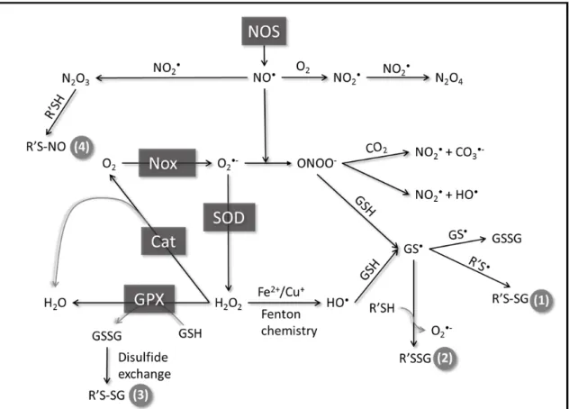
It has previously been observed that peroxynitrite addition to BAECs and VSMCs promotes the formation of glutathiolated Ras at Cys118. Moreover, glutathiolation was concluded to be an activating modification as the population of activated Ras was also glutathiolated (71, 77, 96). As exposure of Ras to peroxynitrite in cells could generate RasC118• and RasSSG, it is difficult to distinguish which modification, the thiyl radical or glutathione, leads to Ras activity changes under the experimental conditions used. To assess whether glutathiolation alters Ras guanine nucleotide binding and/or GTP hydrolysis, we performed nucleotide dissociation and GTP hydrolysis assays on unmodified and glutathiolated hRas 1-166 in vitro. As shown in Figure 4a and b, the intrinsic rates of GDP dissociation (kobs) for RasWT and RasSSG (Figure 4a-b) were determined to be similar (3.9 ± 0.1×10⁻⁵ s-1 vs. 4.7 ± 0.5×10⁻⁵ s-1). As the intrinsic rate of nucleotide dissociation for Ras is slow, interactions with GEFs are required to facilitate nucleotide exchange in vivo. Therefore, we measured the rate of GDP dissociation in the presence of SOS, a Ras GEF, as previously described (108). When the minimal catalytic domain of SOS (SOScat) was added to Ras, the rate of nucleotide dissociation for RasSSG and RasWT was similar (18.5 ± 0.5×10⁻⁴ s-1 vs. 20.2 ± 0.7×10⁻⁴ s-1, respectively).
Figure 4. Biochemical characterization of glutathione-modified Ras.
turnover hydrolysis assays, the intrinsic rate of GTP hydrolysis for RasWT was determined to be 0.6 ± 0.04×10⁻⁴ s-1 at 25˚C (Figure 4c-d), similar to previously reported values of 4.4×10⁻⁴ s-1, which were performed at a higher temperature of 30˚C (118). The rates of GTP hydrolysis determined using the FlipPi sensor compared to previously reported values, indicating that this method is suitable for comparing GTP hydrolysis rates. The intrinsic rate of GTP hydrolysis for RasSSG was determined to be ~4-fold slower (0.14 ± 0.06×10⁻⁴ s-1) than for RasWT. However, as the intrinsic rate of GTP hydrolysis is too slow to be biologically relevant and requires catalysis by GAPs in vivo to regulate Ras activity, we measured the rate of hydrolysis in the presence of catalytic amounts of p120GAP. The rate of GAP-mediated GTP hydrolysis for RasWT and RasSSG were observed to be similar (20.6 ± 2.3×10⁻⁴ s-1 and 19.4 ± 1.7×10⁻⁴ s-1, respectively; Figure 4c-d). Thus, glutathiolation of RasC118 does not affect p120GAP-mediated GTP hydrolysis.
Glutathiolation at Cys118 does not perturb Ras structure
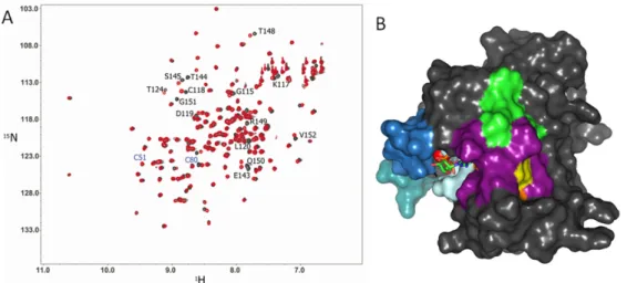
As it was previously suggested that Ras glutathiolation alters the structure of Ras and leads to increased nucleotide exchange (71), we glutathiolated 15N-enriched hRas1-166 and performed 2D NMR HSQC analyses. A 2D 1H-15N NMR HSQC allows for observation of backbone and side chain N-H groups and provides a site-specific probe for every residue in a protein aside from proline. An overlay of the RasWT and RasSSG HSQC spectra is shown in
Figure 5. 2D NMR 1H-15N HSQC comparison of RasWT and RasSSG.
onto the surface of the Ras structure (Figure 5b), only residues proximal to the modified cysteine show chemical shift changes. Interestingly, the chemical shift perturbations for RasSSG (green in Figure 5B) compared to RasSNO (orange; overlap is shown in purple, Cys118 in yellow) are similar, and we have previously shown that RasSNO does not alter Ras structure (66). Thus, the limited chemical shift changes close to the site of glutathiolation and
similarity between the HSQC spectra of RasWT, RasSSG, and RasSNO suggest minimal
structural perturbation by glutathiolation, consistent with our findings that the biochemical properties of Ras are unaltered. Furthermore, the residues important for Ras recognition by regulatory factors and effectors, including the p-loop (residues 10-17), Switch I, and Switch II, do not show chemical shift changes upon glutathiolation. These data are consistent with our findings that GEF- and GAP-mediated stimulation of GDP dissociation and GTP hydrolysis were unaffected by Ras glutathiolation.
Glutathiolation impedes redox-mediated nucleotide dissociation
Previous studies have shown that treatment of Ras with NO2• enhances the rate of nucleotide dissociation, whereas nitrosation of Ras at this site prevents radical-mediated nucleotide dissociation (69). To determine whether Ras glutathiolation, like nitrosation, impedes free radical-mediated dissociation in Ras, the rate of nucleotide dissociation was determined using a MANTGDP-dissociation assay in the presence of the NO•-releasing agent DEANO (1). As shown in Figure 6a, we observe an enhanced rate of RasWT-GDP
dissociation in the presence of DEANO (greater than 200-fold faster). In contrast, the rate of GDP dissociation for the redox inactive RasC118S variant was insensitive to the presence of DEANO. Furthermore, RasSSG was resistant towards DEANO-mediated nucleotide
Figure 6. MANTGDP nucleotide dissociation assays in the presence of the NO-generating agent DEANO.
be formed. Thus, our results show that glutathiolation impedes free radical-dependent Ras GDP dissociation, whereas glutathiolation does not significantly affect Ras structure, activity, or interactions with modulatory proteins.
Discussion
A number of studies have reported Ras activity modulation by ROS and RNS (94). Most of these studies point to RasC118 as the redox-sensitive site because this cysteine has been shown to be oxidized by a number of cysteine-modifying agents and is required for redox-mediated regulation of Ras activity (57). Two of the most common oxidative modifications identified for Ras in vivo are nitrosation and glutathiolation. We have previously reported that radical-mediated nitrosation of Ras leads to increased guanine nucleotide dissociation (68), whereas non-radical-mediated nitrosation of RasC118 does not affect Ras guanine nucleotide binding (66). Given these observations, we were intrigued by reports that glutathiolation of Ras alters Ras activity (71, 77, 96, 100).
Figure 7. Effect of oxidized glutathione on nucleotide binding in Ras.
important consideration that must guide in vitro experimentation with peroxynitrite is that in the absence of CO2, peroxynitrite reacts directly with many biochemical targets,
particularly thiols. This oxidative reaction produces predominately non-radical
oxidativeproducts and is faster than its homolysis to HO• and NO2•. Therefore, peroxynitrite reactions are heavily context dependent. Importantly, given the high glutathione
concentration in cells, once peroxynitrite is decomposed and the free radical chain reactions are completed, glutathiolated Ras will predictably be a major end product that is
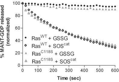
indistinguishable by all means from the end product produced directly by the reaction of oxidized glutathione with RasWT. Therefore, we postulate that the exogenous addition of peroxynitrite and angiotensin II promotes Ras thiyl radical-mediated oxidation of the bound guanine nucleotide, leading to Ras activation. Further, the resulting thiyl radical can react with glutathione to produce RasSSG•, which aerobically decays to RasSSG and O2•-. This route of activation is shown in Figure 8A, and based on our analysis, is the likely pathway of glutathiolation that would result in Ras activation in vivo. Interestingly, Ras glutathiolation appears to protect Ras from further free radical-mediated events. We evaluated guanine nucleotide binding of RasWT, RasC118S, and RasSSG in the presence of the NO-releasing agent DEANO. In these experiments (Figure 6), glutathiolated Ras was protected from free radical-mediated nucleotide dissociation. Therefore, glutathione, which likely modifies Ras after an initial activating event, could protect Ras from over-oxidation in cells under conditions of oxidative stress.
the bound nucleotide after 300 s. In our experiments, we also tested RasC118S and observed identical levels of nucleotide dissociation, indicating that glutathione does not promote guanine nucleotide release from Ras. We also present GEF-induced dissociation of RasWT and RasC118S within Figure 4 to highlight the difference in GSSG-mediated dissociation and GEF-mediated dissociation. Furthermore, as the experimental conditions used in Clavreul et al (71) were not conducive to peroxynitrite-mediated radical formation [7], glutathiolation is unlikely to result in Ras activation. Thus, results from this study do not support Ras activity modulation due to the presence of the glutathione moiety on Ras.
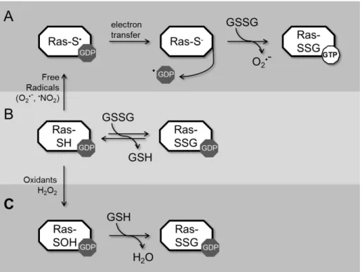
Figure 8. Modification of Ras by glutathione can proceed by three different mechanisms. In pathway A, redox agents capable to generating a Ras thiyl radical can induce radical formation on RasC118; electron transfer results in guanine-radical formation, guanine-base oxidation, and release of the oxidized base from Ras. Given the ratio of GTP/GDP in cells (~10:1), release of GDP promotes GTP binding. Free radical-induced oxidation leaves Ras in the thiyl anion form, which is more reactive to oxidation and can result in glutathione modification. In pathway B, Ras can react with oxidized glutathione through disulfide exchange to form glutathiolated Ras. Given the slow rate of this reaction and concentration of GSSG in cells, this pathway is unlikely to have a large contribution to Ras glutathiolation in vivo. In pathway C, Ras is oxidized by a non-radical oxidant, such as H2O2, which results in sulfenic acid formation at RasC118. Sulfenic acids are an
oxidation is likely too slow to be physiologically relevant. It is more likely that glutathiolation occurs through radical-mediated interactions in cells. However, as Ras glutathiolation can proceed through pathways that do not alter Ras activity, it is difficult to use Ras
glutathiolation as a marker of Ras activity regulation unless Ras radical formation can be detected coincident with Ras glutathiolation.
Conclusions
We have previously shown that NO2• can increase the rate of nucleotide dissociation in Ras and that modification of Ras by NO• renders Ras less sensitive to radical-mediated oxidation (66). Consistent with these observations, RasSSG does not affect the structure or activity of Ras. When considering the faster reaction rates of radical vs.
Supplemental Figure 1. Intact mass spectra resulting from the reaction of RasWT and RasC118S with GSSG.
Supplemental Figure 2. Detection of glutathiolated Ras at Cys118.
Chapter 3: Characterization and Nitrosation of Oncogenic K-RasG12C
Introduction
Ras GTPases are part of the Ras superfamily of GTPases, which play a major role in many cellular signaling pathways, including gene expression, cell growth, transport, and apoptosis (84). These proteins function as molecular switches by cycling between the active and inactive GDP-bound states to regulate activity. Ras alternates between the GTP-bound and GDP-GTP-bound forms through regulation by GTPase activating proteins (GAPs) and guanine nucleotide exchange factors (GEFs). GAPs bind and stimulate GTP hydrolysis to GDP, which inactivates the GTPase, whereas GEFs promote activation by facilitating the exchange of GDP for GTP (127).
Mutations to Ras family proteins account for approximately 30% of all oncogenic mutations (128). There are three isoforms of Ras in the cell, H-Ras, N-Ras, and K-Ras. However, K-Ras is the most frequently mutated isoform in cancer (128). Oncogenesis due to Ras most commonly arises from single mutations at Gly12, Gly13, and Gln61. Gly12 and Gly13 are within the phosphoryl binding loop (p-loop), a structural motif in Ras to interacts with the phosphate moieties of the bound nucleotide. Gln61 functions as the catalytic residue of GTP hydrolysis and serves as the catalytic base in cleavage of the γ phosphate of GTP (90). Mutations to these specific residues often result in loss of GAP sensitivity and constitutive activation (93, 129). For K-Ras, the majority of the mutations occur at Gly12 and account for the majority of mutations in lung and pancreatic cancers; the most common Gly12 mutations in K-Ras are K-RasG12C, K-RasG12D and K-RasG12V (128, 130).
characterization of the variant RasC118S, which was found to be insensitive to oxidants but otherwise behaved identically as RasWT (21).
Methods
Purification of Ras proteins
Protein purification was performed exactly as previously described (131). K-Ras was a gift of Genentech and is in the pET52b vector, which contains a 6x-His purification tag and a TEV (tobacco etch virus) protease site to remove the His tag. This vector was expressed in BL21 (DE3) Rosetta 2 pLysS cells, purified following the Qiagen nickel NTA purification protocol, and verified by SDS-PAGE.
ABD-F modification assays
ABD-F modification buffer (15 mM MES [(2-(N-morpholino)ethanesulfonic acid], 5 mM MgCl2, 30 mM NaCl, 200 µM DTPA, pH 8.0) was prepared with 10 mM dithiothreitol (DTT). An Amicon centricon was used to buffer exchange K-Ras and the protein was reduced for 30 minutes on ice. At the same time, ABD-F modification buffer without DTT was
sparged with N2 gas to remove dissolved oxygen. The protein was exchanged into this buffer to remove DTT from the sample prior to reaction with ABD-F.
In a black 96-well plate, 20 µM K-Ras was added in 100 µL of reaction buffer (100 mM MES, 100 mM HEPES, 5 mM MgCl2, 200 µM DTPA) with a pH that was predetermined. To a separate plate, ABD-F was added to 2 mM in 100 µL of reaction buffer at the same pH as the corresponding well in the original plate. Using a multi-channel pipette, ABD-F was added to the K-Ras plate to start the reaction. The fluorescence of the reaction was
monitored using a Spectramax M5 plate reader over a pH range of 5.8 to 8.5. The excitation wavelength for ABD-F is 389 nm, and the emission wavelength is 513 nm.
Nucleotide exchange and hydrolysis assays
K-Ras was loaded with mantGDP (2’-/3’-O-(N’-methylanthraniloyl)-GDP) for nucleotide exchange as previously described (108, 109, 131). K-Ras was added to 1 µM in 1 mL of
mM MgCl2, and 1 mM DTPA (pH 7.5) to neutralize the acidic pH and remove free metal ions aside from Mg2+ (132). The concentration of CysNO was determined from an absorbance measurement taken at 336 nm (ε = 900 M-1cm-1).
The reactions with RNS were performed with either CysNO or DEANO NONOate (diethylammonium(Z)-1-(N,N-diethylamino)dia-zen-1-ium-1,2-diolate). DEANO was added in an excess of 40:1 to the protein to induce nucleotide dissociation by free radical oxidation. Nucleotide dissociation assays with CysNO were performed with 100:1 CysNO to induce modification by two-electron/non-radical oxidation. The addition of the oxidants was performed in the presence of 2 mM unlabeled GDP.
MgSO4 (pH 7.45) and 10 μM GDP. Far UV scans were from 200 nm to 250 nm. Thermal denaturation of RhoA and the selected variants were monitored at a 221 nm to estimate the protein melting temperature. The temperature ramp rate was 1˚C/min and data points were collected every 1˚C. All data are reported in units of mean residue ellipticity, which was calculated as follows:[θ]MRE=(θraw×MRW)/(10×c×l), where θraw is the ellipticity in degrees, MRW is (Molecular Weight (Da))/((no.of residues)-1), c is the protein concentration in g/ml, and l is the pathlength of the cuvette in cm, according to (134). For nitrosated Ras, Ras was reacted with 50× CysNO for 10 min, which was prepared as previously stated, prior to performing the CD analysis.
NMR analysis
NMR experiments were collected on a Varian Inova 700 MHz spectrometer at 25˚C. The 2D 1H–15N heteronuclear single quantum coherence spectroscopy (HSQC) experiments were performed with pulsed field gradient and water flip-back methods as previously described (115). Uniformly labeled 15N-enriched Ras was purified as previously described (116). 2D 1H-15N HSQC experiments were acquired on 0.8 mM 15N-enriched RasWT and Ras mutants with 1,024 x 128 complex data points and a spectral width of 8,000 Hz for the 1H dimension and 1,709 Hz for the 15N dimension. Buffer contained 10 mM maleate (pH 6.5), 5 mM MgCl2, 40 mM NaCl, 20 μM GDP, and 10% D2O. NMR data were processed and
analyzed using NMR PIPE (117) and NMR ViewJ (One Moon Scientific; Newark, NJ).
Results
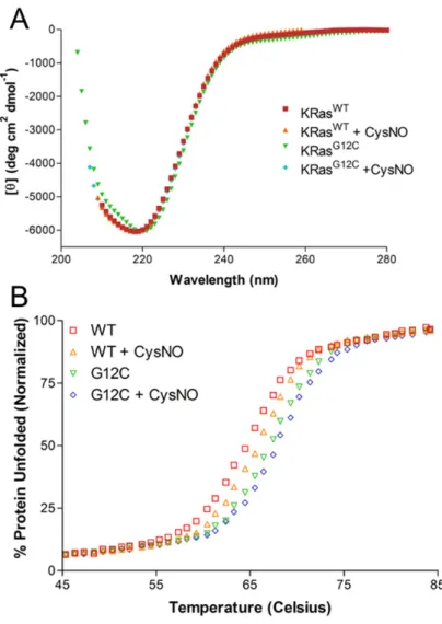
The thermal stability of K-RasG12C is not altered
As shown in Figure 9A, the CD spectra for K-RasG12C shows that there were no significant changes in the secondary structure of oncogenic K-Ras. Interestingly, the
K-RasG12C has an altered pKa
Figure 10 shows that K-RasG12C is susceptible to modification by ABD-F in a pH-dependent manner. Figure 10B shows that there is a shift in the pKa of K-RasG12C with respect to K-RasWT. The pKa for K-RasG12C and H-RasG12C was found to be ~7.5; however, the pH screen used for the ABD-F assay did not extend far enough to allow for the pH plot of K-RasWT to reach a plateau. As Ras has been shown to be unstable above pH 8.5, it was not possible to determine the pKa of Cys118. However, this cysteine is not predicted to have an altered pKa as it is solvent accessible and has no intra-protein contacts.
Figure 9. CD spectroscopy of K-RasWT and K-RasG12C.
Nitrosation of K-RasG12C
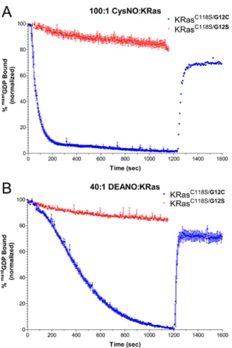
It is important to note that although Ras is specifically sensitive to radical oxidation (21, 62), the rates of dissociation shown here cannot be directly compared for computational
purposes for several reasons. As shown in Figure 11, the amount of CysNO and DEANO added to react with K-Ras were not identical, so equimolar reactions did not occur. In addition, DEANO is initially slower to modify K-Ras than CysNO because it must first decompose and recombine with O2 in solution to form NO2• that can oxidize G12C. As shown in Figure 11, modification at G12C by RNS (CysNO and DEANO) causes complete loss of mantGDP fluorescence over the course of the reaction. The ratios of oxidant to protein were selected to ensure complete modification of K-Ras on an appropriate timescale. Higher and lower ratios of oxidant to protein were also tested; however, the ratios plotted here were sufficient in showing a moderate rate of modification.
After completion of the NO reaction, DTT was added in equimolar concentrations to the CysNO/DEANO reaction cuvettes to reduce the protein to determine whether the
reaction was reversible (resulting in nucleotide re-binding). This additional step resulted in a 60-70% recovery of fluorescence in the CysNO and DEANO reactions. However, because these assays were performed with an excess of GDP present in the solution (about 1000:1 excess GDP compared to mantGDP), a fluorescence recovery of this magnitude should not be expected as this implies that K-Ras is selectively rebinding with the released mantGDP.
Mutations at Gly12 result in a loss of GAP-mediated hydrolysis
Figure 10. Nitrosylated K-Ras and nucleotide binding.
Figure 11. Nucleotide exchange and hydrolysis kinetics of RasG12C and RasG12S.
Figure 12. NMR analysis of Ras position 12 mutants.
GAP-dead.
HSQC analysis of RasG12C shows minimal chemical shifts
As the RasG12C structure has not been reported, we wished to determine whether the structure of this mutant was significantly different from other Ras mutants. However, as many Gly12 mutants have been solved, including RasG12D (135), RasG12V (136), and RasG12P (135), we opted against determining yet another Ras structure. Instead, we analyzed the 2D HSQC data of Ras and two variants, RasG12C and RasG12D, to determine whether the RasG12C variant was likely to have a significantly different structure. As the RasG12D structure had been previously determined, this mutant was used to compare the chemical shifts of a mutant with a structure known to be similar to RasWT. Thus, the data show that mutation of RasG12C has a similar spectra to RasG12D, which was expected, and helps confirm that the RasG12C mutant does not have a significantly altered structure.
Discussion
Activating mutations in Ras lead to the development of cancer in humans. Mutations at positions 12, 13, and 61 alter GAP-mediated nucleotide hydrolysis, which leads to Ras activation in cells. Gly12 mutants are present in developmental disorders, such as Costello syndrome and Cardio-facio-cutaneous (CFC) syndrome (137), and have been associated with an increased occurrence of tumorigenesis. Mutations at this site lead to non-small cell lung cancer occurring in 30-50% of human lung cancer patients (138, 139) and any mutation at this position (except proline) leads to Ras activation (140, 141).
displayed a higher occurrence of hyperplasia and adenomas (138).
Redox agents have been shown to modulate Ras superfamily GTPases at the NKCD motif (62, 69, 84, 86, 145-148). In particular, ROS and RNS activate Ras superfamily GTPases by increasing the rate of nucleotide exchange (65, 68, 80, 149, 150). ROS and RNS activate select redox-sensitive GTPases by reacting with the thiol of a solvent-accessible cysteine residue. Distinct from the NKCD motif in Ras (78), Rho GTPases contain a redox-sensitive cysteine at the end of the p-loop (GXXXXGK(S/T)C) (151-153). RasG12C is located in the p-loop similarly to Rho GTPases and given their redox reactivity, we hypothesized that the RasG12C mutant results in the generation of a new redox-sensitive cysteine that may alter the interaction with modulatory proteins (i.e., GAP proteins) and oxidants compared to other Gly12 mutants.
(Figure 10). As a free cysteine in solution has a pKa of ~8.5, this pKa indicates that RasG12C has an altered pKa and is solvent accessible, which are two requirements for a thiol to be redox sensitive.
To test the reactivity of RasG12C to oxidants, we treated mantGDP-loaded RasG12C with CysNO and NO• from DEANO (Figure 11). Exposure to both nitrosation agents resulted in a rapid and complete loss of mantGDP fluorescence. As it has been surmised that RasG12V is more oncogenic than RasG12D because of steric repulsions between the aspartate residue and negatively charged phosphates (156), we postulated that the loss of mantGDP fluorescence was due to the presence of negatively charged oxygen on nitric oxide decreasing the binding affinity for the nucleotide.
Chapter 4: Oxidation of RhoA at Cys20 regulates nucleotide binding
Introduction
RhoA is part of the Rho family of GTPases, which comprise a distinct subclass of Ras GTPases. Rho family GTPases regulate pathways involved in cell growth, differentiation, cell death (145, 157, 158), regulation of cell morphology and motility through cytoskeletal
rearrangements (159-163). Rho GTPases behave as molecular switches and cycle between the GTP-bound ‘on’ and GDP-bound ‘off’ states. Like most Ras superfamily GTPases, Rho GTPases have regulatory proteins that catalyze nucleotide exchange (guanine nucleotide exchange factors; GEFs), which activate the GTPase, and GTP hydrolysis (GTPase
accelerating proteins; GAPs), which deactivate the GTPase by hydrolyzing GTP to GDP. In addition, Rho GTPases are bound to guanine dissociation inhibitors (GDIs) when in the GDP-bound state, which sequesters the GTPase from the membrane and maintains the inactive state.
Thus, oxidation of these cysteines in Rho family GTPases has been shown to effect nucleotide binding and activation.
In addition, the p-loop has been shown to be critical for Mg2+ and nucleotide binding (165). From the crystal structure, many residues within the p-loop make direct interactions with the bound nucleotide. Furthermore, Mg2+ has been shown to stabilize nucleotide binding and play a role in GEF-mediated nucleotide exchange (166). While the Kd of RhoA for GDP and GTPγS do not vary greatly with and without Mg2+, the koff rate of nucleotide dissociation is greatly enhanced in the absence of Mg2+. Therefore, Mg2+ is considered a gatekeeper of nucleotide binding. Thus, oxidation of Cys20 and Cys16 could mediate effects on RhoA activity by regulating Mg2+ binding.
We have previously shown that exposure of RhoA to NO2• in vitro promotes disulfide formation and occludes nucleotide binding (80). Consistent with this observation, RhoA shows decreased GTP binding when exposed to the nitric oxide-generating species PAPA-NONOate in vascular smooth muscle cells (167). In this study, it was shown that RhoA was s-nitrosated using a nitrosocysteine-specific antibody. It is also possible that a disulfide was formed between Cys16 and Cys20, with the nitrosated product an intermediate to disulfide bond formation; however, other oxidation states aside from nitrosation were not tested. Furthermore, RhoA has been shown to be modified by phenylarsine oxide (PAO). Whereas PAO is a thiol-modifying compound that was originally developed as a protein tyrosine phosphatase inhibitor, RhoA can be modified and inactivated after PAO exposure in Caco-2 cells. Analysis by mass spectrometry showed mixed disulfide formation between Cys16 and Cys20 (83), resulting in deactivation of RhoA by occluding nucleotide binding; however, Rac1, which only has one p-loop thiol, was not inactivated upon exposure to PAO.
In contrast to the studies showing RhoA inactivation by oxidants, Cys16 and Cys20 of RhoA have been shown to be critical for peroxide-mediated activation of RhoA in REF52 cells (81). In this study, RhoA and a redox-insensitive variant, C16A/C20A, were ectopically expressed and exposed to exogenous and endogenous peroxide. RhoAWT was activated by peroxide under serum free conditions, whereas RhoAC16A/C20A was not activated under identical conditions. RhoA activation by peroxide concentrations as low as 0.1 μM was observed using the Rhotekin-RBD pull-down assay and microscopy to observe stress fiber formation, which is indicative of RhoA activation; however, RhoAC16A/C20A was not activated due to exogenous or endogenously produced peroxide (through antimycin A).
While there are a numerous studies that have observed regulation of RhoA activity through oxidation, many of these studies have shown that RhoA activity is indirectly
Thus, a carefully designed redox-insensitive variant that functions similarly to RhoAWT would aid in determining whether the effects of oxidants on RhoA are direct, or the result of indirect activation.
Methods
RhoA protein purification
Truncated human RhoA (RhoA1-181) was cloned into the pQlinkH vector (Addgene; Cambridge, MA), which contains an N-terminal 6x-His purification tag followed by a Tobacco Etch Virus protease cleavage site for removal of the affinity tag. The hypervariable region of RhoA, including the C-terminal CAAX box, was removed as this region does not undergo post-translational lipid modification in bacteria, is unstructured, and its removal does not affect guanine nucleotide binding or GTP hydrolysis in Ras GTPases (106). All proteins were expressed in BL21 Rosetta2 cells (Millipore; Darmstadt, Germany) and purified following the Qiagen Nickel NTA purification protocol (Germantown, MD). RhoA was further purified using size exclusion chromatography (Superdex-75 10/300 GL column; GE Life Sciences; Piscataway, NJ) and judged greater than 95% pure by SDS-PAGE analysis.
Nucleotide exchange and hydrolysis assays
The rate of GDP dissociation from Rho proteins was measured using 2'-/3'-O-(N'-methylanthraniloyl)guanosine-5'-O-diphosphate (mantGDP) as previously described (108, 109). Briefly, 1 μM RhoA and RhoA variants were preloaded with mantGDP (BioLog; San Diego, CA) and added to 1 mL of degassed assay buffer (50 mM Hepes pH 7.4, 50 mM NaCl, 5 mM MgCl2, 2 mM GDP, and 200 μM diethylenetriaminepentaacetic dianhydride; DTPA), and the rate of guanine nucleotide dissociation was measured by monitoring the change in fluorescence (excitation: 365 nm; emission: 435 nm; 25˚C) over time using a Perkin Elmer LS50B fluorimeter (Waltham, MA). All experiments were performed in triplicate.
Fluorescent nucleotide dissociation curves were fit to a one-phase exponential decay equation using GraphPad Prism version 3.03 (GraphPad Software; San Diego, CA). For GEF-induced RhoA-GDP dissociation assays, the minimal catalytic domain of Dbs
purification, and use of this sensor have been previously reported (113). Briefly, all assays used 10 µM FlipPi with 10 µM GTP-loaded RhoA. GTP loading was performed as previously described with minor modifications (111). Briefly, RhoA was loaded with GTP in loading buffer (20 mM Hepes, 50 mM NaCl, 1 mM EDTA, 20 mM (NH4)2SO4 and 1 mM inosine, pH 8.0) for 3 min at 37˚C. Excess nucleotide was removed using a PD-10 desalting column with hydrolysis buffer (20 mM Hepes, 50 mM NaCl, 100 μM EDTA, and 1 mM inosine, pH 7.4) and placed in a 96-well plate. Hydrolysis was initiated by adding 1 mM MgCl2 to the well. Trace phosphate was removed from all buffers using a ‘phosphate mop’ (114). The rate of GTP hydrolysis was measured by taking the ratio of the 535- and 485-nm emission wavelengths (excitation: 435 nm; 25˚C) of kinetic runs performed in triplicate and fit to a one-phase exponential association curve. For these experiments, the ratio of GAP to RhoA was 1:1000, and the minimal catalytic domain of p50 RhoGAP was used for hydrolysis.
4-fluoro-7-aminosulfonylbenzofurazan (ABD-F) modification of RhoA
The reaction was performed in a Spectramax M5 plate reader using 96-well black plates. All reactions were performed in triplicate in ABD-F reaction buffer at a final volume of 200 μl with 10 μM protein and 1 mM ABD-F. ABD-F was stored at -20˚C in DMSO, and the concentration of ABD-F was determined using the extinction coefficient of 4200 M-1cm-1 at 313 nm. The fluorescence of ABDF was measured at an excitation wavelength of 389 nm and emission wavelength of 513 nm.
Circular Dichroism spectroscopy
Circular Dichroism data were collected on a JASCO J-815 CD spectrometer with a JASCO Peltier device and water bath to control the temperature. Experiments were performed in a 1-mm cuvette at a protein concentration of 15 μM in 10 mM potassium phosphate (pH 6.5) containing 500 μM MgCl2 and 10 μM GDP. Far UV scans were from 200 nm to 250 nm. Thermal denaturation of RhoA and the selected variants were monitored at a 221 nm to estimate the protein melting temperature. The temperature ramp rate was
1˚C/min and data points were collected every 1˚C. CD scans are reported in units of mean residue ellipticity, which was calculated as follows: , where θraw is the ellipticity in degrees, MRW is
. , c is the protein concentration in g/ml, and l is the pathlength of the cuvette in cm, according to (171).
Glutathiolation and oxidation of Rho samples
Mass spectrometry of Rho Samples
To prepare Rho samples for MS, the proteins were first modified with the selected oxidant before denaturation in 100 mM NH4CO3 (pH 7.8), 6 M urea, and 10 mM
cleavages and variable modifications of cysteine glutathiolation and oxidation were used as a protein database search parameter. All peptides were filtered and reported within a mass accuracy of 5 ppm.
NMR spectroscopy
NMR experiments were collected on a Varian Inova 700 MHz spectrometer at 25˚C. The 2D 1H–15N heteronuclear single quantum coherence spectroscopy (HSQC) experiments were performed with pulsed field gradient and water flip-back methods as previously
described (91). Uniformly labeled 15N-enriched Rho was purified as previously described. 2D 1H-15N HSQC experiments were acquired on 0.5 mM 15N-enriched RhoAWT, RhoAC20S,
RhoAC16S/C20S, and RhoAC20D with 1,024 x 128 complex data points and a spectral width of 8,000 Hz for the 1H dimension and 1,709 Hz for the 15N dimension. Buffer contained 10 mM MOPS, 100 mM NaCl, 10 mM MgCl2, 10 μM GDP, 0.1% azide, and 10% D2O. NMR data were processed and analyzed using NMR PIPE and NMR ViewJ (One Moon Scientific; Newark, NJ).
Results
Thiol accessibility and 4-fluoro-7-aminosulfonylbenzofurazan (ABD-F) modification of
RhoA
Due to the observed reactivity of RhoA Cys20 to oxidants, we hypothesized that RhoA Cys20 has an altered pKa. We tested the reactivity of the cysteines in RhoA by monitoring the reaction of RhoA at various concentrations of ABD-F. ABD-F reacts with the thiolate state of cysteine residues with an approximate 10-fold faster rate than the thiol state and shows enhanced fluorescence upon modification.
Table 2. Thiol accessibility in RhoA crystal structures
Cys16 Cys20 Cys83 Cys107 Cys159
RhoAGTP
1A2B 1.9 4.3 0.0 7.9 1.3
RhoA
1A2B 1.9 35.4 0.0 7.9 1.3
RhoAGDP
1FTN 1.8 0.0 0.0 5.4 0.0
RhoA
1FTN 1.8 19.7 0.0 5.4 0.0
RhoA‐RhoGEF
3T06 3.0 33.5 0.1 3.7 0.0
RhoA‐RhoGAP
1OW3 3.5 2.2 0.0 4.0 0.5
RhoA‐GDI
1CC0 1.8 0.0 0.0 4.8 0.0
RhoA‐PKN/PRK
1CXZ 2.3 2.4 0.0 4.9 0.3