APPLICATION OF DYNAMIC CONBINTORIAL CHEMISTRY TO IDENTIFY NEW COMPOUNDS THAT BIND G-QUADRUPLEX DNA
&
PROBING THE ROLE OF THE CATION-PI INTERACTION
BETWEEN THE HP1 CHROMODOMAIN AND METHYLATED LYSINE USING UNNATURAL AMINO ACIDS
Elizabeth Jane Cline
A dissertation submitted to the Faculty of the University of North Carolina at Chapel Hill in partial fulfillment of the requirements for the degree of Doctor of Philosophy in the Department
of Chemistry.
Chapel Hill 2013
ABSTRACT
Elizabeth Jane Cline: Application of Dynamic Combinatorial Chemistry to Identify New Compounds that Bind G-Quadruplex DNA & Probing the Role of the Cation-π Interaction
Between the HP1 Chromodomain and Methylated Lysine Using Unnatural Amino Acids (Under the direction of Marcey L. Waters)
This dissertation discusses two different projects. The first project involves the development of cyclic-peptide acridine conjugates in the effort to identify molecules that can selectively bind to and stabilize G-quadruplex DNA. The second project seeks to probe the role of the cation-π interaction in the binding of the HP1 chromodomain to trimethylated lysine 9 of histone 3 (H3).
quadruplex sequences as well as duplex DNA in order to determine the selectivity of each species.
ACKNOWLEDGEMENTS
First, I would like to thank Marcey Waters for giving me the opportunity to join her lab.
You have been an incredible mentor and friend over the past three years, providing a welcoming
and supportive learning environment. Because of this I have learned an incredible amount and
have grown considerably as a chemist. I am so grateful for the opportunities that have been
given to me since joining your lab.
Additionally, I would like to thank all members of the Waters lab, past and present. It
has been a rewarding experience working next to such a diverse group of scientists and as a
result, I have learned a little from each of you. I would specifically like to thank Dr. Dale
Wilger, Dr. Jes Park and especially Dr. Soumyadip Ghosh for teaching and mentoring me when I
first joined the lab.
I am extremely grateful for the presence of Kaiualani, Amber and Effie in the lab.
Working next to you has been an incredible experience and has made the lab a friendly, positive
and supportive environment to be in. You have been my biggest cheerleaders, sharing my
successes and providing much needed motivation in spite of my failures. I will always be
grateful for that. In addition to playing a positive role in the lab, you have also been amazing
friends outside of the lab as well. I have many experiences with each of you that I will never
forget and I am so lucky to call you my friends.
I have made wonderful friends during my time here in Chapel Hill, providing me with
many people that have made Chapel Hill an enjoyable place to live that it would take too long to
list all of you. But, I would specifically like to thank Bryan and Danielle for being remarkable
friends to me. I know I never would have made it through graduate school without your
friendship and support. Our lives have taken us to different places but I hope our friendships will
continue and grow even stronger.
And, finally I need to thank my family. I would not be the person that I am today without
the support of my family. To James, you have become one of my best friends over the past five
years and I am incredibly proud to call you my brother. I would be absolutely nowhere without
my mom and dad. They have been amazing parents to me throughout my entire life. Mom, you
have always pushed me to be my best even when I didn’t want to and I have become a stronger
and more independent woman because of it. Dad, you have always had my back and supported
me no matter what. You have always been a voice of reason in my life and I will never forget
TABLE OF CONTENTS
LIST OF FIGURES………...……..x
LIST OF ABBREVIATIONS……….…..xii
CHAPTER 1: INTRODUCTION: STRUCTURE AND FUNCTION OF G-QUADRUPLEX DNA AND METHODS FOR TARGETING THE G-QUADRUPLEX STRUCTURE………..……1
A. Significance………..………1
B. G-Quadruplex Structure……….………..……....2
C. Biological Relevance………..……….5
i. G4 Structures at Telomeres………..………..5
ii. Effects of G4 Structures on DNA Replication………...6
D. Methods for Quadruplex Topology and Structure Determination……..….8
i. CD Spectroscopy………...8
ii. X-ray Crystallography and NMR Spectroscopy………...9
E. G-Quadruplex Ligands……….……….…….10
F. Methods to Investigate G-Quadruplex/Ligand Interactions………..……14
i. Melting Temperature Measurements………...14
ii. CD Spectroscopy……….15
iii. Surface Plasmon Resonance………15
CHAPTER 2: DESIGN OF DYNAMIC COMBINATORIAL
LIBRARIES AND EXPERIMENTS……….….21
A. Background………..21
i. Cyclic Peptides……….21
ii. Dynamic Combinatorial Chemistry……….…22
iii. Dynamic Cyclic Peptide Libraries from Thiol-Thioester Exchange...25
B. System Design……….28
i. Dynamic Combinatorial Library………..…28
ii. Biological Targets………29
C. Conclusions……….………...…31
CHAPTER 3: SYNTHESIS OF MONOMERS FOR DYNAMIC COMBINATORIAL LIBRARIES……….…………....34
A. Monomer Synthesis……….34
i. Synthesis of Acridine Ligand………...34
ii. Synthesis of Monomers……….………..36
B. Experimental Section………...39
i. Materials and General Methods………...39
ii. Synthesis of Acridine Ligand………...40
iii. Synthesis of Monomers………...42
CHAPTER 4: DYNAMIC COMBINATORIAL EXPERIMENTS……….…..…...45
A. Background………...…………...45
B. Dynamic Combinatorial Experiments………..………46
ii. Dynamic Combinatorial Experiments……….47
C. Conclusions and Future Directions………..59
D. Experimental Procedure……….…..59
i. Oligonucleotides……….59
ii. Circular Dichroism……….…….……60
iii. DCC Experiments………...61
CHAPTER 5: INTRODUCTION: SIGNIFICANCE OF HISTONE MODIFICATIONS AND METHYLATED LYSINE………...63
A. Significance of This Research………...………..63
B. DNA Packaging………...…64
C. Significance of Histone Modification……….……….65
i. Lysine Methylation on Histones………...66
ii. Lysine 9 of Histone H3 Tail………...…….68
D. Purpose of This Work………..70
CHAPTER 6: PROBING THE ROLE OF CATION-Π INTERACTIONS USING UNNATURAL AMINO ACIDS………...…74
A. Background………..…74
B. Peptide Design and Synthesis……….…….76
i. Native Chemical Ligation………..……….77
ii. Synthesis of HP1 Chromodomain………...….78
C. Future Directions……….81
LIST OF FIGURES
Figure 1.1 G-quadruplex structure………...…….…….2
Figure 1.2 Strand polarities and affect on groove size………..4
Figure 1.3 Role of telomerase in cancer………....6
Figure 1.4 Functional role of G-quadruplex structure during transcription………..…8
Figure 1.5 Acridine ligand and how it targets G-quadruplex structure…………...…11
Figure 1.6 Four broad categories of G-quadruplex binding ligands……..………….12
Figure 1.7 Schematic representing melting temperature experiments………....15
Figure 2.1 Scheme of dynamic combinatorial chemistry………....23
Figure 2.2 Reversible covalent reactions in dynamic combinatorial chemistry……..25
Figure 2.3 Scheme of cyclization of peptides via thiol-thioester exchange………....27
Figure 2.4 General design of monomer library………...29
Figure 3.1 Structure of G-quadruplex acridine ligand………....34
Figure 3.2 Synthesis of acridine ligand………...…35
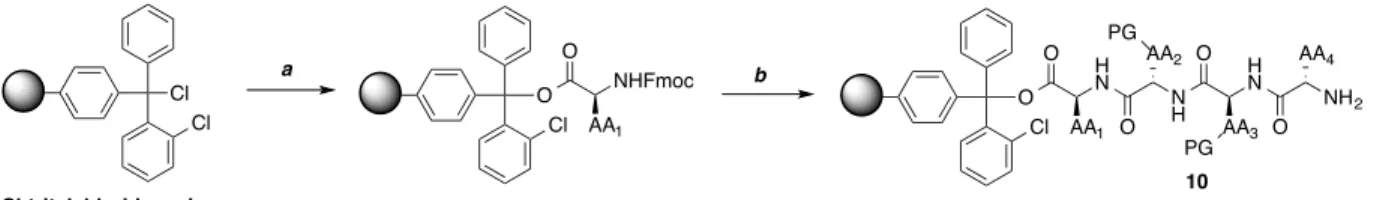
Figure 3.3 Solid phase synthesis of monomer peptides………..36
Figure 3.4 Synthesis acetyl capped monomers………...37
Figure 3.5 Synthesis acridine-peptide monomers………..….39
Figure 4.1 Effect of salt concentration on G-quadruplex folding………...47
Figure 4.2 Chromatograph of LigKCPF, LigQCFR, LigTCFR………..…47
Figure 4.3 Schematic of dynamic combinatorial experiments………...….48
Figure 4.4 Chromatograph of AcWCPR with Htelo……….……..50
Figure 4.5 Chromatograph of AcKCPW with Htelo………...50
Figure 4.7 Chromatograph of LigRCPR, LigKCPF, LigKCPS with Htelo……..….53
Figure 4.8 Chromatograph of LigRCPF, AcTCPR, AcFCPR with Htelo, dsDNA………....53
Figure 4.9 Chromatograph of LigRCPF, AcTCPR, AcFCPR with Htelo, cKit, cMyc, dsDNA………..…………....55
Figure 4.10 Comparison of denaturation using heat or 18-crown-6……….56
Figure 4.11 Schematic of dynamic combinatorial experiments………56
Figure 4.12 Chromatograph of AcFCPR, AcKCPW with Htelo………..…57
Figure 4.13 Sequence of G-quadruplex sequences screened………..……..59
Figure 4.14 CD spectra for G-quadruplex sequences……….……...60
Figure 5.1 Structure of the nucleosome………...………...………..64
Figure 5.2 Methylation states of lysine and arginine……….…….…66
Figure 5.3 Mechanism of lysine 9 methylation……….…..67
Figure 5.4 Aromatic cage of HP1 chromodomain bound to trimethyllysine….…….69
Figure 6.1 Electrostatic potential maps of tryptophan derivatives………….……….75
Figure 6.2 Structure of acetylcholine and fluorination trend of tryptophan..………..76
Figure 6.3 Mechanism of native chemical ligation……….78
Figure 6.4 Sequence of HP1 chromodomain and ligation sequence…………...……79
Figure 6.5 Mechanism of aspartimide formation………..………..79
LIST OF ABBREVIATIONS
AA Amino acid
Ac2O Acetic anhydride
Ach Acetylcholine
AcOH Acetic acid
AdoMet S-adenosylmethionine
Arg, R Arginine
Asp, D Aspartic acid
C Carbon
CD Circular Dichroism
CH2Cl2 Dichloromethane
CN Cyano
CrO3 Chromium(VI) oxide
Cys, C Cysteine
DBU Diazabicycloundecene
DCC Dynamic combinatorial chemistry
DIC Diisopropylcarbodiimide
DIPEA Diisopropylethylamine
DMF Dimethylformamide
DNA Deoxyribose nucleic acid
DSB Double stranded break
EDT Ethanedithiol
ESI-TOF Electrospray Ionization-Time of flight
Et2O Diethyl ether
EtOAc Ethyl acetate
EtOH Ethanol
F Fluorine
Fe(II) Iron(II)
FID Fluorescence indicator displacement Fmoc 9-Fluorenylmethyl chloroformate FRET Fluorescence resonance energy transfer
G Guanine
G4 G-quadruplex
Gln, Q Glutamine
Glu, E Glutamic acid
H2SO4 Sulfuric acid
HBTU O-Benzotriazole-N,N,N’,N’-tetramethyluronium hexafluorophosphate
HCl Hydrochloric acid
HNO3 Nitric acid
HOBt 1-Hydroxybenzotriazole
HP1 Heterochromatin protein 1
HPLC High performance liquid chromatography
Htelo Human telomeric
K Potassium
KCl Potassium chloride
KDM Lysine demethylases
KMT Lysine methyltransferases
LC/MS Liquid chromatography/mass spectrometry
Lys, K Lysine
Me Methyl
MeCN Acetonitrile
Met, M Methionine
N Nitrogen
N2 Nitrogen (g)
Na Sodium
nAChRs Nicotinic acetylchloine receptors NaHCO3 Sodium bicarbonate
NaOH Sodium Hydroxide
NH4OAc Ammonium acetate
NHPPE Nuclease hypersensitive polypurine-polypyrimidine element
NMR Nuclear magnetic resonance
NO2 Nitro
Phe, F Phenylalanine
Pro, P Proline
PTM Post-translational modification
S Sulfur
SnCl2 Tin(II) chloride
SPPS Solid phase peptide synthesis
SPR Surface plasmon resonance
tBu tert-Butyl
TFA Trifluoroacetic acid
TIPS Triisopropylsilane
Tris 2-Amino-2-hydroxymethyl-propane-1,3-diol
Trp, W Tryptophan
TSS Transcriptional start site
Val, V Valine
! ! ! ! !
CHAPTER(1:(INTRODUCTION(
STRUCTURE(AND(FUNCTION(OF(G3QUADRUPLEX(DNA(AND(( METHODS(FOR(TARGETING(THE(G3QUADRUPLEX(STRUCTURE(
! ! A. Significance
B. G-Quadruplex Structure
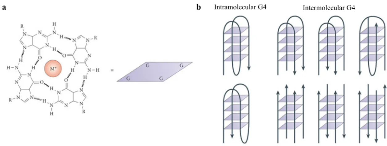
G-rich sequences of nucleic acids can form four-stranded, helical DNA or RNA structures comprised of 4 guanine bases brought together in a square planar arrangement connected by Hoogsten hydrogen bonds (Fig. 1.1a).5,6 The G-rich core typically consists of two or more G-tetrads stacked via π-π interactions with intervening sequences extruded as single-stranded loops (Fig. 1.1b). The sequence and size of the loop regions can vary, with the loops usually containing 1-7 nucleotides. The stacks are joined together by the normal sugar-phosphate backbone with the structure further stabilized by monovalent cations, typically potassium (K+) or sodium (Na+), that occupy the central cavity neutralizing the electrostatic repulsion of the guanine oxygens (Fig. 1.1a).7,8
Figure 1.1 The G-quadruplex structure (a) An illustration of the G-quadruplex G-quartet. The quartet is pictured as a square in the other panels of the figure. M+ denotes a monovalent cation. (b) Schematic of intramolecular (left) and intermolecular (right) G-quadruplex DNA structures. The arrows indicate the direction of the DNA strands.4
The quadruplex structure can be extremely stable. However, the stability can vary due to differences in length and sequence of the loop, strand orientation and alignment and the nature of the binding cations. G-quadruplex structures can be parallel, antiparallel or hybrids of the two and can be made up of one (intramolecular), two or four separate strands (intermolecular) (Fig. 1.1b).6 Loops may link positions on the top (or bottom) of the stacks, forming diagonal (Fig.
Intramolecular G4 Intermolecular G4
Figure 1.2 Structures of the human unimolecular telomeric quadruplex formed by the sequence d[AGGG(TTAGGG)3].6 In each case two views are shown with arrows depicting strand polarities. (a) The Na+ form, determined by NMR containing two anti-parallel and two parallel strands with a diagonal and two lateral loops. (b) K+ form A, determined by crystallography containing all parallel strands with three strand reversal loops resulting in four equal grooves (c) K+ form B, showing the topology determined by NMR, containing three parallel strands with one oriented anti-parallel.
It has been predicted that intramolecular quadruplex structures can form at specific G-rich regions in vivo. These G-rich regions share a common sequence motif of at least four guanines, with each G-tract containing most often at least three guanines. Computational analyses have revealed that there are >375,000 G4 motifs in the human genome including those in ribosomal and telomeric DNA. However, it is still unclear how many of these motifs can form stable G-quadruplex structures in vivo and, if they do, when they form.15-18
a
b
C. Biological Relevance
Computational studies have revealed that G4 motifs are not randomly located within the genome, but tend to cluster in particular genome regions such as promoters. Due to the nonrandom location and evolutionary conservation of the position of G4 motifs within the genome, it is suggested that the G-quadruplex structure plays one or more positive functions within the cell.19,20 Telomeres are regions of the chromosome that contain a high concentration of G4 motifs due to their high GC content and the single-stranded nature of the telomeric overhang. G4 DNA motifs are also common G-rich regions up- and downstream of TSSs and near transcription factor binding sites.
i. G4 Structures at telomeres
Telomeres make up a nucleoprotein complex at the ends of linear chromosomes that protect chromosomes from degradation, end-to-end fusions, and being recognized as DSBs.21 They contain a double-stranded region and a single-stranded G-rich 3’ overhang (Fig. 1.3). In most telomeric DNAs, guanines and cytosines are distributed asymmetrically between two DNA strands with the G-rich strand being longer than its complement, resulting in single-stranded ‘G-tails’ at the termini of the chromosome. Despite differences in the exact sequence, the G-rich strand of various telomeric sequences can usually form stable G4 structures in vitro.22-24
inactive in most somatic cells but is found to be upregulated in many cancers where it is thought to promote the lifespan of malignant cells.25 The G-quadruplex structure can inhibit the activity of telomerase by preventing the enzyme from annealing to G-strand overhangs.26,27 Because telomerase is active in a large number of human cancers and can be influenced by the formation of the G-quadruplex structure, there has been great effort to design a variety of small molecules that can bind and stabilize G4 structures (Fig. 1.3).28
Figure 1.3 Schematic outlining the role of human telomerase in cancer. Telomeric sequences can fold into G-quadruplex structure inhibiting the activity of the reverse transcriptase telomerase. Due to the ability of the G4 motif to inhibit the enzyme telomerase, it has become desirable to design small molecules that can bind to and stabilize the quadruplex structure for use as novel anticancer therapies.
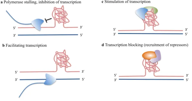
ii. Effects of G4 structures on DNA transcriptional regulation
In addition to their presence in telomeres, a high concentration of G4 motifs have been found in the promoter regions of genes suggesting that G4 structures play a potential role in the
Ac#va#on(of( Telomerase(
Synthe'c)ligand)stabilizes)) G3quadruplex)structure)
Telomerase( lengthens( telomeres( Chromosome)
Double3stranded) region)of)telomere)
Single3stranded) region)of)telomere)
Cancer(
Apoptosis( G7Quadruplex(
structure(inhibits( the(telomerase(
enzyme(
Telomerase)is)ac'vated) in)80–85%)of)human)
cancer)cells)
regulation of gene transcription. Bioinformatics has shown that the promoter region of human
oncogenes and regulatory genes (i.e. transcription factors) are more likely than the average gene
to contain G4 motifs.29 There is an equilibrium between two forms of the DNA during transcription. One side of the equilibrium is the double helix DNA, while on the other side one
strand is separated and has folded up into a G-quadruplex.30 As a result, studies have suggested that the presence of some G4 structures during DNA replication could influence transcription in
positive and negative ways. Depending on which DNA strand contains the G4 motif, the
structure could inhibit transcription (if the motif is on the template strand) or enhance
transcription (if the motif is on the non-template strand) (Fig. 1.4a and 1.4b). Also, proteins that
bind to the G4 structures may also affect transcription (Fig. 1.4c and 1.4d). 31-33 A number of oncogenes have been found to contain the G4 motifs in their promoter region leading to the
possibility of novel therapeutics that target the quadruplex structure. The addition of a
G-quadruplex ligand will energetically favor the G4 structure, thus providing a way to manipulate
Figure 1.4 Proposed functional roles of G-quadruplex structures during transcription.4 (a) G4 structures are thought to block transcription by inhibiting polymerase (blue). (b) G4 structures are postulated to facilitate transcription by keeping the transcribed strand in the single-stranded conformation. (c) G4 structure may stimulate transcription by recruiting proteins (green) that recruit or stimulate polymerase (d) G4 structures are suggested to block transcription by recruiting G4 binding proteins (orange) that can directly or indirectly repress transcription (purple).
D. Methods for Determination of Quadruplex Topology and Structure i. CD Spectroscopy
A number of quadruplex studies have used biophysical chemistry, mainly circular dichroism (CD) to assign topology. CD is sensitive to stereochemical variations, allowing it to be an important technique for studying subtle conformational changes and supramolecular interactions.34 Using CD spectroscopy it is possible to discriminate between quadruplex topologies having differences in parallel and anti-parallel strand orientation due to different arrangements of anti/syn glycosidic angles. Therefore, CD is a useful and rapid method for establishing an overall fold pattern as it requires little sample and is suited to examining a wide range of solution conditions and their influence on quadruplex formation. Classic parallel and
5’#
3’# 5’#3’#
5’#
3’# 5’#3’#
c Stimulation of transcription
d Transcription blocking (recruitment of repressors) 5’#
3’# 5’#
3’# a Polymerase stalling, inhibition of transcription
5’#
3’# 5’#
anti-parallel quadruplexes provide similar traces but with maxima at distinct wavelengths. For
quadruplexes assigned to be parallel-stranded, a maximum is present at ~260 nm and a minimum
at ~240 nm while the maximum and minimum for an anti-parallel quadruplex are typically at
around 290 and 260 nm, respectively.35-37 CD has predominantly been used to study telomeric
sequences and those with regular repeating loop regions. More complex quadruplex-forming
show decreased reliability when assigning topology since they may not conform to observed
telomeric quadruplexes, multiple species cannot be identified by CD and non-telomeric loop
sequences may perturb the CD spectra in unforeseen ways.6,38
ii. X-ray Crystallography and NMR Spectroscopy
X-ray crystallography and high-field NMR spectroscopy are other ways to determine
topological assignment as well as a more detailed atomic-level structure determination.39
However, there can be limitations to each of these methods. In order to successfully determine a
structure by NMR, the sequence needs to form a kinetically stable species in solution while the
presence of multiple species limits the structural information that can be obtained. It is common
to set up a screen of mutated sequences and other variants until one is found that produces a
well-resolved NMR spectrum showing a single species amenable to analysis.40 Similarly,
crystallography uses site mutations and/or sequence screening to find sequences that will
crystallize.6 The various structures formed in solution as variants of the human telomeric
two-repeat sequence show that such mutations and changes cannot always reliably preserve a
particular topology and will inevitably alter the equilibrium between different structures.41
Therefore, generalizations from any one NMR or crystal structure needs to be done carefully and
E. G-Quadruplex Ligands
The G-quadruplex structure has been implicated in several biological dysfunctions that
selectively alter the integrity of cancer cells. As a result, G-quadruplex binders that stabilize the
G4 structure promise to lead to the discovery of novel anticancer agents. A common binding
motif for a ligand is a flat aromatic molecule that binds to the G-quartet of the external face of
the quadruplex resulting in stabilization of the structure through π-π stacking and electrostatic
interactions (Figure 1.5b). The G-quartet has a large flat surface therefore an efficient
quadruplex ligand should feature a large aromatic surface, much larger than that of a duplex
binder, improving the aromatic-aromatic overlap and providing selectivity over duplex
DNA.6,41,42 Electrostatic interactions between positively charged sidechains of the ligands and
the negatively charged phosphate backbone of the G-quadruplex DNA scaffold also contribute to
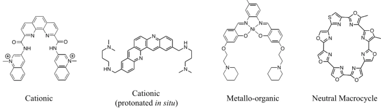
stabilization of the G4 structure (Figure 1.5b). Generally, the ligands that have been synthesized
to date can be classified into four different categories based on their cationic nature, i.e. cationic
(1) upon in situ protonation of an amine appendage, (2) via N-methylation of an aza-aromatic
Figure 1.5 Despite differences in the many ligands developed that successfully target G-quadruplex DNA over duplex DNA, G4 ligands tend to have two common features including a large flat aromatic surface for π stacking with the G-tetrad and protonable side chains for electrostatic interaction with the quadruplex grooves. (a) Shown is an acridine ligand that targets G-quadruplex DNA over duplex DNA and displays the two common characteristics of G4 ligands. The acridine core provides a large flat aromatic surface for binding while the protonable pyrrolidine side chains provide positive electrostatic interactions. (b) X-ray structure of the acridine ligand (red) complexed with a G4 structure (quartet=blue, backbone=grey, loops=orange) illustrating how the ligands can end stack with the G-tetrads of the quadruplex structure.43
The usual way to target G-quadruplex DNA is the introduction of protonable sidearms
around an aromatic core. Neidle, Hurley and co-workers followed this method with the
development of a bisamidoanthraquinone G-quadruplex ligand and telomerase inhibitor.44 While
this class of molecule was shown to have telomerase inhibition activity, these molecules had
insufficient selectivity for G4 DNA over duplex DNA for further biological studies. To improve
the selectivity for quadruplex DNA, Neidle and coworkers progressively modified the core and
sidearms from anthraquinone to fluorenone, then acridone and acridine.45,46 A member of the
3,6-disubstituted acridine series showed particular promise against G-quadruplex DNA (Figure
1.5a).47 This structure displayed hydrophobic-π-stacking interactions between the flat aromatic
core of the acridine and the square planar guanine residues of the G-tetrad in addition to
electrostatic interactions between the two protonable sidechains of the ligand and the quadruplex
grooves. Neidle and coworkers further optimized this class of molecules by designing
BRACO-19, a molecule able to interact simultaneously with three G-quadruplex grooves due to the
N N
H O N
H O
N N
X-ray structure of the complex of disubstituted aminoalkylamido acridine (in red) bound to d(G4T4G4) G-quadruplex
addition of a third side arm.48 The optimized BRACO-19 displayed high levels of quadruplex stabilization as determined by FRET (fluorescence resonance energy transfer)-melting assay and by SPR (surface plasmon resonance) method that revealed 31-fold selectivity for the quadruplex structure. Additionally, BRACO-19 shows a strong potency for telomerase inhibition as well as inhibition of cancer cell proliferation however, pharmacological use of this molecule has been limited by poor membrane permeability or cellular uptake.49 Despite these limitations, the simple acridine motif appears to be very valuable for G-quadruplex recognition and one that we have incorporated into our system design.
Figure 1.6 Molecules exemplifying the four broad categories of G-quadruplex binding ligands.
Despite the number of ligands designed that successfully target G-quadruplex DNA over duplex by targeting the common G-tetrad of the G4 motif, a major challenge still remains to design ligands with specificity for individual quadruplex structures given the wide variety of topologies specific quadruplex sequences can adopt. It may be possible to obtain selectivity by targeting the loops that vary in length and sequence as well as the grooves, which can vary in size due to difference in relative strand orientation. A common strategy that has been employed to achieve greater high selectivity and specificity is conjugation, using a ligand that can target the planar surface of the G-tetrad and appending it to a variable substituent that can contact the loops and grooves of the quadruplex structure.
N N HN O NH O N N N N H N N HN N N O N O O O N N F Ni N O N O N O N O N O S N N O N O
An initial example comes from Pedroso et al., where a library of acridine-oligonucleotide
conjugates were synthesized and studies were carried out to examine their interaction with DNA
quadruplexes.50 The acridine ligand was expected to exhibit selectivity for quadruplex DNA over
duplex while the oligonucleotide attached was complementary to the G-rich telomere strand.
These studies showed that the acridine-oligonucleotides did stabilize the telomeric quadruplex
structure as determined by thermal denaturation experiments; however, no other sequences were
screened against the library to determine selectivity.
Arya et al. published a more recent example of a novel perylene-neomycin conjugate that
was shown to bind preferentially to telomeric G-quadruplex DNA in the presence of other
nucleic acids, including DNA, RNA, DNA-RNA hybrids, and other higher order structures
(single strands, duplexes, triplexes, other G-quadruplexes, and the i-motif).51 The perylene
moiety is known to end stack with the G-tetrads of the quadruplex structure, while recent studies
have shown that neomycin can bind in the wide groove of G4 DNA. Arya et al. showed through
fluorescence intercalator displacement (FID) assays that the conjugated species displayed tighter
binding than each of the parent constituents.
In a final example, Neidle et al. published studies performed with a common G-tetrad
interacting acridine core conjugated to various tetrapeptide substituents that have the ability to
discriminate between different quadruplex types.52 Based on the substitution patterns selectivity
could be seen for the parallel quadruplex derived from the human N-ras gene over the human
telomeric quadruplex while the peptide substituent KRSR proved to show marginal superior
selectivity and affinity over FRHR While linear peptides provided modest binding selectivity, we
F. Methods to Investigate G-Quadruplex/Ligand Interactions
A variety of techniques have been employed to study the interaction of natural or
synthetic compounds with G-quadruplex structures. These methods can vary from evaluating
properties like ligand affinity, to more sophisticated methods used for determining kinetic,
thermodynamic, stoichiometric and conformational data for structure-activity relationship
studies. Methods being used to investigate these interactions should be able to detect and
measure the ligand selectivity for quadruplexes over duplexes or other secondary structures.
Each method has its own advantages and disadvantages therefore more than one method is
usually necessary to obtain complete information about quadruplex DNA/ligand interactions.
i. Melting Temperature Measurements
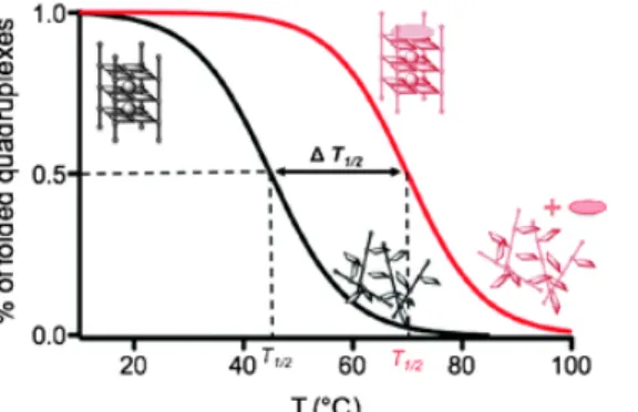
Melting temperatures can be measured, providing information about the stabilization (or
destabilization) of the quadruplex structure by the ligand under investigation. Quadruplex
nucleic acids show a strong absorbance around 295 nm, which is greatly reduced in unstructured
DNA due to the π-stacking of nucleobases in folded quadruplex structures (Figure 1.7).53 There
is a decrease in absorbance at 295 nm upon denaturation by heating. This simple thermal
melting experiment is performed to demonstrate the stabilization or destabilization of a
quadruplex structure by a ligand. Preparation of the sample is straightforward and reasonably
Figure 1.7 Schematic representation of melting temperature experiments using CD at 295 nm.53
ii. CD Spectroscopy
Circular dichroism (CD) spectroscopy is extremely useful in studying the conformation
of nucleic acids. As discussed previously, CD is a widely used technique to study the
G-quadruplex conformation in which the CD signal is influenced by stacking interactions between
adjacent quartets.54 The titration of G4-ligands to quadruplexes can induce changes in the CD
spectrum, correlating to the properties of the ligand under investigation.
iii. Surface Plasmon Resonance
Surface Plasmon Resonance (SPR) has become a popular method to study the kinetic and
thermodynamic parameters of intermolecular interactions. SPR is an optical technique that
utilizes the decrease in intensity of a circular polarized light reflected from the surface of a glass
prism coated with a thing metal film. The minima in reflected light density occur at a specific
angle at which surface plasmons of the metal are excited. The angle at which the resonance
occurs becomes the function of refractive index of the medium in the vicinity of the surface. The
variation of value at this angle is recorded, detecting any interactions involving free species in
solution (ligand) and one that is covalently bound to the surface (DNA). A variety of quadruplex
forming oligonucleotides have been immobilized to sensor chips to study binding interactions
G. Purpose of this work
Given the important regulatory and structural functions of the G4 motif, we aim to
develop quadruplex ligands that exhibit selectively over not only duplex DNA but also over
various quadruplex sequences. In the effort to discover molecules that can discriminate against
assorted quadruplex structures via groove recognition, we have screened libraries of cyclic
peptide-acridine using dynamic combinatorial chemistry (DCC). We chose to focus on cyclic
peptides due to their prevalence in nature and their promise as biologically relevant molecules.
Additionally, the use of cyclic peptides to target the quadruplex structure seemed promising due
to the ability of the macrocyclic, natural product telomestatin to bind G-quadruplex DNA.
Furthermore, in order to screen libraries of cyclic peptides in a high-throughput format, a method
has been developed in our lab using thiol-thioester exchange in DCC. Using this method, we can
generate libraries of cyclic peptides from simple linear peptide building blocks in solution,
within hours, and at a neutral pH. These libraries can efficiently be screened against different
quadruplex sequences as well as duplex DNA in the effort to determine the selectivity of each
species. Once a hit has been discovered, the molecule can easily be resynthesized as a more
References
1. Hurley, L. H. Secondary DNA structures as molecular targets for cancer
therapeutics. Biochemical Society Transactions 2001, 29, 692-696.
2. Wu, Y.; Brosh, R. B. G-Quadruplex nucleic acids and human disease. FEBS
Journal 2010, 277, 3470-3488.
3. Balasubramanian, S.; Neidle, S. G-Quadruplex nucleic acids as therapeutic targets
Curr. Opin. Chem. Biol.2009,13, 345−353.
4. Bochman, M. L.; Paeschke K.; Zakian V. A. DNA secondary structures: stability
and function of G-quadruplex structures. Nature Rev. Genetics2012, 13, 770-780.
5. Huppert, J. L. Structure, location and interactions of G-quadruplexes FEBS
Journal 2010, 277, 3452-4358.
6. Burge, S.; Parkinson, G. N.; Hazel P.; Todd, A. K.; Neidle, S. Quadruplex DNA:
sequence, topology and structure. Nucleic Acids Res. 2006, 34, 5402-5414.
7. Williamson, J. R.; Raghuraman, M. K.; Cech, T. R. Monovalent cation induced structure of telomeric DNA: the G-quartet model. Cell 1989, 59, 871-880.
8. Wong, H. M.; Payet, L.; Huppert, J. L. Function and targeting of G-quadruplexes
Curr. Opin. Mol. Ther. 2009, 11, 146-155.
9. Hazel, P.; Parkinson, G. N.; Neidle, S. Predictive modeling of topology and loop
variations in dimeric DNA quadruplex structures. Nucleic Acid Res. 2006, 34,
2117-2127
10. Hardin, C. C.; Perry A. G.; White, K. Thermodynamic and kinetic
characterization of the dissociation and assembly of quadruplex nucleic acids.
Biopolymers 2000, 56, 147-194.
11. Guedin, A.; Gros, J.; Alberti, P.; Mergny, J. L. How long is too long? Effects of
loops size on quadruplex stability. Nucleic Acids Res. 2010, 38, 7858-7868.
12. Bagaut, A.; Balasubramanian, S. A sequence-independent study of the influence
of short loop lengths on the stability and topology of intramolecular DNA G-quadruplexes. Biochemistry 2008, 47, 689-697.
14. Dingley A. J.; Peterson R. D.; Grzesiek, S.; Feigon, J.; Characterization of the cation and temperature dependence of DNA quadruplex hydrogen bond properties using high-resolution NMR. J. Amer. Chem. Soc. 2005, 127, 1446-14472.
15. Capra, J. A.; Paeschke, K.; Singh, M.; Zakian, V. A. G-quadruplex DNA sequences are evolutionarily conserved and associated with distinct genomic features in Saccharomyces cerevisiae. Plos Comput. Biol. 2010, 6, e1000861
16. Todd, A. K.; Johnston, M.; Neidle, S. Highly prevalent putative quadruplex sequence motifs in human DNA. Nucleic Acids Res. 2005, 36, 144-156.
17. Hershman, S. G. et al. Genomic distribution and functional analyses of potential G-quadruplex forming sequences in Saccharomyces cerevisiae. Nucleic Acids Res. 2008, 33, 144-156.
18. Huppert, J. L.; Balasubramanian, S. Prevalence of quadruplexes in the human genome. Nucleic Acids Res. 2005, 33, 2908-2916.
19. Rawal, P. et al. Genome-wide prediction of G4 DNA as regulatory motifs; role in Escherichia coli global regulation. Genome Res. 2006, 16, 644-655.
20. Eddy, J.; Maizels, N. Gene function correlates with potential for G4 DNA formation in the human genome. Nucleic Acids Res. 2006, 34, 3887-3896.
21. Zakian, V. A. Telomeres: the beginning and ends of eukaryotic chromosomes. Exp. Cell Res. 2012, 318, 1456-1460.
22. Henderson, E. et al. Telomeric DNA oligonucleotides form novel intramolecular structures containing guanine-guanine base pairs. Cell 1987, 51, 899-908.
23. Sundquist, W. I.; Klug, A. Telomeric DNA dimerizes by formation of guanine tetrads between hairpin loops. Nature 1989, 342, 825-829.
24. Sen, D.; Gilbert, W. Formation of parallel four-stranded complexes by gunine-rich motifs in DNA and its implications for meiosis. Nature 1988, 334, 364-366.
25. Shay, J. W.; Wright, W. E. Role of telomeres and telomerase in cancer. Seminars Cancer Biol. 2011, 21, 349-353.
26. Zahler, A. M.; Williamson, J. R.; Cech, T. R.; Prescott, D. M. Inhibition of telomerase by G-quartet DNA structures. Nature 1991, 350, 718-720.
28. Neidle, S. Human telomeric quadruplex: the current status of telomeric G-quadruplexes as therapeutic targets in human cancer. FEBS J. 2010, 277, 1118-1125.
29. Huppert, J. L.; Balasubramanian, S. Prevalance of quadruplexes in the human genome. Nucleic Acids Res. 2005, 33, 2908-2916.
30. Shirude, P. S.; Okumus, B.; Ying, L. M.; Ha, T.; Balasubramanian, S. Single-molecule conformational analysis of G-quadruplex formation in the promoter DNA duplex of the proto-oncogene c-kit. J. Am. Chem. Soc. 2007, 129, 7484-7485.
31. Sun, D.; Hurley, L. H.; The importance of negative superhelicity in inducing the formation of G-quadruplex and i-motif structures in the c-Myc promoter: implications for drug targeting and control of gene expression. J. Med. Chem. 2009, 52, 2863-2874.
32. Brooks, T. A.; Kendrick, S.; Hurley, L. Making sense of G-quadruplex and i-motif functions in oncogene promoters. FEBS J. 2010, 277, 3459-3469.
33. Qin, Y.; Hurley, L. H. Structures, folding patterns, and functions of intramolecular DNA G-quadruplexes found in eukaryotic promoter regions.
Biochemie 2008, 90, 1149-1171.
34. van Dijk, L.; Bobbert, P. A.; Spano, F. C. J. Phys. Chem. B,2010, 114, 817.
35. Balagurumoorthy, P; Brahmachari, S. K.; Mohanty, D.; Bansal, M.; Sasisekharan, V. Nucleic Acids Res., 1992, 20, 4061.
36. Jin, R.; Gaffney, B. L.; Wang, R. A.; Jones, A.; Breslauer, K. J.; Proc. Natl. Acad. Sci., 1992, 89, 8832.
37. Lu, M.; Guo, Q.; Kallenbach, N. R. Biochemistry, 1993, 32, 598.
38. Smith, F.W.; Feigon, J. Nature, 1992, 356, 164.
39. Lane, A. N.; Chaires, B. J.; Gray, R. D.; Trent, J. O. Stability and kinetics of G-quadruplex structures. Nucleic Acids Res. 2008, 36, 5482-5515.
40. da Silva, M. W. NMR methods for studying quadruplex nucleic acids. Methods, 2007, 43, 264-277.
41. Phan, A. T.; Kuryavyi, V.; Luu, K. N.; Patel, D. J. Structure of two intramolecular G-quadruplexes formed by natural human telomerase sequences in K+ solution.
42. Phan, A.T.; Kuryavyi, V.; Patel, D. J. Curr. Opin. Struct. Biol. 2006, 16, 288.
43. Monchaud, D.; Teulade-Fichou, M. P. A hitchhiker’s guide to G-quadruplex ligands. Org. Biomol. Chem. 2008, 6, 627-636.
44. Sun, D.; Thompson, B.; Cathers, B. E.; Salazar, M.; Kerwin, S. M.; Trent, J. O.; Jenkins, T. C.; Neidle, S.; Hurley, L. H. J. Med. Chem. 1997, 40, 2113.
45. Perry, P. J.; Read, M. A.; Davies, R. T.; Gowan, S. M.; Reszka, A. P.; Wood, A. A.; Kelland, L. R.; Neidle, S. J. Med. Chem. 1999, 42, 2679.
46. Harrison, R. J.; Reszka, A. P.; Haider, S. M.; Romagnoli, B.; Morrell, J.; Read, M. A.; Gowan, S. M.; Incles, C. M.; Kelland, L. R.; Neidle, S. Bioorg. Med. Chem. Lett. 2004, 14, 5845.
47. Haider, S. M.; Parkinson, G. N.; Neidle, S. J. Mol. Biol. 2003, 326, 117.
48. Schultes, C. M.; Guyn, B.; Cuesta, J.; Neidle, S. Bioorg. Med. Chem. Lett. 2004, 14, 4347.
49. Lehr, C. –M. et al. Pharm. Res. 2006, 23, 1031.
50. Casals, J.; Debethune, L.; Alvarez, K.; Risitano, A.; Fox, K. R.; Grandas, A.; Pedroso, E. Directing quadruplex-stabilizing drugs to the telomere: synthesis and properties of acridine-oligonucleotide conjugates. Bioconjugate Chem. 2006, 17, 1351-1359.
51. Xue, L.; Ranjan, N.; Arya, D. P. Synthesis and Spectroscopic Studies of the Aminoglycoside (Neomycin)-Perylene Conjugate Binding to Human Telomeric DNA Biochemistry 2011, 50, 2838-2849.
52. Redman, J. E.; Granadino-Roldan, J. M.; Schouten, J. A.; Ladame, S.; Reszka, A. P.; Neidle, S.; Balasubramanian, S. Recognition and discrimination of DNA quadruplexes by acridine-peptide conjugates. Org. Biomol. Chem. 2009, 7, 76-84.
53. Murat, P.; Singh, Y.; Defrancq, E. Methods for investigating G-quadruplex DNA/ligand interactions Chem. Soc. Rev. 2011, 40, 5293-52307.
CHAPTER 2: DESIGN OF DYNAMIC COMBINATORIAL LIBRARIES AND EXPERIMENTS
A. Background
i. Cyclic Peptides
Peptides are well suited to act as modulators of biological function given that all enzyme
sites and protein-protein interactions are comprised of amino acids.1 Additionally, cyclic
peptides can be considered naturally occurring privileged structures due to their ability to mimic
biologically relevant regions of protein diversity, such as β-turns, which are important
recognition elements of peptides and proteins.2,3 The constrained nature of cyclic peptides has
been shown to enhance binding affinity by restricting conformational freedom, causing cyclic
peptides to bind with a higher affinity to targets than their linear counterparts.5 This increase in
binding affinity is due to the entropic advantage gained by the constrained scaffold of cyclic
peptides. Furthermore, cyclic peptides display a higher metabolic stability because without
exposed termini, they are inaccessible to cellular proteases.5 The advantages offered by cyclic
peptides are seen in nature considering many natural product antibiotics, such as tyrocidine A
and gamidin S, are based on cyclic scaffolds.6,7 In addition, the commonly marketed drugs
octreotide and the immunosuppressant cyclosporin A also contain cyclic peptide structures.8
Therefore, given their high affinity, target specificity and metabolic stability, cyclic peptides
continue to hold promise as powerful biological tools.
generating libraries of structurally diverse cyclic peptides in a high-throughput format are
limiting. Methods for synthesizing libraries of cyclic peptides are often difficult and labor
intensive due to the need for orthogonal deprotection strategies as well as time-consuming
purifications at different stages of the syntheses. Syntheses of cyclic peptides often suffer from
low yields due to side reactions and mixtures of cyclic and linear products.5 Due to these
complications, our lab has developed a method using dynamic combinatorial chemistry to
generate libraries of cyclic peptides in situ in a high-throughput format.
ii. Dynamic Combinatorial Chemistry
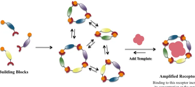
Dynamic combinatorial chemistry is a method for the in situ generation of a complex
mixture of macrocycles from smaller building blocks and is combinatorial chemistry that is
under thermodynamic control with all library constituents under equilibrium.9 This requires that
all library members be able to interconvert through a reversible chemical process, which can
involve either covalent or noncovalent interactions. The composition of the library is determined
via the thermodynamic stability of each of the library members. Thermodynamic control of the
library implies that introduction of changes in conditions of the experiment will induce changes
in the library composition. When a template is introduced and binds to a library member, this
species is stabilized. The bound species is thus amplified, as the remaining building blocks will
establish a new equilibrium increasing the concentration of the selected library member at the
expense of the remaining library members.10-12 Therefore, in theory the amplification should be
Figure 2.1 Scheme representing dynamic combinatorial chemistry as a way to select specific members of a dynamic combinatorial library through noncovalent interactions by the introduction of a separate guest.
An important feature of dynamic combinatorial chemistry is the reversible exchange
reaction that is necessary in order for the building blocks to be capable of interconverting
between library members. A number of requirements are necessary for this reaction: (i) the
reaction needs to be reversible on a reasonable timescale (ii) the reaction needs to be compatible
with the experimental conditions including the functional groups on the building blocks and
template, the solvent, and the pH (iii) the reaction conditions need to be mild so as not to
interfere with the delicate noncovalent interactions involved in molecular recognition (iv) it
needs to guarantee the solubility of all library members so as not to interfere or alter the
equilibrium (v) it should be possible to turn off the reaction and thus kinetically “freeze” the
selected library members enabling their isolation and characterization and (vi) ideally all library
members should be isoenergetic to prevent production of biased libraries.5
Three main different types of reversible reactions have been used including: noncovalent
reversible reactions therefore only reactions most relevant to the Waters lab will be discussed.
The first example is the acid-catalyzed condensation of an amine with a ketone or aldehyde to
form an imine. While imine formation is reversible, imines condense and hydrolyze quickly at
neutral to acidic pH making it an impractical exchange reaction in for biologically relevant
templates.13 On the other hand, hydrazones can be thermodynamically stable in the presence of
water even at lower pHs.14 The hydrazine exchange reaction is reversible and compatible with a
wide range of solvents and functional groups. Most applications of hydrazones in dynamic
combinatorial chemistry involves an acyl hydrazine where the acyl group moderates the
stabilizing influence of the amine subunit in C=N-NRR’.5 The drawback to using hydrazones as
the exchange reaction is that exchange occurs at a pH of 4, which is not compatible with most
biomolecules. Another example of a covalent exchange reaction that is commonly employed in
dynamic combinatorial chemistry is disulfide exchange, which plays an important role in biology
due to its role in the folding of proteins and the redox state of cells.15 The mechanism includes
the nucleophilic displacement of a thiolate anion from the disulfide through attack by another
thiolate anion.16 This process requires deprotonation of the thiol therefore the exchange is highly
pH dependent and allows the process to be halted by lowering the pH. Disulfide exchange can
be carried out at near neutral conditions (pH 7-9) making it compatible with most biomolecules.
However, when a hit has been identified, the disulfide bond is not stable within the cellular
environment and there is no straightforward synthetic replacement. Additionally, this reaction is
Figure 2.2 Reversible covalent reactions used for dynamic combinatorial chemistry. (a) Acid-catalyzed exchange reaction between an amine and an aldehyde to form an imine. (b) Reaction of a hydrazine and aldehyde to form a hydrazone. (c) Disulfide exchange, which occurs between a thiolate and a disulfide bond to release a free thiolate anion.
iii. Dynamic Cyclic Peptide Libraries from Thiol-Thioester Exchange
Our lab has recently published a method utilizing thiol-thioester exchange as a reversible
reaction for DCC to form cyclic peptides.17 The advantage of using thiol-thioester exchange over
other exchange reactions is that it is rapid in aqueous solution at neutral pH and provides a
native-like linkage that is successively replaceable by a more robust amide or ester
functionality.18,19 Equilibrium is reached within 1 to 2 hours depending on the sequence, with no significant hydrolysis of monomers during that time. Previous reports of the use of thioester
exchange in DCC experiments has involved thioesters that were unsubstituted at the α-position,
limiting the diversity of structures possible in a library of cyclic peptides.19,20 Our lab performed systematic studies to determine the reactivity of peptide thiol-thioester exchange to its scope and
limitation in its application in DCC.
Each monomer was a four-residue peptide containing a thioester at the C-terminus and a
thiol at AA2 (Cys). The structures of each monomer were varied at AA1, AA3 and AA4. The
monomers were designed so that AA2-AA3-AA4 form the macrocycle while AA1 remained
aggregation. Proline was initially included in AA3 as a turn residue to favor macrocycles.
Various amino acids were incorporated into AA4 to study their effect on the rate of
macrocyclization, including positively charged amino acids (Lys, Arg), a negatively charged
amino acid (Glu), and a hydrogen-bonding and neutral amino acid (Gln), and hydrophobic and
sterically bulky amino acids (Phe, Val).
Upon dissolution of the monomers in potassium phosphate butter (pH 7), the monomers
underwent facile thiol-thioester exchange to form a mixture of macrocycles, with the dimeric
20-atom macrocyclic hexapeptides as the major product. The reaction mixture was monitored over
time by HPLC, allowing a simple two-step mechanism to be proposed. First, two monomers
react to form an oligodimer followed by a ring closure intramolecular trans thioesterification
reaction. The oligodimer intermediate was not observed to accumulate, indicating that the
intermolecular step is rate limiting.
Thiol-thioester exchange can tolerate a variety of amino acids and structure-function
studies indicated that the rate of macrocycle formation is dependent on the amino acid sequence.
The observed reactivity of monomers with variation at the C-terminus corresponded to
differences in sterics, with the incorporation of β-branched amino acids such as Val resulting in
measurable hydrolysis before cyclization was complete. When Glu was placed at the
C-terminus, it reacted more slowly than its Gln counterpart and can isomerize through anhydride
intermediates. Additionally, the effect of positively charged amino acids (Lys and Arg) at
positions AA1 and AA4 were investigated were found to react significantly faster than their
negatively charged analogues suggesting that positively charged amino acids could stabilize the
buildup of negative charge in the transition state. Finally, Trp was placed at the turn residue to
monomer actually reacted faster than the hydroxyproline (Hyp) analogue. Stereochemistry also
played a role in the rate of macrocyclization as D-Pro reacted considerably faster than Hyp
suggesting that chirality has an influence on the accessibility of the thioester or thiol.
Figure 2.3 Scheme depicting macrocyclization of peptides via thiol-thioester exchange. (a) General design of monomers and major products (b) Sequence of steps toward dimeric cyclic thiodepsipeptide.
In summary, our lab has developed an efficient method to rapidly generate libraries of
macrocyclic thiodepsipeptides at netrual pH for high-throughput screening. Upon mixing two or
more monomers, we can form a complex library of cyclic thiodepsipeptides generated in situ
under thermodynamic control. These libraries can then be screened against a particular target
efficiently within hours. Given the transient stability of thioesters in vivo, a “hit” in the
high-throughput screening can be resynthesized as a more stable analogue by replacing the thioester
group with an amide or ester. Our lab then looked to apply this method of high-throughput
B. System Design
i. Dynamic Combinatorial Library
Our strategy for the discovery of high-affinity ligands with selectivity between
quadruplexes uses a common acridine core to target the planar surface of the G-tetrad, appended
with variable peptide substituents to contact the loops and grooves that distinguish each
quadruplex structure. We chose the acridine ligand due to the high selectivity it provides for
quadruplex DNA over duplex DNA. Previously, Neidle and coworkers screened a number of
linear peptide-acridine ligands in the effort to gain selectivity amongst individual quadruplex
structures. While linear peptides provided modest binding selectivity in Neidle’s work, we felt
that cyclic peptides could deliver greater selectivity and potentially fit nicely into the grooves of
the quadruplex structure. Using methods previously developed in our lab, we wanted to create
libraries of cyclic peptides using thiol-thioester exchange in dynamic combinatorial chemistry.
The dynamic combinatorial libraries consisted of tetrapeptides appended to an acridine core to
target the quadruplex structure with the tetrapeptides designed to undergo thiol-thioester
exchange by incorporation of a Cys at AA2 and a thioester at the C-terminus. Since the loops and
peripheral grooves of the quadruplex structure provide a pattern of hydrogen bonding,
hydrophobic π-surfaces, and negative charges unique to a particular quadruplex sequence, we
chose to focus on the incorporation of amino acids with hydrogen bonding groups, positive
Figure 2.4 General design of monomer library with acridine ligand incorporated at the
N-terminus to target the G-tetrad of the quadruplex structure. Cysteine is incorporated at AA2 along
with a thioester installed at the C-terminus to facilitate thiol-thioester exchange, while a Pro was included at the AA3 as a turn residue to aid in cyclization.
ii. Biological Targets
The goal of this project is to find molecules that can bind to, and differentiate between
various G-quadruplex structures. With this in mind, it was necessary to choose different DNA
sequences to screen our libraries against. The first sequence we chose was the human telomeric
(Htelo) sequence given the extensive studies that have been carried out surrounding the role of
quadruplex formation in the single-stranded telomeric region and its role in the inhibition of the
reverse-transcriptase enzyme telomerase. Secondly, we chose three oncogenes whose promoter
regions are known to contain G-quadruplex folding patterns and structures, cKit21, cMyc22 and
NRas. Finally, we chose a dsDNA sequence containing guanines as a control to determine the
selectivity of our library for quadruplex DNA versus duplex.
The first oncogene we chose to screen is the c-myc oncogene, which is one of the most
commonly malfunctioning genes in human cancers.22 The myc family of oncogenes encodes
phospho-proteins that activate genes and encourage forward cell growth. Normally, the human
c-myc gene is tightly regulated and it is the overexpression of this gene that leads to the
progression of many cancers. The c-myc gene utilizes four promoters with the
nuclease-hypersentive element III1 accounting for 75-85% of total c-myc transcription.23 It is this region of
N N
H N
N H N
O O
HN
N H
H N
O O
O N AA1
SH H N
S O
O
O OMe
the c-myc promoter region that can form G-quadruplexes under physiological conditions thereby
playing an important role in the transcriptional regulation of this oncogene.
The promoter region of c-kit is the second sequence we chose to screen against. c-kit is
an oncogene which codes for a tyrosine kinase receptor and is pivotal for relaying extracellular
signals. When activated, KIT stimulates proliferation, differentiation, migration and survival.24
Overexpression of the c-kit gene results in uncontrolled cell proliferation and is considered the
primary pathogenic event in gastrointestinal stromal tumors. A quadruplex forming
21-nucleotide sequence upstream of the transcription initiation site has been identified on the G-rich
strand, which occupies a site required for core promoter activity.25 Until recently, all cancer
therapies targeting kinases inhibit the kinase protein after expression rather than controlling its
expression. The position of the quadruplex sequence in the promoter region of c-kit makes it an
attractive target for the regulation of c-kit at the transcriptional level.
A final oncogene that we chose to focus on is the NRAS gene that is another of the most
frequently mutated oncogenes detected in human cancer. The NRAS gene encodes for a guanine
nucleotide (GTP/GDP)-binding protein that acts as a signal transducer. Mutated forms of the
gene can result in a protein locked activated state, transmitting constitutively signals for cell
proliferation to the nucleus. The promoter of the NRAS gene contains a nuclease hypersensitive
polypurine-polypyrimidine element (NHPPE) that is essential for transcription and has the ability
to form the G-quadruplex structure.26 Studies have again shown that formation of G4 DNA in the
control region of the gene may contribute to the regulation of expression of the NRAS gene. Due
to its role in the pathogenesis of cancer, the quadruplex structure of the NRAS gene is an ideal
C. Conclusions
The goal of this project is to identify small molecules that can bind to and stabilize the
G-quadruplex structure of DNA. Previous studies have shown modest success in differentiating the
various possible quadruplex structures and topologies through the use of acridine-linear peptides
conjugates. We felt strongly that the use of cyclic peptides could deliver greatly improved
selectivity while also providing the added advantages of mimicking native protein structure,
displaying enhanced metabolic stability and possessing structural preorganization thus reducing
the entropic cost of binding. Despite the therapeutic potential of cyclic peptides, the options for
synthesizing structurally diverse libraries in a high-throughput format are limiting. Using a
strategy that has been developed in our lab, we propose screening libraries of solution-phase
cyclic peptides generated using thiol-thioester exchange for DCC. Our system has been designed
as tetrapeptides containing a thioester installed at the C-terminus with the acridine ligand
attached at the N-terminus. We selected the four quadruplex forming sequences of Htelo, c-kit,
c-myc and NRAS as our biological targets due to their biological importance and implications in a
number of different cancers. The next step was to synthesis the monomers of our library
References
1. Troitskaya, L. A.; Kodadek, T. Peptides as modulators of enzymes and regulatory proteins. Methods, 2004, 32, 406-415.
2. Andrianov, A. M. Mol. Biol. 1999, 33, 534-538.
3. Freidinger, R. M.; Veber, D. F.; Perlow, D. S.; Brooks, J. R. Science, 1980, 210,
656-658.
4. Katz, B; Liu, B.; Cass, R.; Structure based design tools: structural and thermodynamic comparison with biotin of a small molecule that binds to streptavidin with micromolar affinity. J. Am. Chem. Soc. 1996, 118, 7914-7920.
5. Horton, D. A.; Bourne, T. T. Smythe, M. L. Exploring privileged structures: The combinatorial synthesis of cyclic peptides. J. Comput. Aided Mol. Design, 2002,
16, 415-430.
6. Mootz, H. D.; Marahiel, M. A. J. Bacteriol 1997, 197, 6843-6850.
7. Kratzschmar, J.; Krause, M.; Marahiel, M. A.; J. Bacteriol. 1989, 171, 5422-529.
8. Emmel, E. A.; Verweij, C. L.; Durand, D. B.; Higgins, K. M.; Lacy, E.; Crabtree, G. R. Science, 1989, 246, 1617-1620.
9. Corbett, P.; Leclaire, J.; Vial, L.; West, K. R.; Wietor, J. –L.; Sanders, J. K. M.; Otto, S. Dynamic combinatorial chemistry. Chem. Rev. 2006, 106, 3652-3711.
10. Hoss, R.; Vogtle, F. Angew. Chem. Int. Ed. Engl. 1994, 33, 375.
11. Anderson, S.; Anderson, H. L.; Sanders, J. K. M. Acc. Chem. Res. 1993, 26, 469.
12. Busch, D. H. Incl. Phenom. Mol. Recognit. Chem. 1992, 12, 389.
13. Giuseppone, N.; Lehn, J. –M. J. Am. Chem. Soc. 2004, 126, 11448.
14. Carey, F. A.; Sundberg, R. J. Advanced Organic Chemistry Part A, Plenum Press: New York, 1990, p. 451.
15. Gilbert, H. F. J. Biol. Chem. 1997, 272, 29399.
16. Fernandes, P. A.; Ramos, M. J. J. Chem.-Eur. J. 2004, 10, 257.
18. Ura, Y.; Beierle, J. M.; Leman, L. J.; Orgel, L. E.; Ghadiri, M. R. Science, 2009,
325, 73-77.
19. Woll, M. G.; Gellman, S. H. J. Am. Chem. Soc. 2004, 126, 11172-11174.
20. Larsson, R.; Pei, Z. C.; Ramstrom, O. Angew. Chem. Int. Ed. 2004, 43, 3716.
21. Redman, J. E.; Granadino-Roldan, J. M.; Schouten, J. A.; Ladame, S.; Reszka, A. P.; Neidle, S.; Balasubramanian, S. Recognition and discrimination of DNA quadruplexes by acridine-peptide conjugates. Org. Biomol. Chem. 2009, 7, 76-84.
22. Brooks, T. A.; Hurley, L. H. The role of supercoiling in transcriptional control of MYC and its importanct in molecular therapeutics. Nat. Rev. Cancer 2009, 9,
849-861.
23. Gonzalez, V.; Hurley, L. H. The c-MYC NHE III1: function and regulation. Annu. Rev. Pharmacol. Toxicol. 2010, 50, 111-129.
24. Shirude, P. S.; Okumus, B.; Ying, L.; Ha, T.; Balasubramanian, S. Single-molecular conformational analysis of G-quadruplex formation in the promoter DNA duplex of the proto-oncogene c-kit. J. Am. Chem. Soc. 2007, 129, 7484-7485.
25. Rankin, S.; Reszka, A. P.; Huppert, J.; Zloh, M. Parkinson, G. N.; Todd, A. K.; Ladame, S. Balasubramanian, S.; Neidle, S. Putative DNA quadruplex formation within the human c-kit oncogene. J. Am. Chem. Soc. 2005, 127, 10584-10589.
CHAPTER 3: SYNTHESIS OF MONOMERS FOR DYNAMIC COMBINATORIAL LIBRARIES
A. Monomer Synthesis
i. Synthesis of acridine ligand
The monomer system design consisted of a tetrapeptide containing a cysteine residue and
thioester installed at the C-terminus to facilitate thiol-thioester exchange. Additionally, an
acridine ligand was installed at the N-terminus to target the quadruplex structure (Figure 3.1).
The tetrapeptide is synthesized using solid phase peptide synthesis followed by coupling of the
free N-terminus to the acridine ligand. Given the excellent selectivity of the acridine motif for
the quadruplex structure over the duplex DNA, the synthesis of BRACO-19 (1) is well
precedented.1,2 However, the structure synthesized was slightly modified as shown in structure 2
to include a carboxylic acid group off of the phenyl side chain allowing for coupling to the
N-terminus of the peptide substituents.
Figure 3.1 Structure of the G-quadruplex ligand BRACO-19 (1) and the modifications made to the acridine ligand (2) to allow incorporation via solid phase synthesis at the N-terminus of the tetrapeptide monomers.
The synthesis of the acridine ligand began with the nitration of diphenyl methane
followed by oxidation to the ketone (2) using chromium oxide. The nitro groups were reduced in N
HN
R
N H N
H
N N
O O
1 R= -N(Me)2
![Figure 1.2 Structures of the human unimolecular telomeric quadruplex formed by the sequence d[AGGG(TTAGGG) 3 ]](https://thumb-us.123doks.com/thumbv2/123dok_us/8324722.2207162/19.918.345.585.127.520/figure-structures-unimolecular-telomeric-quadruplex-formed-sequence-ttaggg.webp)