Development of a Novel AAV Gene Therapy
Cassette with Improved Safety Features
and Efficacy in a Mouse Model of Rett Syndrome
Kamal K.E. Gadalla,
1,2Thishnapha Vudhironarit,
1Ralph D. Hector,
1Sarah Sinnett,
3,4Noha G. Bahey,
1,5Mark E.S. Bailey,
6Steven J. Gray,
3,4,7and Stuart R. Cobb
11Institute of Neuroscience and Psychology, College of Medical, Veterinary and Life Sciences, University of Glasgow, Glasgow G12 8QQ, UK;2Pharmacology Department, Faculty of Medicine, Tanta University, Tanta 31527, Egypt;3Gene Therapy Center, University of North Carolina at Chapel Hill, Chapel Hill, NC 27599, USA;4Carolina Institute for Developmental Disabilities, University of North Carolina at Chapel Hill, Chapel Hill, NC 27599, USA;5Histology Department, Faculty of Medicine, Tanta University, Tanta 31527, Egypt;6School of Life Sciences, College of Medical, Veterinary and Life Sciences, University of Glasgow, Glasgow G12 8QQ, UK;7Department of Ophthalmology, University of North Carolina at Chapel Hill, Chapel Hill, NC 27514, USA
Rett syndrome (RTT), caused by loss-of-function mutations in the MECP2gene, is a neurological disorder characterized by severe impairment of motor and cognitive functions. The aim of this study was to investigate the impact of vector design, dosage, and delivery route on the efficacy and safety of gene augmentation therapy in mouse models of RTT. Our results show that AAV-mediated delivery of MECP2 to Mecp2 null mice by systemic administration, and utilizing a minimal endogenous promoter, was associated with a narrow therapeu-tic window and resulted in liver toxicity at higher doses. Lower doses of this vector significantly extended the survival of mice lacking MeCP2 or expressing a mutant T158M allele but had no impact on RTT-like neurological phenotypes. Modifying vector design by incorporating an extended Mecp2promoter and additional regulatory 30 UTR elements significantly reduced hepatic toxicity after systemic administration. More-over, direct cerebroventricular injection of this vector into neonatalMecp2-null mice resulted in high brain transduction efficiency, increased survival and body weight, and an amelio-ration of RTT-like phenotypes. Our results show that control-ling levels of MeCP2 expression in the liver is achievable through modification of the expression cassette. However, it also highlights the importance of achieving high brain trans-duction to impact the RTT-like phenotypes.
INTRODUCTION
Rett syndrome (RTT; OMIM 312750) is a neurological disorder char-acterized by a constellation of clinical diagnostic and associated features and with overt onset occurring several months postnatally.1 Typical RTT is almost exclusively caused by de novo germline muta-tions in the X-linked gene,MECP22(as reviewed elsewhere3,4). Several mouse models of RTT have been generated that harborMecp2 dele-tions5–7or knocked-in mutations.8–11Many of these models recapitu-late the principal features that characterize RTT in humans, although there are differences that reflect the phenotypic variability seen in pa-tients.12–14Despite the severity of RTT-like phenotypes, genetic
reac-tivation of silencedMecp2in conditional knockout mice resulted in a robust and enduring reversal of phenotypes.15–17
This inherent reversibility of the phenotype, added to the lack of obvious targets for pharmacotherapy, makes gene therapy an obvious therapeutic strategy in RTT. However, there are significant challenges to a gene transfer approach, including the requirement to transduce sufficient numbers of neurons in the brain16and the avoidance of deleterious overexpression.18
Previous attempts atMECP2gene transfer using AAV9 vectors were confounded by limited brain transduction efficiency and toxicity,19,20 while efficacy in other studies using self-complementary adeno-asso-ciated virus (AAV) (scAAV)21may have been compromised by the use of a construct exceeding the packaging capacity of the vector.
The aim of the present study was to assess the therapeutic impact of dose, route of administration, and expression cassette design in mice modeling RTT. Our results show that modification of the vector design by incorporating more regulatory elements is able to reduce peripheral expression of vector-derived MeCP2 and prevent liver toxicity. We also show that using the same vector design by direct brain injection in mouse neonates resulted in higher brain transduc-tion and improved the RTT-like phenotype.
RESULTS
Dose Escalation with AAV/MECP2Revealed a Narrow Therapeutic Window following Systemic Administration
In order to explore the relationship between vector dose and thera-peutic benefits, we conducted a dose escalation experiment in which
Received 24 February 2017; accepted 19 April 2017; http://dx.doi.org/10.1016/j.omtm.2017.04.007.
Correspondence:Stuart Cobb, Institute of Neuroscience and Psychology, College of Medical Veterinary and Life Sciences, West Medical Building, Glasgow, G12 8QQ UK.
an scAAV2/9 vector was used to deliver a Myc-tagged human
MECP2_e1 cDNA under the control of a short, 229-bp region of the murineMecp2endogenous core promoter (MeP229),19,22 herein-after referred to as the “first-generation vector”. Juvenile male
Mecp2/y and wild-type (WT) mice were injected at the age of
4–5 weeks in the tail vein either with vehicle or with 11011(low dose), 11012(moderate dose), or 11013(high dose) viral ge-nomes (vg) per mouse (dose range,11013–11015vg/kg). As
expected from previous studies of this knockout line,6,7,15onset of RTT-like phenotypic signs in control vehicle-treated Mecp2/y mice15was observed from 4 to 5 weeks of age, and severity progres-sively increased until death or censoring of all mice by 20 weeks of age (Figures 1A–1C).Mecp2/ymice treated with the low dose were indistinguishable from vehicle-treatedMecp2/ymice in terms of sur-vival (median sursur-vival = 9.36 weeks versus 11.64 weeks, respectively; p = 0.2, Mantel-Cox test,) and severity score (Figures 1A and 1C).
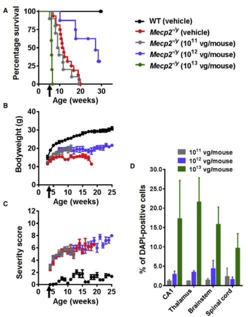
Figure 1. Systemic Delivery of the First-Generation Vector toMecp2–/yMice Revealed Therapeutic
Efficacy and a Narrow Therapeutic Window (A) Kaplan-Meier survival plot forMecp2/ymice injected
with different doses (11011
[n = 10], 11012 [n = 8], and 11013
[n = 5] vg per mouse] of first-generation vector compared to vehicle-treated animals (WT; n = 9,
Mecp2/y; n = 16). The median survival period in Mecp2/ymice treated with 11012
vg per mouse was significantly higher than that in vehicle-treated controls (27.14 versus 11.64 weeks; p = 0.001, Mantel-Cox test). (B and C) Plots showing mean (B) body weight and (C) aggregate severity scores forMecp2/ymice treated with 11011
and 11012
vg per mouse or vehicle. Arrows indicate age at injection; data are presented as mean± SEM. (D) Dose-dependent transduction efficiency (Myc-positive nuclei as a proportion of DAPI-(Myc-positive nuclei) across different brain regions. Data are presented as mean±SEM (n = 3 mice per group). CA1 indicates hip-pocampal region CA1.
However, when measured at 11 weeks (the median survival time for the control vehicle-treatedMecp2/ymice), the mean body weight of the treatedMecp2/ymice was significantly (p < 0.05) higher than that of the Mecp2/y vehicle controls (Figure 1B).
In contrast, Mecp2/y mice treated with the moderate dose (11012) showed significantly increased survival and body weight compared to the vehicle controls (median survival = 27.3 weeks versus 11.64 weeks; p = 0.001, Mantel-Cox test [Figure 1A]; p < 0.05 for mean body weight measured at 11 weeks of age [Figure 1B]). However, there was no differ-ence in the RTT-like phenotype severity score at this dose (Figure 1C). Finally, the cohort receiving the highest dose showed acute toxicity and lethality at 10–15 days post-injection (Figure 1A). Overall, vehicle-treated WT mice differed fromMecp2/ycohorts across all measures (all ps < 0.001).
Patterns of transduction in treated Mecp2/y mice were assessed within the CNS by anti-Myc antibody immunofluorescence labeling (Figure S1), which revealed vector-derived MeCP2 protein expression distributed in a punctate pattern within cell nuclei corresponding to that observed for endogenous MeCP2 in WT mice. Samples from the low-dose cohort revealed low transduction efficiencies across brain regions (0.5% to 1%). The moderate dose resulted in3%– 5% transduction efficiency, whereas the efficiency for the high dose was 10%–22% (Figure 1D).
earlier. The low and moderate doses were tolerated and had no observable effect on body weight or phenotypic severity score ( Fig-ures S2A–S2C). However, WT mice treated with the high dose exhibited the acute toxicity and rapid lethality observed in the knockout mice (Figures S2A–S2C). Quantification of cellular levels of MeCP2 in mice given this high dose revealed that transduced hip-pocampal pyramidal cells expressed vector-derived MeCP2 at a mean level equivalent to 120% of the endogenous level, resulting in total cellular levels of MeCP2 just over 2-fold higher than normal for these cells (Figures S2D–S2F).
Systemic Delivery of First-Generation Vector Resulted in Liver Toxicity
To further investigate toxic effects encountered after systemic injec-tion of the first-generation vector at high doses, levels of vector-derived MeCP2 expression were tested in a range of peripheral tissues. Bio-distribution of the vector genome in different organs was
quanti-fied using qPCR at the end of experiment (Figure S3) and revealed, along with immunohistochemistry, that the proportion of Myc-pos-itive cells in the liver was high (Figure S4). Endogenous MeCP2 levels are known to be much lower in liver cells than in brain neurons23,24 and are typically below the detection threshold for immunohisto-chemistry using available antibodies (Figure S4A). However,
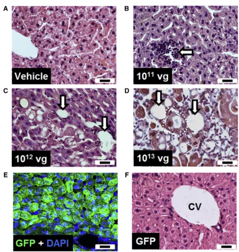
vector-Figure 2. Intravenous Injection of the First-Generation Vector Resulted in Pathological Changes in the Liver
(A–D) Representative H&E-stained liver sections from WT mice injected with (A) vehicle or (B–D) different doses of vector. (E) Liver section from a mouse injected intrave-nously with a GFP control vector, counterstained with DAPI. (F) Representative H&E-stained liver section from a GFP vector-treated mouse. Arrows indicate mononuclear cell infiltration, vacuolation, and/or loss of hepatocytes. Dashed white line indicates cellular swelling. Scale bars indicate 20mm. CV, central vein.
derived MeCP2 levels in a subset of liver cells (using anti-Myc-immunolabeling) of treated WT mice were found to be higher than MeCP2 levels seen in neurons (Figures S4B and S4C) and were thus20 times higher than levels found endogenously in such cells. Myc-positive cells were detected also in the heart, kidney, and other peripheral tissues in treatedMecp2/ymice (data not shown).
Histological investigation of liver sections from mice injected with vehicle or a low dose of the vector showed a largely normal liver structure with occasional areas of mononuclear infiltration (Figures 2A and 2B). In contrast, mice injected with higher doses of the vector showed a dose-dependent increase in pathological features, including cellular destruction and vacuolation, loss of hepatocytes, and mononuclear cell infiltration (Figures 2C and 2D).
To address whether the observed liver pathology was due to the high copy number of viral particles per se or was a consequence of MeCP2 overexpression, we injected mice with a vector driving expression of GFP but otherwise identical to thefirst-generation vector. Despite detection of widespread GFP expression in the liver (Figure 2E), his-tological examination of liver sections revealed no evidence of cellular damage or immune cell infiltration (Figure 2F). In addition, no changes in RTT aggregate severity score were observed with this vec-tor (data not shown).
Systemic Administration of First-Generation Vector Improves Survival inMecp2T158M/yKnockin Mice
An important question for gene transfer in RTT is whether the pres-ence of endogenous mutant MeCP2 might reduce the therapeutic ef-fect of vector-derived wild-type MeCP2. Male mice expressing native MeCP2 tagged with GFP as a fusion protein and harboring the common RTT-causing p.T158M mutation,9Mecp2T158M/y, display a
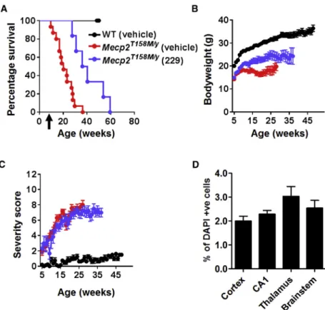
Intravenous delivery of a moderate dose (11012vg per mouse) of thefirst-generation vector to 4- to 5-week-oldMecp2T158M/ymice re-sulted in significantly increased survival (Figure 3A; median survival = 38.3 weeks in vector-treated mice versus 20.3 weeks in vehicle-treated mice; p = 0.0019, Mantel-Cox test, n = 8–15 per group). There was a modest increase in body weight in the vector-treated cohort ( Fig-ure 3B; p < 0.05, one-way ANOVA using data at 20 weeks of age). However, there was no difference in RTT-like aggregate severity score between groups (Figure 3C), consistent with a low brain transduction efficiency (2%–4%) as revealed by anti-Myc labeling (Figure 3D). Overall, vehicle-treated WT mice differed fromMecp2T158M/ycohorts across all measures (all ps < 0.01).
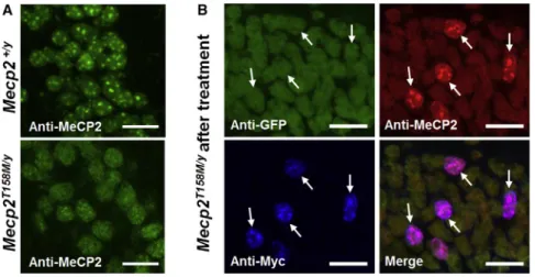
The p.T158M mutation affects the chromatin-binding capacity of MeCP2, leading to loss of the punctate element of MeCP2 labeling in the nucleus (Figure 4A).9 Immunolabeling of hippocampal neurons from treated Mecp2T158M/y mice showed WT patterns of MeCP2 expression, with restored localization to DAPI bright spots, only in transduced (Myc-positive) cells (Figure 4B). This is consistent with vector-derived MeCP2 being able to localize normally to heter-ochomatin, despite the presence of mutant endogenous MeCP2 pro-tein within the same nucleus.
Development of a Second-Generation Vector that Reduced Liver Toxicity after Systemic Administration
In light of the data described earlier, it was evident that a higher AAV vector dose is required to achieve therapeutically relevant levels of
Figure 3. Improved Survival and Body Weight of
Mecp2T158M/YMice after Systemic Delivery of the
First-Generation Vector
(A) Survival plot for treatedMecp2T158M/ymice. Arrow indicates
age at injection. (B and C) Plots of (B) body weight and C) aggregate severity score, respectively, forMecp2T158M/ymice
treated with 11012
vg per mouse of first-generation vector and control groups (Mecp2T158M/y and WT) treated with
vehicle. Data presented as mean±SEM. (D) Transduction ef-ficiency in the brain of treated mice (Myc-positive nuclei as a proportion of DAPI-positive nuclei; n = 3 mice).
brain transduction after systemic delivery. However, severe toxicity after delivery of high doses of our
first-generation cassette necessitated a new design. We tested a range of modifications to the expression cassette and capsid that were predicted to result in lower cellular expression levels and/or reduce liver tropism. This included the use of expression cas-settes utilizing (1) an alternative, compact, and, pre-sumably, weaker JeT promoter25; (2) a short syn-thetic polyadenylation (SpA) signal (Figure S6A)26; and (3) the original first-generation expression cassette packaged in a scAAV9.47 capsid, which emerged from an in vivo screen for liver de-targeted capsid sequences relative to AAV9.27,28Systemic in-jection of these vectors at the moderate dose (11012vg per mouse) into 4- to 5-week-old Mecp2/y mice resulted in significantly extended survival and improved body weight, but there was no impact on the RTT-like aggregate severity score (Figure S6B). In sum-mary, none of these modifications resulted in any significant im-provements over thefirst-generation vector (p > 0.05 for all measures, ANOVA and Mantel-Cox tests). Importantly, these modified vectors all caused the development of liver pathology similar to that observed with the first-generation vector (as previously shown in Figure 2;
Figure S6C).
In order to test the therapeutic efficacy of the second-generation vec-tor, a moderate dose (11012vg per mouse) was injected intrave-nously into 4- to 5-week-oldMecp2/ymice. There was a significant extension of survival in the vector-treated mice compared to the vehicle-treated mice (median survival = 29.9 weeks and 11.6 weeks, respectively; p < 0.0001, Mantel-Cox;Figure 5B). There was also sig-nificant improvement in body weight at the age of 11 weeks (p < 0.05, one-way ANOVA, with Tukey’s post hoc pairwise comparison test;
Figure 5C). In contrast, there was no effect on RTT-like aggregate severity score (Figure 5D). The second-generation vector, thus, showed no therapeutic advantages over the first-generation vector after systemic delivery (Figures 5B–5D). Again, vehicle-treated WT mice differed fromMecp2/ycohorts across all measures (all p < 0.001). In order to compare this vector head-to-head with the
first-generation vector in terms of liver safety, mice were injected intravenously with either thefirst- or the second-generation vector at a dose of 11012vg per mouse. These mice were sacrificed after 30 days, and tissues were analyzed for vector-derived MeCP2 expres-sion (using anti-Myc tag antibody) and signs of liver pathology ( Fig-ure 6). There was no significant difference in transduction efficiency between vector constructs (Figure 6B), but cellular levels of vector-derived MeCP2 (anti-Myc) in mice treated with first-generation vector were significantly higher than those in mice treated with sec-ond-generation vector (Figure 6C; p < 0.001, unpaired t test). Analysis of the distribution of cellular MeCP2 expression levels in transduced cells showed that MeCP2 expression was more tightly regulated in mice injected with the second-generation vector (Figure 6D), with fewer cells exhibiting very high expression levels. Moreover, there was none of the disrupted hepatic architecture or vacuolation previ-ously observed with thefirst-generation vector (Figure 6E). The den-sity of inflammatory foci was significantly higher in liver samples from mice injected withfirst-generation vector than from those in-jected with the second-generation vector (Figure 6F).
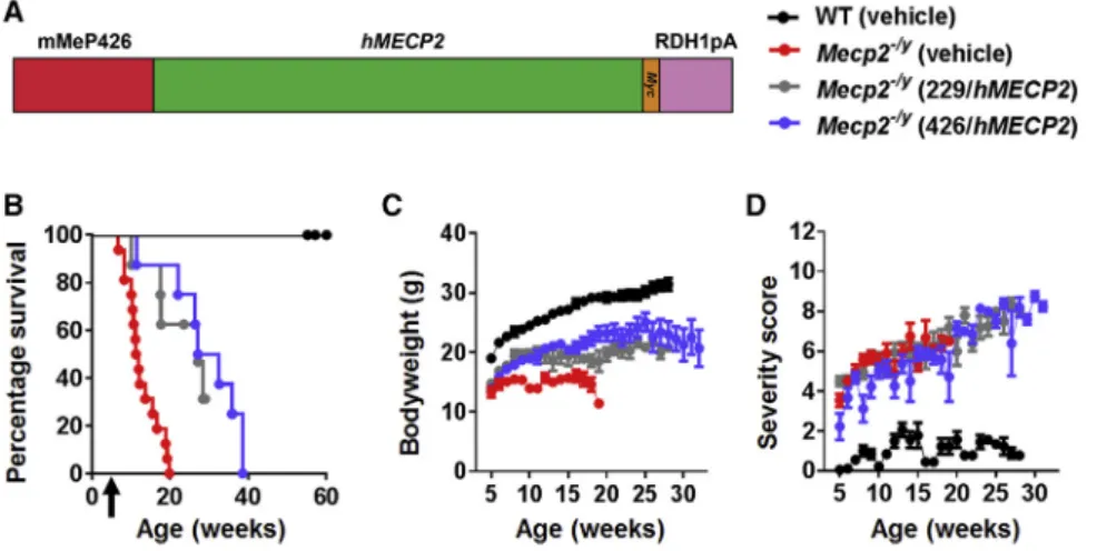
Neonatal Cerebroventricular Injection of the Second-Generation Vector Improved the RTT-like Aggregate Severity Score
The lack of impact on the phenotype after systemic administration is consistent with the low brain transduction efficiencies observed, as it
has been established that phenotype severity and degree of improve-ment after gene restoration correlate with the proportion of MeCP2-expressing cells in the brain.16 Therefore, we decided to test the second-generation vector by direct cerebroventricular injection in mouse neonates, a delivery route that is known to afford widespread transgene expression.19When delivered at a dose of 11011vg per mouse (Figure 7A), there was a pronounced extension in the survival of Mecp2/y mice treated with the second-generation vector in comparison to vehicle-treated mice (median survival = 38.5 and 12.4 weeks, respectively; p < 0.0001, Mantel-Cox test; Figure 7B). While there was a negligible effect of vector on body weight ( Fig-ure 7C), an important observation was the clear improvement in the RTT-like aggregate severity score compared to that of vehicle-treatedMecp2-null mice (Figure 7D; p < 0.01 at 11 weeks, one-way ANOVA, with Tukey post hoc pairwise comparison). Vector-derived MeCP2 (revealed by anti-Myc tag immunolabeling) was detectable in all brain regions, with transduction efficiencies across brain regions ranging from10%–40% (Figures 7E and 7F). Distribution analysis revealed that the modal cellular MeCP2 level in transduced cells in cortex was approximately twice that of endogenous MeCP2 (consis-tent with a vector-derived expression level equal to the endogenous level), with some cells expressing higher levels of vector-derived MeCP2 (Figure 7G).
DISCUSSION
The reversal of a wide range of RTT-like phenotypes in mice following the delayed unsilencing ofMecp2provides a strong ratio-nale for gene transfer as a therapeutic strategy in RTT.15,16There are likely to be a variety of barriers to translational success that will need to be identified and addressed in order to secure optimal out-comes in human clinical trials. In the present study, we identified particular challenges associated with the systemic delivery of aMECP2-bearing gene therapy vector in terms of a narrow therapeu-tic window driven by low brain transduction efficiency and the appearance of peripheral overexpression toxicity upon further dose escalation. However, peripheral overexpression can be reduced by refining the cassette design. We show that direct brain delivery of vec-tor in neonatal mice can achieve therapeutically relevant levels of
Figure 4. Nuclear Localization of MeCP2 in Untreated and TreatedMecp2T158M/YMice Representative confocal images of the CA1 region of the hippocampus. (A) Endogenous MeCP2 exhibits heterochromatin-enriched localization in WT nuclei, while GFP-tagged MeCP2 exhibits decreased heterochromatin localization (i.e., more diffuse labeling) in nuclei from
Mecp2T158M/ymice. (B) Images demonstrating
hetero-chromatin-enriched localization of vector-derived MeCP2 in nuclei of transduced cells inMecp2T158M/ymice treated
transduction that result in phenotype amelioration. We also show that the vector has similar effectiveness in mice expressing the most common RTT-causing mutation, suggesting that the presence of existing mutant forms of MeCP2 is unlikely to be an obstacle to trans-lational success. These results are consistent with experiments in transgenic mice expressing both mutant and WT forms of the protein.36
Recent attempts to deliverMECP2exogenously in mouse models of RTT used widely varying vector doses but are difficult to compare based on additional differences in cassette design and other variables, including viral production, dosing protocol, and phenotype mea-sures.19–21In the present study, we used our previously published cassette design (humanMECP2_e1, under the control of a MeP229 core promoter fragment)19to directly investigate the effect of dose in terms of efficacy and safety. A notable finding was the overall lack of efficacy across the range of doses tested in terms of an effect on RTT-like phenotype severity score. This is not due to such pheno-types being inherently resistant to reversal15,16but is instead most likely explained by the low levels of brain transgene expression af-forded by this route of delivery. In contrast to the phenotype severity score, there was a clear dose-response relationship for survival, with the intermediate dose causing a modest increase in mean body weight and a significant extension in survival. It is not clear whether the sur-vival and body weight effects are due to sufficient (if low) transduction levels in critical brain regions or to expression of MeCP2 in peripheral tissues relevant to mortality. Recent evidence suggests that MeCP2 levels in peripheral tissues can subtly affect body weight,23and it is possible that this may indirectly affect survival measures, as we are obliged to use the acute loss of body weight as an endpoint criterion. However, there was a clear divergence in survival between the 1011 -and 1012-vg doses without overt differences in mean body weight between groups (Figure 1). Another potential explanation is that we were underestimating levels of transduction efficiency related to sur-vival, based on the sensitivity of our immunohistochemical detection. However, vector biodistribution validation using qPCR was consis-tent with our measurements, confirming very modest transduction efficiencies following systemic delivery. Only the highest dose tested
produced appreciable levels of brain transduction (>10%–20%), and, unfortunately, the severe liver pathology and lethality associated with this dose precluded assessment of the potential for brain-specific therapeutic effects in this situation. Liver cells normally express rela-tively low levels of MeCP2 compared to neurons,23and identical doses of a GFP-expressing vector were not toxic, so the dose-depen-dent liver pathology is likely to be attributed to the overexpression of vector-derived MeCP2. The difference in the severity score observed in WT mice treated with the moderate dose (which showed no apparent toxicity) and with the high dose (which showed high levels of lethality) can potentially be explained in terms of the cellular levels of MeCP2 that can be tolerated by liver cells—this tolerability threshold may lie between the levels of MeCP2 achieved by the two vector doses.
Previous preclinical RTT gene therapy studies19–21have focused on using theMecp2/ymodel to screen for vector efficacy and potential toxicity. However, the presence of mutant endogenous MeCP2 could potentially produce a quasi-dominant negative action on the vector-derived MeCP2. We have shown here that, although this knockin line9 exhibits RTT-like phenotype severity scores similar to those observed inMecp2/ymice, it also exhibits prolonged survival, thus indicating that the mutant allele may produce MeCP2 with some residual function. Interestingly, AAV-mediated systemic delivery of
MECP2to these mice resulted in a therapeutic effect similar to that achieved in theMecp2/ymice treated with the same vector dose. Therefore, we conclude that presence of the mutant protein does not impede the functionality of vector-derived MeCP2. Thisfinding supports the potential translational application of augmentation gene therapy in patients with missenseMECP2mutations.
Our initial attempts to lower toxic MeCP2 expression and/or reduce liver tropism involved modifications to the expression cassette and capsid. However, the use of putative weaker synthetic promoters and polyadenylation signals was not sufficient to avoid liver toxicity. Surprisingly, the use of an AAV9.47 capsid, which is purported to de-target the liver relative to AAV9,27,28 resulted in liver pathology similar to that seen with AAV9. Therefore, we focused efforts on a
Figure 5. Therapeutic Efficacy of Second-Generation Vector after Systemic Delivery to
Mecp2–/yMice
(A) Design features of our second-generation vector summarized (seeResultsandFigure S7for details). (B) Survival plot forMecp2/ymice treated intravenously with
11012
vg per mouse of the second-generation vector (median survival = 29.9 weeks) or an identical dose of first-generation vector (median survival = 27.1 weeks) or vehicle (median survival = 11.6 weeks). Arrow indicates age at injection. (C and D) Plots showing (C) mean body weight and (D) aggregate severity scores, respectively, of
Mecp2/y
second-generation vector, whose design was based on the inclusion of endogenous regulatory elements that may better regulate levels of vector-derived MeCP2 in transduced cells. This included the
incorpo-ration of an extended endogenous promoter and an endogenous 30 UTR fragment. Studies analyzing the well-conserved human
MECP2and mouseMecp2promoter regions indicated the presence
Figure 6. Reduced Expression of Vector-Derived MeCP2 in the Livers of Mice Treated with Second-Generation Vector
of a number of putative regulatory elements within a 1-kb window immediately upstream of the transcription start site.29–31 Conse-quently, our extended endogenous promoter (426 bp) in the sec-ond-generation vector comprised a putative silencer element at posi-tion274 to335, with respect to the RefSeq transcription start site (Figure S7).
An endogenous 30UTR was also incorporated, containing the distal
MECP2polyadenylation signal and a number of clustered putative regulatory elements.37–39In addition, we performed an analysis of
Figure 7. Direct Brain Delivery of Second-Generation Vector to NeonatalMecp2–/yMice
Revealed Therapeutic Efficacy
(A) Experimental design. KO, knockout. (B) Survival plot showing extended survival of neonatally treatedMecp2/y
mice (median survival = 38.6 weeks; p < 0.0001, Mantel-Cox test) compared with vehicle-treated animals (median survival = 12.4 weeks). (C and D) Plots showing mean (C) body weight and (D) aggregate severity scores, respec-tively, for the mice shown in (B). (E) Representative confocal images from the cortex of injected wild-type mice. White arrows indicate transduced cells; arrowheads indicate non-transduced cells; scale bars indicate 20mm. (F) Graph showing transduction efficiency in different brain regions (n = 3 mice). (G) Frequency distribution of MeCP2 levels in transduced and non-transduced (“native”) cells in the mouse cortex (n = 3 mice; 954 transduced cells) data presented as mean±SEM.
miRNA binding sites in the 30 UTR of
compared to genetic reversal experiments (up to 90%), and (2) the possible deleterious counteracting effects of overexpressing MeCP2 in a proportion of transduced cells. Analysis of MeCP2 levels, indeed, indicates a significant pool of cells overexpressing MeCP2, presum-ably transduced with multiple copies of vector delivering MECP2. This may also account for the slightly elevated severity score in vec-tor-treated WT mice (Figure 7D) in the form of mild hindlimb clasp-ing. We cannot rule out very subtle consequences of MeCP2 overex-pression that may be revealed by fine-grained behavioral testing. Overall, the proof-of-concept experiments involving direct brain de-livery in neonatal mice suggest that, if transduction efficiency across the brain can reach sufficiently high levels, then a behavioral improve-ment is conferred by this vector design.
Conclusions
The results of the present study highlight the challenges associated with both systemic and direct brain delivery ofMECP2.Thefindings suggest that achieving widespread brain expression, while at the same time maintaining cell-type appropriate control of MeCP2 levels, will be essential requirements for the successful development of a transla-tional therapy. The development of expression cassettes capable of producing effective and sub-toxic levels of MeCP2 may overcome issues of cellular overexpression and enable direct delivery via the cerebrospinalfluid compartment. While AAV9 appears to be insuffi -ciently efficient in terms of brain transduction after systemic delivery ofMECP2to achieve the desired therapeutic benefit, combining the safer second-generation cassette together with capsids with improved brain penetrance43may effectively pair effective CNS gene transfer with safe levels of peripheral MeCP2 transgene expression. Such a combination would hold enhanced translational promise.
MATERIALS AND METHODS
Animals
All experiments were carried out in accordance with the European Communities Council Directive (86/609/EEC) and with the terms of a project license under the UK Scientific Procedures Act (1986). TheMecp2null,Mecp2tm1.1Bird, andMecp2T158Mmice, originally pro-vided as a kind gift from Professor Adrian Bird, were maintained on a C57BL/6 background. Animals were maintained on 12-hr:12-hr light/ dark cycles with free access to normal mouse food. Mice were geno-typed as described previously.9,15
Viral Vector Preparation
Recombinant AAV vector particles were generated at the University of North Carolina (UNC) Gene Therapy Center Vector Core facility. The scAAV particles (AAV2 ITR [inverted terminal repeat]-flanked genomes packaged into AAV9 or AAV9.47 serotype capsids) were produced from suspension HEK293 cells transfected using polyethy-leneimine (Polysciences) with helper plasmids (pXX6-80 and pGSK2/ 9) and a plasmid containing the appropriate ITR-flanked transgene construct. All MeCP2-expressing constructs utilized the human
MECP2_e1coding region with a C-terminal Myc epitope tag unless stated otherwise. Virus production was performed as previously described,44and the vectors were prepared in afinal formulation of
high-salt PBS (containing 350 mmol/L total NaCl) supplemented with 5% sorbitol.
scAAV Vector Injection and Mouse Phenotyping
Frozen scAAV9 viral particle aliquots were thawed and diluted to 100mL in PBS/350 mmol/L NaCl containing 5% sorbitol. Control in-jections were made using the same diluent lacking vector (“vehicle control”). For direct brain injection into mouse neonates, littermates were sexed at birth, and direct bilateral injections of virus (3mL per site) were delivered into the neuropil of unanesthetized males at post-natal day (P)0–P3, as described previously.19The injected pups were returned to the home cage containing their non-injected female litter-mates. Genotyping was carried out at 3 weeks, at which time pheno-typing was initiated. For injection into juvenile male mice, injections were made via the tail vein at 4–5 weeks of age. Following injection, all mice were weighed weekly. Phenotyping was carried out, blind to ge-notype and treatment, twice a week. Mice were scored on an aggregate severity scale using an established protocol (mice were scored for RTT-like phenotypes comprising mobility, gait, breathing, hindlimb clasping, tremor, and general condition).15,16,19,21For survival anal-ysis, mice were censored after natural death or if body weight losses exceeded 20% of peak body weight.
Vector Biodistribution Analysis
For these analyses, mice were sacrificed, blood was collected transcar-dially, and organs were harvested for DNA purification. Genomic DNA was recovered from tissues using the DNeasy Blood and Tissue Kit (QIAGEN). A Qiacube (QIAGEN) was used to carry out automated purifications. Genomic qPCR reactions and analysis were performed on a Roche Lightcycler 480, following the manufacturer’s instructions. For the quantification of vector biodistribution, the amount of vector genome present in each sample was standardized against an amplicon from a single-copy mouse gene, Lmnb2, amplified from genomic DNA. Lmnb2 primers and“universal”MECP2primers (that amplify mouse and humanMECP2) were published previously.19,45
Immunohistochemistry
goat anti-mouse (Jackson ImmunoResearch Laboratories, 112-495-003JIR). Finally, sections were incubated with DAPI nuclear stain (Sigma; 1/1,000) for 30 min at room temperature before mounting with Vectashield (Vector Laboratories).
H&E Staining
Liver samples were rinsed with 0.1 mol/L PBS and then dehydrated through ascending grades of ethanol, and they were then cleared in amyl acetate using an automated tissue processor. Specimens were embedded in Paraplast, and sections (10mm thick) were collected on APES (aminopropyltriethoxysilane)-coated slides and dried over-night in the oven at 37C. Sections were then deparaffinized through two changes of Histo-Clear (Agar Scientific) for 15 min and rehy-drated through descending grades of alcohol (100%, 90%, and 70%). The sections were stained with Mayer’s hematoxylin for 8 min and then rinsed using tap water. The nuclei were stained blue by placing the slides into Scott’s solution for 1 min and were then rinsed using tap water. Sections were then stained with 1% eosin for 2 min and washed by water. Finally, the sections were dehydrated through ascending grades of alcohol and Histo-Clear before being mounted with DPX. Images were captured using an AxioCam MRc (Zeiss) mounted on a light microscope (Zeiss).
Image Analysis
Analysis of expression patterns, transduction efficiency, and quantifi -cation of vector-derived MeCP2 levels within nuclei was carried out on image stacks captured using a Zeiss LSM710 or Zeiss Axiovert LSM510 laser confocal microscope (Zeiss). The z series were taken at 1-mm in-tervals through the section of interest using a 40objective. To ac-count for antibodies’penetrability, stack images were taken close to the surface of sections to a maximum depth of 20mm. To estimate transduction efficiency, images were captured as described earlier, and the ratio of Myc-immunopositive nuclei to DAPI-stained nuclei was calculated for randomfields (n = 12 images per region:4 images from each of three mice) from sections of hippocampus (CA1 region), layer 5 of primary motor cortex, thalamus, hypothalamus, brain stem, and striatum. To quantify levels of vector-derived MeCP2 per nucleus in WT mice, confocal stacks (20mm thick) were obtained as described earlier, and ImageJ software (http://rsbweb.nih.gov/ij/) was used to determine mean MeCP2-channelfluorescence intensity within trans-duced (Myc +ve) and non-transtrans-duced (Mycve) cells. Fluorescence in the DAPI channel was used to define the nuclear boundary.
Statistical Analysis
Tests for differences between treatment groups were carried out in GraphPad PRISM using one-way ANOVA, Student’s t test, and the Mantel-Cox test (survival curves), as appropriate. p < 0.05 was used to define statistical significance. In group comparisons, multi-ple testing correction for pairwise tests among groups was applied using Tukey’s post hoc analysis.
SUPPLEMENTAL INFORMATION
Supplemental Information includes sevenfigures and can be found with this article online athttp://dx.doi.org/10.1016/j.omtm.2017.04.007.
AUTHOR CONTRIBUTIONS
Conceptualization, S.R.C., M.E.S.B., and S.J.G.; Methodology, S.R.C., K.K.E.G., N.G.B., and R.D.H.; Investigation, K.K.E.G., T.V., S.S., N.G.B., and R.D.H.; Writing – Original Draft, S.R.C., K.K.E.G., R.D.H., M.E.S.B., and S.J.G.; Funding Acquisition, S.R.C., S.J.G., M.E.S.B., and K.K.E.G.; Resources, R.D.H. and S.S.; Supervision, S.R.C. and M.E.S.B.
CONFLICTS OF INTEREST
S.J.G. declares a conflict of interest with Asklepios Biopharma, from which he has received patent royalties for intellectual property that is not used in this study.
ACKNOWLEDGMENTS
This work was funded by grants from the Rett Syndrome Research Trust (to S.R.C., M.E.S.B., and S.J.G.), the Chief Scientist Office (ETM/334 to S.R.C. and M.E.S.B.) and the NIH (4T32HD040127-15 to S.S.). We are also grateful to the Rett Syndrome Association Scotland, the R.S. MacDonald Charitable Trust, the Stoneygate Trust, and the Rosetrees Trust for additional support (to S.R.C., M.E.S.B., and K.K.E.G.). T.V. received a Thailand government studentship. In-direct administrative support for S.J.G. was provided by Research to Prevent Blindness to the UNC-CH Department of Ophthalmology. The authors thank John Craig at Glasgow University and Daphne Chen, Clifford Heindel, and Violeta Zaric at UNC for their technical assistance. We also thank the B. Kaspar and G. Mandel labs and other members of the Rett Syndrome Research Trust Gene Therapy Con-sortium for helpful discussions.
REFERENCES
1.Neul, J.L., Kaufmann, W.E., Glaze, D.G., Christodoulou, J., Clarke, A.J., Bahi-Buisson, N., Leonard, H., Bailey, M.E., Schanen, N.C., Zappella, M., et al.; RettSearch Consortium (2010). Rett syndrome: revised diagnostic criteria and nomenclature. Ann. Neurol.68, 944–950.
2.Amir, R.E., Van den Veyver, I.B., Wan, M., Tran, C.Q., Francke, U., and Zoghbi, H.Y. (1999). Rett syndrome is caused by mutations in X-linked MECP2, encoding methyl-CpG-binding protein 2. Nat. Genet.23, 185–188.
3.Bienvenu, T., and Chelly, J. (2006). Molecular genetics of Rett syndrome: when DNA methylation goes unrecognized. Nat. Rev. Genet.7, 415–426.
4.Lyst, M.J., and Bird, A. (2015). Rett syndrome: a complex disorder with simple roots. Nat. Rev. Genet.16, 261–275.
5.Chen, R.Z., Akbarian, S., Tudor, M., and Jaenisch, R. (2001). Deficiency of methyl-CpG binding protein-2 in CNS neurons results in a Rett-like phenotype in mice. Nat. Genet.27, 327–331.
6.Guy, J., Hendrich, B., Holmes, M., Martin, J.E., and Bird, A. (2001). A mouse Mecp2-null mutation causes neurological symptoms that mimic Rett syndrome. Nat. Genet.
27, 322–326.
7.Shahbazian, M., Young, J., Yuva-Paylor, L., Spencer, C., Antalffy, B., Noebels, J., Armstrong, D., Paylor, R., and Zoghbi, H. (2002). Mice with truncated MeCP2 reca-pitulate many Rett syndrome features and display hyperacetylation of histone H3. Neuron35, 243–254.
8.Goffin, D., Allen, M., Zhang, L., Amorim, M., Wang, I.T., Reyes, A.R., Mercado-Berton, A., Ong, C., Cohen, S., Hu, L., et al. (2011). Rett syndrome mutation MeCP2 T158A disrupts DNA binding, protein stability and ERP responses. Nat. Neurosci.15, 274–283.
phenotypic severity among common missense mutations causing Rett syndrome. Hum. Mol. Genet.25, 558–570.
10.Lyst, M.J., Ekiert, R., Ebert, D.H., Merusi, C., Nowak, J., Selfridge, J., Guy, J., Kastan, N.R., Robinson, N.D., de Lima Alves, F., et al. (2013). Rett syndrome mutations abolish the interaction of MeCP2 with the NCoR/SMRT co-repressor. Nat. Neurosci.16, 898–902.
11.Pitcher, M.R., Herrera, J.A., Buffington, S.A., Kochukov, M.Y., Merritt, J.K., Fisher, A.R., Schanen, N.C., Costa-Mattioli, M., and Neul, J.L. (2015). Rett syndrome like phenotypes in the R255X Mecp2 mutant mouse are rescued by MECP2 transgene. Hum. Mol. Genet.24, 2662–2672.
12.Archer, H., Evans, J., Leonard, H., Colvin, L., Ravine, D., Christodoulou, J., Williamson, S., Charman, T., Bailey, M.E., Sampson, J., et al. (2007). Correlation be-tween clinical severity in patients with Rett syndrome with a p.R168X or p.T158M MECP2 mutation, and the direction and degree of skewing of X-chromosome inac-tivation. J. Med. Genet.44, 148–152.
13.Ghosh, R.P., Horowitz-Scherer, R.A., Nikitina, T., Gierasch, L.M., and Woodcock, C.L. (2008). Rett syndrome-causing mutations in human MeCP2 result in diverse structural changes that impact folding and DNA interactions. J. Biol. Chem.283, 20523–20534.
14.Leonard, H., Cobb, S., and Downs, J. (2016). Clinical and biological progress over 50 years in Rett syndrome. Nat. Rev. Neurol.13, 37–51.
15.Guy, J., Gan, J., Selfridge, J., Cobb, S., and Bird, A. (2007). Reversal of neurological defects in a mouse model of Rett syndrome. Science315, 1143–1147.
16.Robinson, L., Guy, J., McKay, L., Brockett, E., Spike, R.C., Selfridge, J., De Sousa, D., Merusi, C., Riedel, G., Bird, A., and Cobb, S.R. (2012). Morphological and functional reversal of phenotypes in a mouse model of Rett syndrome. Brain135, 2699–2710.
17.Jugloff, D.G., Vandamme, K., Logan, R., Visanji, N.P., Brotchie, J.M., and Eubanks, J.H. (2008). Targeted delivery of an Mecp2 transgene to forebrain neurons improves the behavior of female Mecp2-deficient mice. Hum. Mol. Genet.17, 1386–1396.
18.Gadalla, K.K.E., Ross, P.D., Hector, R.D., Bahey, N.G., Bailey, M.E.S., and Cobb, S.R. (2015). Gene therapy for Rett syndrome: prospects and challenges. Future Neurol.10, 467–484.
19.Gadalla, K.K., Bailey, M.E., Spike, R.C., Ross, P.D., Woodard, K.T., Kalburgi, S.N., Bachaboina, L., Deng, J.V., West, A.E., Samulski, R.J., et al. (2013). Improved survival and reduced phenotypic severity following AAV9/MECP2 gene transfer to neonatal and juvenile male Mecp2 knockout mice. Mol. Ther.21, 18–30.
20.Matagne, V., Ehinger, Y., Saidi, L., Borges-Correia, A., Barkats, M., Bartoli, M., Villard, L., and Roux, J.C. (2017). A codon-optimized Mecp2 transgene corrects breathing deficits and improves survival in a mouse model of Rett syndrome. Neurobiol. Dis.99, 1–11.
21.Garg, S.K., Lioy, D.T., Cheval, H., McGann, J.C., Bissonnette, J.M., Murtha, M.J., Foust, K.D., Kaspar, B.K., Bird, A., and Mandel, G. (2013). Systemic delivery of MeCP2 rescues behavioral and cellular deficits in female mouse models of Rett syn-drome. J. Neurosci.33, 13612–13620.
22.Gray, S.J., Foti, S.B., Schwartz, J.W., Bachaboina, L., Taylor-Blake, B., Coleman, J., Ehlers, M.D., Zylka, M.J., McCown, T.J., and Samulski, R.J. (2011). Optimizing promoters for recombinant adeno-associated virus-mediated gene expression in the peripheral and central nervous system using self-complementary vectors. Hum. Gene Ther.22, 1143–1153.
23.Ross, P.D., Guy, J., Selfridge, J., Kamal, B., Bahey, N., Tanner, K.E., Gillingwater, T.H., Jones, R.A., Loughrey, C.M., McCarroll, C.S., et al. (2016). Exclusive expression of MeCP2 in the nervous system distinguishes between brain and peripheral Rett syn-drome-like phenotypes. Hum. Mol. Genet.25, 4389–4404.
24.Skene, P.J., Illingworth, R.S., Webb, S., Kerr, A.R., James, K.D., Turner, D.J., Andrews, R., and Bird, A.P. (2010). Neuronal MeCP2 is expressed at near histone-octamer levels and globally alters the chromatin state. Mol. Cell37, 457–468.
25.Tornøe, J., Kusk, P., Johansen, T.E., and Jensen, P.R. (2002). Generation of a synthetic mammalian promoter library by modification of sequences spacing transcription fac-tor binding sites. Gene297, 21–32.
26.Levitt, N., Briggs, D., Gil, A., and Proudfoot, N.J. (1989). Definition of an efficient syn-thetic poly(A) site. Genes Dev.3, 1019–1025.
27.Pulicherla, N., Shen, S., Yadav, S., Debbink, K., Govindasamy, L., Agbandje-McKenna, M., and Asokan, A. (2011). Engineering liver-detargeted AAV9 vectors for cardiac and musculoskeletal gene transfer. Mol. Ther.19, 1070–1078.
28.Karumuthil-Melethil, S., Nagabhushan Kalburgi, S., Thompson, P., Tropak, M., Kaytor, M.D., Keimel, J.G., Mark, B.L., Mahuran, D., Walia, J.S., and Gray, S.J. (2016). Novel vector design and hexosaminidase variant enabling self-comple-mentary adeno-associated virus for the treatment of Tay-Sachs disease. Hum. Gene Ther.27, 509–521.
29.Liu, J., and Francke, U. (2006). Identification ofcis-regulatory elements for MECP2 expression. Hum. Mol. Genet.15, 1769–1782.
30.Adachi, M., Keefer, E.W., and Jones, F.S. (2005). A segment of the Mecp2 promoter is sufficient to drive expression in neurons. Hum. Mol. Genet.14, 3709–3722. 31.Liyanage, V.R., Zachariah, R.M., and Rastegar, M. (2013). Decitabine alters the
expression of Mecp2 isoforms via dynamic DNA methylation at the Mecp2 regulato-ry elements in neural stem cells. Mol. Autism4, 46.
32.Feng, Y., Huang, W., Wani, M., Yu, X., and Ashraf, M. (2014). Ischemic precondition-ing potentiates the protective effect of stem cells through secretion of exosomes by targeting Mecp2 via miR-22. PLoS ONE9, e88685.
33.Jovicic, A., Roshan, R., Moisoi, N., Pradervand, S., Moser, R., Pillai, B., and Luthi-Carter, R. (2013). Comprehensive expression analyses of neural cell-type-specific miRNAs identify new determinants of the specification and maintenance of neuronal phenotypes. J. Neurosci.33, 5127–5137.
34.Klein, M.E., Lioy, D.T., Ma, L., Impey, S., Mandel, G., and Goodman, R.H. (2007). Homeostatic regulation of MeCP2 expression by a CREB-induced microRNA. Nat. Neurosci.10, 1513–1514.
35.Visvanathan, J., Lee, S., Lee, B., Lee, J.W., and Lee, S.K. (2007). The microRNA miR-124 antagonizes the anti-neural REST/SCP1 pathway during embryonic CNS devel-opment. Genes Dev.21, 744–749.
36.Heckman, L.D., Chahrour, M.H., and Zoghbi, H.Y. (2014). Rett-causing mutations reveal two domains critical for MeCP2 function and for toxicity inMECP2 duplica-tion syndrome mice. eLife3, e02676.
37.Coy, J.F., Sedlacek, Z., Bächner, D., Delius, H., and Poustka, A. (1999). A complex pattern of evolutionary conservation and alternative polyadenylation within the long 300-untranslated region of the methyl-CpG-binding protein 2 gene (MeCP2) sug-gests a regulatory role in gene expression. Hum. Mol. Genet.8, 1253–1262. 38.Bagga, J.S., and D’Antonio, L.A. (2013). Role of conservedcis-regulatory elements in
the post-transcriptional regulation of the human MECP2 gene involved in autism. Hum. Genomics7, 19.
39.Newnham, C.M., Hall-Pogar, T., Liang, S., Wu, J., Tian, B., Hu, J., and Lutz, C.S. (2010). Alternative polyadenylation of MeCP2: Influence of cis-acting elements and trans-acting factors. RNA Biol.7, 361–372.
40.Lewis, B.P., Burge, C.B., and Bartel, D.P. (2005). Conserved seed pairing, often flanked by adenosines, indicates that thousands of human genes are microRNA targets. Cell120, 15–20.
41.Vorozheykin, P.S., and Titov, I.I. (2015). [Web server for prediction of miRNAs and their precursors and binding sites]. Mol. Biol. (Mosk.)49, 846–853.
42.Rehmsmeier, M., Steffen, P., Hochsmann, M., and Giegerich, R. (2004). Fast and effective prediction of microRNA/target duplexes. RNA10, 1507–1517.
43.Deverman, B.E., Pravdo, P.L., Simpson, B.P., Kumar, S.R., Chan, K.Y., Banerjee, A., Wu, W.L., Yang, B., Huber, N., Pasca, S.P., and Gradinaru, V. (2016). Cre-dependent selection yields AAV variants for widespread gene transfer to the adult brain. Nat. Biotechnol.34, 204–209.
44.Clément, N., and Grieger, J.C. (2016). Manufacturing of recombinant adeno-associ-ated viral vectors for clinical trials. Mol. Ther. Methods Clin. Dev.3, 16002. 45.Gray, S.J., Blake, B.L., Criswell, H.E., Nicolson, S.C., Samulski, R.J., McCown, T.J., and