IgG-immune complexes promote B cell memory by inducing
BAFF
1SunAh Kang*,2, Amanda B. Keener*,2, Shannon Z. Jones*,†,2, Robert J. Benschop¶, Alfredo
Caro-Maldonado‖, Jeffrey C. Rathmell‖, Stephen H. Clarke*, Glenn K. Matsushima‡, Jason
K. Whitmire*,§, and Barbara J. Vilen*,3
*Department of Microbiology & Immunology, University of North Carolina at Chapel Hill
†Curriculum in Toxicology, University of North Carolina at Chapel Hill
¶Eli Lilly and Company, Indianapolis, IN
‖Department of Pharmacology & Cancer Biology, Duke University, Durham, NC
‡Neuroscience Research Center, University of North Carolina at Chapel Hill
§Department of Genetics, University of North Carolina at Chapel Hill
Abstract
Memory B cell responses are vital for protection against infections, but must also be regulated to prevent autoimmunity. Cognate T cell help, somatic hypermutation, and affinity maturation within germinal centers (GCs) are required for high affinity memory B cell formation; however, the signals that commit GC B cells to the memory pool remain unclear. In this study, we identify a role for IgG immune complexes (ICs), FcγRs, and BAFF during the formation of memory B cells in mice. We found that early secretion of IgG in response to immunization with a T-dependent antigen leads to IC-FcγR interactions that induce DCs to secrete BAFF which acts at or upstream of Bcl-6 in activated B cells. Loss of CD16, hematopoietic cell-derived BAFF, or blocking IC:FcγR regions in vivo diminished the expression of Bcl-6, the frequency of GC and memory B cells, and secondary antibody responses. BAFF also contributed to the maintenance and/or expansion of the Tfh population, although it was dispensable for their formation. Thus, early antibody responses contribute to the optimal formation of B cell memory through IgG-ICs and BAFF. Our work defines a new role for FcγRs in GC and memory B cell responses.
Keywords
Rodent; B cells; Dendritic Cells; T Cells; Antibodies; Fc Receptors; Memory; Spleen and Lymph Nodes
1This work was supported by NIH R01 AI070984, NIH R21 AI105613, and the Lupus Research Institute; ABK and SZJ were supported by 5T32AI07273-27, SZJ was also supported by a minority supplement to NIH AI070984.
HHS Public Access
Author manuscript
J Immunol. Author manuscript; available in PMC 2017 January 01.
Published in final edited form as:
J Immunol. 2016 January 1; 196(1): 196–206. doi:10.4049/jimmunol.1402527.
Author Manuscript
Author Manuscript
Author Manuscript
Introduction
Adaptive immunity requires the commitment of activated B cells to either the memory or plasma cell (PC) compartments, the differentiation of CD4+ T cells to follicular helper T cells (Tfh), and coordinated expression of chemoattractant receptors to position T and B cells within the follicle for cognate interactions (1, 2). The specialized microenvironment of the germinal center (GC) provides a site for rapid expansion and selection of B cell clones whose somatically mutating immunoglobulin (Ig) V regions compete for a limiting amount of antigen displayed on follicular dendritic cells and limited availability of T cell help (3–5). Although many steps in the cyclic process of somatic hypermutation and clonal selection are defined, the events that dictate activated B cell fate to GC or to the memory B cell pool are incompletely understood.
During the GC response, Tfh cells are critical effectors that provide help to B cells (6, 7). Tfh cells engage activated B cells at the T:B border, and their secreted cytokines promote Ig isotype switching and the selection of cells with high affinity B cell receptors in GCs (1, 5, 8). The expression of CXCR5, ICOS, PD-1, and the secretion of IL-21 distinguish Tfh cells from other CD4+ T cell subsets (6, 9). The formation of Tfh cells is dependent on the expression of Bcl-6, a process linked to ICOS expression on CD4+ T cells (10) and influenced by IL-2 (11, 12). This commits primed T cells to the Tfh pool and inhibits their differentiation to other T cell subsets (13–16). Bcl-6 is also required for GC B cell formation (17–19). In activated B cells, Bcl-6 downregulates Blimp-1, directing B cells away from PC differentiation and toward the memory pathway (20, 21). Cytokines such as IL-6 and IL-21 have been shown to affect Bcl-6 expression in B and T cells (22–24); however the loss of either cytokine is not enough to eliminate GCs and memory B cells, and a more complete picture of the events upstream of Bcl-6 expression are of interest in understanding B and T cell differentiation in GC responses.
BAFF plays an essential role in controlling the development and survival of B2 and marginal zone B cells (25, 26), enhancing the survival of plasmablasts (27) and affinity-matured B cells in the GC (5). Earlier studies in which BAFF was neutralized or deleted suggest BAFF plays a role in the GC response; however, interpretations of those results are complicated by the global loss of B cells associated with BAFF depletion (28–31). Others have shown that BAFF and anti-CD40 increase ICOSL expression on B cells (32, 33), and that TACI serves to limit the expression of ICOSL and the expansion of Tfh cells and GC B cells (34). Thus, BAFF has been implicated in events that contribute to GC responses; however, how BAFF is induced and where it acts in the GC response remains unclear.
In this study we identify a previously unrecognized role for IgG-ICs, CD16 (FcγRIII), and BAFF in the formation of B cell memory. We found that early production of anti-NP-IgG promotes the formation of ICs that activate DCs through CD16. This induces the secretion of BAFF, which acts at or upstream of Bcl-6 to promote the formation of GC B cells and proper memory cell formation. Although BAFF is not involved in the formation of Tfh cells, it plays a role in stabilizing and/or expanding the population at the peak of the GC response. Thus, IgG-ICs and CD16, through BAFF, act at or upstream of Bcl-6 expression in GC B
Author Manuscript
Author Manuscript
Author Manuscript
cells and in the maintenance and/or expansion of Tfh cells to support the optimal formation of B cell memory during NP-specific immune responses.
Materials and Methods
Animals
B6-Ly5.2 congenic mice were purchased from NCI, BAFF−/− mice (29), and CD16-2−/− (FcγRIV) mice (35) were obtained from, Glenn Matsushima, and Charles Jennette at UNC-Chapel Hill. CD16−/− and CD64−/− mice (36) were obtained from Dr. Anne Sperling at the University of Chicago, and tissue from BAFF Tg mice (37) from Jeffrey Rathmell at Duke University (MMRRC strain #36508, B6.Cg-Tg(CD68-Tnfrsf13c)MB21Nemz/Mmucd). CD32−/− mice (38) were purchased from Jackson labs. Mice were used at 8–12 weeks of age and maintained in an accredited animal facility.
Reagents and Antibodies
Antibodies against mouse CD4, CD19, CD95, GL-7, ICOS, ICOSL, PD-1, and B220-647 were purchased from Biolegend, CXCR5, B220, IgG1, IgG2a, IgG2b, and IgG3 antibodies from BD Biosciences, Bcl-6, XBP-1, and IRF-4 from Santa Cruz, and BAFF (1C9) from Enzo. Anti-μ F(ab)2 was purchased from Jackson ImmunoResearch. Anti-μ (clone B7.6), anti-NP (clones H33L and B1-8), 2.4G2 (FcγRIIb /FcγRIII block) and Ac38 idiotype antibodies were purified from hybridoma supernatants. (Ac38 is an idiotype antibody that recognizes B-1-8 specificities generated during NP immunization). Recombinant mouse BR3-Fc and isotype control protein were generated using mammalian expression systems and standard purification protocols. H33L and B1-8 were gifts from Dr. Garnett Kelsoe (Duke University). IL-4 and IL-5 were purchased from Peprotech, recombinant BAFF (rBAFF) from R&D Systems, NP-OSu from Biosearch Technologies, KLH and PNA-biotin from Sigma Aldrich, alum from Thermo Scientific, and streptavidin-Alexa 488 and Alexa 647 from Invitrogen. Streptavidin-alkaline phosphatase and anti-IgG alkaline phosphatase were purchased from Southern Biotech. The Fc-binding TG19320 peptide was synthesized as described (39, 40).
B cell purification and bone marrow derivation of MFs and DCs
Splenic B cells were isolated from B6 mice by negative selection (StemCell Technologies) and were 95–99% pure, as determined by flow cytometry. Splenic DCs were purified by positive selection of CD11c+ cells (Miltenyi) from enriched low-density cells (OptiPrep; Sigma). Purified cells were 80% CD11c+.
Bone marrow-derived DCs (BMDCs) and bone marrow-derived MFs (BMMFs) were prepared from single cell suspensions from the tibias and femurs of B6, CD64−/−, CD32−/−, CD16−/−, and BAFF−/−mice. Following RBC lysis, cells were cultured 7 days in a 24 well low-cluster plate (Costar 3471) with 10 ng/ml GM-CSF and IL-4 to derive DCs and in 20 ng/ml M-CSF to derive MFs.
Author Manuscript
Author Manuscript
Author Manuscript
Cell Culture
We pre-formed immune complexes by stimulating B6 B cells (1.5 × 10⁵) with an excess of anti-μ (B7.6; IgG1; 30 µg/ml). The polyclonal IgM produced after seven days forms a complex with the excess anti-μ resulting in IgG1-IgM ICs in the supernatant. These B cell supernatants were used as a source of ICs in preparing DC conditioned medium (DC CM).
DC CM was prepared by culturing 2 × 10⁴ BMDCs (day 7) in a 96 well plate in the presence of IC-containing supernatants (see above; 20% of volume), IL-4 (25 ng/ml), and IL-5 (25 ng/ml). After seven days, supernatants were harvested and frozen at −80°C.
For in vitro co-cultures, 1.5 × 10⁵ purified B6 B cells were co-cultured with 1 × 10⁴ BMDCs or ex vivo DCs in a 96 well plate stimulated with IL-4 (25 ng/ml), IL-5 (25 ng/ml) and 30 µg/ml anti-μ with or without recombinant murine BAFF (5 ng/ml) or DC CM (20% of total volume). Intracellular Bcl-6 was assessed by flow cytometry after 48 hours.
ELISAs
NP-specific IgG levels were quantitated from serum using microtiter plates coated with NP13BSA and blocked with 0.5% BSA. Serially diluted serum samples were incubated overnight at 4°C. Anti-NP was detected using an alkaline phosphatase conjugated rabbit anti-mouse IgG antibody (1/1000 dilution) followed by phosphatase substrate. Optical density (OD) values were converted to concentration based on standard curves using the H33L (anti-NP) hybridoma.
ELISpot
For the analysis of NP-specific B cells, multiscreen ELISpot plates (Millipore) were coated with NP13BSA in PBS and blocked with 1% BSA. Single cell suspensions of spleen were prepared from immunized or naïve B6 mice. After RBC lysis, cells were plated in serial dilutions on washed ELISpot plates. Anti-NP IgG-secreting spots were detected with anti-IgG-biotin and streptavidin-HRP (BD Biosciences). Plates were developed with 3-amino 9-ethylcarbazole.
To enumerate BAFF-secreting DCs, CD11c+ cells (1 × 10⁶) were purified from spleens and cultured for 60 hours on BR3-Fc coated ELISpot plates. BAFF-secreting cells were detected using anti-BAFF (clone 1C9). To enumerate BAFF secreting cells from BMDCs, day 7 cells (2.5 × 10⁵) were plated on ELISpot plates as above and incubated 24 hours with preformed ICs (IgM + anti-μ or NP-OVA + anti-NP IgG monoclonal Ab, H33L) prior to addition of 1C9. Anti-μ ICs were made by combining the supernatant from stimulated B cells (20 ng of IgM) with anti-μ (5 µg) or by combining anti-NP IgG with NP-OVA. In some experiments, TG19320 was added at 50 µg/ml to inhibit IgG binding to FcγRs.
Bone Marrow Chimeras
B6-Ly5.2 congenic mice (6–8 weeks of age) were lethally irradiated (10.5 Gy; 1050 rads) and reconstituted with 8 × 10⁶ bone marrow cells from either B6 (B6 control chimeras) or BAFF−/− (BAFF−/− chimeras) mice. After 8 weeks, we monitored reconstitution by assessing the frequency of CD45.1+ and CD45.2+ splenocytes by flow cytometry.
Author Manuscript
Author Manuscript
Author Manuscript
Immunization and Adoptive Transfers of BMMF/BMDCs
B6, BAFF−/− bone marrow chimeras, and CD16−/− mice (8–10 weeks of age) were
immunized by i.p. or s.c. injection with 100 µg of NP14KLH precipitated in an equal volume of alum (Imject® Thermoscientific). Mice were boosted by i.v. injection with the same dose of soluble NP14KLH at day 35. To assess the contribution of DCs or MFs in the secretion of BAFF, 8 × 10⁶ BAFF Tg or BAFF−/− BMDCs or BMMFs were injected at the time of s.c. immunization. Draining lymph nodes were harvested on day 7 for flow cytometry analysis.
TG peptide injections
B6 mice were immunized with 100 µg NP14KLH in alum (1:1) via i.p. injection and administered three (i.p.) injections (15–30 mg/kg) of Fc blocking peptide (TG19320) or equal amount of unrelated control peptide over the course of seven days.
Flow Cytometry
GC B cells and Tfh were analyzed on day 7 post-immunization and were defined as CD19+, GL-7+, CD95+ and CD4+, CXCR5+, PD-1+. Ac38 was used to define NP-specific GC B cells. NP-specific memory B cells were defined as Ac38+ IgG+ double positive CD19+ lymphocytes. The lymphocyte gate was determined by forward and side scatter properties. To gate on Tfh populations, initially used isotype control antibody staining for CXCR5. To gate on GC B cells, we used fluorescence minus one CD95 (CD19 PB + GL7 FITC+) and for GL7 (CD19 PB + CD95 PE+). All subsequent gating was based on untreated B6 mice. To quantitate expression of intracellular IRF-4, Bcl-6 and XBP-1, splenocytes from immunized B6, CD16−/−, B6 control chimeras and BAFF−/− chimeras mice were washed, fixed (4% paraformaldehyde), and permeabilized with methanol for a minimum of 24 hours at −20°C. Fixed cells were washed and blocked with 2.4G2 before staining. Data are expressed as fold change in MFI/isotype control MFI.
Real Time PCR
Splenic B cells from B6 and BAFF−/− chimeras were purified after NP14KLH immunization. mRNA was isolated from 5–10 × 106 purified B cells and cDNA synthesized using
Superscript VILO cDNA Synthesis Kit (Invitrogen). DNA was subsequently amplified using FastStart Universal SYBR Green Master mix (Roche). Relative values were compared using the 2−ΔΔCΤ method. 18s rRNA was used as an internal control in all experiments. Primers included: murine Aicda forward 5’GGGAAAGTGGCATTCACCTA3’, murine Aicda reverse 5’GAACCCAATTCTGGCTGTGT3’ murine 18s rRNA forward
5’TCAAGAACGAAAGTCGGAGGTT3’, murine 18s rRNA revese 5’-GGACATCTAAGGGCATCACAG-3’.
Germinal Center Staining and Counting
Spleens were harvested from B6 or CD16−/− mice on days 7, 14, and 21 after immunization and flash frozen in OCT (Optimum Cutting Temperature; Fisher). Tissue sections (6 micron) were fixed in 1:1 MeOH/Acetone, blocked with 10% FBS in PBS containing 2.4G2, and stained with PNA-biotin and B220-Alexa647, and Streptavidin-Alexa488. Germinal centers were defined as PNA+ cell clusters within B220+ follicles (41). The number of germinal
Author Manuscript
Author Manuscript
Author Manuscript
centers per mm2 of B220+ area was determined by dividing the number of germinal centers counted in a field by the area of B220+ follicles in the same field. This accounted for follicles that were only partially represented in a given field (42). This was done for 10–30 fields per mouse, totaling 30–100 follicles per mouse at each time point.
Microscopy
Macroscope images were obtained on a Leica MX16FA fluorescence stereo microscope/ macroscope (0.63× objective; numerical aperature of 1.0). Other images were obtained using an Olympus Fluoview 500 (10× objective; numerical aperature of 0.45).
Statistics
The one-sided, or one-sample t test was used to compare changes in transcription factor levels, and ELISpot. Two-sample student t test was used to assess statistical differences between cell populations measured by flow cytometry, and serum antibody secretion. Analyses were performed in GraphPad Prism.
Results
BAFF−/− bone marrow chimeras exhibit reduced secondary responses
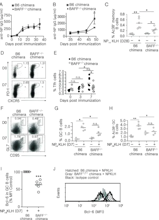
Previous studies have linked BAFF with an enhanced response to vaccination, suggesting that it plays a role in memory responses (43–45). To assess this, we generated BAFF−/− bone marrow chimeras by engrafting irradiated B6 mice with B6 (BAFF+/+) or BAFF−/− bone marrow. This approach limits BAFF deficiency to hematopoietic cells, allowing other sources of BAFF to maintain the peripheral B cell population (46). No differences in the spleen cellularity between B6 and BAFF−/− bone marrow chimeras were observed
(Supplemental Figure 1A), and the basal level (d0) of Tfh and GC B cells were not different. In BAFF−/− chimeras, we found that the primary IgG response to NP14KLH (Figure 1A) was comparable to B6 control chimeras. However, 7 days after secondary immunization (day 42), BAFF−/− bone marrow chimeras showed a 1.4-fold reduction in the levels of IgG, and 14 days after secondary immunization (day 49) the levels of IgG were reduced 2-fold compared to B6 chimeras (Figure 1B). Diminished production of IgG during the secondary response could reflect diminished class switch since BAFF can induce AID expression (47, 48). However, AID mRNA levels in B cells from B6 control and BAFF−/− chimeras were not different (Supplemental Figure 1B), suggesting that BAFF has a role other than in class switch.
BAFF−/− chimeras exhibit defects in the frequency of memory B, Tfh cells, and GC B cells
Rapid, high titer secondary immune responses require the activation of memory B cells (49). Although most IgG memory B cells do not require BAFF for maintenance (50), it is not known whether BAFF is important for their formation. To determine whether BAFF affects the frequency of memory B cells, we immunized (i.p.) BAFF−/− chimeras with NP14KLH and measured the frequency of NP-specific memory B cells (CD19+IgG+Ac38 Id+) on day 28 post-immunization. We found that immunization significantly increased the frequency of memory B cells in B6 and BAFF−/− bone marrow chimeras; however, the magnitude of the response was significantly lower in BAFF−/− chimeras (Figure 1C).
Author Manuscript
Author Manuscript
Author Manuscript
Tfh cells are critical in the early GC response and required for the differentiation of memory B cells (51, 52). It was possible that BAFF affected memory responses by influencing Tfh cells that in turn affected GC responses. To assess whether BAFF affects formation and/or maintenance of Tfh cells, B6 and BAFF−/− chimeras were immunized and the frequencies and numbers of Tfh cells (CXCR5+PD-1+CD4+) were quantitated on days 3 and 7 post-immunization (Figure 1D, 1E, and Supplemental Figure 1C). On day 3, the frequency of Tfh cells in B6 chimeras increased by 1.5-fold while in BAFF−/− chimeras it increased 1.2-fold. This suggests that BAFF does not play a significant role in the formation of Tfh cells. However on day 7 post-immunization, the frequency and number of Tfh cells in B6 chimeras increased an additional 2 fold, whereas their frequency in BAFF−/− chimeras did not change. This suggests that BAFF may support the expansion of Tfh cells after pre-Tfh cells transition to Tfh.
Germinal centers are necessary for the formation of high affinity, class-switched memory B cells (53, 54). To determine whether BAFF impacted the frequency of GC B cells, we enumerated CD19+GL-7+CD95+ GC B cells 7 days after immunization. In BAFF−/− chimeras, the frequency and number of total GC B cells (Figure 1F, 1G, and Supplemental Figure 1D), and the frequencies of Ag-specific (Figure 1H; CD19+Ac38+ GL-7+CD95+) GC B cells in BAFF−/− chimeras were significantly lower than those in B6 chimeras. Thus, BAFF significantly contributes to optimal antigen-specific GC responses.
BAFF acts at, or upstream of, Bcl-6 expression in B cells
Bcl-6 plays a critical role in initiating GC responses and committing activated B cells to a memory cell phenotype (17, 19). Thus, one possibility was that the loss of BAFF negatively affected Bcl-6 expression. To test this, we measured Bcl-6 levels in GC B cells after immunization. We found that on day 7, the levels of Bcl-6 in GC B cells from immunized BAFF−/− chimeras were decreased 40% compared to B6 chimeras (Figure 1I and 1J). The data show that hematopoietic cell-derived BAFF acts at, or upstream of, Bcl-6 expression in B cells. Collectively, our data indicate that BAFF impacts the formation of GC and memory B cells by increasing the expression of Bcl-6 in GC B cells, and indirectly through
maintaining the Tfh cell populations during the GC response.
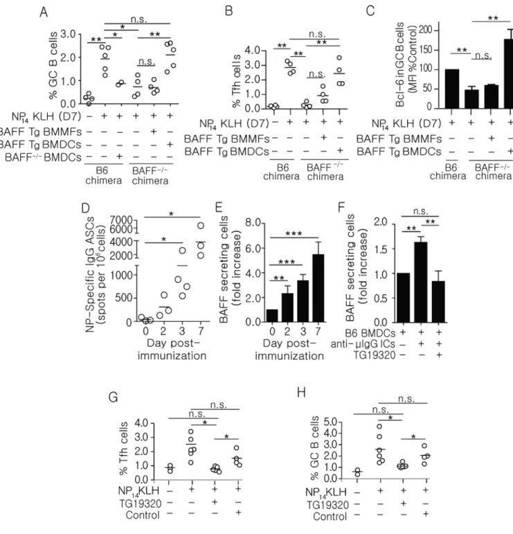
DC-derived BAFF regulates the frequency of GC B and Tfh cells
The BAFF−/− chimera model is characterized by the absence of BAFF in all hematopoietic cells. Among bone marrow-derived cell types that are capable of producing BAFF, myeloid cells are a major source of BAFF following infection or immunization (27, 43, 55). To determine whether myeloid cells are a sufficient source of BAFF during GC B cell and memory B cell fate decisions, we adoptively transferred bone marrow-derived dendritic cells (BMDCs) and bone marrow-derived macrophages (BMMFs) from BAFF transgenic (Tg) mice into BAFF−/− chimeras by s.c. injection at the time of immunization. We previously established that 70% of s.c. injected BMDCs migrated to the inguinal lymph nodes, and that the magnitude of the s.c. anti-NP response was comparable to i.p. immunization (data not shown). We found that constitutive expression of BAFF by Tg DCs, but not Tg MFs, restored the frequencies of GC B cells (Figure 2A), Tfh cells (Figure 2B), and the levels of Bcl-6 in GC B cells (Figure 2C) in BAFF−/− chimeras. Since these BAFF Tg MFs secrete
Author Manuscript
Author Manuscript
Author Manuscript
more BAFF, than BAFF Tg DCs (37), the responses were not due to higher production of BAFF by Tg DCs. Conversely, transfer of BAFF−/− DCs did not increase the frequencies of GC B cells (Figure 2A) indicating that the effects of the transfer on GC B cells were not due to an increased number of DCs, or to secretion of other cytokines made by the transferred DCs. This suggests that in addition to DCs presenting antigen during GC responses (56) DC-derived BAFF is also be important in directing the differentiation of GC cell populations.
The binding of immune complex to FcγRs induces BAFF secretion
Previous studies showed that exogenous ICs induce BMDCs to secrete a number of cytokines, including BAFF (57, 58). In another study, mice lacking the common gamma chain of the FcγRs (Fcγc) exhibited diminished secondary immune responses (59). Because our data suggest that DCs may promote secondary responses via BAFF, we postulated that ICs formed by the early IgG antibody response might induce DCs to secrete BAFF. This model requires that the early IgG response occur concurrently with, or precede BAFF secretion. To test this, we harvested spleens from B6 mice on days 2, 3, and 7 following NP14KLH immunization and used ELISpot to measure the numbers of antibody (IgG) secreting cells (ASCs) and BAFF-secreting DCs. NP-specific (IgG) ASCs were increased 7-fold by day 2 post-immunization and expanded to 90-7-fold over the course of 7 days (Figure 2D). Similarly, the number of splenic CD11c+ DCs that secreted BAFF increased 6-fold between days 2 and 7 (Figure 2E). Thus, secretion of Ig by B cells and production of BAFF by DCs occur concomitantly, beginning approximately 2 days following immunization. This supports the idea that IgG-ICs formed early in immune response contribute to the production of BAFF, which is required to optimize GC responses.
To further test the idea that IgG-ICs induce DCs to secrete BAFF, we blocked IgG-Fc:FcγRs interactions in vitro and assessed whether this impacted BAFF secretion by DCs.
Stimulation of B6 BMDCs with pre-formed IgG-ICs (IgM bound by anti-μ of IgG1 isotype) induced a 1.8-fold increase in BAFF secretion (Figure 2F). This was not unique to IgM/IgG ICs because preformed anti-NP ICs (NP-OVA bound by anti-NP of IgG1 isotype) induced a dose dependent 2.5-fold increase in BAFF secretion (Supplemental Figure 1E). Co-culture with a tetrameric tripeptide (TG19320) that blocks IgG Fc regions (39, 40) reduced the number of BAFF-secreting DCs to levels indistinguishable from unstimulated cells. To assess whether blocking Fc:FcγR interactions affected the adaptive immune response in
vivo, we administered TG19320 to B6 mice at the time of immunization and measured the
frequencies of GC B and Tfh cells. We found that co-administration of TG19320 with NP14KLH blocked an increase in GC B and Tfh cells on day 7 (Figure 2G and 2H). This indicates that the interactions between IgG-ICs and FcγRs are necessary for optimal GC responses and for the maintenance and/or expansion of newly formed Tfh cells in response to immunization. Collectively, these data identify a mechanism wherein IgG-ICs formed early in the immune response ligate FcγRs on DCs to induce BAFF secretion, which in turn contributes to optimal GC responses.
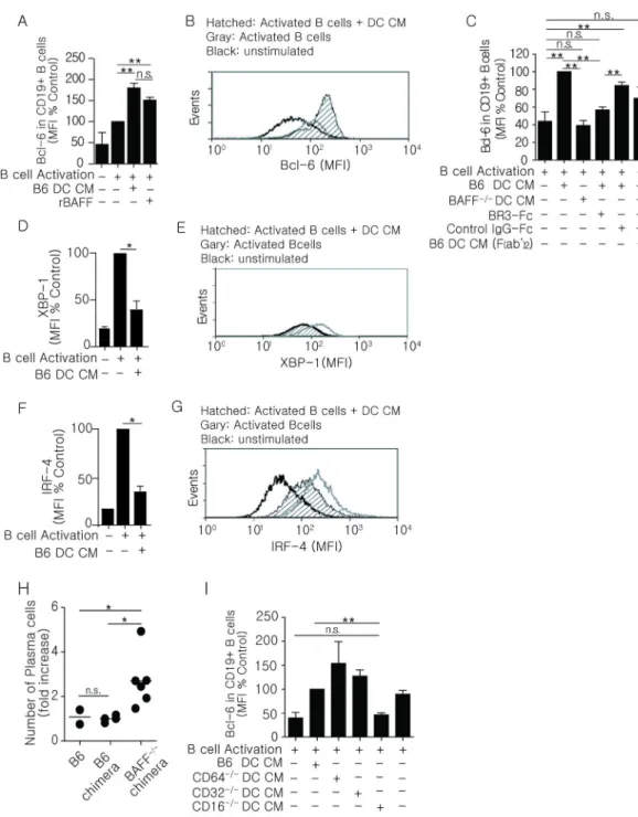
BAFF regulates the expression of Bcl-6 in activated B cells in vitro
To define whether BAFF plays a role in committing activated B cells to the memory compartment, we established an in vitro reconstitution system using the expression of Bcl-6
Author Manuscript
Author Manuscript
Author Manuscript
as a marker of memory B cell commitment. In this in vitro system, we used conditioned medium prepared from B6 BMDCs (DC CM) stimulated with preformed IgG1-ICs (Figure 2F) as a source of BAFF. This was based on in vivo findings that GC responses were dependent of BAFF produced by DCs (Figure 2A–C). To generate activated B cells, we used purified splenic B cells (B6) stimulated with anti-μ in combination with IL-4 and IL-5 to induce a low level of Bcl-6 expression (Figure 3A/B; B cell activation). Thus, changes in Bcl-6 as a consequence of DC CM or recombinant BAFF could be measured. We found that DC CM increased Bcl-6 expression in activated B cells approximately 2-fold compared to cells cultured in the absence DC CM (Figure 3A and 3B). These levels were comparable to those achieved with recombinant BAFF. Further, CM from BMDCs stimulated with F(ab’)2 -containing ICs was not as efficient as CM stimulated with IgG-ICs (intact Fc regions) at inducing Bcl-6. In this in vitro system, Bcl-6 expression was not elevated in B cells cultured with DC CM where BAFF was neutralized with BR3-Fc, or where DC CM was made from BAFF−/− BMDCs (Figure 3C). These data showed that DC-derived BAFF, induced by IgG-ICs, directly affects Bcl-6 expression in B cells activated in vitro.
As Bcl-6 levels become elevated, the PC program is attenuated (60, 61). To further validate the in vitro model, we measured intracellular IRF-4 and XBP-1 and found that DC CM diminished the levels of both transcription factors by 2.2-fold (Figure 3D–G), indicating that BAFF acts at or upstream of Bcl-6, directing B cell differentiation away from a PC fate. To test whether BAFF reduces PC differentiation in vivo, we enumerated splenic PCs from immunized B6 chimeras and BAFF−/− chimeras at day 5 (Figure 3H), a time when PCs normally appear in the spleen (62). We found that CD138+B220− cell numbers in BAFF−/− chimeras were increased 2.7-fold compared to B6 chimeras. This indicates that in vivo, BAFF decreases PC differentiation as the memory response initiated.
IgG-ICs bind CD16 during the anti-NP response
We reasoned that if FcγR stimulation promoted BAFF secretion, loss of the FcγR that binds IgG1-anti-NP ICs would negatively affect the ability of DC CM to promote Bcl-6. To assess this, we tested whether CMs from BMDCs derived from B6 mice, or mice deficient in CD64 (FcγRI−/−), CD32 (FcγRIIb−/−), CD16 (FcγRIII−/−), or CD16-2 (FcγRIV−/−) induced Bcl-6 in the in vitro reconstitution system described above. DC CM from CD16−/− mice failed to induce Bcl-6 expression, while CMs from all other FcγR deficient mice induced Bcl-6 to levels comparable to, or above those induced by B6 DC CM (Figure 3I). This suggests that ligation of CD16 on DCs is required for the expression of Bcl-6 in activated B cells during the anti-NP response.
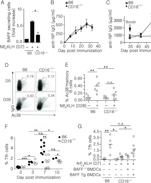
Impaired secondary responses in CD16−/− mice
Our in vitro data indicate that CD16 is responsible for inducing DCs to secrete BAFF after NP14KLH immunization. To test this in vivo, we quantitated the number of splenic BAFF secreting DCs in B6 and CD16−/− mice 7 days after immunization (Figure 4A). In the absence of CD16, we found that the number of BAFF secreting DCs was markedly diminished compared to wild type mice. This is consistent with the idea that CD16 is the FcγR responsible for initiating the IgG1-dominant immune response to NP14KLH (54). It is
Author Manuscript
Author Manuscript
Author Manuscript
likely that other Fc receptors would be utilized during immune responses that generate antibodies of IgG subclasses other than IgG1.
To test whether loss of CD16 in vivo impaired adaptive immune responses, we measured primary and secondary antibody responses in B6 and CD16−/− mice following NP
14KLH immunization. As in the BAFF−/− chimeric mice, the primary IgG response in CD16−/− mice was comparable to B6 mice (Figure 4B). However, in the secondary response, the levels of IgG in B6 mice increased 1.7-fold on day 42 (7 days after boost), and 4.5-fold on day 49 (14 days after boost), while IgG levels in the CD16−/− mice did not increase on days 42 or 49 (Figure 4C). This was not an indirect consequence of altered spleen cellularity or changes in the splenic cell populations due to CD16 deficiency since the frequencies of DCs, T, and B cells in CD16−/− mice were not different than B6 mice, and the number of splenocytes from B6 and CD16−/− mice were comparable (Supplemental Figure 1F). This indicates that CD16 plays a role in generating memory B cells and secondary immune responses to NP14KLH.
CD16−/− mice exhibit defects in forming GC and memory B cells and maintaining Tfh cells
The loss of DC-derived BAFF in CD16−/− mice supports a role for CD16 in the memory response to NP14KLH. To assess whether the diminished secondary response in CD16−/− mice reflects a reduction in memory B cells, we assessed the frequency of NP-specific memory B cells (CD19+Ac38+IgG+) in B6 and CD16−/− mice 28 days following immunization. CD16−/− mice showed a 2.4-fold decrease in the Ac38 Id+ IgG memory B cell population compared to immunized B6 mice (Figure 4D and 4E), indicating that CD16 regulates memory responses in part through BAFF production.
Data from immunized BAFF−/− chimeras suggests that BAFF contributes to B cell memory by affecting the GC B and Tfh cell populations. If the binding of IgG-ICs to CD16 were the predominate source of BAFF, then loss of CD16 would also diminish the GC B and Tfh pools. To assess this possibility, we quantitated Tfh cells (CD4+CXCR5+PD-1+) from immunized B6 and CD16−/− mice. After 3 days, CD16−/− mice had a comparable frequency of Tfh cells compared to immunized B6 mice (Figure 4F). However on day 7, CD16−/− mice had 3-fold fewer Tfh cells, similar to the defect in maintaining Tfh cells observed in
immunized BAFF−/− chimeras (Figure 1E). After 10 days, frequencies of Tfh cells decreased 2-fold in B6 mice, while the frequencies in CD16−/− mice were not changed compared to day 7. This suggests that BAFF plays a role in the expansion and/or
maintenance of Tfh cells during early GC responses, but that other factors also contribute. Adoptive transfer of BAFF transgenic, but not BAFF−/− BMDCs into immunized CD16−/− mice, restored the frequency of Tfh cells on day 7 to levels seen in B6 mice (Figure 4G), suggesting that the defect in CD16−/− mice was a consequence of reduced BAFF. These data demonstrate that CD16 ligation by IgG-ICs induces DCs to secrete BAFF and that CD16 is necessary for memory responses. Though our findings collectively show that binding of IgG-ICs to CD16 contributes to B cell memory through the effects of BAFF on Bcl-6 expression in GC B cells, and maintaining and/or expanding Tfh cells, sources of BAFF other than CD16, or factors other than BAFF may also play a role since loss of either BAFF or CD16 did not completely ablate these populations.
Author Manuscript
Author Manuscript
Author Manuscript
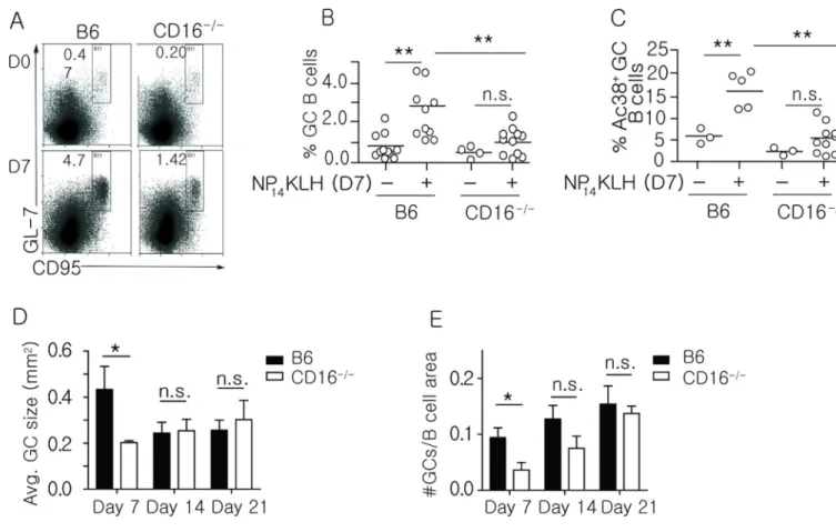
Loss of CD16 diminishes germinal center responses
To assess whether loss of CD16 diminished GCs, we measured the frequency of splenic GC B cells (CD19+GL-7+CD95+) and Ac38 Id+GC B cells 7 days after immunization. We found that both populations of GC B cells were significantly reduced in CD16−/− mice (Figure 5A– C). Consistent with the diminished frequency of GC B cells, we found that the GCs in CD16−/− mice were 54% smaller on day 7 post-immunization, but as the size of the B6 GCs declined (by day 14 and 21) the sizes became similar (Figure 5D and Supplemental Figure 2). The smaller size of the CD16−/− GC on day 7 was not the result of delayed kinetics because by day 28, the GCs in both B6 and CD16−/− mice were diminished, suggesting a comparable duration of the response (data not shown). We also found that immunized CD16−/− mice displayed a 2.3-fold reduction in the number of GCs on day 7; however, the differences were less apparent on day 14. By day 21, the CD16−/− mice had a similar number of GC structures (Figure 5E). Thus, although GCs form in the absence of CD16, they are reduced in number and size early during the immune response. This suggests that stimulation of CD16 is an early event that enhances the GC response and subsequently optimizes the formation of memory B cells.
CD16 and BAFF are required for the expression of Bcl-6 and the formation of GC and memory B cells
Our data indicate that CD16 and IgG-ICs are required for DCs to make BAFF in response to NP14KLH (Figure 4A). To address whether DC-derived BAFF was sufficient to restore the GC and memory B cell pools in the CD16−/− mice, we adoptively transferred BMDCs from BAFF Tg mice at the time of immunization. We found that BAFF Tg DCs restored the frequency of GC B cells on day 7 (Figure 6A), and the frequency of Ac38 Id+ memory B cells on day 28 (Figure 6B). This suggests that the lack of B cell memory responses in CD16−/− mice was due to lack of BAFF, and not to intrinsic defects in B cells. In contrast, BMDCs derived from BAFF−/− chimeras were unable to restore GC or memory B cells (Figure 6A and 6B). These results also emphasize that the defects observed in CD16 deficient mice are mediated by BAFF, since DCs from BAFF−/− chimeras have intact CD16.
Our data suggest that DC-derived BAFF acts at or upstream of Bcl-6 (Figure 1I–J, 2C, 3A– C) and downstream of CD16 (Figure 4A). Thus, the absence of CD16 should also lead to diminished Bcl-6 expression after NP immunization, and restoring DC-derived BAFF by BMDC transfer should restore Bcl-6 levels. To test this, we adoptively transferred BAFF Tg or BAFF−/− BMDCs into CD16−/−mice at the time of immunization. We found that 7 days after immunization, the expression levels of Bcl-6 in CD16−/− GC B cells were 60% lower compared to B6 controls. Transfer of BAFF Tg BMDCs, but not BAFF−/− BMDCs, restored Bcl-6 levels in CD16−/− GC B cells (Figure 6C). Thus, loss of CD16 reduces BAFF, which impacts Bcl-6 expression in GC B cells. Collectively, the data show that IgG-ICs induce DC-derived BAFF through ligation of CD16. BAFF promotes optimal B cell memory responses by inducing Bcl-6 expression in GC B cells and by maintaining and/or expanding Tfh cells.
Author Manuscript
Author Manuscript
Author Manuscript
Discussion
Interactions between T cells, B cells, and DCs are necessary for the proper execution of the adaptive immune response. This study identifies a previously unappreciated role for FcγRs, and BAFF in the early events of the GC response. We show that ICs formed during the early IgG response to NP14KLH induced the production of BAFF by DCs. BAFF acted at, or upstream of Bcl-6 to promote the optimal formation of GC B cells and to maintain and/or expand newly formed (day 7) Tfh cells. This series of events depends on the formation of IgG-ICs, suggesting that productive early antibody responses contribute to the optimal formation of B cell memory. This mechanism may ensure that an antigen-specific antibody response has occurred prior to initiating the events that promote B cell memory.
In the absence of CD16, the diminished formation of GC and memory B cells, and reduced expansion of Tfh cells (Figures 4–6), coupled with observations that mice deficient in the Fc common gamma chain (Fcγc) exhibit diminished secondary antibody responses (59) imply that IgG is required for optimal adaptive immune responses. We detected antigen-specific IgG by ELISpot within 2–3 days of immunization. The source of this IgG could be
extrafollicular PCs because these cells class switch (63) and produce significant local levels of IgG early in the immune response (64, 65). By days 14 and 21, the sizes and numbers of GCs were similar in B6 and CD16-deficient mice, suggesting that this mechanism is involved in pre- or early GC responses. This may also reflect a reduction in available antigen, which would reduce IC formation and make any differences mediated by IC:FcγR interactions less apparent. Consistent with a role for early GC events in the formation of B cells memory, we also found that in the absence of CD16, the average GC size and the number of GCs were diminished on day 7. Further, others showed that in the absence of soluble IgG, the kinetics of secondary GCs were disrupted (66). Thus, IgG plays a role in both primary and secondary GC responses.
Temporally, anti-NP IgG production by ASCs was coincident with BAFF production by DCs (Figure 2D–E). Although both DCs and MFs are among the major producers of BAFF (46, 67–69), we found that transfer of BAFF-producing DCs, but not MFs restored the numbers of GC B cells in BAFF−/− chimeras and CD16−/− mice. This may reflect the tripartite co-localization and interactions between DCs, T cells and B cells during adaptive immune responses. Though they are most well-known for presenting antigen to and activating T cells, DCs also directly modulate B cells through production of IC-induced cytokines like IFN, BAFF and IL-12 (55, 56, 71). Our data do not rule out a role for other immune cell types expressing CD16, like neutrophils, but highlight a key role for DCs as BAFF producers in T-dependent adaptive immune responses.
Because GC B cells, Tfh cells, or memory B cells were not completely absent in the BAFF−/− chimeras or the CD16−/− mice, it is also possible that BAFF from
non-hematopoietic cell sources contributes to the response, or that BAFF is not the only factor induced by IgG-ICs that is involved in early GC events. Early in the adaptive immune response, B cells interact with cognate T cells and co-stimulatory signals induce AID activation and class switch (70). These signals include CD40L, which is also required for GC formation (71). We found that BAFF−/− chimeras had normal AID expression and
Author Manuscript
Author Manuscript
Author Manuscript
primary IgG production, suggesting that cognate interactions mediated by CD40L were intact, possibly accounting for the small number of GC and memory B cells that remain despite the absence CD16 or BAFF. It is unlikely that BAFF and CD40L are completely redundant since diminished memory B cell responses were noted in both BAFF−/− chimeras (Figures 1 and 2) and CD40L deficient mice (72, 73).
It is unclear whether the observed role of BAFF occurs before, during, or after the initial interaction between cognate B and T cells. Because AID induction is intact in BAFF−/− chimeras, BAFF likely acts downstream of initial T:B interactions. Our in vitro data show that recombinant BAFF directly induced Bcl-6 in activated B cells, and DC CM (elicited under conditions that induce BAFF) diminished the expression of XBP-1 and IRF-4. This is supported by our in vivo data where increased numbers of PCs were evident in BAFF−/− chimeras. This suggests that BAFF acts on activated B cells at the time the PC phenotype is being diminished. Therefore, BAFF may act on B cells after cognate T cells help to reduce the PC phenotype, and either commit cells to the memory or GC pathways, or support the survival of precursors that will enter those pathways. This is consistent with studies showing that DC secretion of the PC-inducing cytokine, IL-12, is dampened by IC signaling (74).
Another signal required for a productive GC reaction is IL-21. It is possible that BAFF acts in concert with IL-21 to induce and/or sustain expression of Bcl-6 in GC B cells (75, 76). T cell-secreted IL-21 acts on B cells both during initial T:B interactions and after GCs are formed to promote either Blimp-1 or Bcl-6 expression, depending on the context (77). BAFF may serve as a contextual signal early in the adaptive response to direct B cells toward a GC fate. BAFF may also maintain Bcl-6 in B cells destined for the GC, allowing IL-21 from Tfh cells to take over as the response progresses.
Previous studies showed that cells with a Tfh phenotype appear by day 3 after immunization (10, 78). These cells migrate towards follicles where they interact with B cells at the T-B border (79) to promote continuous expression of Bcl-6, and entry of Tfh cells into the GC (15, 80, 81). Our data suggest that B cells and IgG-ICs are not involved in the formation of Tfh cells (day 3). This is consistent with previous studies showing that on days 1–3 post-immunization, the expression of Bcl-6 and CXCR5 in CD4+ T cells is independent of B cells, and hence independent of IgG (10, 56). Instead, BAFF acted downstream of the formation of Tfh cells, and stabilized and/or expanded the population between days 3 and 7. Thus, previous studies showing a role for B cells in maintaining Tfh cells (22, 78, 82) might reflect the need for B cell-elicited Ig, ICs, and DC-derived BAFF. The role of BAFF in maintaining the Tfh population may be direct, or may involve GC B cells. One possibility is that BAFF promotes the expression of ICOSL on B cells. Previous studies showed that signaling through BAFF-R regulates the expression of ICOSL on B cells (32, 33), thereby sustaining interaction between Tfh cells and B cells at the T:B border and within GCs (10, 11). This interaction could also stabilize the expression of Bcl-6 and the downstream molecules CXCR5 and PD1 (14, 15, 83) to maintain the “Tfh phenotype”. This possibility is supported by studies showing that the absence of ICOSL on B cells reduces the frequency of CXCR5+ CD4+ cells after immunization (15), that ICOS:ICOSL interaction prolong the engagement between B cells and Tfh cells (80), and that follicular bystander B cells support
Author Manuscript
Author Manuscript
Author Manuscript
the formation and/or maintenance of Tfh cells by providing ICOSL in an antigen-independent manner (84).
Because the interaction between GC B cells and Tfh cells is bidirectional, the maintenance of Tfh cells may have also had a role in sustaining GC B cells. Recent work described Tfh as a source of local BAFF within GC B cells that is required for affinity maturation (5). Tfh-derived BAFF, however, is not required for GC initiation or maintenance (5), and its role in memory B cell development remains unknown. Our work indicates that early production of BAFF is upstream of GC formation and memory B cell development. Interestingly, we observed a more complete loss of antigen-specific GC B cells (Ac38+) than total GC B cells in BAFF−/− chimeras and CD16−/− mice. This may reflect a loss of local BAFF within the GC due to diminished maintenance and/or expantion of Tfh cells. However, more work would be needed to determine whether affinity maturation is altered in these models.
Overall, our studies highlight a novel role for IgG-ICs and DC-derived BAFF in the GC response. Elucidating the events that initiate GC responses may impact our understanding of ICs and BAFF in autoimmunity. Systemic lupus erythematosus (SLE) is an autoimmune disease characterized by elevated levels of BAFF, autoantibody/autoantigen ICs, and multi-organ pathology. The formation of autoreactive memory is thought to be instrumental in driving long-lived PCs and sustaining autoantibody production (85, 86); however, the mechanisms that regulate memory formation to self-antigens are unclear. Our findings suggest that chronically high levels of ICs containing self-antigens could contribute to a break in B cell tolerance at the GC checkpoint. In SLE, elevated levels of circulating ICs may elevate BAFF and promote the GC response (85, 87, 88). This suggests that neutralization of BAFF in patients with SLE may affect both B cell survival and GC responses that are necessary in the formation of autoreactive B cells, T cell, and memory cells (67, 89, 90).
Supplementary Material
Refer to Web version on PubMed Central for supplementary material.
Acknowledgements
The authors would like to thank J. Sjef Verbeek for the CD16- and CD64-deficient mice, and Jeff Ravetch for CD16-2-deficient mice. We also thank Jin Kim (CDC/OID/NCIRD) for helpful discussions, the Flow Cytometry Core (NCI P30CA06086), and the Microscopy Services Laboratory (CA 16086-26) for their support.
References
1. Allen CD, Okada T, Cyster JG. Germinal-center organization and cellular dynamics. Immunity. 2007; 27:190–202. [PubMed: 17723214]
2. Victora GD, Nussenzweig MC. Germinal centers. Annu Rev Immunol. 2012; 30:429–457. [PubMed: 22224772]
3. Tew JG, Phipps RP, Mandel TE. The maintenance and regulation of the humoral immune response: persisting antigen and the role of follicular antigen-binding dendritic cells as accessory cells. Immunol Rev. 1980; 53:175–201. [PubMed: 7009404]
Author Manuscript
Author Manuscript
Author Manuscript
4. Victora GD, Schwickert TA, Fooksman DR, Kamphorst AO, Meyer-Hermann M, Dustin ML, Nussenzweig MC. Germinal center dynamics revealed by multiphoton microscopy with a photoactivatable fluorescent reporter. Cell. 2010; 143:592–605. [PubMed: 21074050] 5. Goenka R, Matthews AH, Zhang B, O'Neill PJ, Scholz JL, Migone TS, Leonard WJ, Stohl W,
Hershberg U, Cancro MP. Local BLyS production by T follicular cells mediates retention of high affinity B cells during affinity maturation. J Exp Med. 2014; 211:45–56. [PubMed: 24367004] 6. Crotty S. Follicular helper CD4 T cells (TFH). Annu Rev Immunol. 2011; 29:621–663. [PubMed:
21314428]
7. Vinuesa CG, Cyster JG. How T cells earn the follicular rite of passage. Immunity. 2011; 35:671– 680. [PubMed: 22118524]
8. Nutt SL, Tarlinton DM. Germinal center B and follicular helper T cells: siblings, cousins or just good friends? Nat Immunol. 2011; 12:472–477. [PubMed: 21739669]
9. Breitfeld D, Ohl L, Kremmer E, Ellwart J, Sallusto F, Lipp M, Forster R. Follicular B helper T cells express CXC chemokine receptor 5, localize to B cell follicles, and support immunoglobulin production. J Exp Med. 2000; 192:1545–1552. [PubMed: 11104797]
10. Choi YS, Kageyama R, Eto D, Escobar TC, Johnston RJ, Monticelli L, Lao C, Crotty S. ICOS receptor instructs T follicular helper cell versus effector cell differentiation via induction of the transcriptional repressor Bcl6. Immunity. 2011; 34:932–946. [PubMed: 21636296]
11. Ballesteros-Tato A, Leon B, Graf BA, Moquin A, Adams PS, Lund FE, Randall TD. Interleukin-2 inhibits germinal center formation by limiting T follicular helper cell differentiation. Immunity. 2012; 36:847–856. [PubMed: 22464171]
12. Johnston RJ, Choi YS, Diamond JA, Yang JA, Crotty S. STAT5 is a potent negative regulator of TFH cell differentiation. J Exp Med. 2012; 209:243–250. [PubMed: 22271576]
13. Johnston RJ, Poholek AC, DiToro D, Yusuf I, Eto D, Barnett B, Dent AL, Craft J, Crotty S. Bcl6 and Blimp-1 are reciprocal and antagonistic regulators of T follicular helper cell differentiation. Science. 2009; 325:1006–1010. [PubMed: 19608860]
14. Yu D, Rao S, Tsai LM, Lee SK, He Y, Sutcliffe EL, Srivastava M, Linterman M, Zheng L, Simpson N, Ellyard JI, Parish IA, Ma CS, Li QJ, Parish CR, Mackay CR, Vinuesa CG. The transcriptional repressor Bcl-6 directs T follicular helper cell lineage commitment. Immunity. 2009; 31:457–468. [PubMed: 19631565]
15. Nurieva RI, Chung Y, Martinez GJ, Yang XO, Tanaka S, Matskevitch TD, Wang YH, Dong C. Bcl6 mediates the development of T follicular helper cells. Science. 2009; 325:1001–1005. [PubMed: 19628815]
16. Kusam S, Toney LM, Sato H, Dent AL. Inhibition of Th2 differentiation and GATA-3 expression by BCL-6. J Immunol. 2003; 170:2435–2441. [PubMed: 12594267]
17. Dent AL, Shaffer AL, Yu X, Allman D, Staudt LM. Control of inflammation, cytokine expression, and germinal center formation by BCL-6. Science. 1997; 276:589–592. [PubMed: 9110977] 18. Fukuda T, Yoshida T, Okada S, Hatano M, Miki T, Ishibashi K, Okabe S, Koseki H, Hirosawa S,
Taniguchi M, Miyasaka N, Tokuhisa T. Disruption of the Bcl6 gene results in an impaired germinal center formation. J Exp Med. 1997; 186:439–448. [PubMed: 9236196]
19. Ye BH, Cattoretti G, Shen Q, Zhang J, Hawe N, de Waard R, Leung C, Nouri-Shirazi M, Orazi A, Chaganti RS, Rothman P, Stall AM, Pandolfi PP, Dalla-Favera R. The BCL-6 proto-oncogene controls germinal-centre formation and Th2-type inflammation. Nat Genet. 1997; 16:161–170. [PubMed: 9171827]
20. Tunyaplin C, Shaffer AL, Angelin-Duclos CD, Yu X, Staudt LM, Calame KL. Direct repression of prdm1 by Bcl-6 inhibits plasmacytic differentiation. J Immunol. 2004; 173:1158–1165. [PubMed: 15240705]
21. Shaffer AL, Lin KI, Kuo TC, Yu X, Hurt EM, Rosenwald A, Giltnane JM, Yang L, Zhao H, Calame K, Staudt LM. Blimp-1 orchestrates plasma cell differentiation by extinguishing the mature B cell gene expression program. Immunity. 2002; 17:51–62. [PubMed: 12150891] 22. Poholek AC, Hansen K, Hernandez SG, Eto D, Chandele A, Weinstein JS, Dong X, Odegard JM,
Kaech SM, Dent AL, Crotty S, Craft J. In vivo regulation of Bcl6 and T follicular helper cell development. J Immunol. 2010; 185:313–326. [PubMed: 20519643]
Author Manuscript
Author Manuscript
Author Manuscript
23. Linterman MA, Beaton L, Yu D, Ramiscal RR, Srivastava M, Hogan JJ, Verma NK, Smyth MJ, Rigby RJ, Vinuesa CG. IL-21 acts directly on B cells to regulate Bcl-6 expression and germinal center responses. J Exp Med. 2010; 207:353–363. [PubMed: 20142429]
24. Zotos D, Coquet JM, Zhang Y, Light A, D'Costa K, Kallies A, Corcoran LM, Godfrey DI, Toellner KM, Smyth MJ, Nutt SL, Tarlinton DM. IL-21 regulates germinal center B cell differentiation and proliferation through a B cell-intrinsic mechanism. J Exp Med. 2010; 207:365–378. [PubMed: 20142430]
25. Batten M, Groom J, Cachero TG, Qian F, Schneider P, Tschopp J, Browning JL, Mackay F. BAFF mediates survival of peripheral immature B lymphocytes. J Exp Med. 2000; 192:1453–1466. [PubMed: 11085747]
26. Mackay F, Schneider P, Rennert P, Browning J. BAFF AND APRIL: a tutorial on B cell survival. Annu Rev Immunol. 2003; 21:231–264. [PubMed: 12427767]
27. Balazs M, Martin F, Zhou T, Kearney J. Blood dendritic cells interact with splenic marginal zone B cells to initiate T-independent immune responses. Immunity. 2002; 17:341–352. [PubMed: 12354386]
28. Rahman ZS, Rao SP, Kalled SL, Manser T. Normal induction but attenuated progression of germinal center responses in BAFF and BAFF-R signaling-deficient mice. J Exp Med. 2003; 198:1157–1169. [PubMed: 14557413]
29. Schiemann B, Gommerman JL, Vora K, Cachero TG, Shulga-Morskaya S, Dobles M, Frew E, Scott ML. An essential role for BAFF in the normal development of B cells through a BCMA-independent pathway. Science. 2001; 293:2111–2114. [PubMed: 11509691]
30. Vora KA, Wang LC, Rao SP, Liu ZY, Majeau GR, Cutler AH, Hochman PS, Scott ML, Kalled SL. Cutting edge: germinal centers formed in the absence of B cell-activating factor belonging to the TNF family exhibit impaired maturation and function. J Immunol. 2003; 171:547–551. [PubMed: 12847217]
31. Yan M, Marsters SA, Grewal IS, Wang H, Ashkenazi A, Dixit VM. Identification of a receptor for BLyS demonstrates a crucial role in humoral immunity. Nat Immunol. 2000; 1:37–41. [PubMed: 10881172]
32. Watanabe M, Takagi Y, Kotani M, Hara Y, Inamine A, Hayashi K, Ogawa S, Takeda K, Tanabe K, Abe R. Down-regulation of ICOS ligand by interaction with ICOS functions as a regulatory mechanism for immune responses. J Immunol. 2008; 180:5222–5234. [PubMed: 18390703] 33. Hu H, Wu X, Jin W, Chang M, Cheng X, Sun SC. Noncanonical NF-kappaB regulates inducible
costimulator (ICOS) ligand expression and T follicular helper cell development. Proc Natl Acad Sci U S A. 2011; 108:12827–12832. [PubMed: 21768353]
34. Ou X, Xu S, Lam KP. Deficiency in TNFRSF13B (TACI) expands T-follicular helper and germinal center B cells via increased ICOS-ligand expression but impairs plasma cell survival. Proc Natl Acad Sci U S A. 2012; 109:15401–15406. [PubMed: 22949644]
35. Nimmerjahn F, Lux A, Albert H, Woigk M, Lehmann C, Dudziak D, Smith P, Ravetch JV. FcgammaRIV deletion reveals its central role for IgG2a and IgG2b activity in vivo. Proc Natl Acad Sci U S A. 2010; 107:19396–19401. [PubMed: 20974962]
36. Hazenbos WL, Gessner JE, Hofhuis FM, Kuipers H, Meyer D, Heijnen IA, Schmidt RE, Sandor M, Capel PJ, Daeron M, van de Winkel JG, Verbeek JS. Impaired IgG-dependent anaphylaxis and Arthus reaction in Fc gamma RIII (CD16) deficient mice. Immunity. 1996; 5:181–188. [PubMed: 8769481]
37. Gavin AL, Duong B, Skog P, Ait-Azzouzene D, Greaves DR, Scott ML, Nemazee D. deltaBAFF, a splice isoform of BAFF, opposes full-length BAFF activity in vivo in transgenic mouse models. J Immunol. 2005; 175:319–328. [PubMed: 15972664]
38. Takai T, Ono M, Hikida M, Ohmori H, Ravetch JV. Augmented humoral and anaphylactic responses in Fc gamma RII-deficient mice. Nature. 1996; 379:346–349. [PubMed: 8552190] 39. Fassina G, Verdoliva A, Palombo G, Ruvo M, Cassani G. Immunoglobulin specificity of
TG19318: a novel synthetic ligand for antibody affinity purification. J Mol Recognit. 1998; 11:128–133. [PubMed: 10076825]
Author Manuscript
Author Manuscript
Author Manuscript
40. Marino M, Ruvo M, De Falco S, Fassina G. Prevention of systemic lupus erythematosus in MRL/lpr mice by administration of an immunoglobulin-binding peptide. Nat Biotechnol. 2000; 18:735–739. [PubMed: 10888840]
41. Han S, Zheng B, Takahashi Y, Kelsoe G. Distinctive characteristics of germinal center B cells. Semin Immunol. 1997; 9:255–260. [PubMed: 9237932]
42. Holl EK, O'Connor BP, Holl TM, Roney KE, Zimmermann AG, Jha S, Kelsoe G, Ting JP. Plexin-D1 is a novel regulator of germinal centers and humoral immune responses. J Immunol. 2011; 186:5603–5611. [PubMed: 21464091]
43. Bergamin F, Vincent IE, Summerfield A, McCullough KC. Essential role of antigen-presenting cell-derived BAFF for antibody responses. Eur J Immunol. 2007; 37:3122–3130. [PubMed: 17935087]
44. Kanagavelu SK, Snarsky V, Termini JM, Gupta S, Barzee S, Wright JA, Khan WN, Kornbluth RS, Stone GW. Soluble multi-trimeric TNF superfamily ligand adjuvants enhance immune responses to a HIV-1 Gag DNA vaccine. Vaccine. 2012; 30:691–702. [PubMed: 22146759]
45. Tertilt C, Joh J, Krause A, Chou P, Schneeweiss K, Crystal RG, Worgall S. Expression of B-cell activating factor enhances protective immunity of a vaccine against Pseudomonas aeruginosa. Infect Immun. 2009; 77:3044–3055. [PubMed: 19364838]
46. Gorelik L, Gilbride K, Dobles M, Kalled SL, Zandman D, Scott ML. Normal B cell homeostasis requires B cell activation factor production by radiation-resistant cells. J Exp Med. 2003; 198:937– 945. [PubMed: 12975458]
47. He B, Santamaria R, Xu W, Cols M, Chen K, Puga I, Shan M, Xiong H, Bussel JB, Chiu A, Puel A, Reichenbach J, Marodi L, Doffinger R, Vasconcelos J, Issekutz A, Krause J, Davies G, Li X, Grimbacher B, Plebani A, Meffre E, Picard C, Cunningham-Rundles C, Casanova JL, Cerutti A. The transmembrane activator TACI triggers immunoglobulin class switching by activating B cells through the adaptor MyD88. Nat Immunol. 2010; 11:836–845. [PubMed: 20676093]
48. Kim HA, Seo GY, Kim PH. Macrophage-derived BAFF induces AID expression through the p38MAPK/CREB and JNK/AP-1 pathways. J Leukoc Biol. 2011; 89:393–398. [PubMed: 21169521]
49. Ahmed R, Gray D. Immunological memory and protective immunity: understanding their relation. Science. 1996; 272:54–60. [PubMed: 8600537]
50. Scholz JL, Crowley JE, Tomayko MM, Steinel N, O'Neill PJ, Quinn WJ 3rd, Goenka R, Miller JP, Cho YH, Long V, Ward C, Migone TS, Shlomchik MJ, Cancro MP. BLyS inhibition eliminates primary B cells but leaves natural and acquired humoral immunity intact. Proc Natl Acad Sci U S A. 2008; 105:15517–15522. [PubMed: 18832171]
51. Tangye SG, Ma CS, Brink R, Deenick EK. The good, the bad and the ugly - TFH cells in human health and disease. Nat Rev Immunol. 2013; 13:412–426. [PubMed: 23681096]
52. McHeyzer-Williams M, Okitsu S, Wang N, McHeyzer-Williams L. Molecular programming of B cell memory. Nat Rev Immunol. 2011; 12:24–34. [PubMed: 22158414]
53. Blink EJ, Light A, Kallies A, Nutt SL, Hodgkin PD, Tarlinton DM. Early appearance of germinal center-derived memory B cells and plasma cells in blood after primary immunization. J Exp Med. 2005; 201:545–554. [PubMed: 15710653]
54. Takahashi Y, Dutta PR, Cerasoli DM, Kelsoe G. In situ studies of the primary immune response to (4-hydroxy-3-nitrophenyl)acetyl. V. Affinity maturation develops in two stages of clonal selection. J Exp Med. 1998; 187:885–895. [PubMed: 9500791]
55. Schneider P. The role of APRIL and BAFF in lymphocyte activation. Curr Opin Immunol. 2005; 17:282–289. [PubMed: 15886118]
56. Goenka R, Barnett LG, Silver JS, O'Neill PJ, Hunter CA, Cancro MP, Laufer TM. Cutting edge: dendritic cell-restricted antigen presentation initiates the follicular helper T cell program but cannot complete ultimate effector differentiation. J Immunol. 2011; 187:1091–1095. [PubMed: 21715693]
57. Boule MW, Broughton C, Mackay F, Akira S, Marshak-Rothstein A, Rifkin IR. Toll-like receptor 9-dependent and -independent dendritic cell activation by chromatin-immunoglobulin G
complexes. J Exp Med. 2004; 199:1631–1640. [PubMed: 15197227]
Author Manuscript
Author Manuscript
Author Manuscript
58. Li X, Su K, Ji C, Szalai AJ, Wu J, Zhang Y, Zhou T, Kimberly RP, Edberg JC. Immune opsonins modulate BLyS/BAFF release in a receptor-specific fashion. J Immunol. 2008; 181:1012–1018. [PubMed: 18606652]
59. Goins CL, Chappell CP, Shashidharamurthy R, Selvaraj P, Jacob J. Immune complex-mediated enhancement of secondary antibody responses. J Immunol. 2010; 184:6293–6298. [PubMed: 20439912]
60. Vasanwala FH, Kusam S, Toney LM, Dent AL. Repression of AP-1 function: a mechanism for the regulation of Blimp-1 expression and B lymphocyte differentiation by the B cell lymphoma-6 protooncogene. J Immunol. 2002; 169:1922–1929. [PubMed: 12165517]
61. Shaffer AL, Yu X, He Y, Boldrick J, Chan EP, Staudt LM. BCL-6 represses genes that function in lymphocyte differentiation, inflammation, and cell cycle control. Immunity. 2000; 13:199–212. [PubMed: 10981963]
62. Todd DJ, McHeyzer-Williams LJ, Kowal C, Lee AH, Volpe BT, Diamond B, McHeyzer-Williams MG, Glimcher LH. XBP1 governs late events in plasma cell differentiation and is not required for antigen-specific memory B cell development. J Exp Med. 2009; 206:2151–2159. [PubMed: 19752183]
63. William J, Euler C, Christensen S, Shlomchik MJ. Evolution of autoantibody responses via somatic hypermutation outside of germinal centers. Science. 2002; 297:2066–2070. [PubMed: 12242446]
64. Chan TD, Gatto D, Wood K, Camidge T, Basten A, Brink R. Antigen affinity controls rapid T-dependent antibody production by driving the expansion rather than the differentiation or extrafollicular migration of early plasmablasts. J Immunol. 2009; 183:3139–3149. [PubMed: 19666691]
65. Toellner KM, Gulbranson-Judge A, Taylor DR, Sze DM, MacLennan IC. Immunoglobulin switch transcript production in vivo related to the site and time of antigen-specific B cell activation. J Exp Med. 1996; 183:2303–2312. [PubMed: 8642339]
66. Anderson SM, Hannum LG, Shlomchik MJ. Memory B cell survival and function in the absence of secreted antibody and immune complexes on follicular dendritic cells. J Immunol. 2006;
176:4515–4519. [PubMed: 16585539]
67. MacLennan I, Vinuesa C. Dendritic cells, BAFF, and APRIL: innate players in adaptive antibody responses. Immunity. 2002; 17:235–238. [PubMed: 12354377]
68. Nardelli B, Belvedere O, Roschke V, Moore PA, Olsen HS, Migone TS, Sosnovtseva S, Carrell JA, Feng P, Giri JG, Hilbert DM. Synthesis and release of B-lymphocyte stimulator from myeloid cells. Blood. 2001; 97:198–204. [PubMed: 11133761]
69. Schneider P, MacKay F, Steiner V, Hofmann K, Bodmer JL, Holler N, Ambrose C, Lawton P, Bixler S, Acha-Orbea H, Valmori D, Romero P, Werner-Favre C, Zubler RH, Browning JL, Tschopp J. BAFF, a novel ligand of the tumor necrosis factor family, stimulates B cell growth. J Exp Med. 1999; 189:1747–1756. [PubMed: 10359578]
70. Vinuesa CG, Linterman MA, Goodnow CC, Randall KL. T cells and follicular dendritic cells in germinal center B-cell formation and selection. Immunol Rev. 2010; 237:72–89. [PubMed: 20727030]
71. Xu J, Foy TM, Laman JD, Elliott EA, Dunn JJ, Waldschmidt TJ, Elsemore J, Noelle RJ, Flavell RA. Mice deficient for the CD40 ligand. Immunity. 1994; 1:423–431. [PubMed: 7882172] 72. Renshaw BR, Fanslow WC 3rd, Armitage RJ, Campbell KA, Liggitt D, Wright B, Davison BL,
Maliszewski CR. Humoral immune responses in CD40 ligand-deficient mice. J Exp Med. 1994; 180:1889–1900. [PubMed: 7964465]
73. Grewal IS, Xu J, Flavell RA. Impairment of antigen-specific T-cell priming in mice lacking CD40 ligand. Nature. 1995; 378:617–620. [PubMed: 8524395]
74. Kim SJ, Caton M, Wang C, Khalil M, Zhou ZJ, Hardin J, Diamond B. Increased IL-12 inhibits B cells' differentiation to germinal center cells and promotes differentiation to short-lived
plasmablasts. J Exp Med. 2008; 205:2437–2448. [PubMed: 18809711]
75. Linterman MA, Beaton L, Yu D, Ramiscal RR, Srivastava M, Hogan JJ, Verma NK, Smyth MJ, Rigby RJ, Vinuesa CG. IL-21 acts directly on B cells to regulate Bcl-6 expression and germinal center responses. J Exp Med. 2010; 207:353–363. [PubMed: 20142429]
Author Manuscript
Author Manuscript
Author Manuscript
76. Zotos D, Coquet JM, Zhang Y, Light A, D'Costa K, Kallies A, Corcoran LM, Godfrey DI, Toellner KM, Smyth MJ, Nutt SL, Tarlinton DM. IL-21 regulates germinal center B cell differentiation and proliferation through a B cell-intrinsic mechanism. J Exp Med. 2010; 207:365–378. [PubMed: 20142430]
77. Zotos D, Tarlinton DM. Determining germinal centre B cell fate. Trends Immunol. 2012; 33:281– 288. [PubMed: 22595532]
78. Baumjohann D, Okada T, Ansel KM. Cutting Edge: Distinct waves of BCL6 expression during T follicular helper cell development. J Immunol. 2011; 187:2089–2092. [PubMed: 21804014] 79. Kitano M, Moriyama S, Ando Y, Hikida M, Mori Y, Kurosaki T, Okada T. Bcl6 protein
expression shapes pre-germinal center B cell dynamics and follicular helper T cell heterogeneity. Immunity. 2011; 34:961–972. [PubMed: 21636294]
80. Baumjohann D, Preite S, Reboldi A, Ronchi F, Ansel KM, Lanzavecchia A, Sallusto F. Persistent antigen and germinal center B cells sustain T follicular helper cell responses and phenotype. Immunity. 2013; 38:596–605. [PubMed: 23499493]
81. Crotty S, Johnston RJ, Schoenberger SP. Effectors and memories: Bcl-6 and Blimp-1 in T and B lymphocyte differentiation. Nat Immunol. 2010; 11:114–120. [PubMed: 20084069]
82. Kerfoot SM, Yaari G, Patel JR, Johnson KL, Gonzalez DG, Kleinstein SH, Haberman AM. Germinal center B cell and T follicular helper cell development initiates in the interfollicular zone. Immunity. 2011; 34:947–960. [PubMed: 21636295]
83. Akiba H, Takeda K, Kojima Y, Usui Y, Harada N, Yamazaki T, Ma J, Tezuka K, Yagita H, Okumura K. The role of ICOS in the CXCR5+ follicular B helper T cell maintenance in vivo. J Immunol. 2005; 175:2340–2348. [PubMed: 16081804]
84. Xu H, Li X, Liu D, Li J, Zhang X, Chen X, Hou S, Peng L, Xu C, Liu W, Zhang L, Qi H. Follicular T-helper cell recruitment governed by bystander B cells and ICOS-driven motility. Nature. 2013; 496:523–527. [PubMed: 23619696]
85. Dorner T, Giesecke C, Lipsky PE. Mechanisms of B cell autoimmunity in SLE. Arthritis Res Ther. 2011; 13:243. [PubMed: 22078750]
86. Odendahl M, Jacobi A, Hansen A, Feist E, Hiepe F, Burmester GR, Lipsky PE, Radbruch A, Dorner T. Disturbed peripheral B lymphocyte homeostasis in systemic lupus erythematosus. J Immunol. 2000; 165:5970–5979. [PubMed: 11067960]
87. Dong W, Zhu P, Wang Y, Wang Z. Follicular helper T cells in systemic lupus erythematosus: a potential therapeutic target. Autoimmun Rev. 2011; 10:299–304. [PubMed: 21111068] 88. Luzina IG, Atamas SP, Storrer CE, daSilva LC, Kelsoe G, Papadimitriou JC, Handwerger BS.
Spontaneous formation of germinal centers in autoimmune mice. J Leukoc Biol. 2001; 70:578– 584. [PubMed: 11590194]
89. Liu Z, Davidson A. BAFF and selection of autoreactive B cells. Trends Immunol. 2011; 32:388– 394. [PubMed: 21752714]
90. Coquery CM, Wade NS, Loo WM, Kinchen JM, Cox KM, Jiang C, Tung KS, Erickson LD. Neutrophils contribute to excess serum BAFF levels and promote CD4+ T cell and B cell responses in lupus-prone mice. PLoS One. 2014; 9:e102284. [PubMed: 25010693]
Author Manuscript
Author Manuscript
Author Manuscript
Figure 1. BAFF−/− chimeras have defective memory and GC B cells, diminished Bcl-6 levels and decreased frequency of Tfh cells
(A) B6 and BAFF−/− chimeric mice were immunized (i.p) with 100 mg NP14KLH in alum. Serum IgG anti-NP responses in B6 and BAFF−/− chimeric mice were measured by ELISA on days 7, 14, 21, 28, and 35 post immunization. n=4–6 mice per group over 3 experiments. (B) Serum IgG anti-NP levels measured on days 39, 42, and 49 (4, 7, and 14 days) following boost of 100 mg of soluble NP14KLH on day 35. n = 3–6 mice over 3 experiments. (C) The frequency of CD19+Ac38+ IgG+ B cells was determined by flow cytometry from the spleens
Author Manuscript
Author Manuscript
Author Manuscript
of B6 and BAFF−/− chimeras immunized for 28 days. The number of splenocytes from B6 and BAFF−/− chimeras was comparable. n = 4–6 mice over 4 experiments. (D and E) The frequency of CD4+CXCR5+PD-1+ T cells from B6 and BAFF−/− chimeric mice was measured on days 0, 3 and 7 following immunization. n = 3–6 mice per time point over 2 experiments. (F and G) The frequency of CD19+, GL-7+, CD95+ GC B cells was measured by flow cytometry on day 7 post-immunization. n = 3–5 mice over 3 experiments. (H) The frequency of CD19+Ac38+CD95+GL-7+ B cells was measured on day 7 post-immunization. n = 3–5 mice over 3 experiments. (I and J) Relative expression of Bcl-6 in GC B cells (CD19+CD95+GL-7+) from B6 and BAFF−/− chimeras measured 7 days after NP
14KLH immunization (i.p.). n = 4–6 mice per group over 2 experiments. Bars display mean (C, E, G, H, and I), error bars indicate standard deviation (A and B). ** p ≤ 0.01, *p ≤ 0.05, n.s. = not significant.
Author Manuscript
Author Manuscript
Author Manuscript
Figure 2. Secretion of BAFF by DCs restores Bcl-6 levels and the frequency of GC B and Tfh cells
(A–C) BAFF Tg BMDCs, BMMFs, or BAFF−/− BMDCs (8 × 10⁶) were injected (s.c.) into B6 or BAFF−/− chimeric mice that were simultaneously immunized (s.c.) with NP14KLH. On day 7, inguinal lymph nodes were harvested and the frequencies of (A) GC B cells (GL-7+CD95+ cells in CD19+ B cells), (B) Tfh cell (CXCR5+PD-1+ cells in CD4+ T cells), and (C) Bcl-6 expression in GC B cells from inguinal lymph nodes was measured by flow cytometry. n = 3–5 over 3 experiments. (D) On days 2, 3, and 7 following immunization, 1 × 10⁶ splenocytes from B6 mice were plated on NP-BSA-coated ELISpot plates. After 18
Author Manuscript
Author Manuscript
Author Manuscript
hours, NP-specific ASCs were quantitated. n = 3–4 mice per time point over 3 experiments. (E)1 × 10⁶ purified CD11c+ cells from B6 mice immunized for 2, 3, and 7 days were plated on BR3-Fc coated ELISpot plates. After 60 hours, the number of BAFF secreting cells was enumerated. n = 3–6 mice per time point over 3 experiments. BAFF ELISpots ranged from 32-137 (D0-D7). (F) BAFF secreting cells enumerated from B6 BMDCs (2.5 × 10⁵) cultured for 24 hrs in the presence or absence of anti-μ ICs, with or without Fc blocking peptide (TG19320; 50 µg/ml). n = 3–10 over 6 experiments. BAFF ELISpots ranged from 531–1051 (untreated and treated with IgG-ICs ± TG19320). (G–H) B6 mice were
immunized with NP14KLH and dosed with 15–30 mg/kg of Fc blocking peptide (TG19320) or an unrelated scrambled peptide (control) via i.p. injection. On day 7, the frequency of Tfh cells (CD4+CXCR5+PD1+, G) and GC B cells (CD19+GL7+CD95+, H) was determined. n = 3–6 over 2 experiments. Bars display mean (A, B, D, G, and H), and error bars indicate standard error of the mean (C, E, and F). **p ≤ 0.01, *p ≤ 0.05, n.s. = not significant.
Author Manuscript
Author Manuscript
Author Manuscript
Figure 3. DC-derived BAFF regulates the expression of Bcl-6 in cultured B cells
Purified B6 B cells (1 × 10⁵) stimulated with anti-μ (30 µg/ml), IL-4 (25 ng/ml), and IL-5 (25 ng/ml) (designated as B cell activation) were co-cultured with DC CMs. Intracellular levels of key transcription factors (Bcl-6, XBP-1, IRF-4) were measured by flow cytometry on day 2. (A) Bcl-6 expression in B cells co-cultured with B6 DC CM or rBAFF (5 ng/ml). Flow analysis was restricted to live cells. (B) Representative histogram of Bcl-6 expression in B cells activated by DC CM as in (A). n = 3–4 mice over 4 experiments. (C) Purified B6 B cells were co-cultured with CM generated from B6 BMDCs treated with ICs containing
Author Manuscript
Author Manuscript
Author Manuscript
intact IgG (B6 DC CM) or F(ab’)2 of IgG (B6 DC CM (Fab’)2), BAFF−/− DC CM, or B6 DC CM neutralized with BR3-Fc (10 µg/ml) or control IgG-Fc (10 µg/ml). n = 4–7 over 4 experiments. (D–G) B cells were co-cultured with B6 DC CM. Intracellular levels of XBP-1 (D, E) and IRF-4 (F, G) were quantitated. n = 3 mice per group over 3 experiments.
Histograms are representative of 3 experiments and all flow analysis was done after gating on live cells. (H) Numbers of splenic plasma cells (CD138+B220−) were enumerated by flow cytometry in B6, B6 chimera, or BAFF−/− chimeras at day 5 following immunization. n = 1–3 mice per group over 2 experiments. (I) Bcl-6 expression in B cells cultured with DC CM from B6, CD64−/−, CD32−/−, CD16−/−, or CD16-2−/− (FcγRIV) mice. n=6–7 mice per group over 4 experiments. Bars display mean (H), and error bars indicate standard error of the mean (A, C, D, F, and I). **p ≤ 0.01, * p ≤ 0.05, n.s. = not significant.
Author Manuscript
Author Manuscript
Author Manuscript
Figure 4. CD16−/− mice have defective BAFF secretion, secondary antibody responses, frequency of memory B cell and Tfh cells
(A) BAFF secreting cells enumerated from purified CD11c+ cells that were isolated from B6 and CD16−/− mice on day 7. n = 3 over 3 experiments. B6 and CD16−/− mice were
immunized (i.p) with 100 mg NP14KLH in alum. Serum IgG anti-NP (B) primary responses measured by ELISA on days 0, 7, 14, 21, 28, and 35; serum IgG (C) secondary antibody levels measured on days 39, 42, and 49 (days 4, 7, and 14) following boost of 100 mg soluble NP14KLH on day 35. n = 4–8 mice over 3 experiments. (D and E) The frequency of