THE ROLE OF LIPASE MATURATION FACTOR 1 IN THE MATURATION OF LIPOPROTEIN LIPASE
Melissa A. Babilonia-Rosa
A dissertation submitted to the faculty at the University of North Carolina at Chapel Hill in partial fulfillment of the requirements for the degree of Doctor of Philosophy in the
Department of Biochemistry and Biophysics.
Chapel Hill 2016
ii © 2016
ABSTRACT
Melissa A. Babilonia-Rosa: The Role of Lipase Maturation Factor 1 in the Maturation of Lipoprotein Lipase
(Under the direction of Saskia Neher)
iv
ACKNOWLEDGEMENTS
I started graduate school at UNC looking for career development and I am happy
to say that I am headed to a slightly different path than I initially thought because I
discovered something new and exciting for me. During the past six years a lot of people
have helped me attain my goals and I am grateful for the time they allotted to my
training and for their support. First and foremost, I would like to thank my Ph.D. advisor
Dr. Saskia Neher. Her continuous guidance was instrumental to my improvement and
development as a research scientist. I am also thankful for Dr. Neher’s support through
my teaching internship at UNC. In addition to my advisor, I would like to thank the rest
of my thesis committee: Drs. Henrik Dohlman, Brian Kuhlman, Amy Shaub Maddox, and Rosalind Coleman. Thank you for your guidance through troubleshooting and making sure I had the appropriate controls. Additionally, our continued questions forced me to broaden my understanding of not only my own work but of other biochemical processes.
I am also thankful to past and present members of the Neher laboratory that have helped me complete this journey. Lindsey Broadwell has been an exceptional undergraduate during the past year. Most significantly, I injured my hand through repetitive motions collecting samples to wrap up my thesis and Lindsey was of great help finishing the last experiments under my guidance. Christopher (Brian) Garrett, Cassandra Hayne, and Melissa Ritchie were supportive of my happy fruitful science days as well as the not so good science days.
vi
and Robert Bagnell for teaching me about microscopy and data analysis. Additionally,
Stephanie Gupton, Courtney Winkle, and Benjamin Roberts (from the Neher laboratory)
helped with co-localization data analysis.
Special thanks to the current and past members of the Office of Graduate
Education: Joshua Hall, Patrick Brandt, Ashalla Freeman, Jessica Harrell, Rebekah
Layton, and Erin Hopper. They have all supported different aspects of my career
throughout my years in graduate school but I would like to thank Josh and Patrick for
the opportunity to travel twice to Puerto Rico to gain teaching experience by giving
science workshops at the UPR-Humacao while recruiting undergraduate students to
different programs at UNC. Beka gave me the opportunity to be one of the founders and
chairs of Future Science Educators or FuSE, a cohort for graduate students and
post-doctoral fellows interested in teaching intensive careers. Lastly, Ashalla for doing a
spectacular job of maintaining a community of minority students on campus that
supports each other through scientific and social events.
Lastly, I am grateful for my support group outside of graduate school. I would like
to thank Greg Palczewski and Ivy Babilonia for their continued support, especially in
high stress periods. I also thank my family especially Mayra and Nelly Rosa. Your
TABLE OF CONTENTS
LIST OF TABLES ... ix
LIST OF FIGURES ... x
LIST OF ABBREVIATIONS ... xi
INTRODUCTION ... 1
CHAPTER 1: The lipase gene family ... 1
Regulation of LPL activity by interacting proteins ... 7
Post-translational modifications of LPL ... 11
LMF1 is necessary for secretion of dimeric lipases ... 12
Mutations associated with severe hypertriglyceridemia ... 19
Current treatments for absence of LPL in the capillary lumen ... 19
Elevated TG levels and cardiovascular disease ... 20
Summary ... 21
PURIFICATION, CELLULAR LEVELS, AND CHAPTER 2: FUNCTIONAL DOMAINS OF LIPASE MATURATION FACTOR 1 ... 22
Introduction ... 22
Experimental procedures ... 24
Results ... 28
Discussion ... 39
NOVEL BINDING PARTNERS OF LMF1 SUGGEST A CHAPTER 3: ROLE IN OXIDATIVE FOLDING IN THE ER ... 41
Introduction ... 41
viii
Results ... 49
Discussion ... 71
CONCLUSIONS AND FUTURE DIRECTIONS ... 76
CHAPTER 4: Introduction ... 76
Future directions ... 82
Final remarks ... 86
LIST OF TABLES
x
LIST OF FIGURES
Figure 1.1 Model of an LPL monomer. ... 3
Figure 1.2 Role of lipoproteins and LPL in TG transport in the capillary lumen. ... 6
Figure 1.3 Schematic of LMF1 topology and summary of experimentally supported facts. ... 17
Figure 2.1 The N-terminal domain of LMF1 contributes to LPL maturation. ... 30
Figure 2.2 LMF1 truncation variants localize to the ER. ... 32
Figure 2.3 Topology of LMF1 truncations. ... 35
Figure 2.4 LMF1 purification and cellular levels. ... 38
Figure 3.1 LMF1 binds to LPL through its C-terminus. ... 50
Figure 3.2 Identification of new LMF1-interacting partners. ... 53
Figure 3.3 siRNA knockdown of novel LMF1-interacting partners affects LPL secretion. ... 61
Figure 3.4 Validation of novel LMF1 and LPL interacting partners ... 63
Figure 3.5 ERp44, ERdj5 and TRX interact with LMF1 in a thiol-dependent manner. ... 67
Figure 3.6 Cellular TRX levels and activity affect LPL, but not PL, secretion. ... 70
Figure 3.7 Schematic of LMF1 and DsbD. ... 75
Figure 4.1 ERp44 is not responsible for ER retention of LMF1. ... 77
Figure 4.2 LMF1 does not localize to ER exit sites. ... 79
LIST OF ABBREVIATIONS
aa amino acid
ANGPTL angiopoietin-like proteins Apo apolipoproteins
cld combined lipase deficiency
CM chylomicron
DMEM Dubelcco’s modified eagle medium DSP dithiobis-succinimidyl propionate DUF1222 domain of unknown function 1222 EL endothelial lipase
ER endoplasmic reticulum
ERdj5 endopasmic reditculum resident protein ERdj5 ERAD endoplasmic reticulum associated degradation ERp44 endoplasmic reticulum resident protein 44 ERp72 endoplasmic reticulum resident protein 72 FBS fetal bovine serum
FL full length
GAPDH glyceraldehyde-3-phosphate dehydrogenase
GPIHBP1 glycosylphosphatidylinositol anchored high-density lipoprotein binding protein 1
HDL high-density lipoprotein HL hepatic lipase
xii LMF1 lipase maturation factor 1 LPL lipoprotein lipase
MEF mouse embryonic fibroblasts NEM N-Ethylmaleimide
pBpa p –benzoylphenylalanine PDI protein disulfide isomerase PL pancreatic lipase
PPA protease protection assay PR progesterone receptor
SDS-PAGE sodium dodecyl sulfate-polyacrylamide gel electrophoresis TCA trichloroacetic acid
TG triglyceride
TXNIP thioredoxin-interacting protein TRX thioredoxin
UGGT1 glycoprotein glucosyltransferase 1 UGGT2 glycoprotein glucosyltransferase 2 VLDL very low-density lipoprotein
INTRODUCTION CHAPTER 1:
The lipase gene family
Lipases are hydrolases present in organisms ranging from prokaryotes to
humans that mediate the absorption, transport, storage, and mobilization of lipids. Here, we focus on the dimeric lipases lipoprotein lipase (LPL), hepatic lipase (HL), and
endothelial lipase (EL). These three lipases belong to the same gene family1,2. They reside in the capillary lumen, anchored to the endothelial lining where they hydrolyze lipids from lipoproteins, releasing fatty acids that can be absorbed by neighboring tissue for triglyceride (TG) and phospholipid clearance from the plasma. The three lipases differ in the tissues in which they are expressed and in their substrate specificity. LPL is mainly expressed in the heart, adipose tissue, and skeletal muscle but mRNA
expression has been reported in the brain, lung, adrenal gland, and placenta3-5. LPL activity has been detected in heart, adipocytes, muscle, lungs, kidney and brain6. HL is synthetized mainly in the liver but its also expressed in macrophages7. In contrast to LPL and HL which are expressed in different cell types but active at the endothelial lining of the capillary lumen, EL is synthetized by endothelial cells and northern-blot analysis across different human tissues show mRNA expression in the lungs, liver, placenta, and kidneys4. In terms of substrate specificity, LPL primarily hydrolyzes TG, EL is primarily a phospholipase, and HL can hydrolyze both substrates8.
2
Figure 1.1 Model of an LPL monomer.
I-TASSER22 was used to obtain a predicted 3D structure of human LPL. The catalytic triad (Ser132 - Asp156 - His241) is shown in yellow. The lid covering the catalytic triad is shown in magenta with the stick representation of disulfide bond at the end of the lid. Michael Lafferty generated this model based on sequence similarity to pancreatic lipase.
4
Lipoproteins are important for the transport of TGs within the capillary lumen. They consist of a hydrophobic core of TGs and cholesteryl esters, and a hydrophilic surface that consists of phospholipids, cholesterol, and proteins known as
apolipoproteins23,24. There are five types of lipoproteins synthetized in different tissues and categorized according to particle density; a property that changes with lipid and protein composition (Fig. 1.2A). Chylomicrons (CM) are the most buoyant lipoproteins as they have the most TGs in their core. They are synthesized in the small intestines by enterocytes from re-esterified fatty acids generated from the hydrolysis of dietary TGs by PL. Newly synthesized CMs enter the capillary lumen through the lymphatic system to deliver TG to peripheral tissues (Fig. 1.2B). Very-low density lipoprotein (VLDL) is the lipoprotein with the second highest TG content. The liver secretes VLDL to redistribute TG to adipose, heart, and muscle tissue. Upon TG hydrolysis by LPL in the capillary lumen, the TG content of CM and VLDL decreases by ~90% generating lipoproteins25 of higher density. Chylomicrons are released to the capillary lumen in the fed state
whereas VLDL is secreted upon fasting. CM are transformed into CM remnants and VLDL is transformed to intermediate-density lipoprotein (IDL) and subsequently to low-density lipoprotein (LDL). High-low-density lipoprotein (HDL) is critical for reverse
cholesterol transport as it moves cholesterol from peripheral tissues to the liver. Substrate specificity of the lipases expressed at the capillary lumen allows for different lipoprotein preference and in consequence, the role of these lipases in
6
A.
Figure 1.2 Role of lipoproteins and LPL in TG transport in the capillary lumen. A) Characteristics of lipoproteins as modified from Biggerstaff et. al. 2004. The orange cartoons are a visual representation of the particle size. B) CM and VLDL are TG dense lipoproteins synthetized in the small intestines and liver, respectively. Both of these lipoproteins are secreted into the capillary lumen where they interact with LPL for TG hydrolysis. The fatty acids released are used for energy production or storage in adipocytes, heart, and muscle.
B.
Regulation of LPL activity by interacting proteins
LPL is the rate-limiting enzyme for the hydrolysis of TG within the capillary lumen. LPL activity rises in white adipose tissue (WAT) after feeding while it declines in the heart and muscle; this leads to TG storage in adipocytes. The opposite effect is seen during fasting, LPL activity in the muscle and heart increases where the released fatty acids are used for storage or oxidized to generate energy. LPL within the capillary lumen is regulated by interacting partners such as apolipoproteins, angiopoietin-like proteins, and glycosylphosphatidylinositol-anchored high-density lipoprotein binding protein 1 (GPIHBP1).
Plasma lipoproteins contain apolipoproteins that interact with LPL and modulate its activity. Apolipoprotein C-II (ApoCII) and AV (ApoAV) are known to promote LPL activity whereas ApoCI and CIII inhibit LPL31. ApoCII is mainly synthesized in the liver and it is a 79 amino acid peptide constituent of CM, VLDL, LPL and HDL32. In vitro
activity assays with LPL purified from rat and ApoCII purified from VLDL or HDL
revealed that ApoCII is a cofactor for LPL33. Furthermore, individuals with mutations in ApoCII accumulate TGs in the plasma because LPL needs ApoCII for efficient CM and VLDL hydrolysis34,35. The N-terminal domain of ApoCII contains the lipid-binding domain and the C-terminal domain interacts with LPL35. Crosslinking experiments of ApoCII with bovine LPL revealed a region of 11 amino acids in the N-terminus of ApoCII that bind to LPL36.
8
APOCIII loss of function mutations in humans are associated with reduced TG levels and reduced risk for coronary heart disease40-42. However, ApoCI knockout mice only show a trend towards lower TG levels and it seems that ApoCI impairs the binding of VLDL to the VLDL receptor thus resulting in VLDL accumulation in mice overexpressing ApoCI43. Another possible explanation for the inhibition of LPL activity by this
apolipoproteins is LPL displacement from the lipoproteins44. Due to the discrepancy between in vitro and in vivo studies, further studies are needed to clarify the roles of ApoC1 and CIII in TG metabolism.
ApoAV is also predominantly expressed in the liver and it associates with VLDL and HDL45. Transgenic mice overexpressing ApoAV have a 66% reduction in TG levels while mice knockouts display a 4-fold increase in TG levels when compared to
controls46. Supporting the involvement of ApoAV in TG metabolism, loss of function mutations in humans lead to severe hypertriglyceridemia47,48. Two in vitro studies support the hypothesis that ApoA5 increases LPL activity45. Recombinant ApoAV stimulates LPL activity: 1) with addition of ApoCII and 2) with addition of VLDL isolated from mouse plasma. However, other in vitro studies do not support the hypothesis of ApoAV as an LPL activator. The conflicting data might result from utilization of different substrates. Other modes of action proposed for ApoAV have been intracellular
repression of VLDL synthesis or activation of receptor mediated lipoprotein endocytosis in the liver31. Further studies are necessary to further understand the role of ApoAV in TG metabolism.
coiled-coil domain and a C-terminal fibrinogen like domain. Three family members, ANGPTL3, 4 and 8, are known to alter TG metabolism by inhibition of LPL’s activity. ANGPTL3 and 4 are cleaved by protein convertases like FURIN releasing the N-terminal domain, which inhibits LPL. This cleavage is crucial for LPL inhibition. ANGPTL4 functions as a homo-oligomer and studies from our lab have revealed that ANGPTL4 is a reversible inhibitor of LPL50. ANGPTL8 lacks the C-terminal domain characteristic of the family but the N-terminal domain still shares 20% sequence identity with ANGPTL3 and 4,
suggesting similar functions51. This region includes the ~25 residues of ANGPTL3 and 4 known to bind LPL52. Adenoviral expression of ANGPTL8 in mice liver results in
increased plasma TG levels whereas co-expression of ANGPTL8 and 3 further
exacerbate TG accumulation; expression of ANGPTL3 alone does not alter TG levels53. Additionally, co-immunoprecipitation studies from plasma of these mice show that ANGPTL8 interacts with the N-terminal domain of ANGPTL3. Lastly, the N-terminal domain of ANGPTL3 is released into the media of hepatocytes co-expressing
ANGPTL8 and 3 whereas cells expressing only ANGPTL3 release full-length protein. Taken together, these findings suggest that ANGPTL8 inhibits LPL in an ANGPTL3-dependent manner.
ANGPTL3, 4, and 8 mediate LPL inhibition but they do so in different tissues and at different times in the fed-fasting cycle54. ANGPTL4 inhibits LPL in WAT while
ANGPTL4 and 8 work in the heart and skeletal muscle. Fasting upregulates ANGPTL4, which downregulates LPL’s activity in adipose tissue. Conversely, fasting
10
providing fatty acids to be oxidized for energy. After a meal, ANGPTL4 is downregulated while ANGPTL8 is upregulated. In consequence, LPL remains active in WAT and
inactive in the heart/muscle.
The mechanism by which LPL reaches the capillary lumen remained a mystery until GPIHBP1 was discovered in 201055. Immunohistochemical studies with a
fluorescently labeled antibody against GPIHBP1 revealed that GPIHBP1 is expressed in the capillary lumen in tissues from WT mice. In WT mice, LPL co-localizes with the GPIHBP1 and can travel to its site of action in the capillary lumen of these tissues. In mice lacking GPIHBP1, LPL becomes stuck in the interstitial spaces surrounding myocytes and adipocytes. This finding explains why GPIHBP1 deficient mice have severe hypertriglyceridemia56; LPL is not transported to the capillary lumen for
hydrolysis of chylomicrons and VLDL. Confirmation of the role of GPIHBP1 in transport came from transwell assays using rat heart microvessel endothelial cells expressing GPIHBP157. When LPL is added to the basolateral chamber of the transwells, it is
translocated to the apical chamber only in the presence of GPIHBP1. The acidic domain of GPIHBP1 is known to bind LPL58. A final transwell experiment confirmed that
Post-translational modifications of LPL
In order for LPL to be secreted in its active dimeric form, necessary
post-translational modifications include subunit assembly, glycosylation, and disulfide bond formation60. LPL is active as a noncovalent homodimer in a head to tail subunit
arrangement61,62. Similarly, HL and EL are also homodimers with a head to tail orientation63,64 while PL is active as a monomer65. Dimerization of LPL, HL, and EL occurs in the ER where two populations of lipases can be found: 1) aggregated
monomers that are inactive and eventually degraded and 2) active dimers that can exit the ER66,67.
Like other glycosylated proteins68, members of the lipase gene family contain a glycan chain covalently attached to the consensus sequence Asn-X-Ser/Thr.
Asparagine mutations at positions 43, 56, and 62 of LPL, HL, and EL respectively, result in missfolding defects leading to ER retention and abolishment of lipase activity69-71.
Sequence alignment of PL, LPL, HL, and EL shows these lipases share ten cysteines that are conserved across species1,4 suggesting these residues have a role in the structure and function of these lipases. Disulfide bond pairing was determined sequencing disulfide linked tryptic peptides confirming the formation of 5 disulfide bonds72. Site directed mutagenesis of eight conserved cysteines demonstrates that six of the cysteines are critical for LPL activity73. The first conserved cysteine pair,
12
activity when compared to WT. However, C418 is important for interaction with GPIHBP1 although disulfide bond formation is not required for the interaction75. In agreement with these findings, individuals with the C418Y LPL mutant have severe hypertriglyceridemia76.
LMF1 is necessary for secretion of dimeric lipases
The relationship between dimeric lipases and lipase maturation factor 1 (LMF1) was discovered in 1983 in mice with an autosomal recessive mutation named combined lipase deficiency (cld)77. Mice homozygous for the cld mutation (cld/cld) appear normal at birth but they die within 48 hrs postpartum due to progressive triglyceride
accumulation. However, heterozygous littermates (cld/wt) do not share this phenotype. Activity measurements of LPL and HL from heterozygous vs. homozygous cld mice revealed diminished lipase activity; subsequently impairment of EL activity was also demonstrated9. To understand why lipase activity levels are diminished in the plasma of
cld/cld mice, mRNA levels of LPL and HL were measured from heart and liver and compared to heterozygous littermates78. Because mRNA levels are comparable for homozygous and heterozygous mice, the cld mutation was thought to affect protein synthesis or post-translational modifications. Pulse-chase experiments in heart and liver slices demonstrate that the rate of [35S] methionine incorporation was slightly slower when comparing homozygous and heterozygous mice. However, cld/cld mice also showed lower rates of overall protein synthesis as well which seemed to be a result of the inability of these mice to use dietary triglycerides78. The next step to determine if the
mannose oligosaccharides as proteins emerge from the translocon. In the Golgi, high mannose units are removed and replaced by more complex sugars that are resistant to Endo H cleavage. As a result, Endo H treatment is used to probe accessibility of
mannose units, which is correlated to the location of the glycosylated protein in the secretory pathway. Glycosylated proteins that have moved past the medial/trans-Golgi compartment are Endo H resistant whereas glycosylated proteins in the ER are Endo H sensitive79. Endo H treatment of liver immunoprecipitated LPL revealed that cld/cld mice only have LPL sensitive to Endo H. Thus LPL in these mice does not make it to the medial/trans-Golgi. However, LPL in the cld/+ mice is both Endo H sensitive and
resistant, representing protein being processed in the ER (Endo H sensitive) and protein that has entered the secretory pathway (Endo H resistant). Similarly, HL
immunoprecipitations from homozygous and heterozygous livers followed by Endo H treatment reveal that HL also has a secretion defect in cld/cld mice; all the HL is Endo H sensitive78. Thus mRNA and protein levels of LPL and HL in cld mice are not affected but newly synthetized protein is retained in the ER, preventing Golgi processing and lipase secretion. However, Endo H testing of a different glycoprotein demonstrated that the cld mutation does not affect all N-linked glycoproteins thus the cld mutation does not confer a global failure in the glycosylation pathway78.
14
t haplotypes were key to remove early acting lethal mutations and avoid recombination suppression. These studies revealed that the cld mutation localizes to the haplotype tw73, a region containing 149 genes. Genes to be tested were narrowed down by two methods. First, preference was given to genes involved protein folding or protein retention in the ER because it was known from the Endo H studies that LPL and HL in
cld/cld mice does not move to the medial/trans-Golgi. Second, preference was given to genes demonstrating lower mRNA levels in homozygous vs. heterozygous cells
because nonsense or missense mutations might alter cld mRNA levels80. Eight
candidates were selected for co-expression with LPL in the cld/cld cell line derived and immortalized from mice hepatocytes and fibroblasts (MEF)81. Only one, Tmem112,
rescued LPL and HL activity at levels comparable to heterozygous mice and as a consequence it was renamed to lipase maturation factor 1 (LMF1). Confirming this designation, EL activity in mouse-derived cld fibroblasts is rescued by transfection of LMF19. Reverse transcription-PCR in MEFs from cld mice revealed an insertion of a murine retrovirus resulting in a C-terminal truncation in LMF1 of 214 residues81. The C-terminal domain of LMF1 contains a conserved domain of unknown function 1222 (DUF1222) but co-localization of LMF1 with the ER-membrane bound calnexin shows that LMF1 resides in the ER. This localization is consistent with a role in protein folding in the ER in agreement with the Endo H data from cld mice and the site of LPL
maturation.
The topology for mouse and human LMF1 was elucidated using transmembrane prediction methods in combination with biochemical methods82. There are five
C-terminus facing the ER lumen (Fig. 1.3). Consequently, C-terminal truncation variants of LMF1 were generated to study the role of the DUF1222 domain in LPL maturation as well as the interactions of LPL with the ER facing portions of LMF1 (loop A and C see Fig. 1.3). C-terminal truncations that entirely or partially remove the DUF1222 domain of LMF1 demonstrate these truncations still localize to the ER-lumen. However, neither construct restores LPL activity in the cld/cld cell line. Affinity purification of LPL or PL in complex with wild type (WT) or LMF1 variants shows that both lipases bind to loop C but not loop A. The studies with the C-terminal truncations of LMF1 confirmed the role of the DUF1222 domain in processing of LPL in the ER. Additionally, we learned that loop C does not confer LPL activity when it is in combination with loop A but it is necessary to bind LPL. It is likely that the loop C-LPL interaction is necessary for the folding role of the C-terminus of LMF1 on LPL. Lastly, affinity purification of EL and LMF1 complexes from HEK293 cells demonstrate that EL also physically interacts with LMF19 although it remains unknown if EL binds to loop C. In contrast to dimeric lipases, the activity of the monomeric PL is not reduced in cld cells, and PL does not bind to LMF19,83. Thus it has been suggested that LMF1 is important for the homodimerization of dimeric lipases.
16
localizes to residue W464. Similar to mice, the homozygous defect confers lipid accumulation whereas the subject’s son is heterozygous for the mutation and has normal TG levels. Functional analysis of both nonsense mutations in cld MEFs showed that the W464X mutation restores more LPL activity than the Y439X mutant, resulting in slightly better LPL secretion but not enough to prevent severe hypertriglyceridemia84.
Figure 1.3 Schematic of LMF1 topology and summary of experimentally supported facts.
18
Despite the similarity of the phenotype from the cld mice with LPL and LMF1 deficiency in humans, development of null LMF1 mice was needed to truly understand LMF1 deficiency in mice. The naturally occurring cld mutation in mice localizes to a variant of chromosome 17 known as the t haplotype, a region that contains mutations affecting several genes. As a result, the phenotype of the cld mice might include effects from other genes. Furthermore, the cld truncation variant of LMF1 expresses in HEK293 cells and localizes in the ER. Thus, the allele could be hypomorphic, which would allow some expression of ~60% of LMF1. In agreement with cld mice, LMF1 null mice
develop severe hypertriglyceridemia within 24 hrs after birth86. These mice also have a reduction in LPL and HL activities in the plasma. Their plasma lipoprotein profile
demonstrates the high TG levels are due to accumulation of chylomicrons and VLDL; TG accumulation as well the impairment of lipase activity is not observed in
Mutations associated with severe hypertriglyceridemia
Severe hypertriglyceridemia is a condition characterized by plasma triglyceride levels greater than 1,000 mg/dL (>11.3 mmol/liter)87. LPL deficiency results in
accumulation of CM and VLDL that leads to severe hypertriglyceridemia. LPL deficiency is an autosomal recessive disorder caused by loss of function mutations in the LPL gene. The frequency of LPL deficiency is 1 in half a million and it typically presents at childhood with abdominal pain, eruptive xanthomas, lipemia retinalis, and recurrent pancreatitis. Nearly 100 LPL mutations have been identified in humans. These
mutations can abolish LPL’s catalytic function or prevent interaction with ApoCII, ApoAV or GPIHBP1 and hence result in severe hypertriglyceridemia88-90. Mutations in genes that interact with LPL also result in severe hypertriglyceridemia. These include ApoCII, ApoAV, GPIHBP1, and LMF181,89.
In contrast to the great majority of LPL mutations found to date, population-based studies of LPL variants have revealed a cardioprotective effect for the nonsense mutant S447X. This mutation is found in 20% of the population where it is associated with reduced plasma TG and increased HDL cholesterol91,92. As discussed in the next section, this mutant is used as treatment for LPL deficiency.
Current treatments for absence of LPL in the capillary lumen
The usual treatment for people with LPL deficiency is a restricted low fat diet and the utilization of lipid lowering drugs like statins and fibrates. Statins are the main
20
mg/dl94. Fibrates are ineffective for LPL deficiency treatment because they promote LPL expression and repression of ApoC-III through activation of the peroxisome proliferator-activated receptors (PPAR-α)95. Additionally, a low fat diet does not eliminate disease progression and pancreatitis episodes. Gene therapy with intramuscular injections of the gain of function LPL mutation S447X has been shown to reduces plasma TG levels in clinical trials96. However, TG levels returned to base line after 18-31 months due to an immune response to the AAV1-capsid. A second clinical trial included
immunosupressants. Twenty-six weeks post treatment, LPL protein levels were
detected in 4 out of 7 samples while LPL activity was detected only in 397. Additionally, after 3-12 weeks of treatment, 50% of the subjects had a ≥40 reduction in plasma TG levels while two subjects had no improvement. More importantly, TG levels return to baseline 16-26 weeks post-treatment. Even thoughgene therapy with the LPL variant S447X was approved by the European Commission in 2012, the cost for therapy is over 1 million dollars which is not cost effective considering the fleeting effects of the
treatment98. A more effective treatment is needed for patients with LPL deficiency.
Elevated TG levels and cardiovascular disease
compounds that rescue proteins from degradation in the ER by serving as a folding template, restoring activity and trafficking pathways101,102. Treatment with
pharmacological chaperones for LPL could prevent risk of cardiovascular disease due to high lipid levels.
Summary
22
PURIFICATION, CELLULAR LEVELS, AND FUNCTIONAL DOMAINS CHAPTER 2:
OF LIPASE MATURATION FACTOR 11
Introduction
Lipoprotein lipase (LPL) plays a critical and complex role in lipid metabolism. LPL hydrolyzes triglycerides (TGs) from two classes of circulating lipoproteins, VLDLs and chylomicrons, in order to distribute free fatty acids to peripheral tissues. Biochemical deficiency of LPL activity is one well-established cause of hypertriglyceridemia, which is associated with increased risk of atherosclerosis, acute pancreatitis, and presence of metabolic syndrome103. Mutations in both LPL and its interacting partners can result in biochemical deficiency of LPL activity. Here we investigate how one of these interacting partners known as LMF1 promotes LPL activity.
Deleterious mutations in the gene for LMF1 result in severe
hypertriglyceridemia81,104. LMF1’s precise genetic location was only recently discovered81, but its role in promoting LPL activity is well established. Mice with a recessive mutation on chromosome 17 were severely deficient for the activity of LPL and the very closely related hepatic lipase (HL)77. This mutation was termed cld, for combined lipase deficiency77. Mice with homozygous disruptions in the LPL gene had phenotypes that were nearly indistinguishable from cld/cld mice (death within 48 hours of birth with extreme elevations of serum triglycerides85. However, the cld mutation
1The work referenced in this chapter has been published in: Babilonia-Rosa MA and Neher SB. (2014) Purification, cellular levels, and functional domains of lipase maturation factor 1.
clearly did not affect the LPL and HL structural genes, as these genes mapped to different chromosomes105. Furthermore, the amount of LPL protein present in tissues was not reduced, but its activity was106. LPL activity was reduced because the majority of the LPL was retained in the ER as inactive aggregates in cld/cld cells83. Recently, the
cld mutation was mapped to a gene coding for an ER-resident, transmembrane protein, and renamed LMF1, for lipase maturation factor 181. Subsequently, LMF1 was found to be important for the activity of a third dimeric lipase, endothelial lipase9.
Although LMF1 is vital for secretion of active, dimeric lipases, it is not clear how it promotes the exit of dimeric lipases from the ER. It is therefore important to determine which domains of LMF1 contribute to dimeric lipase maturation. Mapping studies of LMF1's domain architecture reveal that it has a total of five transmembrane domains with its N-terminus in the cytosol and its C-terminus in the ER lumen82. The loops
connecting these transmembrane domains are labeled A-D and are diagramed in Figure 1A. Recent data suggest that loop C and the C-terminus of LMF1 are important for dimeric lipase maturation82. The importance of LMF1's C-terminal, ER resident domain was established in studies of the original cld mutation and in patients with LMF1
24
Here, we sought to determine which of LMF1’s domains are essential for its function and how LMF1 interacts with LPL by measuring the cellular levels of both proteins. To determine if the C-terminal portions of LMF1 were sufficient to promote dimeric lipase maturation, we made N-terminal LMF1 truncation variants. We show that these LMF1 truncations are properly localized and oriented in ER membrane. However, expression of these constructs in cld/cld cells show that the entire LMF1 protein is required for maturation of LPL. We generated a high-affinity, polyclonal antibody using purified LMF1. We found that endogenous LMF1 levels are very low, and each LMF1 molecule promotes the maturation of at least 50 molecules of LPL.
Experimental procedures
Expression constructs
Constructs for the expression of CD3δ-YFP and CFP-CD3δ107 and mCherry-KDEL (mCh-mCherry-KDEL)108 have been described. The coding sequence of human LPL was amplified from pCMV-SPORT6-LPL (Open Biosystems) with a C-terminal V5 epitope tag and inserted into the NheI and XbaI sites of pIRES-EGFP (Addgene). For LMF1 variants, the cDNA for human LMF1 was obtained from Open Biosystems (ID
was amplified with an added C-terminal polyhistidine tag and inserted into the Spe1 and Xba1 sites of pFastbac1 (Invitrogen).
Cell lines, transfection, and media collection
COS-7, cld/cld and cld/wt110 cell lines were maintained at a split ratio of 1:10 in Dubelcco’s modified Eagle’s medium, 10% fetal bovine serum, 1%
penicillin/streptomycin, and 1% L-glutamine (complete medium). Transfections were performed with 2 mg of DNA and X-treme gene (Roche Applied Science) according to manufacturer’s instructions. COS-7 cells were transfected using Fugene 6 (Promega) with 0.3 mg of mCh-KDEL, 1.7 mg of LMF1 constructs, and 1 mg of CD3δ-YFP and CFP-CD3δ. Cells were transfected 24 hours after plating and harvested 24 h
post-transfection. For secretion experiments, the media of cld/cld or cld/wt cells was changed to complete media but with 1% FBS and 15 u/mL of heparin 3 hours prior collection, 600 µL media was used per 9.5 cm2 well.
Western blot analysis
26
Biotech) was used at 1:20,000. Rabbit anti-progesterone receptor (PR, Santa Cruz) was used at a 1:100 dilution. HRP-conjugated secondary antibodies were used at 1:5000. Westerns were developed using ADVANSTA WesternBright reagent (Bioexpress).
Indirect Immunofluorescence
COS-7 cells were plated to 70% confluency on glass coverslips. Twenty-four hours post-transfection, the cells were fixed with 4% paraformaldehyde in PBS for 15
min. Cells were washed three times with PBS then incubated for 10 min with 0.1% triton
X-100 and 100 mM glycine. Coverslips were washed three times with PBS, then
blocked with 2% BSA for 30 minutes, and incubate with anti-his antibody (1:200 in 2%
BSA) for 1 hour at room temperature. Secondary antibody (Alexa Fluor 488-conjugated
anti-mouse, Molecular Probe) was diluted 1:800 in 2% BSA and incubated for 1 hour at
room temperature in darkness. DAPI (Sigma-Aldrich) was used at 0.8 mg/mL for 10
minutes. After further washing in PBS, coverslips with cells were mounted facedown
onto glass slides (Fisher) using ProLong Gold Antifade (Molecular Probes). Cells were
examined at room temperature under a Zeiss LSM 710 confocal microscope with a 63X
oil/1.4 Plan Apo.
Protease protection assay (PPA)
Abcam) antibodies, respectively, at 1:5000. HRP-conjugated secondary antibodies were used at 1:20,000.
Protein purification
LPL-V5 was purified essentially as previously described61. LMF1 was expressed in SF9 cells. Bacmids generated as per manufacture’s instructions (Invitrogen) were transfected into SF9 cells using Xtreme gene (Roche). Baculovirus was amplified for three passages and used to infect SF9 cells. Infected cells were harvested after 72 hours, resuspended in Buffer 1 (20 mM Tris pH 7.5, 100 mM NaCl, and 5 mM
28
Antibody production
Antibodies against purified LMF1 were raised in chickens according to standard protocol (Covance) and purified as described112.
Genomic PCR
DNA was isolated from cld cell lines with DNeasy kit (Quiagen) according to manufactures directions. The primer sequences used to distinguish between WT LMF1 and cld LMF1 have been described81.
Results
The C-terminus of LMF1 is not sufficient for dimeric lipase maturation
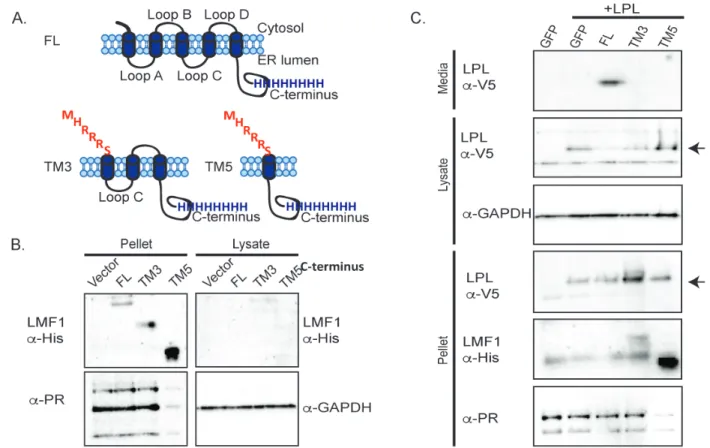
was highly expressed and so 5-fold less protein was loaded to allow detection of the other LMF1 variants. No LMF1 was detected in the soluble fraction, showing that the truncation versions localize to the membrane as expected.
Next, C-terminally V5-tagged LPL was co-expressed with each of the three LMF1 constructs in cld/cld cells to determine the minimal domain of LMF1 sufficient for LPL maturation. Heparin was added to the media to induce release of LPL from the cell surface into the media. LPL is expressed in cells with all LMF1 constructs, as it is detected in the lysate and pellet fractions (Fig. 2.1C). However, LPL is only secreted when FL LMF1 is present (Figure 2.1C, media fraction). Taking into account that only 1/5th of the TM5 pellet was loaded, there is more LPL in the pellets of TM3 and TM5 than in the FL LMF1 pellet fraction. This suggests that when FL LMF1 is not present, LPL can’t fold properly and is not able to exit the ER, as shown for cells lacking
LMF1113.Taken together, the release of LPL into the media only with FL LMF1 and the accumulation of LPL in the pellets when co-expressed with the TM3 and TM5
30
Figure 2.1 The N-terminal domain of LMF1 contributes to LPL maturation. A) A schematic of full length (FL) LMF1’s topology as well as both N-terminal
truncations. The MHRRRS sequence was added as an ER retention signal. B) Western blots against the C-terminal His tag show that FL, TM3 and TM5 truncations are
Localization of LMF1 constructs by fluorescence microscopy
32
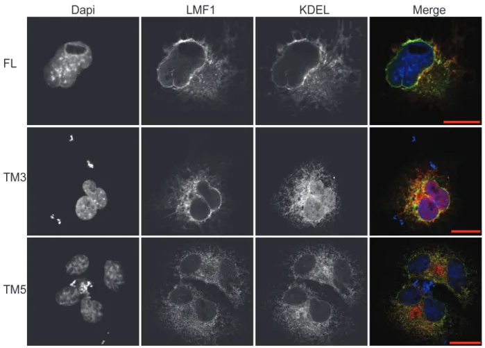
Figure 2.2 LMF1 truncation variants localize to the ER.
Immunocytochemistry of the LMF1 variants (green) was compared to the ER marker mCh-KDEL (red) in COS-7 cells. All LMF1 constructs show perinuclear staining
Membrane topology of LMF1 constructs
Truncation of the N-terminus of LMF1 could result in insertion of the protein into the ER with an incorrect orientation. We therefore performed a protease protection assay (PPA) to determine the orientation of the LMF1 truncations in the ER. In this assay, the plasma membrane is selectively permeablized with digitonin followed by trypsin treatment111. Trypsin will cleave membrane domains not protected within the lumen of organelles. COS-7 cells transfected with CD3δ-YFP (CD3δ with a YFP tag exposed to the cytosol) served as a positive control for trypsin cleavage (Fig. 2.3A, left panel). CD3δ with a CFP tag protected by the ER lumen (CFP-CD3δ) was a negative control to ensure that the organelles were preserved at the digitonin conditions used. Figure 3A shows complete loss of CD3δ-YFP signal within 2 minutes of trypsin
exposure whereas incubation with digitonin alone does not result in YFP degradation. The right panel of Figure 2.3A shows no degradation of the 50 kDa CFP-CD3δ band, confirming that the assay conditions leave the ER membrane intact.
34
Figure 2.3 Topology of LMF1 truncations.
A) A schematic of the CD3δ constructs used as positive and negative controls for trypsin cleavage, CD3δ-YFP and CFP-CD3δ respectively. The middle and right panels show the western blots for expression and the PPA. Lane 1 was mock (M) transfected, lane 2 shows untreated cells (U), lane 3 has 120 µM digitonin (D) addition for 3 minutes, and lane 4 has 20 µM of trypsin (T) for 2 minutes (after a 1 minute incubation with
36
Low level LMF1 expression is sufficient for LPL secretion.
The relative expression levels of LMF1 mRNA have been compared across different tissue types, but little is known about LMF1 protein levels81. We generated an antibody against LMF1 to better understand its cellular levels. LMF1 was expressed in SF9 cells and purified to homogeneity as shown by coomassie staining and gel filtration (Fig. 2.4A). Purified protein was used to raise antibodies against LMF1 in chickens. This antibody could detect as little as 17.5 fmoles of purified LMF1 (Fig. 2.4B).
However, Western blots using this antibody failed to detect a specific band for LMF1 in the cld/wt vs. cld/cld cells (Fig. 2.4B). When we transfected a plasmid containing LMF1
under the control of a CMV promoter into the cells, a band at the expected molecular weigh of LMF1 appeared (Fig. 2.4B).
Because we could not detect endogenous LMF1 in cld/wt cells, we wanted to ensure that both cell lines had the expected genotype. We confirmed the correct
genotype of these cells by analysis of the genomic DNA. Genomic DNA was harvested and tested by PCR for insertion of the murine endogenous retrovirus into intron 7 of
LMF1, which defines the cld mutation (Fig. 2.4C)81. Bands of the expected size were observed (Fig. 2.4C) indicating that LMF1 is present in the cld/wt but not the cld/cld
cells, as expected.
We cannot report the exact number of LMF1 molecules per cell, but can calculate an upper limit. The assays in figure 4B used 1 x 10⁴ cells. Additionally, the membrane fraction of a greater number (1 x 10⁶) of cld/cld and cld/wt cells was harvested,
38 Figure 2.4 LMF1 purification and cellular levels.
A) Gel filtration trace of purified LMF1 shown with a coomassie stained gel. B) Western blot showing that an antibody raised against LMF1 can detect as little as 17.5 fmole of the purified protein. Cells are mock transfected (M) or transfected with a plasmid
Discussion
Previous studies on LMF1 have focused on the C-terminus. In patients with defective LMF1, mutations are located in the C-terminus and in vivo co-IP’s show that loop C of LMF1 binds to LPL81,82,104. Based on these findings, we hypothesized that LMF1’s terminus is not sufficient for the maturation of dimeric lipases but the C-terminus in combination with loop C would promote lipase maturation. To test this hypothesis, we made truncations of LMF1 starting from the N-terminus. Because LPL must be properly folded to enter the secretory pathway83, we tested LPL secretion into the media in cells expressing only the N-terminally truncated variants of LMF1. These data show that FL LMF1 is required for the maturation process of LPL. Because the ER retention signal of LMF1 is not known, we used the ER retention signal of Iip33 to
ensure ER localization of the truncated variants. To demonstrate that the TM3 and TM5 LMF1 truncations did not compromise ER localization and the luminal orientation of the C-terminus, we co-localized LMF1 with an ER marker and performed a protease
protection assay. The results of these combined experiments demonstrate that TM3 and TM5 LMF1 have the same localization and topology as FL LMF1. However, they cannot promote the maturation of LPL, indicating that the N-terminus has an important, but unknown function.
40
present at a relatively low level compared to general chaperones like GRP78, calnexin, and calreticulin, which are present at over 1,000,000 molecules per cell, it must perform a specialized function114,115. Additionally, LMF1 has a catalytic role because each
molecule can efficiently promote the maturation of multiple molecules of LPL.
It is widely accepted that LMF1 is required for maturation of dimeric lipases, but it is not known if LMF1 coordinates the activities of other interacting partners. FL LMF1 is required for lipase maturation, but only loop C and the C-terminus have a known role. This suggests that other domains of LMF1 could interact with binding partners required for lipase maturation. Supporting this idea, we were unable to detect a strong, direct interaction between purified LPL and LMF1 in vitro (data not shown). Additionally, real time-PCR analysis shows that LMF1 mRNA is expressed at higher levels in tissues that lack dimeric lipases, such as the pancreas and testis, compared to tissues expressing dimeric lipases81. Future studies will be needed to determine if LMF1 requires
NOVEL BINDING PARTNERS OF LMF1 SUGGEST A ROLE IN CHAPTER 3:
OXIDATIVE FOLDING IN THE ER
Introduction
LPL is a secreted, dimeric lipase that hydrolyzes triglycerides present in the lipoproteins VLDL and chylomicrons. Because LPL is rate limiting for clearance of triglycerides from the plasma, LPL deficiency results in severe hypertriglyceridemia. As a secreted protein, LPL transits the ER where its two N-linked glycans and five disulfide bonds are processed. LPL also requires a specialized factor, LMF1, for proper folding and exit from the ER81. When LMF1 is absent, LPL forms disulfide-bonded aggregates in the ER, resulting in a phenotype similar to LPL deficiency83,116. LMF1 assists in the maturation of not only LPL but also the related, dimeric lipases endothelial lipase (EL) and hepatic lipase (HL)81. Pancreatic lipase, a sequence-related, but monomeric lipase, does not require LMF1.
LMF1 is a ER-resident membrane protein that has 5 membrane-spanning domains, resulting in two loops and a large C-terminal domain in the ER lumen117. Known deleterious mutations that prevent LPL secretion are located in the C-terminal, ER resident domain81,84. In vivo pull down experiments showed that loop C interacts with LPL117. Finally, our N- and C–terminal truncation experiments show that every ER resident loop of LMF1 is required for LPL maturation118.
42
(C216/C239, C264/C275, C278/C283)73. Protein folding and quality control in the endoplasmic reticulum (ER) requires not only oxidation of thiols to form disulfide bonds but also reduction of disulfide bonds to facilitate the degradation of misfolded proteins. Members of the disulfide isomerase (PDI) family mediate this thiol-disulfide exchange in eukaryotes119. Although glutathione powers the reduction of disulfide bonds120,
eukaryotes have long been hypothesized to utilize another source of reducing equivalents. The bacterial transmembrane electron transporter, DsbD transfers two electrons from thioredoxin to the protein disulfide isomerase DsbC for formation of nonconsecutive disulfide bonds121. DsbC must remain in a reduced state to initiate isomerization and DsbD is required to maintain DsbC in a reduced state122.
In order to understand how LMF1 promotes LPL maturation we took two approaches. First, we developed lipase chimeras with the LMF1-independent PL to determine what region of LPL interacts with LMF1. The chimeras revealed that the C-terminus of LPL binds to LMF1 and this interaction is important for dimeric lipase secretion. Second, we utilized crosslinkers and proteomics to identify binding partners of LMF1. We uncovered a number of ER-resident oxidoreductases as well as
thioredoxin (TRX), a cytosolic protein. We further show that TRX, ERp44, and ERdj5 bind to LMF1 in the absence of a crosslinker. In combination these data suggest that LMF1 acts like the bacterial protein DsbD, which shuttles electrons across the
Experimental procedures
Expression constructs
Constructs for wild type LMF1 and the terminal truncations with an 8X C-terminal polyhistidine tag were previously described123. Site directed mutagenesis of LMF1-His was employed to obtain C to A mutations via two single-primer reactions in parallel124. The orthogonal pair of suppressor tRNA (B. stearothermophilus) and tRNA synthetase (S. cervisiae) for photocrosslinking were provided by P. Schultz (The
Scripps Research Institute)125. AvrII and BsrGI sites were used to remove GFP from the pSWAN-GFP37TAG and insert LMF1 (Open Biosystems) with an 8X C-terminal
polyhistidine tag. The amber codon was incorporated in the pSWAN-LMF1 in residue F262 using site directed mutagenesis. Human LPL was excised from pCMV-SPORT6-LPL (Open Biosystems), a C-terminal V5 tag was added, and it was inserted into a
pcDNA5⁄FRT vector (Thermo Fisher Scientific) using HindIII/BcII sites. To clone the
lipase chimeras, thepcDNA5⁄FRT vector was cut with HindIII/Bcl1 and LMF1 was
inserted with an N-terminal polylinker containing XmaI, Pme1, and Cla1 sites. The
Pme1 and Cla1 sites were used to insert an IRES from the vector pIRES-EGFP
(Addgene) into pcDNA5⁄FRT. Human LPL or PL with a C-terminal V5 tag were inserted
using NheI/Xho1 or NheI/Xma1 respectively. PCR-driven overlap extension126was used
to create a chimera containing the N-terminal of LPL with the C-terminal of PL (LPL/PL)
and the reverse construct (PL/LPL). Both chimeras contained a C-terminal V5 tag and
were inserted in the modified pcDNA5⁄FRT. To make the plasmid for expression of
TXNIP, the cDNA sequence of human TXNIP was synthesized as a GBlock by IDT. It
44
Cell lines, transfection, and media collection
HEK 293 Flp-In™ (Thermo Scientific) cells were transfected with LPL-His or LMF1-His using Fugene 6 (Promega) and selected with 200 µg/mL of hygromycin. Regular HEK 293 cells were transiently transfected with Fugene 6 and 1 µg of DNA for LMF1-His and the respective lipase construct for the DSP crosslinking experiment in Figure 3.1C. Transfection of cld/cld cells on six well plates was performed with 2 µg of DNA and X-treme Gene HP127 according to manufacturer’s instructions. All cell lines were maintained in Dubelcco’s modified Eagle medium with 10% fetal bovine serum, 1% penicillin/streptomycin, and 1% L-glutamine (complete media).
siRNA Knockdowns
siRNA transfections were performed with 20 nM of RNA oligonucleotide and 7 µL of Lipofectamine RNAiMax (Life Technologies) per six-well plate and samples were collected 48 hours post-transfection. Validated Silencer Select siRNAs were obtained from Life Technologies: negative control No. 1 (4390843), ERp44 (s22965), PDI (s439), ERp72 (s18446), UGGT1 (132932), UGGT2 (112074), and ERdj5 (132773).
Knockdown was quantitated using a ChemiDoc MP Imaging System from Biorad and quantitated using Image Lab Software. The percent of remaining gene expression was calculated with the following formula:
Crosslinking and pull down assays
For pull down assays of LMF1-His from HEK Flp-In stable cell lines, two T-75 flasks per condition were seeded with 7.5 X 10⁶ cells. LMF1-His expression was induced with 2 µg/mL tetracycline the next morning. The next day, cells were trypsinized, washed with 1X PBS, crosslinked with 2 mM DSP (ProteoChem) with agitation for 30 minutes at room temperature, and quenched with 150 mM Tris pH 8. The cells were lysed with by douncing in buffer 1 (250 mM NaCL, 10 mM Tris pH 8, and 1 mM PMSF). Following centrifugation for 20 minutes at 13,000 RPM, the pellets were re-suspended by douncing with 1.4 mL of buffer 1. Lysates were incubated overnight with agitation and the addition of 20 mM fos-choline 12 (Anatrace). After centrifugation as above, 5% glycerol and 40 mM imidazole were added to the supernatant as well as Ni-NTA agarose beads (Qiagen). Binding was allowed for an hour at 40C with agitation. The beads were washed 3X with 4 mL of buffer 2 (1M NaCl, 10 mM Tris pH 8, 3 mM fos-choline 12, 5% glycerol, 40 mM imidazole, and 1 mM PMSF) in 11 mL poly-prep chromatography columns (Bio-Rad). LMF1 complexes were eluted with buffer 3 (250 mM NaCL, 10 mM Tris pH 8, 3 mM fos-choline 12, 400 mM imidazole, and 1 mM
PMSF). Eluate fractions were loaded on 8% SDS-PAGE, transferred to PVDF (Bio-Rad) for 75 minutes at 100V, and blocked with 5% nonfat milk or BSA.
IP’s of LPL-His from HEK Flp-In stable cell lines were carried out similarly.
Purification LMF1-His with lipases were carried out similarly but at a smaller scale. Cells were seeded at 6.5 X 10⁵ cells per well of 6 well plates (Denville); 3 wells were
46
For photocrosslinking with pBpa, HEK 293 Flp-In™ stably expressing LPL-V5 were plated in six-well plates at 10.5 X 10⁵ cells per well; four wells were utilized per sample. The next day, 1 µg of pBpa plasmid or LMF1-amber construct were transfected with Fugene 6. After 24 hours, the media was replaced with media containing 2 µg/mL of tetracycline, 0.5 mM pBpa (Chem-Impex International), and 0.3% of DMSO. Control wells lacked pBpa but contained 0.3% of DMSO. Cells were grown with pBPA for 24 hours. The media was changed to 1X PBS and the cells were kept on ice and
crosslinked with a UV lamp at a wavelength of 365 nm for 30 minutes at 2.5 cm from the lamp. LMF1 was purified from cells as described above.
Lipase secretion
HEK 293 Flp-In stables expressing lipase constructs were induced with 2 ug/mL of tetracycline 24 hrs before media collection. Three hours before media collection, lipase secretion was promoted by addition of 600 µL of DMEM with 1% FBS and 15 u/mL of heparin per 9.5 cm2 well. For cld/cld MEFs, lipase secretion was promoted 24 hrs post-transfection. Samples from lipase secretion experiments were separated using 12% SDS-PAGE.
Antibodies
HRP-conjugated anti-mouse or rabbit antibodies from Southern Biotech. Mouse anti-V5 antibody from Bio-Rad was diluted 1:5,000.
Mass Spectrometry
Gel slices containing purified LMF1 with pBPA or DSP-crosslinked binding partners were delivered to the Duke Proteomics facility for analysis by mass
spectrometry. Coomassie stained SDS-PAGE bands were subjected to standardized in-gel trypsin digestion (
48
Raw LC-MS/MS data files were processed in Proteome Discoverer (Thermo Scientific) and then submitted to independent Mascot searches (Matrix Science) against an SwissProt database (Human taxonomy) containing both forward and reverse entries of each protein (20,322 forward entries). Search tolerances were 5 ppm for precursor ions and 0.02 Da for product ions using trypsin specificity with up to two missed
cleavages. Carbamidomethylation (+57.0214 Da on C) was set as a fixed modification, whereas oxidation (+15.9949 Da on M) and deamidation (+0.98 Da on NQ) were
considered dynamic mass modifications. All searched spectra were imported into Scaffold (v4.3, Proteome Software) and scoring thresholds were set to achieve a peptide false discovery rate of 1% using the PeptideProphet algorithm.
NEM Modification
BSO, DTT, and tunicamycin sensitivity
To assess cell sensitivity to buthionine sulphoximine (BSO), DTT and
tunicamycin, first 1x10⁵ cld/cld or cld/wt were plated in a 6-well plate. The next day either a buffer control, 10 mM L-BSO, 2.5 µg/mL tunicamycin or 5 mM DTT was added. After 24 (tunicamycin or BSO) or 12 (DTT) hours of treatment, cells were removed from the plate with trypsin, stained with trypan blue and live cells were manually counted using a hemocytometer.
Results
LMF1 Binds to the C-terminus of LPL
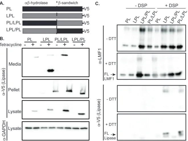
To determine the LMF1 binding site in LPL we generated two LPL-PL chimeras. These chimeras are diagramed in Figure 3.1A. The PL-LPL chimeras consist of the N-terminal domain of PL fused to the C-N-terminal domain of PL, whereas the LPL-PL
chimera is comprised of the N-terminal domain of LPL fused to the C-terminal domain of LPL. Both chimeras, as well as the WT LPL and PL controls, have C-terminal V5
epitope tags for uniform detection. As shown in Figure 3.1B, all of the lipase constructs are stably integrated into HEK293 cells under the control of a tetracycline inducible promoter. All of the proteins express, as shown in the lysate panel. However, only PL, LPL, and PL/LPL are secreted. Each variant was co-expressed with His tagged LMF1, crosslinked with the amine reactive crosslinker dithiobis-succinimidyl propionate (DSP), and affinity purified via LMF1’s His tag to detect interacting lipase constructs. Western blots showed that PL/LPL but not LPL/PL binds LMF1 (Fig. 3.1C). Thus, LMF1
50
Figure 3.1 LMF1 binds to LPL through its C-terminus.
Identification of novel LMF1 binding Partners
To better understand LMF1’s function we next set out to determine which
proteins it interacts with in the cell. We used two crosslinking reagents to covalently link LMF1 and its partners during extraction from the membrane. DSP is a homobifunctional, amine reactive crosslinker with a cleavable disulfide bond in its spacer arm. We also used a method for site-specific incorporation of an artificial amino acid into LMF1128. In this system, an orthogonal tRNA and aminoacyl-tRNA synthetase pair incorporates an unnatural amino acid into LMF1 at a nonsense codon in a position of our selection. We used the artificial amino acid p-benzoylphenylalanine (pBpa), which crosslinks to nearby proteins when UV light is applied to the cells129.
For DSP crosslinking, HEK293 cells stably expressing LMF1-His under the
52
Figure 3.2 Identification of new LMF1-interacting partners.
A) Western blot of LMF1-partners after DSP crosslinking and affinity tag purification using LMF1’s C-terminal His tag. Tetracycline induces LMF1 expression. Loading dye with 50mM DTT was used to break the disulfide bonds between LMF1 and its
54
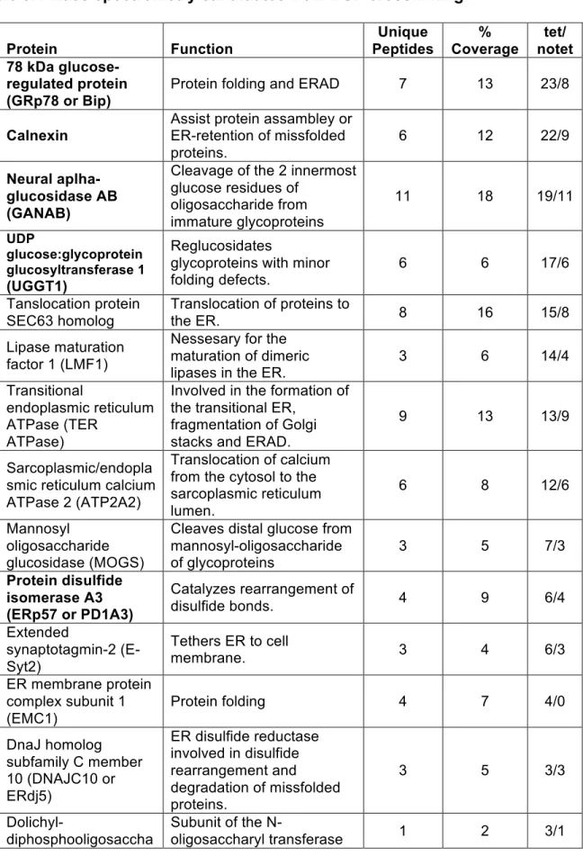
Over 300 proteins in total were identified in the two samples. We selected
candidates identified with 95% or greater confidence with more peptides identified in the LMF1-containing sample than a control sample. Additionally, for pBpa the predicted molecular weight of the LMF1-candidate protein complex had to fall within or exceed the molecular weight range corresponding to LMF1 plus the molecular weight of the
candidate. For both crosslinkers, we focused on proteins annotated as residing in the ER. Tables 3.1 and 3.2 list protein candidates that meet these criteria for DSP and pBpa respectively. Of note, we found SEL1L, a known LMF1-interacting protein, through our DSP crosslinking131. Additionally, in agreement with a proteomics study that isolated interacting partners of hepatic lipase, we found proteins associated to the
Table 3.1 Mass spectrometry candidates from DSP crosslinking. Protein Function Unique Peptides % Coverage tet/ notet 78 kDa
glucose-regulated protein (GRp78 or Bip)
Protein folding and ERAD 7 13 23/8
Calnexin
Assist protein assambley or ER-retention of missfolded proteins.
6 12 22/9
Neural aplha-glucosidase AB (GANAB)
Cleavage of the 2 innermost glucose residues of
oligosaccharide from immature glycoproteins
11 18 19/11
UDP
glucose:glycoprotein glucosyltransferase 1 (UGGT1)
Reglucosidates
glycoproteins with minor folding defects.
6 6 17/6
Tanslocation protein SEC63 homolog
Translocation of proteins to
the ER. 8 16 15/8
Lipase maturation factor 1 (LMF1)
Nessesary for the maturation of dimeric
lipases in the ER. 3 6 14/4
Transitional
endoplasmic reticulum ATPase (TER
ATPase)
Involved in the formation of the transitional ER,
fragmentation of Golgi stacks and ERAD.
9 13 13/9
Sarcoplasmic/endopla smic reticulum calcium ATPase 2 (ATP2A2)
Translocation of calcium from the cytosol to the sarcoplasmic reticulum lumen.
6 8 12/6
Mannosyl oligosaccharide glucosidase (MOGS)
Cleaves distal glucose from mannosyl-oligosaccharide
of glycoproteins 3 5 7/3
Protein disulfide isomerase A3 (ERp57 or PD1A3)
Catalyzes rearrangement of
disulfide bonds. 4 9 6/4
Extended
synaptotagmin-2 (E-Syt2)
Tethers ER to cell
membrane. 3 4 6/3
ER membrane protein complex subunit 1 (EMC1)
Protein folding 4 7 4/0
DnaJ homolog subfamily C member 10 (DNAJC10 or ERdj5)
ER disulfide reductase involved in disulfide rearrangement and degradation of missfolded proteins.
3 5 3/3
Dolichyl-diphosphooligosaccha
Subunit of the
56 Protein Function Unique Peptides % Coverage tet/ notet ride protein glycosyltransferase subunit 1 (RPN1)
(OST) complex which catalyzes the transfer of a high mannose
oligosaccharide to glycosilated proteins. Protein disulfide
isomerase A4 (ERp72 or PDIA4)
Catalyzes rearrangement of
disulfide bonds. 3 6* 3/0
Glucosidase 2 subunit
beta (GLU2B) N-glycan processing 3 7 3/0
Protein disulfide isomerase (PDIA1)
Catalyzes the formation, breakage and
rearrangement of disulfide bonds.
2 8 2/2
UDP
glucose:glycoprotein glucosyltransferase 2 (UGGT2)
Reglucosidates
glycoproteins with minor folding defects.
1 1 2/0
Protein sel-1 homolog 1 (Sel1L)
Involved in ER-associated
degradation (ERAD) 1 3 1/0
Protein disulfide isomerase A6 (ERp5 or PDIA6)
Catalyzes rearrangement of
disulfide bonds. 1 3 1/1
Coatomer subunit beta (COPB1)
Essential for the retrograde Golgi-to-ER transport of dilysine-tagged proteins
1 1 1/0
Exostosin-2 (EXT2) Protein glycosilation 1 1 1/0
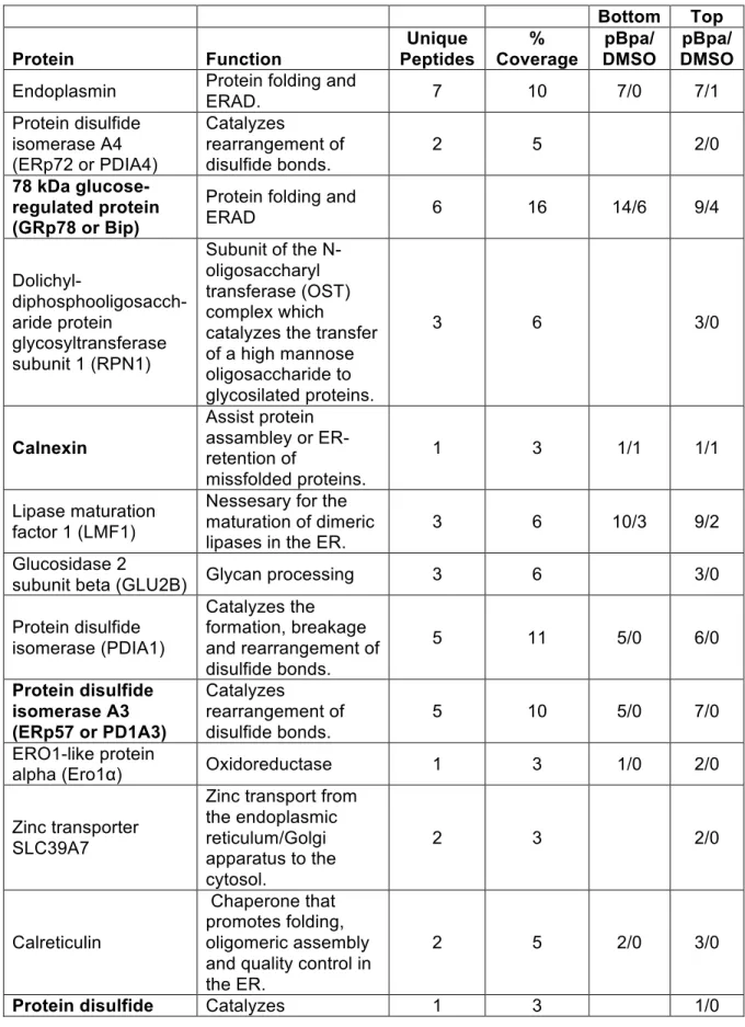
Table 3.2 Mass spectrometry candidates from pBpa crosslinking.
Bottom Top
Protein Function
Unique Peptides % Coverage pBpa/ DMSO pBpa/ DMSO
Endoplasmin Protein folding and ERAD. 7 10 7/0 7/1
Protein disulfide isomerase A4 (ERp72 or PDIA4)
Catalyzes
rearrangement of disulfide bonds.
2 5 2/0
78 kDa glucose-regulated protein (GRp78 or Bip)
Protein folding and
ERAD 6 16 14/6 9/4
Dolichyl- diphosphooligosacch-aride protein
glycosyltransferase subunit 1 (RPN1)
Subunit of the N-oligosaccharyl transferase (OST) complex which catalyzes the transfer of a high mannose oligosaccharide to glycosilated proteins.
3 6 3/0
Calnexin
Assist protein assambley or ER-retention of
missfolded proteins.
1 3 1/1 1/1
Lipase maturation factor 1 (LMF1)
Nessesary for the maturation of dimeric lipases in the ER.
3 6 10/3 9/2
Glucosidase 2
subunit beta (GLU2B) Glycan processing 3 6 3/0
Protein disulfide isomerase (PDIA1)
Catalyzes the formation, breakage and rearrangement of disulfide bonds.
5 11 5/0 6/0
Protein disulfide isomerase A3 (ERp57 or PD1A3)
Catalyzes
rearrangement of disulfide bonds.
5 10 5/0 7/0
ERO1-like protein
alpha (Ero1α) Oxidoreductase 1 3 1/0 2/0
Zinc transporter SLC39A7
Zinc transport from the endoplasmic reticulum/Golgi apparatus to the cytosol.
2 3 2/0
Calreticulin
Chaperone that promotes folding, oligomeric assembly and quality control in the ER.
2 5 2/0 3/0
58
Bottom Top
Protein Function
Unique Peptides % Coverage pBpa/ DMSO pBpa/ DMSO isomerase A6 (ERp5
or PDIA6)
rearrangement of disulfide bonds. ER resident protein
44 (ERp44)
Oxidative protein
folding. 2 5 2/0
Serpin H1 Chaperone that binds to collagen. 2 5 2/0
Peptidyl-prolyl cis-trans isomerase B (PPIaseB)
Catalyzes the cis-trans isomerization of proline.
2 9 2/0 4/0
Ras related protein Rab-1A
RAB1A regulates vesicular protein transport from the ER to the Golgi and on to the cell surface
3 18 3/0
Peptidyl prolyl cis-trans isomerase A (PPIA)
Catalyzes the cis-trans isomerization of proline.
4 28 4/0 4/0
Peptidyl-prolyl cis-trans isomerase FKBP1A
Catalyzes the cis-trans isomerization of proline.
1 12 1/0
Thioredoxin
Protein reduction by thiol-disulfide exchange.
60
Knockdown of Candidate LMF1-interacting Partners Reduces LPL secretion
We next tested the role of our novel LMF1 binding partners in LPL secretion. PL, which does not depend on LMF1 for exit from the ER, served as a negative control for changes to the ER protein-folding environment. siRNAs against ERp44, PDI, ERp72, UGGT2, ERdj5 and UGGT1 were compared to a scrambled siRNA control (NC). As shown in the media fraction of Figure 3.3A-B, knockdown of ERp44, ERp72, UGGT2, and ERdj5 decreased secretion of LPL much more dramatically than PL. Knockdown of UGGT1 slightly decreased LPL, but not PL, secretion. Unexpectedly, knockdown of PDI seemed to increase secretion of both LPL and PL. The knockdown of the LMF1
A) and B) HEK293 cells stably expressing LPL-V5 or PL-V5 were transfected with scrambled siRNA (NC) or siRNA against the newly identified LMF1-interacting partners. Lipase secretion into the media was induced with heparin, and media and lysate
fractions were collected for analysis. Media fractions were probed with 41a antibody for LPL and a C-terminus V5 tag for PL. Lysates were also probed for the respective LMF1 interacting-partners. GAPDH was used as a loading control. C) siRNA knockdowns shown in A and B were performed in triplicate. The intensity of bands for each knocked down protein was quantified using Image Lab, and the percent knockdown was
calculated as described in methods. D) Quantification of lipase secretion normalized to the cell number per sample and to secretion with scrambled siRNA. Just like for panel C, band intensity was quantified with Image Lab and error bars indicate the standard error.
Figure 3.3 siRNA knockdown of novel LMF1-interacting partners affects LPL secretion.