The Development of Polycationic Materials for Gene Delivery Applications
Peter Benjamin Uthe
A dissertation submitted to the faculty of the University of North Carolina at Chapel Hill in partial fulfillment of the requirements for the degree of Doctor of Philosophy in the
Department of Chemistry.
Chapel Hill 2010
Approved by
Advisor: Dr. Valerie Sheares Ashby
ii ©
2010
iii ABSTRACT
Peter Benjamin Uthe: The Development of Polycationic Materials for Gene Delivery Applications
(Under the direction of Dr. Valerie Sheares Ashby)
iv
v
TABLE OF CONTENTS
LIST OF TABLES………...…... ix
LIST OF FIGURES……….……. x
LIST OF SCHEMES……….…... xiii
LIST OF ABBREVIATIONS…………..………..…... xiv
LIST OF SYMBOLS.……...……….... xvii
Chapter 1: An Introduction to Gene Therapy... 1
1. Introduction to Gene Delivery... 2
1.1Delivery………...………...2
1.1.1 Naked DNA Delivery………....……….. 3
1.1.2 Viral Delivery……….………. 4
1.1.3 Synthetic Vector Delivery………...……….5
1.2 Non-Degradable Materials………...……..………....…… 8
1.2.1 Poly(ethyleneimine)……...……….………. 8
1.2.1 Poly(methacrylate) and Poly(Methacrylamide)……...……… 14
1.3 Degradable Materials...……...………...………… 19
1.3.1 Poly(β-amino ester)s…...………...… 20
vi
1.4 Biologically Derived Materials………...……….… 25
1.4.1 Chitosan…...……….………... 25
1.4.2 β-cyclodextrin……….………... 26
1.4.3 Sugar alcohol functionalized...………...………..…...27
1.5 Bioactive Modifications………...……….………... 28
2. Conclusions……….………... 29
Reference..………...……….……… 32
Chapter 2. Using Ionization Control to Enhance Gene Delivery... 35
1. Introduction ………... 36
2. Experimental Details………..……….……….. 38
2.1 Materials……….……….……….. 38
2.2 Instrumentation.………...……… 38
2.3 Diene Synthesis………..……... 39
2.4 Polymer Synthesis………...…………..……... 41
2.5 Polymer/Polyplex Characterization………...…………... 43
3. Results and Discussion………..………... 46
3.1 Synthesis………..……….. 47
3.2 Ionization Studies..………...………..50
3.3 DNA Binding Agarose Gels and Transfection………..… 53
3.4 Complex Studies..……….……… 57
3.4 Complex Studies..……….……… 58
vii
References………...……… 60
Chapter 3. Synthesis of Degradable Gene Therapy Materials with Decreased Toxicity... 62
1. Introduction …...…... 63
2. Experimental Details……..……….……….. 67
2.1 Materials….………...……….. 67
2.2 Instrumentation.………...……… 67
2.3 Monomer Synthesis………….……….…………. 68
2.5 Polyester Synthesis……….……….………….. 70
2.5 Polymer/Polyplex Characterization……….….……. 72
3. Results and Discussion………..………... 74
3.1 Synthesis………..……….. 75
3.2Polyplex/Biological Characterization………...……….. 77
4. Conclusions………...……… 81
References………...…….………... 82
Chapter 4. Developing Guanidine Functionalized Materials for Enhanced Cellular Uptake... 84
1. Introduction ………... 85
2. Experimental Details………...……….. 89
2.1 Materials and Instrumentation……….……….. 89
2.2 Bromoisoprene………...……… 89
2.2 Hydroxyisoprene………..………….………..……... 91
viii
3.1 Bromoisoprene……….……….. 95
3.2 Hydroxyisoprene……..……….………. 97
4. Conclusions…………..……….……… 100
References……….……….………... 102
Chapter 5. General Conclusions……….……….………. 104
Appendix A... 110
Appendix B... 174
ix
LIST OF TABLES
Table Page
2.1 Radical Homo- and Copolymerizations of DMAI, DEAI, and DPAI…….... 47 2.2 Calculated values for DMAI and DPAI reactivity ratio determination…….. 48 2.3 Acid/base titrations and polyplex characterization...…………...…………... 52 3.1 Homo- and copolymerization of D2 and D3, with either OD or TEG……... 76 3.2 Complex size and surface charge measurements…....……..……….. 78 4.1 Membrane permeable peptides, the active sequences, and relative
x
LIST OF FIGURES
Figure Page
1.1 Specific and non specific cell membrane interactions that lead to cellular uptake; I-II) receptor mediated; III) electrostatic; IV) hydrophobic…...….. 3 1.2 Transfection pathway from complex formation to gene expression; I)
complex formation; II) translocation; III) endosomal escape; IV)
polymer/DNA dissociation; V) nuclear uptake; VI) gene expression……... 6 1.3 Proton sponge theory of endosomal release; I) early endosome; II)
acidification and chloride accumulation; III) osmotic swelling and
membrane rupture... 7 1.4 Synthesis and structure of a) branched PEI (b-PEI) and b) linear PEI
(l-PEI)………... 9 1.5 a) alkylated PEI; b) acylation of PEI with acetic anhydride………... 11 1.6 Acid labile PEG grafts conjugated to branched PEI……….… 12 1.7 a) Linear PEI containing redox active disulfide linkages in the main
chain; b) Branched PEI with PEG-b-PCL degradable grafts…………... 13 1.8 a) Radical polymerization of N,N-dimethylaminoethylmethacylate
(DMAEMA); b) PDMAEMA-co-PEGMA; C) PDMAEMA-b-PCMA…... 14 1.9 PHEMA with PDMAEMA grafted through a hydrolyzable carbonate
linkage…... 16 1.10 top: thermoresponsive PNIPAM-co-PDMAEMA-co-PBMA; bottom:
Temperature controlled complex formation and deformation utilized for dynamic delivery system... 17 1.11 Thermoresponsive pentablock hydrogel used for persistent DNA
delivery………...…. 18 1.12 Acrylamide monomer HPMA-DMAE and analogues used in
xi
1.13 Conjugative addition polymerization to yield poly(β-amino ester)s
utilizing bis-secondary amines or primary amines...…… 20
1.14 Peptide based delivery vehicles; left: PLL; right: Oligo arginine R7…... 22
1.15 Polyamidoamine (PAMAM) synthesis; a): linear; b): dendritic…...……… 23
1.16 Synthesis of a carboxylic acid and guanidine functionalized PAMAM designed as an RGD mimic………….……….. 24
1.17 Structure of the biologically derived chitosan………... 25
1.18 Polymerization of β-cyclodextrin utilizing a cationic diamidine linkage... 26
1.19 Linear PEI with sugar alcohol end groups………..……...……... 27
2.1 Acid/base titrations for the (a) MePr and (b) EtPr series …………... 50
2.2 pKa as a function of monomer feed; (♦) EtPr series and (■) MePr …….….. 51
2.3 Agarose gel electrophoresis assay; Lanes correspond to various N/P ratios; Lane L: DNA molecular ladder; lane P: Naked DNA; All other lanes correspond to various N/P ratios; a) MePr series and b) EtPr series..…...…. 56
2.4 Transfection experiments of selected materials with HeLa cells at N/P ratios of 2,4, and 10; Values represent mean + SD (n = 3)………...…… 57
2.5 Cytotoxicity of polyplexes in vitro; Values represent mean ± SD (n = 3).... 58
3.1 Polymer structures; a) poly(β-aminoester), b) poly(ethyleneimine) (PEI), c) poly(diethylaminoisoprene) (PEAI)…...…....……… 65
3.2 Cytotoxicity of PII-60, PII-100, and PEI; Values represent mean ± SD (n = 3)... 77
3.3 Transfection experiments of PII-60, PII-100, and PEI with HeLa cells at an N/P ratio of 20 for the polyesters and N/P of 4 for PEI…...………...… 79
xii
5.1 Elaboration of 2-functional isoprenes; the diene is serving as a(n) left:
xiii
LIST OF SCHEMES
Scheme
Page
2.1 Synthesis of dialkylaminoisoprenes and resulting polymers; a MePr-X (X = percent DMAI); b EtPr-Y (Y = percent DEAI)………..……... 46 3.1 top: Diels Alder synthesis of the diacid monomer (D1) and new target
monomer (D2); bottom: amine containing degradable copolymer
synthesis………... 66 3.2 a) Synthesis of 2-(N,N-diethylaminomethyl)-3-methyl-1,3-butadiene and
of 4-((diethylamino)methyl)-5-methylcyclohex-4-ene-1,2-dicarboxylic acid (D2); b) amine containing degradable copolymer synthesis
containing octanediol; c) amine containing degradable copolymer synthesis containing tetraethyleneglycol; (X = percent D2
feed... 74 4.1 Synthesis of Boc protected guanidinoisoprene via a bromoisoprene
intermediate...… 95 4.2 Preparation of hydroxyisoprene utilizing an aryl ether protecting
group……....…...….. 97 4.3 Preparation of hydroxyisoprene utilizing a silyl ether protecting
group……...…… 97 4.4 Synthesis of hydroxyisoprene by carboxylation and reduction of
protected butadiene... 98 4.5 Synthesis of hydroxyisoprene through a grignard reaction between
chloroprene and formaldehyde………...…. 99 4.6 Synthesis of tosylisoprene from ring opening of 2-methyl-2-vinyloxiran... 99 5.1 Quaternization of PEAI from 24% to 50% of the amines………... 106 5.2 Cylindrical brush formation by a) ring opening of caprolactone or b)
xiv
LIST OF ABBREVIATIONS
AIBN 2,2′-Azobis(2-methylpropionitrile) BCA Bicinchoninic acid assay
Boc tert-Butoxylcarbonyl
Boc2G 1,3-(di-tert-butoxycarbonyl)guanidine
BPO Benzoyl peroxide
CD β-Cyclodextrin
mCPBA meta-Chloroperbenzoic acid
D-A Diels-Alder
DBU 1,8-Diazabicyclo[5.4.0]undec-7-ene
DMF N,N-Dimethyl formamide
DPBS Dulbecco’s phosphate buffered saline DSC Differential scanning calorimetry
FBS Fetal bovine serum
GPC Gel permeation chromatography LCST Lowewr critical solution temperature LDA Lithium diisopropylamide
MEM Mimimum essential medium
MTS 3-(4,5-Dimethylthiazol-2-yl)-5-(3-carboxymethoxyphenyl)-2-(4-sulfophenyl)-2H-tetrazolium
xv NLP Nuclear localizing protein NMR Nuclear magnetic resonance
OD 1,8-Octanediol
PCL Poly(ε-caprolactone)
pCMV-Luc Plasmid cytomegalovirus; luciferase PDI Polydispersity index
PAMAM Poly(amidoamine)
PBAE Poly(β-amino ester)
PBS Phosphate buffered solution
PDMAEMA Poly[2-(dimethylamino)ethyl methacrylate] PEG Poly(ethylene glycol)
PEI Poly(ethyleneimine)
b-PEI Branched poly(ethyleneimine) l-PEI linear poly(ethyleneimine) PEO Poly(ethylene oxide)
PHEMA Poly(2-hydroxylethyl methacrylate)
PHPMA-DMAE poly(carbonic acid 2-dimethylamino-ethyl ester 1-methyl-2-(2-methacryloylamino)- ethyl ester)
PLA Poly(lactic acid)
PLGA Poly(lactic acid-co-glycolic acid) PLLA Poly(L-lactic acid)
PMMA Poly(methyl methacrylate)
xvi
PS Polystyrene
RI Refractive index
RLU Relative light units
Sn(Oct)2 Stannous 2-ethylhexanoate
TEA Triethylamine
TEG Tetraethyleneglycol
TGA Thermogravimetric analysis
THF Tetrahydrofuran
Tosyl 4-Toluenesulfonyl chloride
TMP Tetramethylpiperdine
xvii
LIST OF SYMBOLS
‹Mn› Number-average molecular weight ‹Mw› Weight-average molecular weight Tg Glass transition temperature Tm Crystalline melting temperature
2 1. Introduction to Gene Therapy
The ability to activate, silence, or introduce gene expression is a powerful tool for the treatment of diseases.1 One method to accomplish this is to introduce foreign therapeutic DNA to a desired location so that it may affect a target gene. There is a broad range of techniques for delivery including direct introduction of naked DNA, use of modified viruses, or developing synthetic strategies.2 The first uses an external driving force, such as particle bombardment, magnetic fields, or electric fields, to promote membrane translocation. The latter two approaches incorporate vehicles, or vectors, that contain and protect the DNA, promote cellular uptake, and ultimately release the cargo at the nucleus.
1.1 Delivery.
3
translocate into an intracellular compartment (Figure 1.1). If it is taken up through endocytosis, it must escape from the endosome prior to degradation. The next hurdle is passing through the cytosol followed by nuclear uptake and ultimately transcription and translation to produce the therapeutic protein.
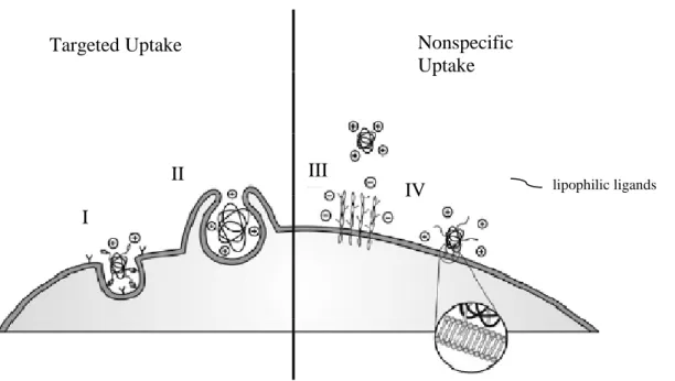
Figure 1.1. Specific and non specific cell membrane interactions that lead to cellular uptake; I-II)
receptor mediated; III) electrostatic; IV) hydrophobic6
1.1.1 Naked DNA Delivery.
The exact pathway for delivery is dependent on the vector chosen, which can be categorized into three main types. The first is direct delivery of naked DNA, the second is viral based delivery, and the third is non-viral delivery. In the case of naked DNA delivery, the DNA enters the cell through an external force that either penetrates the cell membrane or disrupts it to permit the passage of larger molecules. Particle bombardment techniques such as the gene gun or jet injection use high velocity particles or media that
Targeted Uptake Nonspecific
Uptake
I
II III
4
are capable of penetrating a target tissue.2 Cellular uptake of naked DNA has also been enhanced by using electroporation, sonoporation, or use of a magnetic field.2 The mechanism for electroporation consists of two parts. The first is an electric field that induces accumulation near the cell membrane via electrophoretic mobility. The current also disrupts the cell membrane on a microscopic scale which allows for passage across the membrane. Sonoporation causes cavitation around the target site that enhances membrane permeability. 2 Lastly, delivery has been enhanced by tethering the cargo to magnetic particles and applying a magnetic field. In naked DNA delivery there is no protection of the DNA from nuclease degradation and these pathways suffer from low efficiency. Furthermore the application of the delivery is often limited to superficial tissues, small target sites, and/or is accompanied with invasive procedures.
1.1.2 Viral Delivery.
5
nucleus through nuclear localizing proteins (NLP). They then utilize nuclear pores to transfer the cargo into the nucleus.
The most common viral vectors that have been developed are the adeno-associated virus, herpes virus, pox virus, and lentivirus.7 Retroviral vectors randomly insert DNA into the host chromosome and results in persistent gene expression. An issue arises however if the insertion occurs at an integral site, interrupting protein expression, and potentially leading to oncogenic effects. Adenoviral vectors do not insert DNA into the host chromosomes, but as a result only yield short term gene expression. In both systems the body can gain natural immunity and elicit inflammatory or immunological responses. Despite the high transfection efficiency, viral type vectors have resulted in cancer and death, and have not been able to move beyond phase III clinical trials.8,9
1.1.3 Synthetic Vector Delivery.
6
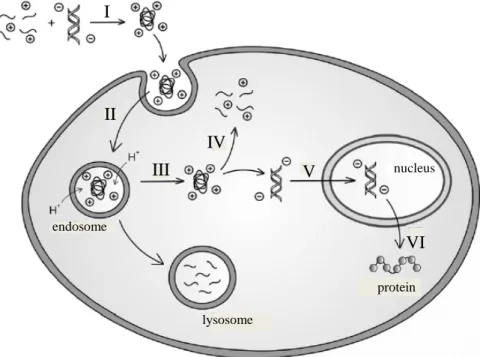
quantities, which aids in commercialization. Various topologies have been utilized including linear, branched, hyperbranched, and dendritic materials as well as liposomes and micelles.4,6,8,9 The greatest disadvantages in synthetic delivery are non-specific toxicity associated with polycationic materials as well as typically low transfection efficiency.
Figure 1.2. Transfection pathway from complex formation to gene expression; I) complex
formation; II) translocation; III) endosomal escape; IV) polymer/DNA dissociation; V) nuclear uptake; VI) gene expression6
As depicted in Figure 1.2, synthetic vectors go through a similar pathway to viral vectors. Attachment of signaling ligands can induce cellular uptake or they can go through nonspecific membrane association via electrostatic or hydrophobic interactions (Figure 1.1). They are then taken up into the cell through endocytosis. Vectors then escape from the endosome, and to achieve this, endosomal disruption proteins or more prominently the ‘proton sponge’ effect have been utilized (Figure 1.3). Briefly, cationic
II
III
IV
endosome
lysosome
nucleus V
VI I
7
materials have a distribution of charged and uncharged functional groups, typically amines. Upon endosomal acidification, the neutral amines become protonated, effectively buffering the endosome and increasing the internal charge potential. This induces chloride diffusion into the vesicle followed by osmotic swelling that leads to membrane rupture and cargo release. Once in the cytosol the cargo must be delivered to the nucleus. Synthetic vectors have been modified with nuclear localizing peptides to take advantage of the natural cellular machinery that actively moves the polyplex to the nucleus. Passive diffusion of large particles in the cytosol in very difficult, and it is generally thought to not be the mode of transport if no NLP is present. To date the exact pathway has not been elucidated to date. It is also important to note that nuclear uptake of synthetic materials typically occurs during cell replication when the nuclear membrane is dissolved, adding another limitation of synthetic vectors by making them cell cycle dependent.
Figure 1.3. ‘Proton sponge’ theory of endosomal release; I) early endosome; II) acidification and
chloride accumulation; III) osmotic swelling and membrane rupture3
8
A variety of synthetic methods has been employed to complex and protect DNA including liposomes, micelles, and functionalized polymers. The first two systems utilize amphiphilic materials that encapsulate the DNA. They have the advantage of low toxicity and facile surface functionalization. The greatest disadvantage in these systems is their lower structural stability of the complex and their susceptibility to disruption and premature DNA release. A great focus of gene therapy has been in the research of micelles and liposomes and many reviews have been written on these subjects.10,11 The remainder of the research reviewed herein will focus on polymeric systems.
1.2 Non-Degradable Materials
1.2.1 Poly(ethyleneimine)
9
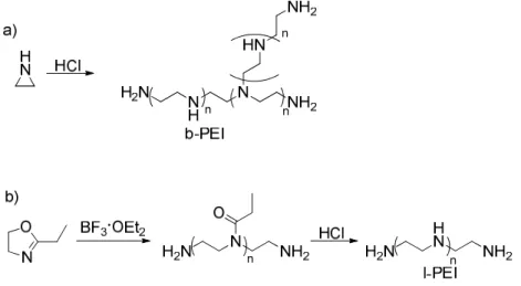
Figure 1.4. Synthesis and structure of a) branched PEI (b-PEI) and b) linear PEI (l-PEI)
The material is one of the most physically and biologically characterized delivery vehicles, and due to the high efficiency, it has become a standard in the field. The branched form contains 1o, 2o, and 3o amines in a ratio of 1:1:1 respectively.13 The linear form has mainly 2o amines. The acid/base profiles for each system show a sloped pH transition between a pH of 5-8 showing multiple pKa values for the amines present in the polymers.14 With a range of pKa values the branched form of PEI has 20% amine protonation at biological pH (pH 7.4) leaving a significant proportion of the amines unprotonated and allowing for a large buffering capacity. This ‘proton sponge’ effect is attributed to efficient endosomal escape and ultimately the high transfection of PEI.
10
tertiary amines that impart a greater buffering potential than l-PEI.14 Despite these properties, l-PEI has shown high in vivo transfection efficiency16,17, and derivatives of this form are more prominent in commercially available gene delivery vehicles, e.g. ExGen500 and jetPEI. The high efficiency has been attributed to the ability of l-PEI to mediate cell-cycle independent nuclear delivery18 as well as possessing lower complex stability that permits DNA dissociation once the target site has been reached.
11
Figure 1.5. a) alkylated PEI; b) acylation of PEI with acetic anhydride
To capitalize on the high bioactivity and reduce the toxicity, modifications such as N-alkylation and N-acylation, pegylation, or degradation have been used. Klibanov has shown that N-alkylation of the 1o and 2o amines (Figure 1.5) decreased the transfection efficiency of 25 kDa branched PEI, but alkylation of the 3o amines enhanced activity.22 This observation was attributed to the position of alkylation with the 1o and 2o amines located at the periphery of the material and the 3o amines more internalized. In solution the hydrophobic alkyl groups placed at the surface collapse the structure, shielding the interior amines. Since the 3o amines are already internalized, alkylation of the 3o amines preserved the overall structure and increased transfection efficiency. Akinc et al. demonstrated that quaternization of all of the amines enhances complex formation and cell internalization, but due to the loss of buffering ability, overall transfection was greatly diminished.23 The transfection of this material increased when the endosomal buffering compound, chloroquin, was present and was one example that supports the ‘protein sponge’ theory for endosomal release.
12
partial acetylation of up to 57% of the primary amines of b-PEI with acetic anhydride, has a 58-fold increase in transfection efficiency in HEK293 cells.24,25 Despite a decrease in buffering capacity, the bioactivity was enhanced. This was attributed to a decrease in binding strength, facilitating DNA dissociation upon delivery. On the other hand, Thomas et al. demonstrated that in commercially available 25 kDa l-PEI, the material is only 89% deacylated as received.26 When the remaining 11% of the amides were hydrolyzed, the efficiency was enhanced by 21-fold in vitro, and 10,000-fold in vivo. The buffering capacity of l-PEI at biological pH was known to be less than b-PEI, and it was concluded that the enhanced transfection efficiency stemmed from its increase. Alkylation and acylation demonstrated the balance of charge, toxicity, binding, and buffering that is required in developing vector. Branched PEI was enhanced by decreasing the number of the more toxic and stronger binding 1o and 2o amines, while linear PEI, with primarily 2o amines, benefited from increasing the buffering capacity.
Figure 1.6. Acid labile PEG grafts conjugated to branched PEI
13
DNA resulting in charge neutralization. PEG then orients itself to the periphery forming a micelle and internally shields the polycation/DNA complex from premature clearance via macrophage or the reticuloendothelial system. When no other modifications to the block copolymers are used such as degradable groups or targeting ligands, PEG decreases surfaces interactions, which results in higher circulation times and lower toxicity, but also prevents membrane association and internalization.27 To avoid the limited transfection ability of PEGylated complexes, ether chains have been conjugated to PEI via labile groups that facilitate deshielding of the polyplex once internalized into the cell (Figure 1.6).28,29 This strategy coupled with attaching targeting ligands to promote cellular uptake has been a promising for gene delivery. Shuai et al. have synthesized a series of PEG-polycaprolactone grafted b-PEIs (Figure 1.7).30 The transfection efficiency was directly related to the PEI molecular weight as well as to the grafting density with higher molecular weight PEI (25 kDa vs 800 Da) and lower grafting density showing optimal bioactivity. The toxicity of the degradable materials was less than the nondegradable parent materials, but also higher concentrations were required to reach comparable or better transfection efficiency.
Figure 1.7. a) Linear PEI containing redox active disulfide linkages in the main chain; b)
14
Another strategy for reducing the toxicity of PEI has been crosslinking or chain extension of low molecular weight PEI (< 10 KDa) with a degradable moiety. Lee et al. synthesized linear PEI (800 Da) that contained a reducible disulfide group in the backbone (Figure 1.7). They found that the reducible PEI was nontoxic, but the transfection efficiency was less than the 25 kDa PEI control used in the experiments.31 There are numerous examples of modifications similar to those reported above, and some of those can be found in recent reviews.3,4,11
1.2.2 Poly(methacrylate) and Poly(methacrylamide)
O O N O O N O O N O O O OH n m O O N O O O P n O O O N m
a) b) c)
(NH4)2S2O8
Figure 1.8. a) Radical polymerization of N,N-dimethylaminoethylmethacylate (DMAEMA); b) PDMAEMA-co-PEGMA; c) PDMAEMA-b-PCMA
15
ammonium peroxydisulfate initiator, a <Mn> of 45 x103 g/mol and <Mw> of 36 x 104 g/mol were attained (Figure 1.8).32 It was found that the transfection efficiency was optimized at a w/w ratio of 6-13/1 and the efficiency was serum independent.33 At the optimum concentration the transfection was comparable to commercially available Lipofectin®. Despite having promising activity, focus has been on developing copolymers to decrease the toxicity.
16
Figure 1.9. PHEMA with PDMAEMA grafted through a hydrolyzable carbonate linkage
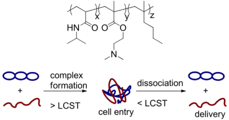
17 + complex formation > LCST + dissociation < LCST
cell entry delivery
HN O O O
N
x y z
Figure 1.10. top: Thermoresponsive PNIPAM-co-PDMAEMA-co-PBMA; bottom: Temperature
controlled complex formation and deformation utilized for dynamic delivery system
18
copolymer was enhanced by using the thermal program, and efficiencies greater than poly(L-lysine), but lower than PDMAEMA were observed.
Figure 1.11. Thermoresponsive pentablock hydrogel used for persistent DNA delivery
19 HN O O O O N HN O O O O N HN O O O O N HN O O O O N N N HN O O O O N HN O O O O N O
HPMA-DMAE HPMA-DMAPr HPMA-DEAE HPMA-DBMPAP HPMA-MPPM HPMA-NHEM
Figure 1.12. Acrylamide monomer HPMA-DMAE and analogues used in hydrolysable
polyacrylamide vectors
Another acrylamide based polymer, poly(carbonic acid 2-dimethylamino-ethyl ester 1-methyl-2-(2-methacryloylamino)-ethyl ester) (PHPMA-DMAE), was developed by Funhoff et al. that incorporated hydrolysable grafts functionalized with tertiary amines (Figure 1.12).41 PHPMA-DMAE gave promising toxicity and transfection results in the absence of serum but had limited transfection efficiency while serum was present. Luten et al. elaborated on this structure by synthesizing acrylamide monomer analogues of HPMA-DMAE to examine how subtle structural changes affected the rate of hydrolysis as well as serum dependence on transfection. (Figure 1.12).42 DEAE, HPMA-MPPM, and HPMA-BDMP showed less toxicity than poly(methacrylate) materials that did not contain a rapidly hydrolysable group, and transfection was serum independent.
1.3 Degradable Delivery
20
decrease toxicity by either masking the cationic sites or by integrating biodegradable groups that leads to formation of less toxic oligomers. Degradable sites can be installed in pendant functionality as seen in some of the examples already discussed but with only limited result in decreasing toxicity. Alternatively, degradable groups can be placed on the main chain of the polymer. Typically this has been done using poly(ester) or poly(amide) based materials. The advantage of chain degradation is that the polymer can be fully digested as opposed to only pendant groups degrading, leaving the main chain intact. The degradation products can be designed to be further metabolized by the body, or the starting monomers can be chosen from a range of biocompatible starting materials. Polymerization typically occurs via ring opening or step-growth polymerization. The former generally yields high molecular weight polymers, and the latter lends itself to facile functionalization and access to large material libraries.
Figure 1.13. Conjugative addition polymerization to yield poly(β-amino ester)s utilizing bis-secondary amines or primary amines
21
22
opportunity to enhance cellular uptake and activity through new functionality and better controlled degradation.
Figure 1.14. Peptide based delivery vehicles; left: poly(L-Lysine); right: Oligo arginine R7
1.3.2 Poly(amide)s
23
Other functionalities have been used to enhance delivery. One of the greatest adaptations that viral vectors possess is membrane-permeable peptides that enhance membrane association and uptake.47,48 A common characteristic of these peptides is a high concentration of cationic amino acids, most notably the guanidine containing arginine. It has been proposed that the cationic domain (protein transduction domain, PTD) on these peptides interacts with heparin sulfate proteoglycans that promote cellular uptake. 47 To mimic the PTD oligo arginine peptides have been used as delivery vehicles or conjugated onto other delivery vehicles to enhance cellular uptake (Figure 1.14).49,50 Utilizing the guanidine rich groups was shown to enhance delivery and oligos of around 7 to 9 arginine repeat units were shown to be optimal.
Figure 1.15. Polyamidoamine (PAMAM) synthesis; a): linear; b): dendritic
24
addition to diacrylamides in an analogues synthesis to that of the PBAE materials.11 Utilizing this modular synthetic approach, installation of a large range of functionalities, including more amino groups, carboxylic acids, alcohols, as well as peptides, was possible. PAMAMs are less toxic than PLL, and including acids and alcohols further decreases their toxicity.
Figure 1.16. Synthesis of a carboxylic acid and guanidine functionalized PAMAM designed as
an RGD mimic
Ferruti has developed linear PAMAM materials by reacting an acid functionalized diacrylate with agmatine (Figure 1.16).51-53 Agmatine is a decarboxylated analogue of arginine and contains a guanidinium group. The repeat unit containing the acid and guanidine moiety mimics the cell active RGD sequence which promotes cellular uptake. In an acid base titration, three acid dissociation constants were observed, 2.25, 7.45, and
25
transfection efficiency was higher than other common PAMAMs and comparable to commercially available JetPEI.
1.4 Biologically Derived Materials
Figure 1.17. Structure of the biologically derived chitosan
1.4.1 Chitosan
26
increase efficiency. Quaternization increased transfection efficiency while simultaneously increasing toxicity. The imidazole moiety also increased transfection by increasing the buffering capacity, lending itself to endosomal release via the ‘proton sponge’ effect.57
Figure 1.18. Polymerization of β-cyclodextrin utilizing a cationic diamidine linkage
1. 1. 1.
27
Another carbohydate derivative, β-cyclodextrin (CD), have also been shown to increase biocompatibility and ultimately efficiency. Synthesis of these polymers was achieved by stepgrowth polymerization between a diaminocyclodextrin and diimidate compounds (Figure 1.18).58 Transfection was optimized at an N/P ratio >10 with low toxicity. The transfection was greatest when the linkage between CD segments was 6-8 methylenes. Greater than 8 methylenes diluted the charge density of the material resulting in solubility issues and low transfection efficiency.59 It was also found that these materials did not escape from the endosome via amine protonation, demonstrating that the transfection pathway is greatly dependent on the material being studied.
Figure 1.19. Linear PEI with sugar alcohol end groups
1.4.3 Sugar alcohol functionalized
28
transfection efficiency was highly dependent on the stereochemistry of the carbohydrates. This was due to changes in the complex stability. The toxicity was also shown to be dependent on the distance from the cationic site and the carbohydrate, with further separation resulting in increased toxicity.
1.5 Bioactive Modifications
Further delivery enhancement has been achieved in the presence of targeting ligands, endosomolytic compounds, and nuclear localizing proteins. A range of targeting ligands has been used such as RGD and folic acid where cellular uptake was initiated by receptor binding.6 The degree of specificity greatly depends on the ligand chosen. For example the RGD receptor is present on many types of cells and little specificity is gained. Other receptors, such as the folate receptor, are over expressed in some cell lines, and these systems have much higher specificity. The effectiveness of targeting is greatly dependent on the chemistry involved in conjugation to the polymer, with the spacer between the targeting head group and the polymer being important, as well as the density of the ligation.3 It is critical for the site of conjugation between the polymer and ligand not to interfere with recognition or binding on the cell surface receptors. In polycationic materials there is also nonspecific binding via electrostatic interactions and specificity can only be attained when competition from nonspecific electrostatic interactions is minimized (charge neutrality).
29
approach however could not be used in vivo.3 Another strategy has been the conjugation of inactivated adenovirus particles. Lastly, natural and synthetic fusogenic peptides have been attached to the delivery vehicle where upon acidification the proteins go through a structural transition. These structural changes lead to membrane interaction and disruption, ultimately enhancing delivery efficiency from one to three orders of magnitude.
Once endosomal escape has been achieved there are many barriers present in the cytosol. Diffusion of large molecules in the cytosol is extremely slow and inefficient. Techniques such as nuclear localizing proteins (NLP) are used to take advantage of the natural cell machinery and promote accumulation at the nucleus.3 Most NLPs are short cationic protein sequences, usually arginine or lysine rich. The cationic nature of synthetic delivery vehicles are proposed to partially mimic NLPs, but with only limited efficiency. Few complexes reach the nucleus, and little has been elucidated regarding the mechanism post endosomal release, to nuclear localization, and ultimately nuclear uptake and complex dissociation.
6. Conclusions
30
number, density, type, and pKa of the amines. The transfection of b-PEI was enhanced by minimizing binding strength of the material by reducing charge density and number of primary and secondary amines. This showed that the binding strength and buffering capacity of the material was greater than needed for transfection prior to acetylation, and the number of amines could be reduced to limit DNA binding strength without being detrimental to delivery. On the other hand, l-PEI had lower buffering capacity and binding strength than b-PEI, and it was found that deacetylation of its amines enhanced delivery. The molecular weight of vectors has also been shown to influence delivery. Typically, higher molecular weight materials were more efficient until a weight was reached where toxicity minimized the delivery enhancement. To reduce toxicity, degradable and naturally derived materials have been used. These vectors decrease toxicity, but the concentration needed for delivery is greatly increased with N/P ratios typically 20-70, as opposed to an N/P ratio of 5-10 for nondegradable materials.
31
32 References
(1) Eliyahu, H.; Barenholz, Y.; Domb, A. J. Molecules 2005, 10, 34-64.
(2) Taira, K.; Kataoka, K.; Niidome, T.; SpringerLink (Online service). Springer-Verlag Tokyo: Tokyo, 2005.
(3) Pack, D. W.; Hoffman, A. S.; Pun, S.; Stayton, P. S. Nat Rev Drug Discov 2005, 4, 581-593.
(4) Tiera, M. J.; Winnik, F. M.; Fernandes, J. C. Curr Gene Ther 2006, 6, 59-71. (5) Günther, M.; Wagner, E.; Ogris, M. Current Medicinal Chemistry - Anti-Cancer
Agents 2005, 5, 157-171.
(6) Wong, S. Y.; Pelet, J. M.; Putnam, D. Prog Polym Sci 2007, 32, 799-837. (7) Boeckle, S.; Wagner, E. AAPS Journal. 2006, 8, 83.
(8) Lehrman, S. Nature 1999, 401, 517-518. (9) Marshall, E. Science 1999, 286, 2244-2245.
(10) Martin, B.; Sainlos, M.; Aissaoui, A.; Oudrhiri, N.; Hauchecorne, M.; Vigneron, J. P.; Lehn, J. M.; Lehn, P. Curr Pharm Design 2005, 11, 375-394. (11) Mintzer, M. A.; Simanek, E. E. Chem Rev 2009, 109, 259-302.
(12) Brissault, B.; Kichler, A.; Guis, C.; Leborgne, C.; Danos, O.; Cheradame, H. Bioconjugate Chem 2003, 14, 581-587.
(13) von Harpe, A.; Petersen, H.; Li, Y. X.; Kissel, T. J Control Release 2000, 69, 309-322.
(14) Akinc, A.; Langer, R. Biotechnol Bioeng 2002, 78, 503-508.
(15) Dunlap, D. D.; Maggi, A.; Soria, M. R.; Monaco, L. Nucleic Acids Res 1997, 25, 3095-3101.
(16) Goula, D.; Benoist, C.; Mantero, S.; Merlo, G.; Levi, G.; Demeneix, B. A. Gene Ther 1998, 5, 1291-1295.
(17) Zou, S. M.; Erbacher, P.; Remy, J. S.; Behr, J. P. J Gene Med 2000, 2, 128-134. (18) Brunner, S.; Furtbauer, E.; Sauer, T.; Kursa, M.; Wagner, E. Mol Ther 2002, 5,
80-86.
(19) Godbey, W. T.; Wu, K. K.; Mikos, A. G. J Biomed Mater Res 1999, 45, 268-275. (20) Kunath, K.; von Harpe, A.; Fischer, D.; Peterson, H.; Bickel, U.; Voigt, K.;
Kissel, T. J Control Release 2003, 89, 113-125.
(21) Werth, S.; Urban-Klein, B.; Dai, L.; Hobel, S.; Grzelinski, M.; Bakowsky, U.; Czubayko, F.; Aigner, A. J Control Release 2006, 112, 257-270.
(22) Thomas, M.; Klibanov, A. M. P Natl Acad Sci USA 2002, 99, 14640-14645. (23) Akinc, A.; Thomas, M.; Klibanov, A. M.; Langer, R. J Gene Med 2005, 7,
657-663.
(24) Forrest, M. L.; Meister, G. E.; Koerber, J. T.; Pack, D. W. Pharm Res 2004, 21, 365-371.
(25) Gabrielson, N. P.; Pack, D. W. Biomacromolecules 2006, 7, 2427-2435.
(26) Thomas, M.; Lu, J. J.; Ge, Q.; Zhang, C. C.; Chen, J. Z.; Klibanov, A. M. P Natl Acad Sci USA 2005, 102, 5679-5684.
33
(28) Knorr, V.; Allmendinger, L.; Walker, G. F.; Paintner, F. F.; Wagner, E. Bioconjugate Chem 2007, 18, 1218-1225.
(29) Ahn, C. H.; Chae, S. Y.; Bae, Y. H.; Kim, S. W. J Control Release 2002, 80, 273-282.
(30) Shuai, X. T.; Merdan, T.; Unger, F.; Wittmar, M.; Kissel, T. Macromolecules 2003, 36, 5751-5759.
(31) Lee, Y.; Mo, H.; Koo, H.; Park, J. Y.; Cho, M. Y.; Jin, G. W.; Park, J. S. Bioconjugate Chem 2007, 18, 13-18.
(32) Cherng, J. Y.; vandeWetering, P.; Talsma, H.; Crommelin, D. J. A.; Hennink, W. E. Pharm Res 1996, 13, 1038-1042.
(33) vandeWetering, P.; Cherng, J. Y.; Talsma, H.; Hennink, W. E. J Control Release 1997, 49, 59-69.
(34) Deshpande, M. C.; Garnett, M. C.; Vamvakaki, M.; Bailey, L.; Armes, S. P.; Stolnik, S. J Control Release 2002, 81, 185-199.
(35) Deshpande, M. C.; Davies, M. C.; Garnett, M. C.; Williams, P. M.; Armitage, D.; Bailey, L.; Vamvakaki, M.; Armes, S. P.; Stolnik, S. J Control Release 2004, 97, 143-156.
(36) Rungsardthong, U.; Deshpande, M.; Bailey, L.; Vamvakaki, M.; Armes, S. P.; Garnett, M. C.; Stolnik, S. J Control Release 2001, 73, 359-380.
(37) Lam, J.; Ma, Y.; Armes, S.; Lewis, A.; Stolnik, S. J Pharm Pharmacol 2004, 56, S70-S70.
(38) Jiang, X.; Lok, M. C.; Hennink, W. E. Bioconjugate Chem 2007, 18, 2077-2084. (39) Kurisawa, M.; Yokoyama, M.; Okano, T. J Control Release 2000, 69, 127-137. (40) Agarwal, A.; Unfer, R. C.; Mallapragada, S. K. Biomaterials 2008, 29, 607-617. (41) Funhoff, A. M.; van Nostrum, C. F.; Janssen, A. P. C. A.; Fens, M. H. A. M.;
Crommelin, D. J. A.; Hennink, W. E. Pharm Res 2004, 21, 170-176.
(42) Luten, J.; Akeroyd, N.; Funhoff, A.; Lok, M. C.; Talsma, H.; Hennink, W. E. Bioconjugate Chem 2006, 17, 1077-1084.
(43) Green, J. J.; Langer, R.; Anderson, D. G. Accounts Chem Res 2008, 41, 749-759. (44) Lynn, D. M.; Langer, R. J Am Chem Soc 2000, 122, 10761-10768.
(45) Anderson, D. G.; Akinc, A.; Hossain, N.; Langer, R. Mol Ther 2005, 11, 426-434.
(46) Chen, D. J.; Majors, B. S.; Zelikin, A.; Putnam, D. J Control Release 2005, 103, 273-283.
(47) Futaki, S. Adv Drug Deliver Rev 2005, 57, 547-558.
(48) Suzuki, T.; Futaki, S.; Niwa, M.; Tanaka, S.; Ueda, K.; Sugiura, Y. J Biol Chem 2002, 277, 2437-2443.
(49) Fuchs, S. M.; Raines, R. T. Biochemistry-Us 2004, 43, 2438-2444.
(50) Kim, T. I.; Baek, J. U.; Yoon, J. K.; Choi, J. S.; Kim, K.; Park, J. S. Bioconjugate Chem 2007, 18, 309-317.
(51) Ferruti, P.; Bianchi, S.; Ranucci, E.; Chiellini, F.; Piras, A. M. Biomacromolecules 2005, 6, 2229-2235.
(52) Ferruti, P.; Franchini, J.; Bencini, M.; Ranucci, E.; Zara, G. P.; Serpe, L.; Primo, L.; Cavalli, R. Biomacromolecules 2007, 8, 1498-1504.
34
(54) Domard, A.; Rinaudo, M. Int J Biol Macromol 1983, 5, 49-52.
(55) Huang, M.; Fong, C. W.; Khor, E.; Lim, L. Y. J Control Release 2005, 106, 391-406.
(56) Ishii, T.; Okahata, Y.; Sato, T. Bba-Biomembranes 2001, 1514, 51-64.
(57) Kim, T. H.; Ihm, J. E.; Choi, Y. J.; Nah, J. W.; Cho, C. S. 2003, 93, 389-402. (58) Gonzalez, H.; Hwang, S. J.; Davis, M. E. Bioconjugate Chem 1999, 10,
1068-1074.
(59) Hwang, S. J.; Bellocq, N. C.; Davis, M. E. Bioconjugate Chem 2001, 12, 280-290.
(60) Prevette, L. E.; Kodger, T. E.; Reineke, T. M.; Lynch, M. L. Langmuir 2007, 23, 9773-9784.
36
Abstract. The degree of ionization of amine containing polymers plays a critical role in
gene delivery. In order to develop new materials with optimized properties for transfection, a clear understanding of the influence of ionization needs to be established. We determined the influence of altering pKa values for structurally similar dialkylaminoisoprene polymers to balance the competing properties of complex formation and buffering capacity. Free radical polymerization was used to generate materials with distinct pKa transitions. The degree of ionization at physiological pH was determined and found to directly affect DNA binding, transfection efficiency, and toxicity. It was shown that there is a balance between buffering capacity and complex formation with an ideal ionization in the range above 24% to below 50% amine protonation (pH = 7.4). It was also found that a material with 33% protonation yielded a transfection efficiency (N/P ratio of 2) 100 fold higher than the field standard poly(ethyleneimine).
1. Introduction
A major focus of gene therapy research is the development of synthetic materials 1-13
37
(polyplex), 2) associate to the cell membrane and become internalized through endocytosis, 3) escape from the endosome, 4) transport the cargo to the nucleus, and 5) dissociate from the DNA.20 Most materials to date utilize 1o, 2o, and or 3o amines, which at physiological conditions maintain protonated and unprotonated amines. These properties are fundamental for complex formation and endosomal release, respectively.21
During cell internalization, it is critical for the polyplex to escape from the endosome prior to fusion with a lysosome in order to avoid its harsh degradative environment. The most prominent mechanism for escape is the ‘proton sponge’ effect proposed by Behr in 1995, which asserts upon endosomal acidification the neutral amines become protonated, followed by an influx of chloride ions to balance the charge. Ultimately, there is an increase in osmotic pressure and eventual membrane rupture.22-24 From the early endocytic vesicle to the late endosome, the pH ranges from 7.4 to ~5.25 Poly(ethyleneimine) (PEI) has become a standard in the field due to the material’s high delivery efficiency which is attributed to the range of amines that allow it to buffer over the entire lifetime of the endosome. The result is efficient endosomal escape.24 Many of the reported materials attempt to mimic PEI’s buffering ability by integrating functionality, such as an imidazole moiety26-31, to impart varied pKa values and gain enhanced buffering characteristics.
38
research focuses on the characterization of the buffering capability, DNA/polymer complex formation, and resulting transfection efficiency to further understand structure/biological activity relationships. To this end, homopolymers and copolymers were synthesized with discrete pKa ranges to compare the effects of ionization on delivery efficiency. Through this work insights will be gained into the effects of charge on transfection and property targets will be established for the design and screening of new gene delivery materials.
2. Experimental Details
2.1 Materials. Plasmid DNA (pCMV-Luc) was purchased from Elim
Biopharmaceuticals, Inc. (Hayward, CA). Minimum essential medium (MEM), Hanks’ balanced salt solution (HBSS), Dulbecco’s phosphate buffered saline (PBS), fetal bovine serum (FBS), and a 1 kb DNA ladder were purchased from Invitrogen. CellTiter 96 AQueous one solution cell proliferation assay and luciferase assay system were purchased from Promega Corp. (Madison, WI). BCA protein assay kit was purchased from Pierce (Rockford, IL). Branched poly(ethyleneimine) with a molecular weight of 25,000 g/mol was used as a control in the characterization experiments. All other chemicals were purchased from Sigma-Aldrich and were used without further purification.
39
HR2, HR4, and HR5). Glass transition temperatures were measured with a Seiko 220C differential scanning calorimeter, using a heating and cooling rate of 10 °C/min. The glass transition temperatures were reported based on the second heating. Thermogravimetric analysis was carried out using a Perkin Elmer TGA 7 with a heating rate of 10 ºC/min in a N2 atmosphere. Zeta potential measurements were completed on a Malvern Zeta Sizer Nano Series Nano-ZS instrument using Dispersion Technology Software version 4.20 at a wavelength of 633 nm using a 4.0 mW, solid state He-Ne laser at a scattering angle of 173 oC. Cell culture plates were analyzed on a Molecular Devices SpectraMax M5. Ethidium bromide stained agarose gels were visualized on a Bio Rad VersaDoc imaging system.
2.3 Diene Synthesis. General synthesis of
2-bromo-N,N-dialkylprop-2-en-1-amine. To a solution of N,N-dialkylamine in diethyl ether was added 2,3-dibromopropene dropwise at 0 °C while stirring. The reaction was allowed to warm to room temperature and to react for 18 h. NaOH (1 M) was added until the formed salt dissolved, and the solution was extracted with diethyl ether (3x, 50 mL). The organic layers were combined, washed with brine, dried with MgSO4, and concentrated. The product was purified and isolated by distillation as a colorless liquid.
2-bromo-N,N-dimethylprop-2-en-1-amine. 1H NMR (400 MHz, CDCl3): δ 5.77 (s, 1H), 5.53 (s, 1H), 3.07 (s, 2H), 2.23 (s, 6H). 13C NMR (400 MHz, CDCl3): δ 131.60 (CH2=CBrCH2), 119.12 (CH2=CBrCH2), 68.20 (CH2N), 45.14 (N(CH3)2)
40
NMR (400 MHz, CDCl3): δ 132.64 (CH2=CBrCH2), 116.80 (CH2=CBrCH2), 61.46 (CH2N), 46.67 [N(CH2CH3)2], 11.73 [N(CH2CH3)2]
2-bromo-N,N-dipropylprop-2-en-1-amine. 1H NMR (400 MHz, CDCl3): δ 5.90 (s, 1H), 5.53 (s, 1H), 3.23 (s, 2H), 2.43 (t, 4H, J = 7.6), 1.45 (m, 4H, J = 7.6), 0.88 (t, 6H, J = 7.2). 13C NMR (400 MHz, CDCl3): δ 133.60 (CH2=CBrCH2), 117.80 (CH2=CBrCH2), 63.00 (CH2N), 55.92 [N(CH2CH2CH3)2], 20.78 [N(CH2CH2CH3)2], 12.16 [N(CH2CH2CH3)2]
General synthesis of 2-(N,N-diakylaminomethyl)-1,3-butadiene. To a flame dried 1 L three neck flask was added [1,3-bis(diphenylphosphino)propane]dichloronickel (0.5 mol%), dialkylaminobromopropene, and dry THF under a N2 atmosphere. Vinyl magnesium bromide (1 M in THF) was added dropwise at 0 °C while stirring. The reaction was allowed to warm to room temperature and to react for 24 h. The reaction was quenched with NH4Cl (satd.) and extracted with diethyl ether (3x, 50 mL). The organic layers were combined, washed with brine, dried with MgSO4, and concentrated. The product was purified and isolated by distillation as a colorless liquid.
N,N-dimethyl-2-methylenebut-3-en-1-amine (Dimethylaminoisoprene; DMAI). 1H NMR (400 MHz, CDCl3): δ 6.36 (dd, 1H, J1 = 17.6 Hz, J2 = 11.2 Hz), 5.41 (d, 1H, J = 17.6), 5.14 (s, 1H), 5.11 (m, 1.5H), 5.09 (s, 0.5H), 3.01 (s, 1H), 2.21 (s, 1H). 13C NMR (400 MHz, CDCl3): δ 143.31 (CH2=CCH2N), 137.56 (CH2=CHCCH2N), 117.73 (CH2=CHCCH2N), 114.44 (CH2=CCH2N), 61.86 (CH2N), 45.54 [N(CH3)2]
41
17.6), 5.204 (s, 1H), 5.142 (s, 1H), 5.08 (d, 1H), 3.16 (s, 1H), 2.51 (q, 4H, J = 7.2), 1.02 (t, 6H, J = 7.2). 13C NMR (400 MHz, CDCl3): δ 144.07 (CH2=CCH2N), 137.92 (CH2=CHCCH2N), 116.98 (CH2=CHCCH2N), 113.96 (CH2=CCH2N), 55.25 (CH2N), 47.03 [N(CH2CH3)2], 11.67 [N(CH2CH3)2]
N,N-dipropyl-2-methylenebut-3-en-1-amine (Dipropylaminoisoprene; DPAI). 1H NMR (400 MHz, CDCl3): δ 6.38 (dd, 1H, J1 = 17.6 Hz, J2 = 10.8 Hz), 5.45 (d, 1H, J = 17.6), 5.22 (s, 1H), 5.12 (s, 1H), 5.06 (d, 1H, J = 10.8), 3.15 (s, 1H), 2.35 (t,4H, J = 7.6 Hz), 1.46 (m, 4H, J = 7.6 Hz), 0.87 (t, 6H, J = 7.6). 13C NMR (400 MHz, CDCl3): δ
144.22 (CH2=CCH2N), 137.94 (CH2=CHCCH2N), 116.99 (CH2=CHCCH2N), 113.92 (CH2=CCH2N), 56.66 (CH2N), 56.22 [N(CH2CH2CH3)2], 20.18 [N(CH2CH2CH3)2], 11.95 [N(CH2CH2CH3)2]
2.4 Polymer Synthesis. Free Radical Polymerization of
2-(N,N-dialkylaminomethyl)-1,3-butadiene. Monomer and AIBN were added to an ampoule with a magnetic stir bar. After three freeze-pump-thaw cycles, the ampoule was sealed under nitrogen and placed in an oil bath preheated to 70 °C. After 24 h, the polymer was precipitated into acetone at -78 °C and dried under vacuum at room temperature for 5 d.
42
poly(2-(N,N-diethylaminomethyl)-1,3-butadiene) (PEAI). 1H NMR (400 MHz, CDCl3): δ 5.27 (s, 1H), 2.92 (s, 2H, trans), 2.85 (s, 2H, cis), 2.41 (m, 4H), 2.10 (m, 4H), 0.96 (m, 6H). 13C NMR (400 MHz, CDCl3): δ 137.57 (CH2CH=CCH2), 127.72 (CH2CH=CCH2), 59.94 [CH2N(CH2CH3)2, cis], 51.84 [CH2N(CH2CH3)2, trans], 46.43 [N(CH2CH3)2], 36.13 (CH2CH=CCH2), trans), 27.97 (CH2CH=CCH2, cis), 26.61 (CH2CH=CCH2), 11.67 [N(CH2CH3)2]
poly(2-(N,N-dipropylaminomethyl)-1,3-butadiene (PPAI)). 1H NMR (400 MHz, CDCl3): δ 5.28 (s, 1H), 2.93 (s, 2H, trans), 2.85 (s, 2H, cis), 2.27 (m, 4H), 2.12 (m, 4H), 1.42 (m, 4H), 0.85 (m, 6H). 13C NMR (400 MHz, CDCl3): δ 138.00 (CH2CH=CCH2), 127.80 (CH2CH=CCH2), 61.29 [CH2N(CH2CH2CH3)2, cis], 55.65 [N(CH2CH2CH3)2], 53.09 [CH2N(CH2CH2CH3)2, trans], 36.02 (CH2CH=CCH2, trans), 28.04 (CH2CH=CCH2, cis), 26.42 (CH2CH=CCH2), 20.21 [N(CH2CH2CH3)2], 12.04 [N(CH2CH2CH3)2]
Free Radical Copolymerization. The density for each monomer was determined, and varied amounts of monomer were added via micropipette to an ampoule with a magnetic stir bar. After three freeze-pump-thaw cycles, the ampoule was sealed under nitrogen and placed in an oil bath preheated to 70 °C. After 24 h, the polymer was precipitated into acetone at -78 °C and dried under vacuum at room temperature for 5 d.
43
CDCl3): δ 137.10 (CH2CH=CCH2), 128.25 (CH2CH=CCH2), 66.53 [CH2N(CH3)2, cis], 61.21 [CH2N(CH2CH2CH3)2, cis], 58.23 [CH2N(CH3)2, trans], 55.73 [N(CH2CH2CH3)2], 53.04 [CH2N(CH2CH2CH3)2, trans], 45.77 [N(CH3)2], 35.91 (CH2CH=CCH2, dimethyl, trans), 34.56 (CH2CH=CCH2, dipropyl, trans), 29.09 (CH2CH=CCH2, dimethyl, cis), 28.81 (CH2CH=CCH2, dipropyl, cis), 27.98 (CH2CH=CCH2, dimethyl), 26.39 (CH2CH=CCH2, dipropyl), 20.23 [N(CH2CH2CH3)2], 12.04 [N(CH2CH2CH3)2]
poly(2-(N,N-diethylaminomethyl)-1,3-butadiene-co-2-(N,N-dipropylaminomethyl)-1,3-butadiene) (EtPr). 1H NMR (400 MHz, CDCl3): δ 5.28 (s, 1H), 2.94-2.88 (m, 2H), 2.42 (broad, 4H), 2.26 (broad, 4H), 2.11 (broad, 4), 1.43 (broad, 4H), 0.978 (broad, 6H), 0.85 (broad, 6H). 13C NMR (400 MHz, CDCl3): δ 137.90 (CH2CH=CCH2), 127.61 (CH2CH=CCH2), 61.23 [CH2N(CH2CH2CH3)2, cis], 60.16 [CH2N(CH2CH3)2, cis], 55.73 [N(CH2CH2CH3)2], 53.04 [CH2N(CH2CH2CH3)2, trans], 51.92 [CH2N(CH2CH3)2, cis], 46.95 [N(CH2CH3)2], 34.60 (CH2CH=CCH2, diethyl, trans), 34.62 (CH2CH=CCH2, dipropyl,trans), 29.21 (CH2CH=CCH2, diethyl,cis), 28.85 (CH2CH=CCH2, dipropyl, cis), 28.04 (CH2CH=CCH2, diethyl), 26.50 (CH2CH=CCH2, dipropyl), 20.24 [N(CH2CH2CH3)2], 12.00 [N(CH2CH3)2], 11.72 [N(CH2CH2CH3)2]
Reactivity Ratios. Reactivity ratios were determined by 1H NMR with low conversion (< 10%) copolymerizations of DMAI and DPAI. The polymerization setup was identical to the copolymer procedure. Reactions were run for 2 h and samples were added directly to NMR tubes containing CDCl3.
2.5. Polymer/Polyplex Characterization. pH Titration. The pH of the solution
44
20 mg of material into 10 mL of 0.1 M HCl. The solution was titrated with 0.1 M NaOH, and the pH was recorded after equilibration (30 s). A derivative of the curve was used to determine the acid base equilibrium points. A line of best fit was taken through the buffering plateau, and the average of the two equilibrium points was used to determine the inflection point of the titration curve.
Standard Polyplex Formation. To 1 mg of each polymer sample was added 15
µL of glacial acetic acid followed by 485 µL of either opti-MEM or MEM media to make polymer solutions at a concentration of 2 mg/mL. Once dissolved the polymer samples were diluted with the appropriate media to obtain the polymer solutions at concentrations of 0.1 mg/mL. To solutions of 2 µg/mL pCMV-Luc plasmid in media was added the polymer solution and media to form complexes at desired N/P ratios with total volumes of 200 µL.
Agarose Gel Electrophoresis. The standard polyplex formation was used to generate complexes at desired N/P ratios. Electrophoresis was conducted on a 0.75% agarose gel in TAE buffer containing 0.5% ethidium bromide for 45 min. at 120 V. The bands were visualized using ethidium bromide staining.
Cytotoxicity Assay. HeLa cells were grown in 96-well plates at a density of
5×10³ cells/well in 150 µL of growth medium (89% MEM, 10% FBS, 1% L-glutamine). Cells were grown for 24 h. The media was removed, and the cells were washed with 100
45
were removed, and the cells were washed with DPBS. To the cells were added 100 µL of MEM and 20 µL of 3-(4,5-dimethylthiazol-2-yl)-5-(3-carboxymethoxyphenyl)-2-(4-sulfophenyl)-2H-tetrazoleum, inner salt (MTS, from CellTiter 96 AQueous One Solution Cell Proliferation Assay kit). The cells were incubated for 4 h and the absorbance at 490 nm was monitored on a Molecular Devices SpectraMax M5 plate reader.
Transfection Efficiency Assay. HeLa cells were grown in 48-well plates at a
density of 1.5×10⁴ cells/well in 300 µL of growth medium (89% MEM, 10% FBS, 1% L-glutamine). Cells were grown for 24 h. The media was removed, and the cells were washed with 300 µL of HBSS and incubated with 450 µL of opti-MEM and 50 µL of polyplex solution for 4 h. The solutions were removed, and the cells were washed with HBSS and incubated with 500 µL of opti-MEM (89% MEM, 10% FBS, 1% L-glutamine) for 32 h. The growth medium was removed and the cells were washed with 300 µL of HBSS and treated with 150 µL of cell lysis buffer (1X). The resulting mixtures were centrifuged and 20 µL of supernatant were added directly to white opaque 96-well plates. Luciferase activity was quantified by adding 100 µL of luciferase assay reagent, followed by a 2 second delay. Luminescence was quantified over a 1 second read time. The result was normalized to the total protein content using a BCA protein assay whose absorption was monitored at 562 nm on a Molecular Devices SpectraMax M5 plate reader.
46
of the polymer was verified to be 7.4. Using an autotitrator, the polymer concentration was increased by an N/P ratio of 0.25 up to a total ratio of 10, and the ζ-potentials were monitored.
3. Results and Discussion
R2NH
+ Br
Br
0oC - rt
24 h Br NR2 + MgBr NR2 Ni(dppp)Cl2 THF
0oC - rt
NB2
AIBN
70oC
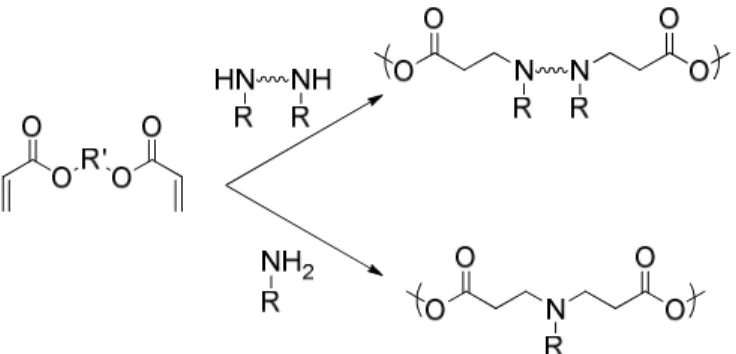
24 h NA2 NB2 n NA2 + R -Me -Et -Pr Monomer DMAI DEAI DPAI A -Me -Et -Pr -Me -Et B -Me -Et -Pr -Pr -Pr Polymer PMAI PEAI PPAI MePr-Xa EtPr-Yb
47
Table 2.1. Radical Homo- and Copolymerizations of DMAI, DEAI, and DPAI
Sample Feed a (%)
Actual Incorp.b (%)
‹Mn›c
(× 10-3 g/mol) PDI
c Tgd (°C)
Decompositione (oC) 5% 10%
DPf
PMAI - - 16 1.50 -29 353 365 109
PEAI - - 15 2.29 -46 337 359 120
PPAI - - 20 1.67 -55 313 352 122
MePr-20 20 17 19 1.59 -46 354 366 117
MePr-40 40 44 17 1.66 -47 330 358 125
MePr-54g 54 54 17 1.74 -44 345 362 140
MePr-60 60 59 19 1.77 -38 345 361 136
MePr-80 80 80 17 1.63 -35 344 362 118
EtPr-20 20 19 19 1.82 -54 316 353 127
EtPr-40 40 38 20 1.92 -47 350 364 117
EtPr-60 60 58 18 2.15 -46 346 360 114
EtPr-80 80 78 17 2.50 -46 345 360 109
a
amine monomer relative to the DPAI; b determined by 1H NMR; c determined by GPC; d determined by DSC; e determined by TGA; f based on the average MW of the monomers incorporated into the polymer; g designed as a pKa mimic of PEAI
3.1 Synthesis
48
To further verify random copolymer formation, the reactivities of DMAI and DPAI monomers were determined by 1H NMR (Table 2.2).33 Monomer concentrations were varied by 0.1 equivalent of DMAI relative to DPAI. The ratio of monomers was determined by normalizing the region of 2.8 to 3.2 ppm for the methylenes extending from the diene or polymer backbone and integrating the peaks at 3.13 and 3.01 ppm, which correspond to DMAI and DPAI, respectively. The normalized monomer concentration (At ) at time t and the starting monomer concentration (A0) were used to determine the mole fraction of DMAI ( M
t
F ) and DPAI ( P t
F ) incorporated into the copolymers (equations 1 and 2; Table 2.2).
] A [A ] A [A A A F P t P M t M M t M M t − + − − = 0 0 0 (1) ] A [A ] A [A A A F M t M P t P P t P P t − + − − = 0 0 0 (2)
Table 2.2. Calculated values for DMAI and DPAI reactivity ratio determination
M t
A * AtP
* M
t
F FtP
Conv.*
(%) F f G H
MePr-10 0.15 1.52 0.15 0.85 8.5 0.18 0.11 -0.51 0.07 MePr-20 0.31 1.37 0.28 0.72 7.5 0.39 0.25 -0.38 0.16 MePr-30 0.48 1.20 0.37 0.62 8.5 0.60 0.43 -0.28 0.31 MePr-40 0.67 1.05 0.46 0.54 7.5 0.87 0.67 -0.10 0.51 MePr-50 0.86 0.86 0.50 0.50 8.5 1.00 1.00 0.00 1.00 MePr-60 1.06 0.70 0.58 0.42 6.0 1.40 1.50 0.43 1.61 MePr-70 1.25 0.52 0.65 0.35 7.5 1.87 2.33 1.09 2.90 MePr-80 1.46 0.35 0.74 0.26 5.5 2.80 4.00 2.57 5.71 MePr-90 1.67 0.18 0.87 0.13 5.5 6.50 9.00 7.62 12.46 *
49
Copolymer incorporation ratios (F) and monomer feed ratios ( f ) were then determined using the following equations:
P t M t F F
F = (3) P
M A A f 0 0
= (4)
The value of F and f were used to determine G and H (Finemann-Ross method; equations 5 and 6; Table 2.2).
) (F F
f
G= −1 (5)
F f H
2
= (6) G=Hrm −rp (7)
50 3.2 Ionization Studies
It is thought that the range of amines or pKa values contained in PEI results in efficient buffering at various pH values, and this property leads to high transfection efficiency.34 Previously, we have seen high efficiency in a diene based material that contains one unique tertiary amine. This finding deviates from the ideology that varied pKa values lead to higher transfection efficiency. Given that a broad buffering capacity
2 3 4 5 6 7 8 9 10 11
9 10 11 12 13
p H NaOH (mL) PPAI MePr-20 MePr-40 MePr-54 MePr-60 MePr-80 PMAI 2 3 4 5 6 7 8 9 10 11 12
9 10 11 12 13
p H NaOH (mL) PPAI EtPr-20 EtPr-40 EtPr-60 EtPr-80 PEAI
Figure 2.1 Acid/base titrations for the (a) MePr and (b) EtPr series
a)
51
range is not required, the question remains, what are the optimum buffering properties that lead to high transfection efficiency?
To examine buffering capacity and ionization, acid-base titrations were carried out to determine the average pKa of the series of aminoisoprene homopolymers in solution and to compare their buffering profiles with the profile for PEI (Figure 2.1). The titration experiments began with dissolution of ~20 mg of material in 10 mL of 0.1 N HCl and slowly titrating the solution with 0.1 N NaOH. The acid/base equilibrium points were then used to determine the inflection point of the curve, giving pKa values for PMAI, PEAI, and PPAI of 7.9, 6.9, and 5.8, respectively. One of the first observations that came from the initial experiments was the small difference of pH between the onset and endpoint of amine protonation. A more drastic change in pH was expected due to the electrostatic depression of amine protonation as the number of charged sites increased. The result of the titration gave an almost distinct buffering transition for each material,
y = 2.0529x + 5.8286 R² = 0.9983
y = 1.09x + 5.8833 R² = 0.9758
5 5.5 6 6.5 7 7.5 8 8.5
0 0.2 0.4 0.6 0.8 1
p
Ka
Monomer Feed
MePr EtPr
52
and the mechanism for which is under further investigation. These titration results (Table 2.3) help to better understand the results of our previous work where it was determined that PMAI and PPAI had low transfection efficiency, but PEAI showed higher efficiency than PEI at an N/P ratio of 2 (N/P ratio = protonizable amines of the polymer to phosphates of the DNA).35 Polyplex formation for PMAI was the most efficient in the series with moderate to low transfection efficiency. With an average pKa of 7.9, PMAI had 76% of its amines protonated at physiological pH, explaining the low concentration needed for complex formation. As such, this left only a small percentage of free amines available for endosomal buffering and release. On the other hand, PPAI had 2.4% of its amines protonated, and it was shown to be an ineffective DNA binder at concentrations below an N/P of 20. PEAI, with a pKa of 6.9, had 24% of its amines protonated and offered the best balance of charge for polyplex formation and buffering for endosomal release. Similar observations have been made by Ahmad et al. where they observed a
Table 2.3. Acid/base titrations and
polyplex characterization
Polymer pKa
Ionization
pH = 7.4
(%)
PMAI 7.9 76
PEAI 6.9 24
PPAI 5.8 2.4
MePr-20 6.3 7.4
MePr-40 6.7 16
MePr-54 6.9 24
MePr-60 7.1 33
MePr-80 7.4 50
EtPr-20 6.2 5.9 EtPr-40 6.3 7.4
EtPr-60 6.6 14