Role of Viral Hemorrhagic Septicemia
Virus Matrix (M) Protein in Suppressing
Host Transcription
Qi Ke,
a*
Wade Weaver,
aAdam Pore,
aBartolomeo Gorgoglione,
aJulia Halo Wildschutte,
a*
Peng Xiao,
b*
Brian S. Shepherd,
cAllyn Spear,
cKrishnamurthy Malathi,
aCarol A. Stepien,
dVikram N. Vakharia,
bDouglas W. Leaman
aDepartment of Biological Sciences, The University of Toledo, Toledo, Ohio, USAa; Institute of Marine and Environmental Technology, University of Maryland Baltimore County, Baltimore, Maryland, USAb; USDA/ARS, School of Freshwater Sciences, University of Wisconsin—Milwaukee, Milwaukee, Wisconsin, USAc; NOAA Pacific Marine Environmental Laboratory (PMEL), Seattle, Washington, USAd
ABSTRACT
Viral hemorrhagic septicemia virus (VHSV) is a pathogenic fish
rhabdovi-rus found in discrete locales throughout the Northern Hemisphere. VHSV infection of
fish cells leads to upregulation of the host’s virus detection response, but the virus
quickly suppresses interferon (IFN) production and antiviral gene expression. By
sys-tematically screening each of the six VHSV structural and nonstructural genes, we
identified matrix protein (M) as the virus’ most potent antihost protein. Only M of
VHSV genotype IV sublineage b (VHSV-IVb) suppressed mitochondrial antiviral
signal-ing protein (MAVS) and type I IFN-induced gene expression in a dose-dependent
manner. M also suppressed the constitutively active simian virus 40 (SV40) promoter
and globally decreased cellular RNA levels. Chromatin immunoprecipitation (ChIP)
studies illustrated that M inhibited RNA polymerase II (RNAP II) recruitment to gene
promoters and decreased RNAP II C-terminal domain (CTD) Ser2 phosphorylation
during VHSV infection. However, transcription directed by RNAP I to III was
sup-pressed by M. To identify regions of functional importance, M proteins from a
vari-ety of VHSV strains were tested in cell-based transcriptional inhibition assays. M of a
particular VHSV-Ia strain, F1, was significantly less potent than IVb M at inhibiting SV40/
luciferase (Luc) expression yet differed by just 4 amino acids. Mutation of D62 to
ala-nine alone, or in combination with an E181-to-alaala-nine mutation (D62A E181A),
dra-matically reduced the ability of IVb M to suppress host transcription. Introducing
either M D62A or D62A E181A mutations into VHSV-IVb via reverse genetics resulted
in viruses that replicated efficiently but exhibited less cytotoxicity and reduced
anti-transcriptional activities, implicating M as a primary regulator of cytopathicity and
host transcriptional suppression.
IMPORTANCE
Viruses must suppress host antiviral responses to replicate and spread
between hosts. In these studies, we identified the matrix protein of the deadly fish
novirhabdovirus VHSV as a critical mediator of host suppression during infection.
Our studies indicated that M alone could block cellular gene expression at very low
expression levels. We identified several subtle mutations in M that were less potent
at suppressing host transcription. When these mutations were engineered back into
recombinant viruses, the resulting viruses replicated well but elicited less toxicity in
infected cells and activated host innate immune responses more robustly. These
data demonstrated that VHSV M plays an important role in mediating both
virus-induced cell toxicity and viral replication. Our data suggest that its roles in these
two processes can be separated to design effective attenuated viruses for vaccine
candidates.
KEYWORDS
MAVS, matrix protein, VHSV, transcriptional inhibition
Received21 February 2017Accepted13 July 2017
Accepted manuscript posted online26 July 2017
CitationKe Q, Weaver W, Pore A, Gorgoglione B, Wildschutte JH, Xiao P, Shepherd BS, Spear A, Malathi K, Stepien CA, Vakharia VN, Leaman DW. 2017. Role of viral hemorrhagic septicemia virus matrix (M) protein in suppressing host transcription. J Virol 91:e00279-17.https://doi .org/10.1128/JVI.00279-17.
EditorDouglas S. Lyles, Wake Forest University Copyright© 2017 American Society for Microbiology.All Rights Reserved. Address correspondence to Douglas W. Leaman, [email protected]. *Present address: Qi Ke, The University of North Carolina at Chapel Hill, Chapel Hill, North Carolina, USA; Julia Halo Wildschutte, Bowling Green State University, Bowling Green, Ohio, USA; Peng Xiao, Institute of Oceanology, Chinese Academy of Sciences, Qingdao, People's Republic of China.
Q.K and W.W. contributed equally to this work. This is PMEL contribution number 4623.
H
igher eukaryotes have evolved complex innate immune systems that serve as the
first line of defense against pathogens like bacteria, fungi, and viruses. Host cells
detect conserved pathogen-associated molecular patterns (PAMPs) via germ
line-encoded pattern recognition receptors (PRRs) (1), which, once activated, initiate
sig-naling cascades to produce antipathogenic factors, such as type I interferons (IFNs) and
other proinflammatory cytokines (2). The retinoic acid-inducible gene 1 (RIG-I)-like
helicases (RLHs), including RIG-I, melanoma differentiation-associated factor 5 (MDA5),
and laboratory of genetics and physiology 2 (LGP2), are cytoplasmic PRRs, expressed in
both immune and nonimmune cells, which are essential for detection of intracellular
RNA products, primarily of viral origin (3). Upon activation, both RIG-I and MDA5 recruit
and activate MAVS (mitochondrial antiviral signaling protein; also called IPS-1/Cardif/
VISA) (4), leading to activation of downstream signaling molecules and induction of
type I IFNs and other double-stranded RNA (dsRNA)/virally regulated genes (5–7).
Secreted IFNs binds to the cognate type I IFN receptor (IFNAR) complex and activate
signal transducer and activator of transcription (STAT)-dependent signaling cascades
that lead to transcription of IFN-stimulated genes (ISGs) (8). ISG proteins impact a
variety of cellular functions, including transcriptional and translational regulation,
pro-and antiapoptotic processes, cell signaling, etc., pro-and work together to establish an
antiviral state (9). Perturbation of the viral detection or IFN response pathways leads to
enhanced sensitivity to most viruses.
The IFN system is highly conserved from mammals down to bony fish (10–12).
Teleost IFNs are similar to mammalian type I IFNs based on coding sequences and
crystalline structure analysis, although the fish genes differ from their mammalian
counterparts by the inclusion of introns (13–15). All of the critical signaling molecules
in the viral detection and IFN response pathways, including RLHs, Janus kinase (JAK),
and STAT signaling molecules, and a number of traditional ISGs, such as Mx (13, 16–18),
have been cloned from multiple fish species. These studies suggest that the innate
antiviral pathways and proteins in teleost fish have many regulatory features in
common with their mammalian orthologues.
Viral hemorrhagic septicemia virus (VHSV) causes severe disease and mortality
among more than 90 marine and freshwater fish species worldwide (19, 20). VHSV is a
bullet-shaped, enveloped, nonsegmented, negative-sense, single-stranded RNA virus in
the
Novirhabdovirus
genus of the
Rhabdoviridae
family (20). Its 11-kb viral genome
contains 6 genes encoding (in order, from 3
=
to 5
=
) nucleoprotein (N), phosphoprotein
(P), matrix protein (M), glycoprotein (G), nonvirion protein (NV), and RNA-dependent
RNA polymerase (L) (21, 22). Replication occurs entirely in the cytoplasm by means of
a combination of virally encoded and host-derived factors. VHSV isolates are classified
in four genotypes (designated I to IV) based on phylogenetic analysis (23). Each group
is endemic to specific geographic regions, with freshwater strains included in
geno-types I and IV, and each appears to infect regional fish species (24). Genotype I is further
divided into five sublineages (Ia to Ie), with the Ia strain being responsible for most
outbreaks in European freshwater rainbow trout farms (20, 23, 25, 26). Genotype IV
viruses are further divided into three sublineages: IVa, IVb, and IVc (24). In 2005,
VHSV-IVb was first isolated from muskellunge (Esox masquinongy) from Lake Ontario
and subsequently found in an archived sample from Lake St. Clair, Ontario, dating back
to 2003 (isolate MI03GL) (27). VHSV-IVb caused massive die-offs among many
freshwa-ter species during the next decade and continues to pose a potential threat to both fish
farming and the sport fishing industry in the Great Lakes watershed (28–30). VHSV-IVb
has been isolated from at least 31 fish species, including muskellunge, yellow perch,
and walleye (28), and has been detected in all five of the Laurentian Great Lakes (31,
32). However, unlike the European Ia sublineage, IVb has low pathogenicity for rainbow
trout (33). Despite intensive management and surveillance, VHSV is still detectable
among many asymptomatic fish in the Great Lakes region (34).
As with other viruses, VHSV must suppress or evade components of the host
antiviral responses in order to propagate. Studies have suggested that NV from the IVb
sublineage suppressed apoptosis (35) and that NV from the Ia sublineage suppressed
innate immune responses (36, 37). M has been implicated in cellular apoptosis and
transcriptional suppression in the related fish novirhabdovirus infectious hematopoietic
necrosis virus (IHNV) (38). Several M variants have been found for VHSV-IVb (39), but
little else is known about VHSV-IVb antihost processes.
We have undertaken a systematic study of VHSV-IVb proteins to identify which
might contribute to antihost activities. VHSV infection of fish cells led to activation
of the virus detection system, but the virus quickly suppressed IFN production and
antiviral gene expression. By screening each of the six viral structural and
nonstruc-tural genes, we have identified M as the most potent antihost protein expressed by
VHSV-IVb. Comparative studies with M isolated from other VHSV strains or other fish
novirhabdoviruses identified mutations that reduced antitranscriptional function. In
particular, point mutations of the aspartic acid residue at position 62 (D62G or D62A)
and/or the glutamic acid residue at position 181 (E181A) had combinatorial effects on
M antitranscriptional potential (D62A E181A). When reverse engineered into
recombi-nant viruses, the single and double mutations in M led to reduced antihost activities
that altered viral cytopathicity and antiviral gene expression. These data suggested that
VHSV M is a pivotal component of the VHSV suppression of host antiviral responses and
that targeting M for mutation has the potential to undermine this antihost function
without destroying the viral replication function of M.
RESULTS
VHSV inhibits IFN antiviral signaling.
To study the effect of VHSV infection on host
innate immune detection and IFN production, epithelioma papulosum cyprinid (EPC)
cells were treated with or without IFN (EPC-derived type I IFN) either 24 h prior to or
16 or 24 h postinfection (h.p.i.). VHSV-IVb was used at a multiplicity of infection (MOI)
of 1, and cellular cytopathic effects (CPE) were assessed at 72 h.p.i. (Fig. 1A). Infection
resulted in significant CPE, which were completely prevented by IFN pretreatment (Fig.
1A). In contrast, IFN added 16 or 24 h.p.i. was ineffective at blocking viral CPE, despite
the fact that no cell death was observed at the time of IFN addition (Fig. 1A). These data
suggest that upon infection, VHSV produces a factor or factors that are capable of
shutting down host IFN-mediated antiviral responses. To further dissect the interplay
between viral infection and cellular IFN production, EPC cells were transiently
trans-fected with a luciferase reporter under the control of the IFN-induced transmembrane
1 (IFITM1) promoter. Cells were left uninfected or infected with VHSV for 24 or 48 h and
untreated or treated with IFN for the final 6 h prior to the luciferase (Luc) assay (Fig. 1B).
As expected, viral infection alone led to a 6- to 10-fold increase in IFN-induced
transcription from the IFITM1 promoter compared to that in uninfected cells (Fig. 1B).
However, this induction was far less than that observed with IFN treatment alone.
More importantly, viral infection suppressed responsiveness to exogenous IFN by
80 to 90% compared to IFN treatment alone (Fig. 1B). These data provide evidence
that VHSV is capable of inhibiting IFN production in host cells and/or blocking the
cells’ response to IFN.
VHSV-IVb M inhibits host gene expression.
To identify viral proteins involved in
the inhibitory effect of VHSV on host IFN responsiveness, each of the six VHSV structural
and nonstructural genes was cloned into the pcDNA3.1 expression plasmid and then
cotransfected along with IFITM1/luc
and EPC-derived MAVS into EPC cells. Ectopic
MAVS expression was sufficient to induce endogenous IFN expression and release,
which fed back on the cells to activate the IFITM1 promoter. Although several of the
viral genes had subtle effects on IFITM1 induction, only the pcDNA3.1 M plasmid
(pCD-M) potently decreased IFITM1 promoter activation (Fig. 2A). Conversely, NV was
able to upregulate luciferase activity from the IFITM1 promoter. Although each of the
Myc-tagged VHSV genes was clearly expressed in the EPC cells, VHSV M was
consis-tently reduced in expression due to the feedback inhibition of the plasmid promoter
(Fig. 2B). To determine whether the impact of M was downstream of IFN expression,
FIG 2VHSV-IVb M inhibits host promoter activation. (A) EPC cells were cotransfected with 0.4g of the IFITM1/lucconstruct, EPC-derived MAVS (0.3g), and plasmids encoding each of the VHSV genes (0.05 or 0.1g), followed by luciferase assay 48 h later. Luciferase values were normalized to those with IFITM1/luc
plus MAVS. Plasmid concentrations in all samples were equalized with an empty vector. (B) EPC cells (1⫻106) were transfected with 2g of expression plasmids
for the indicated VHSV proteins expressed in frame with a C-terminal Myc epitope tag. After 48 h, cell lysates were separated by SDS-PAGE and immunoblotted for protein expression with a Myc MAb. Note that VHSV M is less highly expressed because of its ability to inhibit its own expression from the RNAP II-directed CMV promoter. (C) EPC cells were cotransfected with IFITM1/luc(0.4g) and various concentrations of a VHSV-IVb M expression plasmid (0.5 to 50 ng) for 24 h and then treated or not treated with EPC IFN for 24 h, followed by luciferase assay. Luciferase values were normalized to that for IFITM1/lucplus IFN. (D) EPC cells were cotransfected with human IFN (hIFN)/luc(0.4g), MAVS (0.3g), and various concentrations of a VHSV-IVb M expression plasmid (pCD-M; 0.5 to 50 ng) for 24 h, followed by luciferase assay. Luciferase values were normalized to that for IFN/lucplus MAVS. (E) EPC cells were cotransfected with a SV40/luc
construct (0.4g) and various concentrations of pCD-M (0.5 to 50 ng) for 24 h, followed by luciferase assay. Luciferase values were normalized to that for SV40/lucalone. Ctl, control.
cells were transiently transfected with pCD-M and IFITM1/luc
and then treated with IFN
for 24 h. VHSV-IVb M potently inhibited luciferase expression with a 50% inhibitory
concentration (IC
50) of less than 0.5 ng of plasmid/5
⫻
10
5cells (Fig. 2C). For analysis
of pathways upstream of IFN, EPC cells were cotransfected with MAVS, pCD-M, and an
IFN promoter-luciferase reporter plasmid (IFN/luc) for 24 h. M again potently blocked
induction of luciferase activity in a dose-dependent manner (Fig. 2D), with 80%
inhibition observed at concentrations of just 0.5 ng of pCD-M/5
⫻
10
5cells. The potent
inhibition of both IFN and ISG transcriptional induction suggested that M either
selectively targeted components of both pathways or was a general inhibitor of gene
transcription or translation. To test this, pCD-M was cotransfected with an unrelated,
constitutively active simian virus 40 (SV40) promoter-luciferase reporter (SV40/luc).
VHSV M again potently inhibited SV40 promoter activity in a dose-dependent manner
(Fig. 2E), suggesting that M inhibits a general step in cellular protein expression, either
transcription or translation. To clarify whether M functioned as a transcriptional or
translational inhibitor, cells again were cotransfected with pCD-M and SV40/luc
plas-mids and luciferase mRNA was quantified via reverse transcription-quantitative PCR
(RT-qPCR) (Fig. 3A). M coexpression induced a dose-dependent decrease in luciferase
mRNA expression, strongly indicating an impact on cellular RNA transcription or
half-life. Since the impact of M on luciferase mRNA was not as potent as that observed
when measuring luciferase activity (compare Fig. 3A to Fig. 2E), we reasoned either that
M exhibited secondary effects on mRNA translation or that luciferase mRNA expression
began in cells before the cotransfected M could elicit an inhibitory effect. Thus, to
measure the impact of M on transcriptional initiation instead of preexisting steady
mRNA levels, we cotransfected pCD-M with a regulatable plasmid bearing a mouse
SEAP (secreted embryonic alkaline phosphatase) reporter gene under the
transcrip-tional control of a tetracycline-responsive element (Tet). After 24 h, transfected cells
were left unstimulated or were treated with doxycycline for 24 h prior to RNA extraction
and RT-qPCR analysis of SEAP mRNA levels. Under these conditions, SEAP mRNA
induction was completely inhibited by M coexpression (Fig. 3B). Taken together, these
data suggest that the primary antihost effect of VHSV-IVb M is to inhibit host cellular
transcription during viral infection.
VHSV M blocks nascent cellular RNA synthesis.
Previous studies focused on the
antihost role of vesicular stomatitis virus (VSV) M and demonstrated direct inhibition of
host transcription (40). To determine whether VHSV M behaved similarly, we utilized an
analog of uracil, 5-ethynyl uridine (5-EU), to study active (nascent) cellular RNA
syn-thesis. 5-EU contains an alkyne that can react with an azide-modified fluorophore to
give fluorescent signal (41, 42). EPC cells were left untreated, treated with
␣
-amanitin
(1
g/ml) or actinomycin D (1
g/ml), or infected with VHSV-IVb (MOI, 1) for 24 h and
FIG 3VHSV-IVb M inhibits host mRNA expression. (A) EPC cells (1⫻106) were cotransfected with orthen pulsed with 5-EU for 2 h, labeled with the Click-iT reagent, and imaged. Untreated,
labeled cells showed nuclear RNA staining, with strong puncta representing rRNA
synthesis.
␣
-Amanitin, which inhibits predominantly RNAP II-mediated transcription at
the dose used (43), showed strongly suppressed nuclear labeling, albeit with residual
rRNA nucleolar staining (Fig. 4A). In contrast, actinomycin D, which inhibits RNAP I, II,
and III and thus served as a control for complete transcriptional inhibition, suppressed
all RNA synthesis in treated cells. 5-EU staining in VHSV-infected cells mimicked the
pattern observed with
␣
-amanitin treatment, although residual nucleolar staining in
VHSV-infected cells was demonstrably less than in
␣
-amanitin-treated cells (Fig. 4A).
These data suggested that VHSV-IVb potently inhibited the activities of all three RNA
polymerases to various degrees. To assess a direct role for M in the observed
virus-dependent inhibition of host transcription, we cotransfected pCD-M with a
cytomeg-alovirus (CMV)-regulated green fluorescent protein (GFP) plasmid (CMV/GFP, used as a
marker of transfection) into EPC cells for 24 h and then labeled cells with 5-EU.
M-transfected cells exhibited decreased nascent cellular RNA staining compared to
GFP-only-transfected control cells (Fig. 4B). The nascent cellular RNA reduction mirrored
that observed upon viral infection, implicating M as a contributing viral protein.
To more precisely define which transcriptional responses might be inhibited by M,
we tested VHSV M inhibitory potency on RNAP I, II, and III promoters in cell-based
luciferase assays. The SV40/luc
reporter was used again as an indicator of RNAP
II-dependent transcription. A human U6/luc
reporter was employed to monitor RNAP
FIG 4VHSV M blocks nascent cellular RNA synthesis. (A) EPC cells were left untreated, treated with 2g/ml␣-amanitin or 1g/ml actinomycin D, or infected with VHSV-IVb (MOI⫽1) for 24 h and then cultured in 100M 5-EU for 2 h. 5-EU was visualized with Alexa Fluor 594 via a click chemistry reaction. VHSV-transfected cells were visualized using a polyclonal anti-VHSV antibody, followed by goat anti-rabbit FITC secondary staining. (B) EPC cells were cotransfected with 0.4g GFP and 0.1g IVb M for 24 h and then labeled with 100M 5-EU for 2 h. 5-EU was visualized with Alexa Fluor 594 via a click chemistry reaction. (C) EPC cells were transfected with an SV40/luc, ITS1/luc, or U6/lucreporter construct with 0.25g of pCD-M per 1⫻106cells for 24 h, followed by total RNA extraction and reverse transcription to cDNA. Luciferase mRNA levels
were quantified by RT-qPCR and normalized to EPC-actin mRNA levels. (D) The data in panel C are expressed as a percentage of the control values for each construct, demonstrating that relative levels of inhibition were similar for all.
III-dependent transcription, and a rainbow trout rRNA intergenic sequence region
(ITS-1/luc) reporter was used to assess RNAP I-dependent transcription. Luciferase
mRNA levels were measured by RT-qPCR since RNAP I and III do not promote efficient
protein production. VHSV-IVb M inhibited the activities of all three promoters, but the
RNAP II-regulated promoter appeared to be slightly more sensitive to the inhibitory
effects of M than were the RNAP I and RNAP III promoters (Fig. 4C and D). Previous
studies had implicated VSV M in suppression of all three RNA polymerases, with
different efficacies (44), and our studies suggest that VHSV-IVb M is similarly effective
in blocking RNAP I- to III-dependent transcription.
Subcellular localization studies using the Myc/His-tagged M revealed both nuclear
and cytoplasmic localization, consistent with a proposed role in both viral packaging
and host transcriptional suppression (Fig. 5A and B). Previous studies on VSV M had
indicated a role for M in blocking the nuclear export of mRNAs (45–48), in addition to
its direct inhibition of cellular transcription. To determine whether VHSV infection alters
the subcellular distribution of cellular mRNAs, EPC cells (5
⫻
10
6) either were left
uninfected or were infected with the VHSV-IVb strain (MOI, 1) for 24 h. Cells were
separately treated with leptomycin B (LMB) for 3 h as a control for nuclear export
inhibition. After treatment or infection, cell pellets were spiked with 1
⫻
10
5human
embryonic kidney 293 (HEK 293) cells to serve as an internal control. The cell mixtures
then were separated into nuclear and cytoplasmic fractions, and RNA was isolated from
each. RT-qPCR was then performed to assess the subcellular distribution of fish

-actin
mRNA in the uninfected, infected, or LMB-treated cells. To control for variability in
FIG 5VHSV IVb M localizes to the nucleus and cytoplasm and alters host mRNA dynamics. (A) Cells transiently transfected with pCD-M were fractionated into nuclear membrane (lane NM), soluble nuclear (lane SN), mitochon-drial (lane M), and cytoplasmic (lane C) fractions using differential centrifugation. Lysates were run on a 15% SDS-PAGE gel and transferred to PVDF. IVb M was detected with a 1:1,000-diluted Myc antibody (Cell Signaling). (B) EPC cells were transfected with pCD-M for 24 h. Fixed/permeabilized cells were incubated with anti-Myc primary antibody and then goat anti-rabbit secondary antibody conjugated with FITC. Cells were counterstained with propidium iodide (PI) and viewed using a confocal microscope (magnification,⫻100). (C) EPC cells were left uninfected or were infected with VHSV-IVb virus (MOI of 1) for 24 h or treated with leptomycin B (LMB) for 3 h. After infection/treatment, the EPC cell pellets were spiked with 1⫻105HEK 293 cells before fractionation, which wasfractionation fidelity or RNA extraction efficiency, human GAPDH
(glyceraldehyde-3-phosphate dehydrogenase) mRNA was quantified in parallel and used to normalize the

-actin mRNA values. Since the human cells had not been infected or treated with LMB,
the human GAPDH mRNA distributions in the spiked HEK 293 cells were expected to be
identical for all samples, and the human primers were designed and validated not to
cross-react with EPC GAPDH cDNA (data not shown). VHSV infection resulted in a
decrease in overall

-actin mRNA levels but had only a moderate impact on the ratio
of nuclear to cytoplasmic

-actin mRNA compared to that in uninfected cells (Fig. 5C
and D). In contrast, LMB altered dramatically the proportion of mRNA in the nucleus
relative to that in the cytoplasm (Fig. 5C and D). From these data, we cannot rule out
a minor role for M in altering mRNA subcellular localization, but its primary antihost
effect appears to be the inhibition of nascent transcription.
VHSV-IVb M disrupted RNAP II activity and recruitment.
To gain further insight
into the mechanism by which M inhibits host transcription, we used chromatin
immu-noprecipitation (ChIP) to assess the impact of VHSV-IVb M on the recruitment of RNAP
II to a core promoter sequence. EPC cells were cotransfected with Tet-mSEAP (as
described for Fig. 3) and pCD-M for 24 h and then left untreated or treated with
doxycycline for 4 h, after which ChIP analyses were performed using an RNAP II
antibody and primers representing TATA-proximal primers. RNAP II was constitutively
bound to the promoter, but binding was augmented upon doxycycline treatment (Fig.
6A). With pCD-M cotransfection, both uninduced and induced RNAP II binding was
significantly decreased (Fig. 6A).
The C-terminal domain (CTD) of RPB1, the largest RNAP II subunit, consists of 25 to
52 tandem copies of a conserved YSPTSPS heptapeptide repeat (49). During transcript
FIG 6VHSV-IVb M blocks RNAP II recruitment and activation. (A) EPC cells were transfected with a Tet-regulated SEAP plasmid and pCD-M for 24 h and were then left untreated or were induced with 2g/ml doxycycline for 4 h. After cross-linking, chromatin was immunoprecipitated with antibodies to IgG and RNAP II and analyzed by RT-qPCR using primers specific for the minimal CMV promoter. RNAP II recruitment was normalized to that in the IgG control.*,P⬍0.05;**,P⬍0.01;***,P⬍0.005. (B) EPC cells were infected with VHSV-IVb virus (MOI⫽5) for 0 to 72 h. Cell lysates were separated by PAGE, and immunoblots were probed with antibodies recognizing the RNA polymerase II CTD repeat YSPTSPS (␣-RNAPII-Ps2), total RNAP II,-actin, and VHSV structural proteins, followed by HRP secondary antibod-ies and chemiluminescent detection. The bottom panel shows just the M band from the VHSV immunoblot.
elongation, the RNAP II CTD is phosphorylated, predominantly at the Ser2 and Ser5
residues, and these modifications indicate different phases of transcription and RNAP II
activity (50–52). Early in the transition from preinitiation to elongation, the CTD is
phosphorylated primarily on Ser5 residues. During elongation, phosphorylation occurs
mainly on Ser2 residues to generate elongation-proficient RNAP II, and by the 3
=
end
of the gene, CTD phosphorylation is dominated by Ser2 residues. To address which
phase of transcription was impacted, we infected EPC cells with VHSV for 0 to 72 h and
assessed total and Ser2-phosphorylated RNAP II levels by immunoblotting. Ser2
phos-phorylation decreased during the course of a VHSV infection, and interestingly, total
RNAP II exhibited a mobility shift from a slower-migrating form to a more rapidly
migrating band over the course of the infection (Fig. 6B). Together, these data suggest
that VHSV-IVb M inhibits host cellular transcription to suppress host immune responses
by disrupting RNAP II recruitment and subsequent phosphorylation, thereby
prevent-ing transcriptional initiation and elongation.
Comparative studies on fish rhabdoviral M proteins.
Many of the observed
VHSV-IVb M effects on EPC cells mirrored those observed previously with VSV M in
mammalian cells (44). To extend our studies beyond IVb M, we assessed the effects of
M proteins from various VHSV sublineages, as well as M proteins from the related fish
rhabdoviruses infectious hematopoietic necrosis virus (IHNV), spring viremia of carp
virus (SVCV), and snakehead rhabdovirus (SHRV), in cell-based luciferase inhibition
assays. Each M gene was cloned into pcDNA3.1 and cotransfected with SV40/luc
into
EPC cells for 24 h, after which time luciferase assays were performed. Like VHSV-IVb M,
all rhabdoviral M proteins inhibited luciferase expression to various degrees, although
M proteins from SVCV and SHRV exhibited less activity than M proteins in VHSV and
IHNV (Fig. 7A) and, in the case of SHRV, required higher plasmid concentrations to
exhibit inhibition in these cells (data not shown). Subsequently, Western blotting was
used to confirm the expression of rhabdoviral M proteins (Fig. 7B). Although all VHSV
strain M proteins had activities similar to that of IVb M, one important exception was
a variant M from a VHSV-Ia strain (F1), which exhibited significantly less inhibition than
M from the IVb strain when nascent RNA synthesis was monitored (Fig. 7C). This Ia M
variant differed from IVb M at only four amino acid positions (T9I, D62G, E181A, and
V198A). In order to determine which of these changes impacted antitranscriptional
activity, we mutated these same residues in various combinations within the VHSV-IVb
M background. The two residues that impacted activity the most were at positions 62
and 181. Reverse mutations (G62D, G62D, and A181E) within the Ia (F1) backbone
rescued the antitranscriptional efficacy of M (data not shown), whereas IVb M proteins
that were mutated singly at position 62 or doubly at positions 62 and 181 (D62A, D62A,
and E181A) each experienced about a 90% reduction in efficacy (Fig. 8A and B). The
effect of these mutations on global host transcription was also assessed by measuring
RNAP II phosphorylation. For these studies, EPC cells were transfected with either
wild-type (WT) or mutant M Myc-His-encoding plasmids for 48 h, and RNAP II CTD
serine-2 phosphorylation was assessed using an RNAP II phosphoserine 2-specific
antibody (anti-RNAP II-Ps2) in immunofluorescence experiments. Cells expressing either
of the VHSV M constructs were visualized using an anti-Myc monoclonal antibody
(MAb) and fluorescein isothiocyanate (FITC)-conjugated goat anti-mouse antibody.
These studies showed that cells expressing the mutant M proteins exhibited
more-prominent RNAP II-Ps2 staining than the RNAP II-Ps2 staining seen in WT-M-expressing
cells (Fig. 8C). These data suggested that the D62A or D62A E181A M mutants were not
as effective as WT M at inhibiting RNAP II phosphorylation.
time point. A viral-yield assay was used to compare the abilities of mutant viruses to
replicate to the ability of the rWT virus to replicate by determining their titers in the
medium from each time point in 1:10 serial dilutions on Bluegill Fry (BF-2) cells. At an
MOI of 0.1, replication of the rWT virus peaked at 54 h, while that of M D62A and M
D62A E181A viruses peaked at 96 h and 72 h, respectively (Fig. 9A). M D62A virus
replication was similar to that of the rWT virus at 72 h and then exceeded rWT titers at
96 h (Fig. 9A). M D62A E181A virus yielded titers that were several orders of magnitude
less than that of the rWT or M D62A virus (Fig. 9A). However, at an MOI of 1, all three
FIG 7Comparison of fish rhabdoviral M proteins. (A) M proteins from various VHSV strains and substrains (IVb, MI03GL; KRRV, isolate of a IVa strain from Japan; F1, isolate of a Ia strain from Egtved, Denmark; Bog, NA-5 isolate from the Bogachiel River, WA; EB, EB 7 isolate from Elliot Bay, WA) and from IHNV, SVCV, and SHRV were cotransfected with SV40/lucinto EPC cells for 24 h, after which time luciferase assays were performed. (B) Immunoblot analysis of transfected viral M proteins (1.0g per 1⫻106cells) in EPClysates, detected using anti-myc monoclonal antibody followed by HRP goat anti-mouse antibodies and visualization using chemiluminescence. *, IHNV M was so effective at shutting down transcription (including its own) that subsequent studies with higher protein amounts were required to detect its expression (data not shown). (C) EPC cells were left untreated or transfected with 0.1g of VHSV-IVb M or VHSV-Ia (F1) M expression plasmids for 24 h and then labeled with 100M 5-EU for 2 h. 5-EU was visualized with Alexa Fluor 594 via a Click-iT chemistry reaction.
viruses reached similar titers after 96 h (Fig. 9B), suggesting that other factors affecting
viral replication, such as viral cytotoxicity and restriction by the innate immune
re-sponse, were impacted by these mutations.
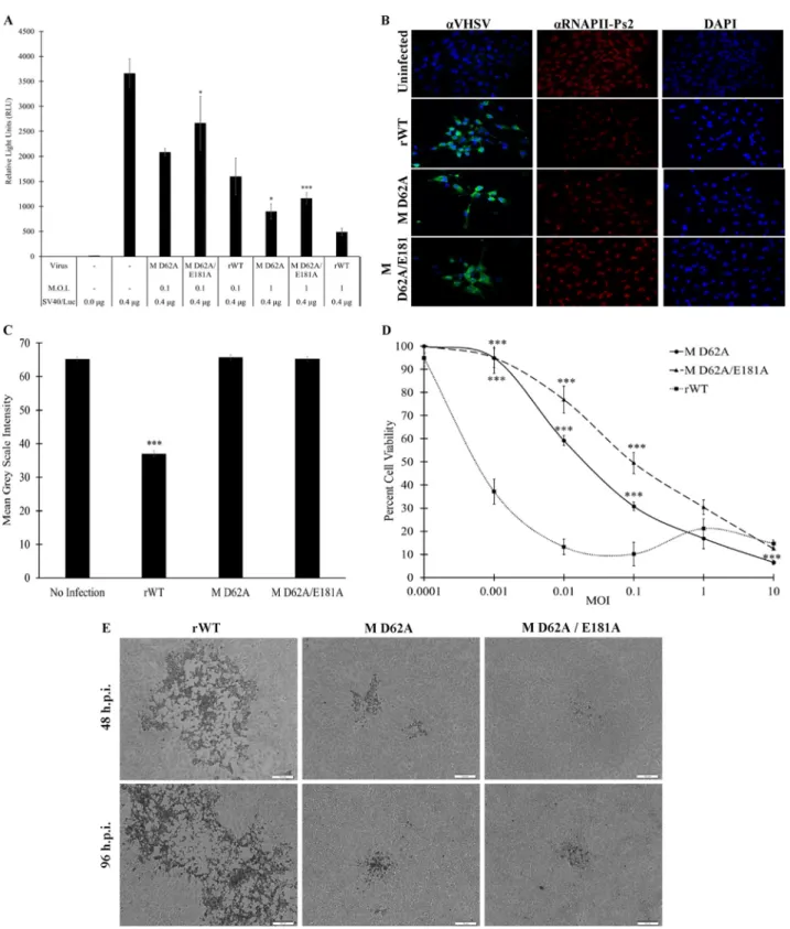
a mean grey-scale intensity within the nuclei (Fig. 10B and C). Anti-VHSV staining,
carried out in parallel cultures (due to secondary-antibody conflict), indicated that
virtually all cells were infected. Only the rWT virus significantly inhibited RNAP II
phospho-serine-2 staining, suggesting that the antitranscriptional effects of M D62A
and M D62A E181A viruses were markedly reduced compared to that of the rWT VHSV.
Cytopathic effects of rWT VHSV and mutants.
The reduced transcriptional
inhib-itory activities of the M D62A and M D62A E181A mutant viruses suggested that
induction of host cell cytopathicity was likely to be less severe for the mutant viruses
than for rWT VHSV. To assess the impact of viral titer on cytopathicity, EPC cells were
infected with each of the viruses for 96 h at MOIs ranging from 0.0001 to 10. After that
time, cells were fixed in trichloroacetic acid and stained with sulforhodamine B to
quantify viable cells. This assay showed that the mutant viruses induced significantly
fewer cytopathic effects at an MOI of
⬍
1.0 (Fig. 10D). Interestingly, in EPC cells infected
with mutant M viruses, plaques that initially appeared similar to those for rWT virus
formed, but over the course of the experiment, the mutant virus plaques remained
smaller than those generated by the rWT virus and in some cases refilled with cells after
initial formation (Fig. 10E).
To monitor global cellular RNA synthesis, we again used the Click-iT chemistry
reaction with Alexa Fluor 594 (AF-594) and 5-EU and then assessed incorporation with
both fluorescence microscopy and flow cytometry. EPC cells were left untreated or
were treated with actinomycin D (1 h prior to 5-EU addition) or infected with mutant
or WT viruses (24 h). The 5-EU was added for the last hour of treatment/infection, after
which labeled cells were subjected to flow cytometry to quantify 5-EU-positive versus
-negative cells (Fig. 11). To ensure robust infection, cells on coverslips from each
treatment were subjected to immunofluorescence microscopy using an anti-VHSV
primary and an FITC-conjugated goat anti-rabbit secondary antibody after the Click-iT
reaction was completed (data not shown). Flow cytometric data suggested that the
mutant-M-containing viruses were roughly 80% less effective at inhibiting host
tran-scription than rWT VHSV.
Innate immune activation by rWT and mutant VHSVs.
To determine how
differ-ences in transcriptional inhibition impacted antiviral activity release, media collected
from control or infected cells were UV irradiated to inactivate live virus and subjected
to IFN bioassay (Fig. 12). Both mutant M viruses elicited enhanced antiviral activity
(expressed in units of IFN [uIFN] per ml) compared to that of rWT VHSV. The mutant
with M D62A induced 1.9- and 22.5-fold-more uIFN/ml than rWT at MOIs of 0.1 and 1,
respectively, whereas the M D62A E181A virus induced 2.25- and 49.4-fold-more
uIFN/ml than rWT virus at MOIs of 0.1 and 1, respectively (Fig. 12).
FIG 9Replication of wild-type versus M D62A and M D62A E181A mutant viruses. Viral titers from media harvested from cells infected at the indicated time points at an MOI of 0.1 (A) or 1.0 (B) are shown. Error bars reflect SEM.*,P⬍0.05;**,P⬍0.01;***,P⬍0.001 for mutant virus results in a comparison with the rWT virus results at the same time point.
To determine whether the enhanced antiviral activity correlated with altered innate
immune gene expression, EPC cells were infected with the M D62A, M D62A E181A, or
rWT virus for 96 h at MOIs of 0.1 and 1, with both media and cells collected at 0, 4, 18,
36, 54, 72, and 96 h.p.i. cDNA was made from RNA extracted from the collected cells
that had been spiked with 1.0 ng of
in vitro-transcribed GFP RNA for normalization,
since virus infection alone leads to suppression of host RNA synthesis and impacts
reference gene expression. Expression of IFN, IFN-responsive Mx-1, and VHSV RNA was
quantified with RT-qPCR (Fig. 13). At both MOIs, the M D62A virus induced more IFN
and Mx-1 transcription than the M D62A E181A and rWT viruses. However, when these
values were normalized to the amount of viral RNA, the M D62A E181A virus induced
5- and 4.5-fold-more IFN and Mx-1 mRNA, respectively, than the M D62A virus at an MOI
of 1. The mutant M viruses also consistently induced 10-fold-more IFN mRNA than rWT
virus at MOIs of 0.1 and 1, respectively. These data suggested that the mutant M viruses
FIG 11Mutant M viruses elicit reduced transcriptional inhibition. EPC cells were left untreated and unstained (A), untreated and stained (B), treated with actinomycin D (C), or infected with the indicated viruses at an MOI of 1 for 24 h prior to 5-EU labeling of nascent RNA (D to F). 5-EU was conjugated to Alexa Fluor 594 by a Click-iT reaction and flow data were collected. The percentage of cells scored as AF-594 negative and AF-594 positive are shown as an inset within each histogram.
FIG 13Regulation of innate immune genes by the wild-type versus the M D62A and M D62A E181A mutant viruses. RT-qPCR results from RNA harvested at the indicated times postinfection at an MOI of 0.1 (A to C) or an MOI of 1 (D to F). Synthesized cDNAs were assessed for VHSV RNA (A, D), EPC type I IFN (B), (E), and fish Mx-1 (C, F). Data were normalized to those for a spiked internal control as described in Materials and Methods and are presented as a fold changes in expression from expression in untreated controls. Error bars reflect SEM.*,P⬍0.05;**,P⬍0.01;***,P⬍0.001 in a comparison of mutant virus results and rWT results at the same time point.
led to greater expression of IFN throughout the course of infection than did the rWT
virus.
DISCUSSION
Most viruses have evolved mechanisms to inhibit the expression or functions of host
innate immune genes during virus replication (54, 55). Inhibition of host transcription
is a common strategy used by RNA viruses that replicate entirely within cytoplasm.
Shutting down host transcription not only frees up cellular translational machinery that
can be used for the biosynthesis of viral gene products but also inhibits host antiviral
responses by preventing the synthesis of antiviral proteins. One clear example of an
antihost protein found within the rhabdovirus family is VSV M, which potently inhibits
host gene expression, thereby suppressing host antiviral responses, including
upregu-lation of type I IFNs (56, 57). VSV M also may block mRNA export from the nucleus (47,
48, 58). These antihost functions of VSV M are separable from its critical role in viral
assembly and budding but are still essential for efficient viral replication (59–61).
Salmonid rhabdovirus IHNV M also inhibits host-directed gene expression (38).
Here we report that VHSV-IVb infection suppressed host IFN-mediated antiviral
responses, with expression of M alone capable of inhibiting MAVS-mediated IFN
expression, as well as IFN-mediated transcriptional responses, in cell-based luciferase
assays (Fig. 2). Importantly, viral infection or transfected VHSV M also inhibited
tran-scription from a constitutively active SV40 promoter-driven luciferase construct (Fig. 3),
suggesting that VHSV M acts similarly to other rhabdoviral M proteins in shutting down
general transcription and/or posttranscriptional events. This possibility was enforced
and clarified by the observation that VHSV infection or ectopic expression of M led to
decreased nascent RNA transcription by the host. Our results support the hypothesis
that VHSV M inhibits host transcription as a means of promoting viral dissemination.
VSV M blocked host transcription directed by all three host RNA polymerases (RNAP
I to III [44]). We tested the inhibitory potency of VHSV M on three different luciferase
constructs driven by the RNAP I-dependent Atlantic salmon ITS1 promoter, the RNAP
II-dependent SV40 promoter, and the human RNAP III-dependent U6 promoter,
respec-tively. VHSV M cotransfection inhibited transcription mediated by all three RNA
poly-merases (Fig. 4). Interestingly, 5-EU staining of nascent RNA synthesis in VHSV-infected
and M-transfected cells resembled the pattern of
␣
-amanitin treatment (Fig. 4). Since
␣
-amanitin targets RNAP II and III, but not RNAP I, this suggests that M may target all
three host RNAPs through a common mechanism, which may not be equally efficacious
in all instances. Since continued rRNA synthesis benefits the virus, it is possible that
residual RNAP I activity is an evolutionary outcome of otherwise-indiscriminate host
RNAP suppression.
M proteins from a variety of VHSV strains and several closely related fish viruses,
including IHNV, SHRV, and SVCV, all inhibited SV40 promoter activity in EPC cells, which
is consistent with the suppression of host protein expression being a conserved role of
M among rhabdoviruses. Interestingly, a novel M clone isolated from a VHSV F1 strain
(Ia substrain) sample was less potent than other M proteins in inhibiting transcription
(Fig. 7). When comparing this F1 M sequence with that of WT VHSV-IVb M, just four
amino acid differences (T9I, D62G, E181A, and V198A) were present. We tested a range
of targeted mutations at these positions and found that G62D conversion in the Ia M
clone enhanced function to approximately that of IVb M (data not shown). To further
investigate the impact of the M alternatives (using more-traditional alanine
substitu-tions), two mutant M constructs (M D62A and M D62A E181A) were made within the IVb
M background. Both exhibited decreased ability to inhibit cellular gene expression
compared to that of WT VHSV-IVb M (Fig. 8). The predicted secondary structure of VHSV
M is quite similar to that of VSV M, even though amino acid conservation between the
two is low (data not shown). The aspartic acid residue at position 62 is conserved across
multiple VSV and VHSV strains (62). In VSV M, this aspartic acid (D92) is located between
helices
␣
1 and
␣
2 and is surface exposed (62). It may play a critical role in the antihost
function of M by serving in a structural capacity or as a site for protein-protein
interactions with host factors involved in regulating transcription. Regardless,
conser-vation of this aspartic acid residue across viruses is striking and worthy of additional
investigations.
The two M point mutants (M D62A and M D62A E181A) that exhibited significantly
reduced antitranscriptional potential in cell-based studies (Fig. 8) were incorporated
into an rWT VHSV backbone, using a reverse genetic system. The primary goal was to
determine if the M mutants would support M proteins critical functions in viral
replication and, if so, whether antihost functions of the resulting viruses were impacted.
Both the M D62A and M D62A E181A viruses were viable, which allowed us to
investigate the abilities of these mutant viruses to replicate, elicit cytotoxic effects,
inhibit host transcription, and/or otherwise modulate host gene expression. The M
D62A E181A virus exhibited reduced replicative capabilities at an MOI of 0.1 compared
to those of the WT and D62A viruses (Fig. 9). These data suggested that the D62A E181A
mutation, but not the D62A mutation alone, elicited a restrictive effect on propagation,
perhaps implicating E181 in viral packaging or budding. For VSV M, F208 is structurally
homologous to VHSV M E181 and helps coordinate the positioning of a superiorly
located
␣
-helix that forms the border of the hydrophobic pocket that VSV M utilizes in
multimerization during viral skeleton formation (63). Thus, loss of electron density at
this position might allow for a larger degree of freedom in the superior
␣
-helix, leading
to a malformed hydrophobic pocket, deficient polymerization, and thus reduced
replication. However, the most striking effects of the D62A and D62A E181A mutants
were their reduced abilities to suppress transcriptional responses in EPC cells (Fig. 8, 10,
11, and 13). As such, an alternative explanation for the reduced replication of the
double mutant might be that enhanced IFN production in infected cells was capable of
restricting propagation so effectively that the virus could not spread far beyond the
initially infected cells, particularly if replication was even slightly delayed.
Although the D62A virus fared better than the double mutant in viral-yield assays,
its inability to suppress gene expression led to decreased cytopathicity and plaque
spread at low MOIs (Fig. 10). Since the D62 residue is highly conserved among
rhabdoviruses, we predict that it is structurally or functionally important. Molecular
modeling predicted that residue D62 of VHSV M is in a random coil located proximal
to the globular domain (64). Mutations within this region impacted the localization of
VSV M (65), suggesting that the D62A mutation may have affected the ability of M D62A
mutants to localize properly, as opposed to causing a more general loss in structural
integrity. Other functions have been ascribed to the orthologous region of VSV M,
including regulation of membrane association (66) and protein turnover (59), although
both of those studies utilized larger deletions or multiple amino acid mutations than
the point mutation used here. Outside this specific coil domain, the N terminus of VSV
M has been implicated in other aspects of host inhibition, including association with
RAE1/NUP98 (45, 48) or suppression of eukaryotic initiation factor 2
␣
(eIF2
␣
)
phosphor-ylation (M51R) (67). Whether VHSV M also engages these conserved proteins and/or
whether D62A mutations impact these same cellular functions, directly or indirectly, will
have to await future studies.
Overall, our data provide insight into the various and critical roles of VHSV M in viral
replication and host suppression. The findings are consistent with many previous
studies of VSV M and confirm a conserved role for M in host suppression among many
rhabdoviruses. Our results show that, as with VSV M, the antihost functions of VHSV M
can be uncoupled from the viral packaging functions and as such lay the groundwork
for studies directed toward developing disabled or attenuated recombinant viruses
useful in host response and/or immunization studies, as well as more in-depth
func-tional analyses of the mechanisms behind the observed biological effects of VHSV M
and its mutants.
MATERIALS AND METHODS
Cell lines and culture conditions.Epithelioma papulosum cyprinid (EPC) cells were purchased from the American Type Culture Collection (ATCC, Rockville, MD; CRL-2872). The cells were grown in Eagle’s minimum essential medium (EMEM) (Fisher) supplemented with 10% fetal bovine serum (FBS) (Invitro-gen, Carlsbad, CA) and 1% penicillin-streptomycin (Invitrogen) (complete EMEM) at 22°C in a 5%-CO2
-enriched environment. Human embryonic kidney 293 (HEK-293) cells were purchased from the ATCC (CRL-1573) and grown in complete Dulbecco’s MEM at 37°C in a 5%-CO2-enriched environment. ␣-Amanitin (Santa Cruz Biotechnology, Inc., CA) and actinomycin D (Sigma-Aldrich, St. Louis, MO) were used at a final concentration of 1g/ml, while leptomycin B (LMB) was used at 10g/ml.
Plasmids.Expression vectors for EPC MAVS and EPC IFN were obtained from Michel Brémont (French National Institute for Agricultural Research, Jouy-en-Josas, France), the VHSV L expression plasmid was reported previously (53), the Tet-mSEAP construct was obtained from Fan Dong (University of Toledo, Toledo, OH, USA), and the IFN-luciferase reporter was obtained from John Hiscott (Istituto Pasteur-Fondazione Cenci Bolognetti, Rome, Italy). Other plasmids used included the IFN-induced transmem-brane 1 (IFITM1)/lucplasmid (derived from pDW9-27CD2), SV40/-galactosidase (-Gal) (Promega), and SV40/luc(modified from SV40/-Gal). VHSV-IVb M, NV, G, and N coding sequences were PCR amplified with appropriate primers (Table 1). All fragments were cloned into pcDNA 3.1(⫺)myc/His A (Invitrogen) or p3xFLAG-CMV-14 (Sigma-Aldrich). IHNV, SVCV, and SHRV M cDNAs were PCR cloned from viral stocks using targeted primers (Table 1).
Luciferase driven by a human U6 promoter reporter construct (pGL3-U6-Luc) was generated by subcloning the human U6 promoter from the pLKO.1 puro vector (Addgene plasmid 10879) into the pGL3 luciferase reporter vector (Promega E1751) upstream of the luciferase gene using Gibson Assembly (NEB E5520). The rainbow trout RNAP I promoter luciferase construct (pGL3-ITS1-Luc) was generated by subcloning rainbow trout rRNA intergenic sequence region 1 (ITS1) into the pGL3 luciferase reporter vector between two HindIII sites.
Recombinant virus production.Construction of a full-length infectious clone of VHSV has been described earlier (68). This clone was used as a backbone to introduce desired mutations in the M gene, which is flanked by unique NheI and PvuII restriction sites in the full-length clone. To construct plasmid pVHSV-D62A, primers were designed to effect the Asp-to-Ala mutation at amino acid position 62 (D62A) in M and used in PCR along with the flanking NheI forward and PvuII reverse primers to amplify a PCR fragment of 712 bp. The obtained PCR product was subjected to DNA sequencing to confirm the presence of the introduced mutation (D62A). This fragment was doubly digested with NheI and PvuII restriction enzymes and cloned between the NheI-PvuII sites of the full-length VHSV clone. To construct plasmid pVHSV-D62A E181A, another set of primers was used to effect the Glu-to-Ala mutation at position 181 of M. Using the single mutant cDNA fragment as a template, another PCR was carried out with the flanking NheI forward and PvuII reverse primers to obtain a PCR product of 712 bp. DNA of the PCR product was sequenced to confirm the presence of two mutations (D62A E181A). This product was doubly digested with NheI-PvuII and cloned into the full-length VHSV clone, as described above.
transcription to obtain cDNA fragments of the VHSV genome, which were sequenced completely to confirm the presence of introduced mutations in the M gene.
VHSV amplification and purification.The VHSV-IVb MI03GL isolate was kindly provided by James Winton (United States Geological Survey, Seattle, WA). The VHSV F1 strain was kindly provided by Gale Kurath (United States Geological Survey, Seattle, WA). VHSV-IVb isolates and recombinant viruses were amplified for subsequent purification by infecting a confluent monolayer of BF-2 cells in a 15-cm tissue culture dish with a 1:1,000 (vol/vol) dilution of unpurified virus stock in serum-free EMEM. Viral adsorption was allowed to proceed for 1 h before virus-containing medium was replaced with complete EMEM. Virus was cultured until the onset of cytopathicity was observed (72 h). Virus-containing media and attached cells were subjected to a freeze-thaw cycle before the removal of cell debris by low-speed centrifugation (4,000⫻g, 30 min). The resulting supernatant was clarified using a 0.22-m syringe tip filter and then subjected to ultracentrifugation through a 25% (wt/vol) sucrose pad at 25,000 rpm for 3 h at 4°C. The virus-containing pellet was resuspended overnight at 4°C in phosphate-buffered saline (PBS). The titers of virus stocks were determined by 1:10 serial dilution using confluent EPC cells and then divided into 100-l aliquots and stored at⫺80°C until use.
Virus yield and IFN bioassays.To assess viral replication, EPC cells were infected for 0, 4, 18, 36, 54, 72, or 96 h at an MOI of 0.1 or 1.0 with either M D62A, M D62A E181A, or rWT VHSV. Media and cells were harvested at each time point. A viral-yield assay was used to compare the abilities of mutant viruses to replicate to the level of rWT virus by determining the titer of the medium from each time point in 1:10 serial dilutions on BF-2 cells. At 72 h.p.i., plaques were counted and a final viral concentration in numbers of PFU per milliliter was calculated for each time point. Antiviral assays were completed using UV-irradiated medium (70 mJ/cm2) from each time point, which was applied overnight to EPC cells in 1:3
TABLE 1Primers for PCR analysis and cloning
Primera Sequence (5=¡3=)
Restriction site
mSEAP se GACCCTGCTCAGGACCCTC
mSEAP as GATTTGCCATCCTCAGCCTTG
U6 promoter se TCTCTATCGATAGGTACCTTTCCCATGATTCCTTCATATTTG KpnI U6 promoter as CAGTACCGGAATGCCAAGCTTCGTCCTTTCCACAAGATATATAAAG HindIII
CMV se CGTTTAGTGAACCGTCAGATCG
CMV as CCGGTGTCTTCTATGGAGGTCA
VHSv IVb M se ACGAATTCATGGCTCTATTCAAAAGAAAGCGCACCATCCTG EcoRI
VHSv IVb M as ACGGTACCCCGGGGTCGGACAGAG KpnI
IVb M mid se ACAAGCTTCAAGATAGCTGAAGC HindIII
IVb M mid as ACAAGCTTGTGATCAGGGTTTTG HindIII
VHSvNV se ACGAATTCATGACGACCCAGTCGGCAC EcoRI
VHSvNV as ACGGTACCTGGGGGAGATTCGGAGCCA KpnI
VHSvN se CAGAATTCATGGAAGGAGGAATC EcoRI
VHSvN as GTGGTACCATCAGAGTCCTCG KpnI
VHSvG se ACGAATTGATGGAATGGAATACTT EcoRI
VHSvG as GTGGTACCGACCATCTGGCT KpnI
VHSvP se CAGAATTCATGACTGATATTGAGAT EcoRI
VHSvP as GTGGTACCCTCTAACTTGTCCA KpnI
F1 M se ACGAATTCATGGCTCTGTTCAAAAGAAAGCGCATCATCC EcoRI
F1 M H as ACAAGCTTGGTACCCCGGGGCCG HindIII
F1 M K as ACGGTACCCCGGGGCCGGGCAGAGGGGG KpnI
D62A se TCTCTGTGAAGCTCAACATCCT
D62A as AGGATGTTGAGCTTCACAGAGA
SVCV M se CAGAATTCATGTCTACTCTAAGAAAG EcoRI
SVCV M as CAGGTACCATCTCCCATGAACAGGGA KpnI
SHRV M se CAGAATTCATGGCAGAATCGATCGAG EcoRI
SHRV M as CAGGTACCCTTTCTTGAGGACTCGTT KpnI
IHNV M se ACGAATTCATGTCTATTTTCAAGAGAGC EcoRI
IHNV M as CTTGGTACCTTTTTCCTTCCCCCGCTTTTCGG KpnI
Virus down se ACGGATCCAAAACGCAGATCAG Virus down as AGGGGTGAGTATACAGTGGAGT
VHS clone se AAGCTAGCACAAAAAACATGGCTCTATTCA NheI
VHS clone as TTCAGCTGGTTGTGTACACAAA PvuII
EPC IFN se TGGGTGGAAAATATCCTGAG
EPC IFN as CTCCTTATGTGATGGCTGGT Fish MX-1 se ATTAACCTGGTTGTGGTGCCATGC Fish MX-1 as TACCACTGTCCCTTCAGTGCCTTT Fish-actin se AGACATCAGGGTGTCATGGTTGGT Fish-actin as GGGGTGCTCCTCTGGGGCAA
GFP se ATGGTGAGCAAGGGCGAGGA
GFP as TAGCGGCTGAAGCACTGCACGCC
ase, sense; as, antisense.
serial dilutions. Cells treated with irradiated medium were subjected to virus challenge using sucrose-purified rWT VHSV for 72 h and then were fixed and stained with crystal violet. Stained wells were dried overnight, and dye was dissolved with 30% (vol/vol) acetic acid for spectrophotometric quantification. Absorbance values of treated wells were normalized to values obtained by untreated and uninfected wells, and 1 unit of IFN was defined as the amount necessary to provide 50% protection from virus CPE. Transfection.EPC cell transfections were performed by using PolyJet reagent (SignaGen, Gaithers-burg, MD) by following the manufacturer’s instructions. Briefly, plasmids were mixed with PolyJet in serum-free EMEM for 20 min and then added to cells in serum-free medium. Media were changed to complete medium after 3 h of incubation. Plasmid concentrations in all transfection experiments were equalized between samples by inclusion of an empty vector.
Click-iT RNA Alexa Fluor 594 imaging and immunofluorescence microscopy.Click-iT reactions were performed using a Click-iT RNA imaging kit (Invitrogen). For fluorescence microscopy, cells were seeded on poly-L-lysine-coated coverslips in a 6-well culture dish and grown to⬃70% confluence. For flow cytometry, cells were plated identically but without coverslips. Wells were then infected with either rWT, M D62A, or M D62A E181A virus at an MOI of 1.0 for 24 h. At 23 h.p.i., an uninfected well was treated with actinomycin D (1g/ml) for 1 h before 5-ethynyl uridine (5-EU) was added to all wells to a final concentration of 1 mM. Cells were allowed to incorporate 5-EU for 1 h before monolayers were washed with PBS and coverslips removed. Cells remaining on the plate were harvested by the addition of Versene (0.02% [wt/vol] EDTA in PBS) and centrifuged at 700⫻gfor 5 min at 4°C. Cells on coverslips and in Eppendorf tubes were fixed and permeabilized by the addition of fixation buffer (4.0% [wt/vol] parafor-maldehyde in Tris-buffered saline with Tween 20, pH 7.5 [TBST]) for 30 min at room temperature. Cells were washed once with 100 mM glycine in PBS to quench paraformaldehyde-induced auto-fluorescence and then once in PBS. Cells were incubated in Click-iT reaction cocktail for 30 min at room temperature in the dark and then washed with Click-iT reaction rinse buffer. For immunofluorescent costaining, cells on coverslips were blocked for 30 min at room temperature (1% bovine serum albumin [BSA] in PBS) and then with primary antibody (in 1% BSA in PBS) for 1 h. Cells were washed in PBS 3 times for 5 min each, and then FITC-conjugated secondary antibody conjugate (in 1% BSA in PBS) was added for 1 h at room temperature. After a PBS wash, the coverslips were mounted to slides with ProLong Gold antifade mountant with DAPI (4=,6-diamidino-2-phenylindole; Life Technologies, Carlsbad, CA) for 24 h and then imaged on an Olympus IX81 inverted fluorescence microscope. For flow cytometric analysis, cells were pelleted and then resuspended in 1% (wt/vol) BSA and incubated at 4°C in the dark until flow cytometry data collection using a BD Scientific LSRFortessa cell analyzer using the allophycocyanin (APC) 650/ 660-nm excitation/emission filter. Flow data were then analyzed using FlowJo single-cell analysis software v.10 (BD Biosciences, San Jose, CA).
Luciferase assays/-Gal assays.Cells were transfected with the appropriate plasmids in 12-well tissue culture plate at a density of⬃70% for 24 or 48 h. After medium removal, cells were washed with PBS twice and then lysed for 15 min on ice in 150l of 5⫻cell culture lysis reagent (diluted to 1⫻in water) (Promega, Madison, WI). Half of the clarified lysate was used to assess luciferase activity, while the other half was used for-Gal activity determination using 50l of-Gal buffer (60 mM Na2HPO4, 40 mM
NaH2PO4, 10 mM KCl, 2 mM MgSO4, 2.6 M ortho-nitrophenyl--D-galactopyranoside, 3.2 l -mercaptoethanol). The mixture was incubated for 1 h at 37°C, and then absorbance was read at 414 nm using a plate reader (SpectraMax; Molecular Devices, Sunnyvale, CA). The luciferase reading was normalized to the -Gal reading. To obtain a fold induction value, the value of each sample was normalized to the value of the negative control.
Cell fractionation.Cells transiently transfected with pCD-M were fractionated into nuclear mem-brane, soluble nuclear, mitochondrial, and cytoplasmic fractions using differential centrifugation. Briefly, three 10-cm plates were transfected with pCD-M. The following day, cells were scraped from the plate and resuspended in 5 volumes of mitochondrial isolation buffer (220 mM mannitol, 68 mM sucrose, 10 mM HEPES, pH 7.4, 10 mM KCl, 1 mM EGTA, 1 mM EDTA, 1 mM MgCl2). Cells were incubated for 5 min
on ice and then Dounce homogenized. Homogenate was centrifuged for 10 min at 1,000⫻g. The supernatant was further centrifuged for 10 min at 10,000⫻g, providing the mitochondrial pellet and cytoplasmic fraction (supernatant). The previous pellet was resuspended with RIPA lysis buffer (150 mM NaCl, 1% NP-40, 0.5% sodium deoxycholate, 0.1% sodium dodecyl sulfate [SDS], 50 mM Tris, pH 8.0, 1 mM phenylmethylsulfonyl [PMS]) and incubated on ice for 15 min. Finally, the lysate was centrifuged at 10,000⫻gfor 10 min to provide the nucleoplasm and nuclear membrane extracts. Both final pellet fractions were washed three times with the buffer used in the previous step.
Immunoblotting.Cell lysate was separated by SDS-polyacrylamide gel electrophoresis (PAGE) as previously described (69). Briefly, samples were run on 12.5% gel. Proteins then were transferred to an Immobilon P polyvinylidene difluoride (PVDF) membrane and blocked in 1% BSA in TBST. Membranes were incubated overnight with primary antibody (1:1,000 in 1% BSA in TBST) at 4°C, washed in TBST, and then incubated with secondary antibody conjugated with horseradish peroxidase (HRP) in 1% BSA in TBST (for 1 h at room temperature). Membranes were washed in TBST and incubated with enhanced-chemiluminescence reagent (Pierce, Rockford, IL) for 2 min and then visualized using a ChemiDoc-It2510
imager (UVP).
to the reaction mixture to serve as an internal control for normalization, since virus infection itself shuts down endogenous RNA synthesis. Reaction mixtures were briefly cooled to 4°C before addition of 13l of an M-MLV–reverse transcriptase mixture (4l of 5⫻M-MLV–reverse transcriptase reaction buffer, 2l of 5 mM deoxynucleoside triphosphates [dNTPs; Invitrogen], 0.5l M-MLV–reverse transcriptase [Pro-mega], and water to 13l). Samples then were incubated for 1 h at 42°C. cDNA samples were diluted 1:10 with water and then subjected to qPCR using GFP, EPC-actin, VHSV M, Mx-1, and EPC IFN primers (Table 1). RT-qPCR was performed using 5l of 2⫻Radiant green from a Lo-ROX qPCR kit (Alkali Scientific, Pompano Beach, FL), 1l of diluted cDNA, 50 ng of each primer, and water to a total volume of 10l. Reactions and data collection were performed with a Bio-Rad C1000 real-time thermocycler for 3 min of initial denaturation at 95°C, followed by 40 cycles of 15 s at 95°C for denaturation and 30 s at 60°C for elongation. Readings were taken at the end of each elongation step. Threshold numbers were obtained by an automated single point threshold within the log-linear range. Samples were normalized to EPC-actin or GFP, and relative gene expression levels were calculated using the 2⫺ΔΔCTmethod,
whereCTis threshold cycle.
ChIP assay.The chromatin immunoprecipitation (ChIP) assay was performed as previously described with the following modifications (70). Briefly, 107cells were cross-linked with 1% formaldehyde for 10
min and then quenched with 125 mM glycine for 5 min at room temperature. Nuclei were prepared in cell lysis buffer (5 mM Tris-HCl, pH 8, 85 mM KCl, 0.5% NP-40, 0.5 mM PMSF, 1⫻protease inhibitor cocktail [PIC; Thermo Scientific]) on ice for 10 min and sonicated to yield chromatin fragments (200 to 700 bp). Immunoprecipitations were performed overnight at 4°C using 1g of anti-RNAP IIa A304-405A (Bethyl Laboratories, Montgomery, TX) or IgG and then incubated with protein A-agarose (EMD Millipore, Billerica, MA), which was preequilibrated with sonicated herring sperm DNA and BSA. Immunoprecipi-tated material was washed extensively, and the cross-links were reversed. DNA from the eluted chromatin was purified with a PCR purification kit by following the manufacturer’s instructions (Qiagen). Differences in DNA enrichment for ChIP samples were determined by qPCR using 4% of the precipitated sample DNA and 1% of the input DNA. The primers used for ChIP assay are listed in Table 1.
Statistics.Data management, analysis, and graphing were done in Microsoft Excel 2016. Analysis was performed by two-tailed, unpaired Studentttests. Graphs represent the statistical means⫾standard errors of the means (SEM).
ACKNOWLEDGMENTS
This work benefitted from critical comments provided by anonymous reviewers. We
also thank Shelby Powell, Tyler Williams, Tyler Popil, and Jacob Blandford for technical
assistance with some of the experiments described herein.
This study was funded by NSF grant DBI-1354806/1354684 (D.W.L, C.A.S., V.N.V.) and
USDA/ARS CRIS project number 5090-31320-004-00D under specific cooperative
agree-ment number 58-3655-9-748 (D.W.L., C.A.S.).
The views contained in this document are those of the authors and should not be
interpreted as necessarily representing the official policies, either expressed or implied,
of the U.S. Government. Mention of trade names, proprietary products, or specific
equipment does not constitute a guarantee or warranty by the USDA and does not
imply its approval to the exclusion of other products that may be suitable.
REFERENCES
1. Janeway CA. 1989. Approaching the asymptote? Evolution and revolu-tion in immunology. Cold Spring Harbor Symp Quant Biol 54:1–13.
https://doi.org/10.1101/SQB.1989.054.01.003.
2. Janeway CA, Jr, Medzhitov R. 2002. Innate immune recognition. Annu Rev Immunol 20:197–216.https://doi.org/10.1146/annurev.immunol.20 .083001.084359.
3. Loo YM, Gale M, Jr. 2011. Immune signaling by RIG-I-like receptors. Immunity 34:680 – 692.https://doi.org/10.1016/j.immuni.2011.05.003. 4. Kawai T, Takahashi K, Sato S, Coban C, Kumar H, Kato H, Ishii KJ, Takeuchi
O, Akira S. 2005. IPS-1, an adaptor triggering RIG-I- and Mda5-mediated type I interferon induction. Nat Immunol 6:981–988.https://doi.org/10 .1038/ni1243.
5. Lin R, Mamane Y, Hiscott J. 1999. Structural and functional analysis of interferon regulatory factor 3: localization of the transactivation and autoinhibitory domains. Mol Cell Biol 19:2465–2474.https://doi.org/10 .1128/MCB.19.4.2465.
6. Seth RB, Sun L, Ea CK, Chen ZJ. 2005. Identification and characterization of MAVS, a mitochondrial antiviral signaling protein that activates NF-kappaB and IRF 3. Cell 122:669 – 682.https://doi.org/10.1016/j.cell.2005 .08.012.
7. Yoneyama M, Suhara W, Fukuhara Y, Fukuda M, Nishida E, Fujita T. 1998. Direct triggering of the type I interferon system by virus infection:
activation of a transcription factor complex containing IRF-3 and CBP/ p300. EMBO J 17:1087–1095.https://doi.org/10.1093/emboj/17.4.1087. 8. Platanias LC. 2005. Mechanisms of type-I- and
type-II-interferon-mediated signalling. Nat Rev Immunol 5:375–386.https://doi.org/10 .1038/nri1604.
9. Stark GR. 2007. How cells respond to interferons revisited: from early history to current complexity. Cytokine Growth Factor Rev 18:419 – 423.
https://doi.org/10.1016/j.cytogfr.2007.06.013.
10. Galabov AS. 1981. Induction and characterization of tortoise interferon. Methods Enzymol 78:196 –208. https://doi.org/10.1016/0076 -6879(81)78118-7.
11. Altmann SM, Mellon MT, Distel DL, Kim CH. 2003. Molecular and func-tional analysis of an interferon gene from the zebrafish, Danio rerio. J Virol 77:1992–2002.https://doi.org/10.1128/JVI.77.3.1992-2002.2003. 12. Schultz U, Kaspers B, Staeheli P. 2004. The interferon system of
non-mammalian vertebrates. Dev Comp Immunol 28:499 –508.https://doi .org/10.1016/j.dci.2003.09.009.
13. Zou J, Tafalla C, Truckle J, Secombes CJ. 2007. Identification of a second group of type I IFNs in fish sheds light on IFN evolution in vertebrates. J Immunol 179:3859 –3871.https://doi.org/10.4049/jimmunol.179.6.3859. 14. Sun B, Robertsen B, Wang Z, Liu B. 2009. Identification of an Atlantic
salmon IFN multigene cluster encoding three IFN subtypes with very