THE EFFECT OF FOOTBALL SHOULDER PAD REMOVAL TECHNIQUE AND EQUIPMENT REMOVAL TRAINING ON CERVICAL SPINE MOTION, TIME TO
TASK COMPLETION, AND PERCEIVED TASK DIFFICULTY
Catherine Shawn Lenhardt
A thesis submitted to the faculty of the University of North Carolina at Chapel Hill in partial fulfillment of the requirements for the degree of Master of Arts in the Department of Exercise and Sport Science (Athletic Training).
Chapel Hill 2014
©2014
ABSTRACT
Catherine Shawn Lenhardt: The Effect of Football Shoulder Pad Removal Technique and Equipment Removal Training on Cervical Spine Motion, Time to Task Completion, and
Perceived Task Difficulty
(Under the direction of Jason P. Mihalik)
Current recommendations for management of cervical spine injury suggest
leaving football equipment in place unless otherwise indicated by the NATA position
statement. We investigated the effect of three shoulder pad removal techniques and the
effect of reinforced training on cervical spine motion, time, and difficulty. The RipKord
shoulder pads were faster than both traditional shoulder pad removal techniques
(P<0.001) and easier (P<0.05) than the flat torso to remove. Less cervical spine range of
motion with the flat torso technique was observed in the sagittal and frontal planes
(P<0.05) during Testing Session II in the reinforced training group. Both traditional
shoulder pad removal techniques were faster during Session II [flat (P=0.001); elevated
(P<0.001)]. The RipKord shoulder pads provided a method for removal with superior
measure of time and difficulty compared with traditional removal techniques. With
ACKNOWLEDGMENTS
A project as meaningful as this one deserves many thanks. Particularly to my
faculty and doctoral advisors, I extend my deepest gratitude to Jason Mihalik and Rob
Lynall for their commitment to guiding, challenging, and mentoring me throughout this
entire process. To my thesis committee: Meredith Petschauer, Erik Swartz, and Melissa
Fraser, thank you each for your specific contributions to the excellence of my research. I
also owe thanks to the faculty, staff, doctoral students, graduate assistants, and
undergraduate students who spent many hours participating and assisting in the
completion of data collection. Thank you to all who contributed thoughtful advice and
support during this process.
I am thankful for the opportunity I had to attend graduate school at the University
of North Carolina at Chapel Hill, for the support from my loving family, for my steadfast
friends and mentors, for everyone who has encouraged me to reach higher than I could
dream, and for God’s faithfulness through each step.
I dedicate this document to Fred Thompson, a football player who lost his life
from an episode of sudden cardiac death. Since his death, my prayer has been that my
research would contribute to best practices in the domain of emergency management of
catastrophic injury with the hope that well-prepared certified athletic trainers would
TABLE OF CONTENTS
LIST OF TABLES ... vii!
LIST OF FIGURES ... viii!
CHAPTER I ... 1!
Variables ... 4!
Research Questions & Hypotheses ... 5!
Operational Definitions ... 6!
Assumptions ... 6!
Delimitations ... 7!
Limitations ... 7!
Clinical Significance ... 7!
CHAPTER II ... 9!
Epidemiology ... 9!
Mechanism of Injury ... 11!
Pathology ... 13!
Recommendations for Management ... 22!
Cervical Spine Injury Research Biomechanics ... 26!
Experience & Training ... 31!
Methodological Considerations ... 34!
Participants ... 37!
Instrumentation ... 37!
Procedures ... 40!
Data Reduction ... 45!
Statistical Analyses ... 46!
CHAPTER IV ... 48!
Introduction ... 48!
Methodology ... 51!
Instrumentation ... 51!
Procedures ... 54!
Data Reduction ... 59!
Statistical Analyses ... 60!
Results ... 61!
Discussion ... 62!
LIST OF TABLES
LIST OF FIGURES
Figure 4.1 – RipKord shoulder pads and wood head block ...73
Figure 4.2 – Starting position for each removal technique ...74
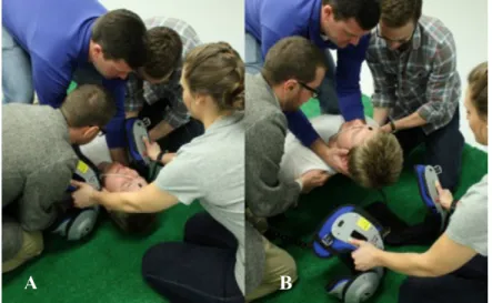
Figure 4.3 – Elevated torso removal technique ...75
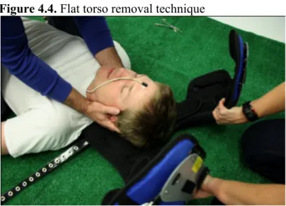
Figure 4.4 – Flat torso removal technique ...76
CHAPTER I
INTRODUCTION
Football athletes are at a high risk for sustaining cervical spine injuries (Mueller & Cantu, 2012). Football produces the highest total number of catastrophic spine injuries of all sports in the United States (Mueller & Cantu, 2012). Currently, the standard of care for a spine injured football athlete is to leave all equipment in place, with the exception of the facemask, while providing rescue care unless one or more of the following conditions are present: 1) access to the airway is not possible or removal of the facemask is
unachievable in an appropriate amount of time; 2) the helmet is not properly fit and therefore securing the helmet to the spine board does not result in sufficient
immobilization of the head and cervical spine; or 3) leaving the helmet on does not result in neutral alignment of the cervical spine (Swartz et al., 2009). If one of the three
assuming a potentially dangerous extension position following helmet removal with the
shoulder pads still in place (Swartz et al., 2009).
Traditionally, the flat torso technique has been employed for shoulder pad
removal. This method involves 2-4 rescuers, and is accomplished by unfastening the side
straps of the shoulder pads, cutting the laces that hold the shoulder pads together
anteriorly, and sliding the remaining portion of the shoulder pads over the top of the
injured athlete’s head (Horodyski et al., 2009; Swartz et al., 2009). If four or more
rescuers are available, the elevated torso technique may be employed. The shoulder pads
are unfastened in the same manner as the flat torso technique. Shoulder pad removal is
achieved after the patient is elevated to 30-40 degrees of trunk flexion, or just enough to
allow for unencumbered removal of the shoulder pads while neutral alignment of the
head, neck, and torso are maintained (Horodyski et al., 2009; Peris, Donaldson, Towers,
Blanc, & Muzzonigro, 2002). One cadaveric study reported the elevated torso technique
as a superior method for moderating the amount of induced cervical spine motion during
shoulder pad removal when compared with the flat torso technique (Horodyski et al.,
2009). However, Dahl et al. note that, while described as a viable option for equipment
removal by the National Athletic Trainers’ Association position statement, the elevated
torso technique (tilt technique as described by the study) results in a greater amount of
vertebral displacement between intact and lesioned cervical spines in cadaveric models
compared to the log roll and five-person lift techniques (Dahl, Ananthakrishnan, Nicandri,
Chapman, & Ching, 2009).
The Riddell™ RipKord technology has recently emerged to address the growing
RipKord shoulder pads are identical to traditional shoulder pads in nearly all aspects,
except for the RipKord itself and its posterior attachment conducive for separation. The
RipKord is a guided cable that, when pulled, allows the shoulder pads to separate into
right and left halves posteriorly. This allows each side of the shoulder pad to slide out
independently from underneath the athlete, provided the anterior attachment is released
(Kordecki, Smith, & Hoogenboom, 2011). In one study investigating this new technology,
the authors found that removing shoulder pads utilizing the Riddell™ RipKord system
resulted in significantly less time to task completion compared to traditional shoulder
pads utilizing the flat torso technique. However, the authors found no significant
differences in cervical spine motion or in perceived difficulty between the two techniques
(Bric, Swartz, S.J., Decoster LC, & J.P, 2013). It is necessary to investigate the
Riddell™ RipKord shoulder pad technology as its design yields the potential for a safer
method of equipment removal by reducing the risk of iatrogenic pathology to a
potentially spine injured athlete. To our knowledge, no investigation had directly
compared each of the 3 equipment removal techniques (elevated torso with traditional
shoulder pads, flat torso with traditional shoulder pads, and RipKord shoulder pad
removal) under a single study design. Furthermore, many institutions complete spine
boarding and equipment removal training sessions each year. However, no studies, to our
knowledge, had explored the effect of reinforced training on the successful application of
these three removal techniques. Thus, further investigation was warranted.
Therefore, the purpose of this study was twofold. The first aim was to compare
induced head motion, time to task completion, and perceived difficulty during football
traditional shoulder pads, 2) flat torso with traditional shoulder pads, and 3) Riddell™
RipKord shoulder pad removal. The second aim of the study was to measure the effect of
reinforced equipment removal training on induced head motion, time to task completion,
and perceived difficulty during football equipment removal.
Variables
Independent variables
1. Testing Session
a. Session I
b. Session II
2. Equipment removal technique
a. Elevated torso with traditional shoulder pads
b. Flat torso with traditional shoulder pads
c. Riddell™ RipKord shoulder pad removal
3. Training group
a. Reinforced training
b. Control
Dependent variables
1. Head to thorax integrated motion and range of motion in degrees
a. Sagittal
b. Frontal
c. Transverse
2. Time to task completion in seconds
Research Questions & Hypotheses
Research Question 1: What is the effect of shoulder pad removal technique (elevated
torso with traditional shoulder pads, flat torso with traditional shoulder pads, and
Riddell™ RipKord shoulder pad removal) on induced head motion, time to task
completion, and rate of perceived exertion (RPE)?
Hypothesis 1.1: We hypothesized that the Riddell™ RipKord shoulder pad
removal would result in less induced head motion, shorter time to task completion,
and a lower RPE than the elevated torso technique with traditional shoulder pads.
Hypothesis 1.2: We hypothesized that the flat torso technique with traditional shoulder pads would result in no differences in induced head motion or RPE, but
would take significantly more time to complete than the Riddell™ RipKord
shoulder pad removal (Bric et al., 2013).
Hypothesis 1.3: We hypothesized that the elevated torso technique with
traditional shoulder pads would result in significantly less induced head motion
(Horodyski et al., 2009), but would take significantly more time to complete with
a higher RPE than the flat torso technique with traditional shoulder pads.
Research Question 2: What is the interaction between reinforced equipment removal
training and control groups on induced head motion, time to task completion, and RPE
across the three shoulder pad removal techniques (elevated torso with traditional shoulder
pads, flat torso with traditional shoulder pads, and Riddell™ RipKord shoulder pad
removal)?
session during each of the three shoulder pad removal techniques (elevated torso
with traditional shoulder pads, flat torso with traditional shoulder pads, and
Riddell™ RipKord shoulder pad removal) compared to the control group.
Operational Definitions
Time to task completion: Time to task completion was measured from the initiation of cervical spine stabilization, to the moment the injured patient model was returned to the
flat surface (following shoulder pad removal) with the head and neck resting in neutral.
Integrated head motion: Measured as the absolute values normalized to time (seconds) of head-to-thorax motion the head passes through in each plane: sagittal, frontal, transverse.
Range of motion: Measured as the maximum and minimum head-to-thorax motion, or excursion, in each plane: sagittal, frontal, transverse.
Equipment intensive sports: When asked on a demographics survey, equipment intensive sports included football, men’s lacrosse, and ice hockey.
Experienced clinicians with equipment intensive sports: Experience with an equipment intensive sport was categorized as greater than or equal to 5 years experience with
football, men’s lacrosse, or ice hockey at any level of competition.
Assumptions
1. A prone athlete would have already been log rolled to a supine position.
2. There were no contraindications for alignment of the cervical spine during the log
roll.
3. The helmet would have already been removed.
4. Cervical spine motion was estimated and measured by induced head to thorax
5. The torso was moved in the same manner for each subject pair during each
equipment removal technique.
Delimitations
1. We used one set of Riddell™ RipKord shoulder pads for both the traditional and
RipKord removal techniques. No other shoulder pad equipment was used in this
study.
Limitations
1. Equipment was fit according to manufacturer guidelines for both traditional
shoulder pads and the Riddell™ RipKord shoulder pads. The study design did not
account for athlete alterations of the equipment or wear over time.
2. Participant experience with equipment intensive sports and emergent equipment
removal varied.
3. Induced head motion was used to make conclusions about cervical spine motion.
Motion at the cervical spine was not measured directly.
4. Six injured patient models were utilized for the completion of data collection.
5. Participants were collected as a convenience sample and tested in pairs.
Clinical Significance
Certified athletic trainers must be educated and prepared to initiate football
equipment removal in the event they are responsible for providing medical coverage.
While not all athletic trainers will work in equipment-intensive sports such as football, it
is paramount that those who do are properly trained in football equipment removal
techniques. Differentiating between equipment removal techniques in terms of induced
clinician to select the most appropriate removal technique for their particular setting. We
anticipated our study to yield clinically meaningful information, in order to provide
clinicians with additional information to support best practices for acute management of
CHAPTER II
REVIEW OF LITERATURE
Head down tackling and catastrophic cervical spine injuries continue to occur in American football despite the advent and implementation of spearing rules. It is
necessary that an athlete with a potential cervical spine injury receive the most
conservative, immediate, and appropriate care possible so as to avoid further injury. This thesis project sought to compare induced head motion, time to task completion, and perceived difficulty during football equipment removal between elevated torso with traditional shoulder pads, flat torso with traditional shoulder pads, and the new Riddell™ RipKord shoulder pad removal techniques. Additionally, we measured the effect of reinforced training in equipment removal on the aforementioned clinical measures. The purpose of this literature review was to establish the foundational underpinnings of the proposed project, its research questions, and to provide the rationale supporting the study hypotheses.
Epidemiology
elevated risk of cervical spine injury (Bailes, Petschauer, Guskiewicz, & Marano, 2007).
In 1976, the National Collegiate Athletic Association implemented a rule in which
intentional spearing, or the use of the head as the initial point of contact with the intent of
punishing an opponent, was banned from the sport of football (Chao, Pacella, & Torg,
2010; Mueller & Cantu, 2012). This rule was then changed in 2005 to reflect both
intentional and unintentional use of head-first contact (Mueller & Cantu, 2012).
Prevention of cervical spine injuries that result from axial loads is dependent on
the ability to prevent both intentional and unintentional head-first contact. Instruction in
proper tackling techniques for both defense and offense is a key factor as injuries occur
on either side of ball possession (Mueller & Cantu, 2012). Head-up tackling, where
contact with an opponent is made with the shoulder or the chest, is the safest means of
tackling another player in football (Heck, Clarke, Peterson, Torg, & Weis, 2004). It is
crucial that this technique be coached and practiced at all positions such that it becomes
instinctive to make contact with an opponent with the head held upright (Heck et al.,
2004). Additionally, consistent officiating is necessary for penalties to be
nondiscriminatory. Every head-down contact should be flagged and awarded a penalty to
continue discouragement of head-down tackling for the safety of all participants
regardless of intention (Heck et al., 2004).
Annually from 1997 to 2006, there were approximately 7.8 catastrophic cervical
spine injuries resulting in incomplete recoveries, and 6 incidences of quadriplegia
(Mueller & Cantu, 2010). In 2011, 8 football-related cervical spinal cord injuries were
recorded, 7 in 2010, 9 in 2009, and 14 in 2008 (Mueller & Cantu, 2012). In total, from
324 injuries that have resulted in incomplete recovery, 77.2% of which followed some
sort of tackle (Mueller & Cantu, 2012). Of those who suffered a catastrophic injury from
tackling specifically, 24.8% followed a head down tackling attempt (Mueller & Cantu,
2012).
Despite the rule changes, improvements in coaching methods to increase player
tackling safety, increased time spent on practicing proper tackling technique, and all other
attempts made to eliminate catastrophic spine injuries in football, cervical spine injuries
still occur. Consequently, it is vital to assess and improve all associated variables, in our
effort to lower the annual number of cervical spine injuries. This includes thorough first
responder preparation to ensure optimal initial care of the injured athlete.
Mechanism of Injury
The most common mechanism of injury to the cervical spine is a forceful load to
the top of the head along the longitudinal axis of the cervical spine, commonly called
axial loading (Bailes et al., 2007; Cantu, Li, Abdulhamid, & Chin, 2013; Chao et al.,
2010; Ivancic, 2012). A neutral cervical spine is oriented in a lordotic position such that
when the cervical spine is flexed 30 degrees, the vertebrae lose their normal curvature
and assume a straight alignment. (Chao et al., 2010). Chao et al. describe this position of
the cervical spine stating that the principles of mechanical engineering are the same in the
flexed cervical spine as they are with an architectural segmented column such that
compression will result in elastic instability, buckling, and ultimately failure (Chao et al.,
2010). When the cervical spine is in normal alignment, axial forces are appropriately
dissipated to bone, intervertebral discs, and surrounding soft tissue structures. However,
helmet, the compressive forces from the rapidly decelerating head and accelerating torso
are absorbed primarily by the cervical spine, rather than being properly dispersed to soft
tissue (Clark, Ducker, & Cervical Spine Research Society. Editorial Committee., 1998;
Heck et al., 2004; Ivancic, 2012). When this load exceeds the amount the cervical spine
can bear, soft and hard tissue will fail (Swartz, Floyd, & Cendoma, 2005). This may
result in several different independent or concurrent cervical spine injuries. Intervertebral
disc deformation occurs, causing the cervical spine to move into a further flexed position
(Chao et al., 2010; Torg, Vegso, O'Neill, & Sennett, 1990). If the force is large enough,
this forward “buckling” of the cervical spine will continue, leading to fractures and/or
dislocations of the cervical vertebrae or herniation of the associated intervertebral discs
(Chao et al., 2010; Torg et al., 1990). Unstable fractures with or without a dislocation are
the most common cause of catastrophic cervical spine injury in collision sports (Banerjee
et al., 2004b). Thus, highlighting the danger head down tackling can impose on an athlete.
Cantu et al. note a slightly flexed cervical spine will fail under less than 150 ft lbs of
kinetic energy when tested in a controlled environment. A football player in motion is
capable of exceeding this kinetic energy threshold by as much as 10 times (Cantu et al.,
2013).
In 2012, Ivancic used five cadavers with healthy cervical spines to investigate the
effect an impact to the crown of the head has on the cervical spine. The investigator fixed
the C5 (fifth cervical vertebra) or C6 vertebra of each cadaver and then positioned the
occiput to form a 30 degree angle of the head relative to the cervical spine. This position
mimics a head down tackle in football. All five cadavers underwent a single impact with
4.1 m/s to the crown of the head. The vertebrae experienced high amounts of compressive
forces during the 50-60 millisecond time interval as shown on high-speed video. With the
neck staying in anatomical flexion limits, the large axial load caused the cervical spine to
fail in an “s-shaped curvature”, such that the central cervical vertebrae moved into
extension and two milliseconds later the upper cervical vertebrae moved into flexion.
Following the impact, Ivancic both visually inspected and used fluoroscopy imaging to
inspect the inflicted damage finding fractures of the occiput, first and second cervical
vertebrae, and facet joints (Ivancic, 2012).
To summarize, head down tackling places the cervical spine in flexion. This is a
vulnerable position for the cervical spine as an axial load to the head and neck in this
position may result in catastrophic injury. The abnormal transmission of forces along the
longitudinal axis of the cervical spine increases the risk of cervical spine fractures, joint
dislocations, disc herniations, and resultant spinal cord/nerve root damage. These
phenomena will be later discussed in the literature review.
Pathology
Cervical spine injuries vary in severity, but all potential injuries should be
managed in the same manner. It is important, however, to note various cervical spine
injuries present differently, and the ability of the clinician to recognize the signs and
symptoms of specific injuries allows for appropriate care to be administered. Possible
injuries include soft tissue strains, ligamentous sprains, fracture or dislocations of the
vertebrae and/or associated intervertebral discs, and complete or incomplete spinal cord
are examples of cervical spine injuries and associated symptoms that warrant close
attention by clinicians.
Soft Tissue Injuries
Soft tissue injuries may occur as a result of many different mechanisms, yet most
muscle strains, ligament sprains, or contusions to the cervical spine will not present with
neurological symptoms, deformity, or bony pathology (Cantu et al., 2013). Muscle strains
are graded on a three-degree scale. First-degree muscle strains involve a stretch with
small amounts of damage to the muscle fibers. Primary symptoms of a first degree strain
include pain with muscle contraction, pain with palpation, and minimal swelling at the
site of injury (Starkey, Brown, Ryan, & Starkey, 2010). Second-degree muscle strains
involve more damaged muscle fibers than first-degree strains. Symptoms are similar to
first degree strains with the addition of ecchymosis at the site of the injury (Starkey et al.,
2010). Third-degree muscle strains involve complete tearing of the muscle fibers
resulting in loss of muscle function, weakness, palpable deformity in the muscle fibers,
swelling, discoloration, and pain (Starkey et al., 2010). Ligament sprains are also graded
on a three-degree scale. First-degree sprains result from a stretch of the fibers with little
to no tearing of the ligament. Symptoms of a first degree sprain include local pain,
minimal tenderness to palpation, and a small amount of swelling (Starkey et al., 2010).
Second-degree sprains result in partial tearing of the ligamentous fibers such that joint
laxity may occur when the ligament is stressed during clinical examination. Symptoms of
a second-degree sprain include pain and swelling. Loss of proper joint function may also
occur (Starkey et al., 2010). A ligament that has lost its integrity completely indicates a
laxity with an empty end feel upon clinical evaluation. Symptoms of a third-degree sprain
include swelling, loss of proper joint function, and ecchymosis at or distal to the site of
injury (Starkey et al., 2010). Contusion injuries to soft tissue result from a direct blow.
Symptoms include pain, redness, discoloration, and ecchymosis. Bony contusions are
extremely painful and result from a direct blow to superficial hard tissue (Starkey et al.,
2010). These injuries require treatment and rehabilitation, but can typically be considered
far less severe than other potential injuries at the cervical spine. Athletes who suffer
contusions may return to play when neck pain with and without palpation has resolved,
no symptoms return with and without cervical compression along the longitudinal axis of
the spine, and when full range of motion and strength at the cervical spine have returned
(Cantu et al., 2013).
The intervertebral discs provide shock absorption and load distribution between
the vertebral bodies during weight bearing or loading (Clark et al., 1998). Intervertebral
disc herniations can cause damage to the nearby neurological tissue. Disc herniations
vary in severity ranging from the least severe, protrusion, to the most, sequestration.
Protrusion involves a small amount of the nucleus pulposus encroaching on the annulus
fibrosus, but does not exit through the entire annulus fibrosus structure. Sequestration
involves the nucleus pulposus material passing completely through an opening in the
annulus fibrosus in a pathologic manner. All categories, in some fashion, involve the
extrusion of the nucleus pulposus out of its normal containment by the annulus fibrosus.
Damage to the annulus fibrosus allows this to occur. The intervertebral disc is the
primary structure resisting high load compression (Clark et al., 1998). When the load is
increases the pressure centrally forcing it against the annulus fibrosus. This distributes the
force across the body of the vertebrae and makes the disc rigid and resistant to load. If the
integrity of the annulus fibers is compromised, the disc is unable to resist the same
compression magnitude and the contents of the disc may herniate. This is often the result
of a combination of flexion and lateral bending of the cervical spine. Cervical forward
flexion is restrained by the posterior cervical musculature, the ligamentum flavum,
interspinous ligaments, and supraspinous ligaments. Extension is limited by anterior
cervical musculature and places an increased load on the facet joints of the cervical
vertebrae (Clark et al., 1998). The herniation of disc materials can cause compression on
the adjacent nerve roots as they exit the spinal cord and ultimately cause radicular
symptoms into the cervical region, associated dermatomes, and myotomes of the upper
extremity. Symptoms may include pain, spasm, altered sensation, and weakness (Starkey
et al., 2010).
Nervous Tissue Injury
Nerve root or brachial plexus injury, often referred to as brachial plexus
neuropraxia or burner/stingers, is one of the most commonly occurring injuries in football
(Bell, 2007; Rihn et al., 2009). Two mechanisms of head and neck motion can result in
neuropraxia symptoms. The first involves a traction injury where the head and one
shoulder are forced in opposite directions. This results in elongation of the brachial
plexus on the same side as the involved shoulder, and radicular symptoms in the
associated upper extremity (Rihn et al., 2009). A compression injury is also common in
football and can result in brachial plexus neuropraxia symptoms. This is caused by
nerve roots as they exit the narrowed foraminal canal of the cervical vertebrae (Chao et
al., 2010). Symptoms, resulting from a temporary block in nerve conduction to the
peripheral nerves, may include pain, weakness, tingling, and other paresthesias. It is
common for these symptoms to present unilaterally in the upper extremity (Chao et al.,
2010; Rihn et al., 2009). These symptoms usually resolve in a few minutes, but may take
up to 24 hours to resolve completely (Rihn et al., 2009).
Spinal cord injury can occur following a variety of mechanisms including
hyperflexion, hyperextension, axial loading, or indirectly through other methods (Clark et
al., 1998). Injury to the spinal cord can be classified either as complete or incomplete.
Complete injury to the spinal cord results in full function loss below the level at which
the lesion occurred (Banerjee, Palumbo, & Fadale, 2004a). This may follow a physical
injury to the spinal cord itself, but it most commonly follows a hemorrhage or loss of
blood supply to the cord permanently blocking the transmission of impulses (Banerjee et
al., 2004a). As seen with central cord syndrome, however, not all pathology to the spinal
cord results in permanent loss of function. Central cord syndrome is considered a less
severe injury than its counterparts, but is the most frequently occurring spinal cord injury
(Banerjee et al., 2004a). This syndrome results in incomplete motor loss and weakness
affecting the upper and/or lower extremities, yet it may not affect them both equally with
larger motor deficits typically noted in the upper extremities (Bailes et al., 2007). Central
cord syndrome is most commonly associated with a hyperextension mechanism with no
concurrent cervical fracture. A resultant folding of the ligamentum flavum causes
temporary compression of the spinal cord and potentially the nearby vascular supply as
spinal cord injury with impermanent effects, but its symptoms and presentation upon
initial evaluation would warrant immediate care.
Anterior cord syndrome affects the anterior portion of the spinal cord and the
associated blood supply (Bailes et al., 2007). Second to central cord syndrome, anterior
cord syndrome is the second most common spinal cord injury (Banerjee et al., 2004a).
This syndrome causes complete loss of all motor function and sensation below the level
of the spinal cord lesion. Unlike the uneven deficit distribution characteristic of central
cord syndrome, anterior cord syndrome is nondiscriminatory affecting all extremities
equally (Bailes et al., 2007). This particular injury has been noted following a number of
spinal injury mechanisms with no specific primary mechanism (Bailes et al., 2007).
Regardless, disruption of the blood supply to the anterior spinal cord via the anterior
spinal artery appears to be a large contributing factor (Bailes et al., 2007). Anterior cord
syndrome is seen as a complete spinal cord injury with permanent function and sensation
damage. Due to the concern for blood supply in anterior cord syndrome, it is important
emergency care be initiated immediately to encourage fast and appropriate transportation
to a hospital for further assessment.
Other spinal cord syndromes including Brown- Séquard syndrome and posterior
cord syndrome result from similar mechanisms and present with similar symptoms as the
aforementioned conditions (Bailes et al., 2007). Most spinal cord syndromes present with
motor and sensation loss, and can affect the upper extremity, lower extremity, ipsilateral
side, contralateral side, and a variety of combinations therein. Many incomplete injuries
occur to the spinal cord that do not necessarily fall within each of these defined
sensory component that is not always distributed in a predictable fashion (Bailes et al.,
2007).
Neuropraxia of the spinal cord can occur following hyperextension, hyperflexion,
and even axial compression injuries during football (Rihn et al., 2009) Neuropraxia of the
spinal cord is not the same injury as a brachial plexus or nerve root neuropraxia.
Neuropraxia of the spinal cord, also known as transient quadriplegia, is characterized by
paralysis of motor function, loss of sensation of the extremities depending on the location
of the insult to the spinal cord, burning pain, and paresthesias (Bell, 2007; Chao et al.,
2010; Rihn et al., 2009). Symptoms typically last between 5 and 15 minutes, but can take
up to 48 hours to resolve (Bell, 2007; Chao et al., 2010; Rihn et al., 2009). Similar to
brachial plexus neuropraxia, the symptoms arise with fervor, but resolve completely in a
relatively short period of time.
Permanent quadriplegia is an irreversible spinal cord injury, which typically
occurs following an axial compression mechanism. This particular mechanism, which
will be described in detail, may result in a vertebral fracture or dislocation, most
commonly in the lower cervical spine, leaving the cervical spine inherently unstable
(Banerjee et al., 2004b; Chao et al., 2010). This unstable spine can no longer function as
it would normally to protect the now vulnerable cervical spinal cord. The spinal cord then
experiences dangerous deformation with permanent functional disruption of the
components of the cord that are responsible for impulse transmission (Chao et al., 2010).
It is this disruption of nerve impulse capabilities that results in permanent neurological
damage including complete loss of sensory and motor function below the level of the
The severity of the symptoms associated with cervical spinal cord damage is
dictated by the injury location. Damage at the C3-C4 level may cause complete paralysis
of all four extremities, the abdomen, and the diaphragm as well as sensation loss below
the clavicle (Clark et al., 1998). Loss of diaphragmatic control will likely result in
respiratory compromise due to its crucial role in breathing (Tortora & Derrickson, 2010).
Injury to the C4-C5 spinal cord level will spare function of the trapezius muscle for head
extension and shoulder shrugging. However, paralysis of the upper extremities, lower
extremities, and the trunk will result. The ability to breathe is still present as
diaphragmatic control is spared. Injury to the C5-C6 level will produce diminished
function of the distal upper extremities. Only hyperextension at the wrists is preserved,
while fine motor movements in the fingers are compromised. Motion into elbow flexion
may be weakened and voluntary motion into extension is lost. Pain sensation in the
fingers will be absent as well. Injury at the C7-T1 level will result in the ability to flex the
fingers into a fist but strength is compromised. Upper extremity extension will be weak
and fine motor movements in the fingers will be diminished. Finally, damage below C7
and T1 may spare the upper extremity and trunk depending on the level of the injury.
This injury will likely result in lower extremity paralysis with pain sensation
compromised in the affected myotome distribution. It is important to note that damage at
these levels may affect proximal or distal levels of the spinal cord as a result of
hemorrhage and is likely worsened with poor immediate management and immobilization
Bony Injuries
Permanent quadriplegia and neurological deficits that occur from participation in
football are in large part due to fractures and dislocations (Bailes et al., 2007). Fractures
and dislocations can occur at any cervical spine level, any location within each cervical
vertebra, or at articulations between the superior and inferior vertebrae. Upper cervical
spine fractures and dislocations, however, are rare with the majority of injuries occurring
in the lower cervical spine (Banerjee et al., 2004b). These injuries to the cervical vertebra
can result in both transient and permanent symptoms (Banerjee et al., 2004a). Chao et al.
identified two particular vertebral fractures that both may result from axial loading, the
mechanism of injury mentioned previously. The first is a fracture of the anteroinferior
corner of the vertebrae that does not result in permanent neurological damage. The
second is a fracture in two planes, sagittal and frontal, which typically results in
permanent neurological damage. Neurological symptoms from this injury include
paralysis and loss of sensation distal to the spinal cord fracture (Chao et al., 2010; Tortora
& Derrickson, 2010). It is important to note, however, that cervical spine fractures alone
do not necessarily cause spinal cord damage or neurological deficits. Fractures improve
the likelihood a bony fragment may lacerate the cord or the resultant swelling may place
increasing pressure on the spinal cord producing further damage (Starkey et al., 2010).
Dislocations at the cervical spine are inherently more dangerous to the spinal cord
than cervical vertebral fractures. When the cervical spine in flexed and rotated the facet
joints become incongruent and a dislocation may result. The normal congruency of the
cervical spine is compromised with the pathological vertebra now encroaching the
cord pressure and may result in signs and symptoms similar to that of a brachial plexus
injury. Unlike brachial plexus neuropraxia, the symptoms of a cervical spine dislocation
do not rapidly diminish (Starkey et al., 2010).
Respiratory Compromise
Unmanaged or unsuccessful management of respiratory emergencies may result
in the most grievous outcome of cervical spine injury, death. Spinal cord injuries
occurring above C5 can result in complete paralysis of the diaphragm and accessory
muscles responsible for both inhalation and exhalation (Brown, DiMarco, Hoit, &
Garshick, 2006). In the event this vital life function is compromised due to a cervical
spinal cord injury, mechanical breathing assistance is necessary to improve the
probability of survival. Furthermore, Claxton et al. found injury at or above C4 is an
independent predictive factor for death following spinal cord injury (Claxton, Wong,
Chung, & Fehlings, 1998). Inherently, death is the most severe potential outcome
following cervical spine injury, thus appropriate management is crucial.
Cervical spine injuries occur on a wide severity spectrum from muscle strains and
ligament sprains to permanent quadriplegia or death. It is necessary that emergency
responders perform a thorough and efficient initial assessment on the field in order to
properly handle these conditions. Maintaining life and immobilizing the cervical spine
should be revered as the primary responsibility of the responder.
Recommendations for Management
It is imperative clinicians limit the amount of cervical spine motion induced
during cervical spine injury management (Bailes et al., 2007). Upon initial evaluation, the
airway, breathing, and circulation are assessed. The goal of the primary survey is to rule
out life threatening injuries (Bailes et al., 2007). The spine injury emergency protocol
should be initiated if the athlete is unconscious or their level of consciousness is altered,
if they display bilateral neurological deficits or abnormalities, if they express cervical
spine or neck pain with or without palpation, or if there is obvious spinal deformity
(Swartz et al., 2009). If the patient is conscious and responsive, it is the responsibility of
the clinician to question the injured athlete inquiring about numbness, abnormal
sensations, and neck pain (Bailes et al., 2007). Should one or more of the aforementioned
signs or symptoms be present upon initial evaluation, treatment should include rapid,
immediate stabilization of the cervical spine. When the injured athlete is a football player
dressed in full equipment, including helmet and shoulder pads, the following protocol
should be conducted. The responding clinician should be positioned at the top of the
injured athlete’s head with their hands placed on either side of the helmet at the level of
the mastoid processes (Bell, 2007; Swartz et al., 2009). A firm grip should limit the
motion of the helmet and, ideally, the motion that occurs at the head and neck. In the
event the injured athlete is prone, the first responder’s arms should be in contact with the
helmet at the same level as if the athlete were supine, but their arms must be crossed upon
initial immobilization of the cervical spine such that they become uncrossed as the
injured athlete is log rolled to a supine position (Swartz et al., 2009). Should the head and
neck not be in anatomical alignment, the cervical spine can be placed in neutral position
for immobilization and securing to the spine board as long as no contraindications for
alignment are present. If alignment compromises the airway or the efficacy of the airway,
results in increased muscle spasm, is physically difficult for the responder to perform, or
if restriction is present upon attempt to align the cervical spine, the head, helmet, and
neck should be immobilized in the last position that resulted in none of the
aforementioned conditions. No further attempt to align the cervical spine should take
place (Swartz et al., 2009).
In terms of equipment removal, the helmet and shoulder pads are to remain in
place in the event of a cervical spine injury, while the facemask is removed in order to
access the airway. To access the chest should cardiopulmonary resuscitation (CPR) or use
of an automated external defibrillator (AED) be indicated, the laces of traditional
shoulder pads are to be cut, the side buckles unbuckled, and the shoulder pads splayed
anteriorly (Swartz et al., 2009). There are, however, three conditions that warrant
removal of football helmet and shoulder pads. If access to the airway is not possible or
removal of the facemask is unachievable in an appropriate amount of time, if the helmet
is not properly fit and therefore securing of the helmet to the spine board does not result
in sufficient immobilization of the head and cervical spine, or if leaving the helmet on
does not result in neutral alignment of the cervical spine, both the helmet and the
shoulder pads are to be removed (Swartz et al., 2009).
In the event that equipment removal is warranted, both the helmet and the
shoulder pads must be removed. This ultimately reduces the risk of iatrogenic pathology
or cervical spine compromise (Waninger, 1998). Decoster et al. measured the amount of
cervical lordosis imposed on the cervical spine during four conditions including helmet
on, helmet off, helmet off and the void filled with towels to the approximate distance the
minutes later. Using x-ray images, the cervical spine angles were measured determining
there was a significant increase in cervical lordosis between the first and second
condition. Additionally, they concluded there was no significant difference between the
full equipment (helmet and shoulder pads on) condition and either condition using the
towel to fill the void (Decoster et al., 2012). This suggests removal of the helmet without
concomitant removal of the shoulder pads moves the cervical spine out of normal
alignment. It is not known how much motion or in what plane may result in further injury
to an already cervical spine compromised athlete, but it is generally accepted the least
amount of motion induced during injury management, the better.
Similarly, Palumbo et al. studied the effect of equipment, both helmet and
shoulder pads, on cervical spine position. Measurements of 15 cadavers were conducted
using radiographs to assess the motion induced at the C5-C6 vertebral junction. The four
conditions consisted of no equipment, helmet only, helmet and shoulder pads, and
shoulder pads only. One image was taken on each cadaver for each condition with an
intact cervical spine at the C5-C6 level. Based on these radiographs, the authors
concluded that there was no significant change in angle at the vertebral level measured
between the no equipment condition and the full equipment condition. They found a
significant decrease in the lordotic angle of these vertebrae between the helmet only
condition and the other 3 conditions. Lastly, a significant increase in cervical lordosis
was noted when the shoulder pads only condition was compared to the no equipment
condition and the full equipment condition (Palumbo et al., 1996). A year later, similar
claims were made by Swenson et al. who concluded no differences between a no
significant increase in cervical lordosis when the helmet only condition was compared to
the no equipment condition (Swenson, Lauerman, Blanc, Donaldson, & Fu, 1997). These
two studies have contributed to the current recommendation of the National Athletic
Trainers’ Association (NATA), that when able, responders are to leave both pieces of
equipment in place unless otherwise contraindicated as removal of one piece without the
other may result unwanted cervical spine motion (Swartz et al., 2009).
In a study of cadavers with induced cervical spine instability, motion at the
cervical spine was monitored with fluoroscopy during helmet and shoulder pad removal.
The authors concluded that simultaneous removal of the helmet and shoulder pads
resulted in less total cervical spine motion than was induced with removal of each piece
of equipment separately (Donaldson, Lauerman, Heil, Blanc, & Swenson, 1998). As
described in several articles, removal of one piece of equipment without removal of the
other places the cervical spine out of neutral alignment (Palumbo et al., 1996; Swenson et
al., 1997). Based on the findings of Donaldson et al. removal of equipment, if indicated,
is to be done in a simultaneous fashion such that removal of one piece does not result in a
delay before removal of the second leaving the cervical spine vulnerable to misalignment
or further injury due to the responder’s management.
Cervical Spine Injury Research Biomechanics
Head and Helmet Motion
As mentioned, stabilization of the helmet should limit head motion and, therefore,
cervical spine motion of the injured athlete. Many studies have made the assumption that
the head and properly fitted helmet move as a unit such that head and helmet motion
& Sturmfels, 2002; Swartz, Belmore, Decoster, & Armstrong, 2010; Swartz, Norkus,
Cappaert, & Decoster, 2005). Toler et al. measured motion at both the head and helmet
during various airway access techniques finding significant differences in head and
helmet motion during certain conditions. The authors concluded inconsistent results,
however, as the pocket mask insertion technique resulted in smaller differences in head
and helmet motion than two other techniques in the study (Toler et al., 2010). This
suggests that the head and helmet may move more congruently in certain response
scenarios and less congruently in others. There is still a lack of consistent evidence to
suggest that stabilization of the helmet does not effectively stabilize the head in other
response conditions, such as equipment removal. Discrepancies in head and helmet
motion may have resulted from the specific airway access technique, which may not
demonstrate a real, on-field scenario. Measurement of head motion is clinically
applicable as the first responder is in direct control of the head, not the cervical spine,
during management of a cervical spine injured athlete (Swartz et al., 2011).
Helmet Removal
Although the focus of this thesis involves shoulder pad removal, this important
step in the field is not possible until the helmet is first removed and, thus, is worthy of
discussion. When equipment removal is indicated, the chin strap must be cut or
unfastened from the helmet, the jaw pads must be removed or deflated, if the helmet
allows for this, all air bladders deflated if applicable, and stabilization of the cervical
spine must be assumed anteriorly by a second responder in a supine injured athlete. This
allows the initial responder to then take the helmet and remove it from the injured athlete.
accomplished by placing one hand on base of the athlete’s occiput and one hand on their
mandible using the thumb and index finger (Bell, 2007). The initial responder is then
responsible for removal of the helmet by spreading each side of the helmet away from the
athlete’s head and pulling it superiorly, from the athlete’s perspective. Due to helmet
design, rotation of the helmet anteriorly, in reference to a supine athlete, may aid removal
of this piece of equipment (Bell, 2007; Swartz et al., 2009).
Swartz et al. compared this current recommendation for manual helmet removal
to a removal system designed to eject the helmet via inflatable bladders. This system, was
designed to be used either in a prophylactic manner or such that it could be inserted
between the helmet and the athlete’s head when needed if helmet removal was necessary.
This tool would be inflated using a handheld device or a specific air-filled cartridge. The
inflatable bladder, once inserted if not already, would be filled enough where the helmet
would be ejected from the athlete’s head. While used primarily in motor sports, this
device had not been investigated for use in football helmets. Thirty-five certified athletic
trainers completed 2 manual helmet removals and 2 eject system helmet removals and the
investigators measured head motion, time to task completion, and difficulty of the tasks.
They concluded that there was no significant difference reported in difficulty between the
two scenarios. Manual helmet removal was shown to be significantly faster than the time
it took for the eject removal system to be completed. Lastly, the eject removal system
resulted in significantly larger head movement throughout the procedure (insertion to
removal) than the manual helmet removal in all three planes measured: frontal, sagittal,
and transverse. Based on this, the authors completed a follow up comparison looking
exclusion of the insertion portion of the eject removal system. They reported significantly
less overall time and motion in the frontal and transverse planes induced by manual
helmet removal. One limitation of this study was that the measurements recorded were of
head motion rather than cervical spine motion specifically. However, not only is this is
the first study that had measured head motion in 3 planes during helmet removal, it is
clinically relevant considering head and helmet motion are what responders are
attempting to control during in-line stabilization of a potentially cervical spine injured
athlete (Swartz et al., 2011). The investigators note that head motion will result in
cervical spine motion. Thus, limiting the amount of head motion during management will
also limit the amount of neck motion, which is the goal of all cervical spine management
(Swartz et al., 2011).
Shoulder Pad Removal
Removal of the helmet is to be completed in conjunction with shoulder pad
removal. If necessary, padding can be utilized as suggested by Decoster et al. to prevent
the head and neck from moving into extension (Bell, 2007; Decoster, Swartz, Cappaert,
& Hootman, 2010; Swartz et al., 2009). Once the helmet is removed, the initial responder
takes over inline stabilization of the head by holding the head and neck in neutral while
the second responder begins shoulder pad removal. To initiate shoulder pad removal, the
responder must cut and splay the jersey, cut the anterior laces and unbuckle or cut the
lateral straps of the shoulder pads. From here, one of two techniques can be used to
complete shoulder pad removal.
Two techniques are noted in the literature as acceptable methods for removal of
technique is an accepted protocol for shoulder pad removal according to the NATA (Peris
et al., 2002). This removal technique involves 4 rescuers and a supine athlete with a
suspected cervical spine injury. To remove the shoulder pads, the above steps are taken
and then the torso of the athlete is elevated approximately 30-40 degrees so the shoulder
pads can be slid out from underneath the athlete (Horodyski et al., 2009; Peris et al.,
2002). The second technique, flat torso, involves a team of 2-4 rescuers. The initial steps
for removal must take place including cutting the necessary attachments. The anterior
portion of the apparatus is then splayed such that it clears the head and can be slid out
from under the athlete cerebrally from the perspective of the injured athlete (Horodyski et
al., 2009; Swartz et al., 2009).
Horodyski et al. compared the elevated torso technique to the flat torso technique
using cadavers with and without induced cervical spine injury. Cadavers with initially
intact cervical spines received each of the two shoulder pad removal techniques, flat torso
and elevated torso, and then underwent each shoulder pad removal technique again
following an experimentally induced cervical spine injury. They found that in the
cadavers with the induced cervical spine instability, the elevated torso shoulder pad
removal technique resulted in significantly less overall cervical spine motion when
compared to the flat torso technique (Horodyski et al., 2009). It is reasonably prudent to
assume a fracture or dislocation, in any athlete that presents with symptoms that would
elicit inline stabilization and initiation of the spine injury emergency protocol.
Additionally, Peris et al. viewed the cervical spine from baseline (a supine athlete in full
equipment) through the elevated torso helmet and shoulder pad removal technique with
translation, or space available for the spinal cord through the duration of the protocol.
Most importantly, no significant change was seen in the normal lordotic posture of the
cervical spine from initial position to the elevated position (Peris et al., 2002). On the
contrary, Dahl et al. note, while described as a viable option for equipment removal by
the National Athletic Trainers’ Association position statement, the elevated torso
technique (tilt technique as described by the study) results in a greater amount of
vertebral displacement between intact and lesioned cervical spines in cadaveric models
compared to the log roll and five-person lift techniques (Dahl et al., 2009). The
contradictions in the literature concerning the viability of the elevated torso technique in
reducing cervical spine motion warrants further investigation.
During initial management of a cervical spine injured athlete, the primary goals of
the responder are to maintain the life of the injured athlete if vital signs are compromised
and provide proper management of the cervical spine. It is imperative the emergency
responders make equipment removal decisions, when indicated, that provide the athlete
with the best care, and in the case of cervical spine injury, the least amount of cervical
spine motion.
Experience & Training
Experience
There is evidence to suggest there is no difference in experience with emergency
response protocols. Toler et al. investigated airway access techniques on football athletes
using certified athletic trainers (3.75 ± 3.95 years certified, 2.67 ± 3.18 seasons working
football) and non-certified athletic training students (2.5 ± 1.36 semesters in the program,
motion there were no differences in clinical measure between the groups. This is to say
that both certified athletic trainers and non-certified students induced approximately the
same amount of head motion suggesting that experience, as defined by certification status,
makes no difference in effectiveness of care (Toler et al., 2010).
Del Rossi et al. measured induced cervical spine motion during the log roll and
lift and slide spine boarding techniques. The participants included certified athletic
trainers, non-certified athletic training students, and emergency medical technicians. The
subjects were randomly assigned into groups to complete each condition and all subjects
watched the same video presentation prior to the familiarization period. During the
familiarization period, all subjects completed both techniques on all 5 cadavers being
used for the study. The study results showed no differences in cervical spine motion
between the two techniques regardless of injury status as the cadavers were initially
measured with healthy cervical spines and then received experimentally induced cervical
spine instability at C5-C6. Although experience was not directly measured in this study,
it is relevant as they used non-certified students as subjects (Del Rossi et al., 2004).
Toler et al., in a study of emergency airway access, analyzed the effect of
experience on a multitude of clinical measures. The authors concluded that neither time
nor head motion was significantly affected by certification status. Certification status was
defined as certified athletic trainer or non-certified athletic training student (Toler et al.,
2010).
It is our understanding that no studies at this time have measured differences in
Training & Retention
To our knowledge, no training retention effects have been investigated with
regard to football equipment removal. In the aforementioned study of airway access
techniques by Toler et al., significant improvements were reported in the amount of time
to task completion and induced cervical spine motion from one trial to a second within
participants. The authors did not find differences between participants, but rather
performance improved from one trial to the next regardless of airway access technique
being used for that trial (Toler et al., 2010). Another study investigated cardiopulmonary
resuscitation (CPR) skill retention. The authors found skill deterioration in multiple
medical professionals as early as two weeks following their initial training session in
CPR (Moser & Coleman, 1992). This skill deterioration may be problematic in an
emergency scenario depending on how distant the event is from the first responder’s last
CPR training. Furthermore, deterioration in lifesaving skills, such as CPR, could
potentially determine the survival of the patient. In light of this studied skill deterioration,
we investigated the effect of training retention in football equipment removal in terms of
induced head motion, time to task completion, and perceived difficulty. Improvements in
these clinical measures following reinforced training, or a deterioration in skills for those
that do not receive reinforced training, may influence the notion of more frequent training
sessions for emergency responding staff. Not only is it necessary to improve the clinical
skills of clinicians completing equipment removal, but it is important skill retention take
place to ensure emergency responders are prepared to deliver the optimal care to a
Methodological Considerations
The Riddell™ RipKord shoulder pads present an alternative method for
equipment removal. Removal of these new shoulder pads requires 2-3 rescuers, unlike the
flat and elevated torso techniques which each require 2-4 (Horodyski et al., 2009;
Kordecki et al., 2011). These shoulder pads are manufactured such that, to remove them,
the rescuer must cut the anterior laces and zip tie, and then pull the anteriorly fastened
RipKord. Removal of the RipKord results in posterior separation of the shoulder pads
into right and left halves. The shoulder pads can then be slid out from either side of the
supine athlete with no need for elevation (Kordecki et al., 2011).
Bric et al. compared use of the Riddell™ RipKord shoulder pads to the flat torso
shoulder pad removal technique on cervical spine motion, time to task completion, and
perceived difficulty of the task. They found shoulder pad removal utilizing the Riddell™
RipKord shoulder pads resulted in a significantly shorter amount of time to task
completion when compared with the flat torso technique. However, the authors found no
differences between the two methods of shoulder pad removal in cervical spine motion
(measured in the frontal, sagittal, and transverse planes). Lastly, no differences were
reported in the perceived difficulty between the two tasks (Bric et al., 2013). To our
knowledge, there has been no comparison of the new Riddell™ RipKord shoulder pad
technology to the elevated torso technique. It is necessary to investigate the induced
cervical spine motion, time to task completion, and perceived difficulty between these
Measurement of Cervical Spine Motion
Head motion has been measured using a variety of measurement tools. Many
studies have used optoelectric motion capture systems with high-speed cameras and
active reflective markers to measure the head motion induced during various emergency
protocols (Swartz et al., 2010; Swartz et al., 2011; Swartz, Nowak, Shirley, & Decoster,
2005). Others have utilized electromagnetic motion capture systems to measure head and
helmet motion, as well as cervical spine motion (Del Rossi et al., 2004; James, Riemann,
Munkasy, & Joyner, 2004; Mihalik, Beard, Petschauer, Prentice, & Guskiewicz, 2008;
Toler et al., 2010).
A pilot study conducted by Morphett et al. examined passive cervical spine
motion using an electromagnetic tracking system. Study subjects were fixed with one
electromagnetic sensor atop a plastic helmet. Head motion was measured via the sensor
relative to the fixed electromagnetic transmitter. Full range of motion was measured from
anatomical neutral in all three planes (sagittal, frontal, and transverse) using the
electromagnetic tracking system. Two sets of measurements were taken, one by an
experienced clinician and one set by an inexperienced clinician. They concluded that the
electromagnetic motion capture system is an accurate measurement instrument for the
objective evaluation of passive cervical spine motion. This system was shown to have
high intraexaminer reliability regardless of experience operating the equipment with
intercorrelation coefficient (ICC) values of 0.97, 0.94, and 0.96 for rotation, lateral
flexion, and flexion/extension, respectively. Interexaminer reliability was shown to be
fair to high with ICC values of 0.94, 0.80, and 0.78 for rotation, lateral flexion, and
reliability values for the electromagnetic tracking system were shown to be good to high
with ICC values of 0.94, 0.89, and 0.90 for rotation, lateral flexion, and flexion/extension,
respectively (Morphett et al., 2003).
An electromagnetic motion capture system, MotionStar (Ascension, Inc.,
Burlington, VT), was used to measure head motion relative to a fixed thorax during
football shoulder pad removal from a supine model. The absolute value of head motion
was measured to achieve resultant head motion in all planes. A Simpson integration was
used to calculate the absolute value of movement in all 3 planes (Toler et al., 2010).
All equipment was fit according to manufacturer guidelines and a 9-volt trigger
was used to time each trial. When depressed by the primary investigator, the signal to the
trigger exceeded 9 volts (9V) and spike in the data marked the time stamp at that moment.
The trigger was activated at the initiation of the trial (onset of cervical spine stabilization)
and at the end of the trial with the injured patient model lying in neutral.
Perceived difficulty, RPE, was measured using a modified Borg CR 10 scale. This
scale has been used frequently in the relevant literature and has been chosen for future
comparisons to other equipment removal protocols (Copeland, Decoster, Swartz, Gattie,
CHAPTER III
METHODOLOGY
Participants
Thirty-two participants were recruited (12 males, 20 females, age = 28.25 ±7.75
years, height = 172.89 ±10.04 cm, weight = 80.95 ± 18.66 kg, years certified as an
athletic trainer = 6.02 ± 7.48 years, experience with equipment intensive sports = 3.35 ±
4.69 years, last training in equipment removal = 3.95 ± 4.80 years) for this experimental
prospective repeated measures study. All participants were certified athletic trainers or
eligible to take the Board of Certification examination. Participants were excluded if they
were younger than age 18, had any current upper extremity injury, a neuromuscular
disorder, or reported any bias toward the study, study participants, or equipment removal
techniques. Each participant was required to read and sign an informed consent approved
by our institution detailing the purpose of the study prior to participation. The participants
then completed a demographic questionnaire and were allowed to ask questions regarding
their participation in the study.
Instrumentation
Injured Patient Model
A research assistant served as the injured patient model. A total of six male
injured patient models were used throughout the entirety of the study (age = 20.83 ±1.72
years, height = 186.09 ± 7.47 cm, weight = 92.50 ± 9.50 kg). The model was fit with
shoulder pads were used for all trials and techniques. For the elevated and flat torso
removal techniques, the participants were asked to disregard the RipKord mechanism and
manage the scenario as though the athlete were wearing traditional shoulder pads. The
same certified athletic trainer verified shoulder pad fit prior to all trials.
Research Assistants
Two research assistants (RA1 and RA2) were employed to assist in the equipment
removal techniques when necessary. For all trials, RA1 removed a wood head block
(described below) once the participant at the head verbally confirmed readiness. This
initiated the trial. Specific roles of the RAs are described in each technique below.
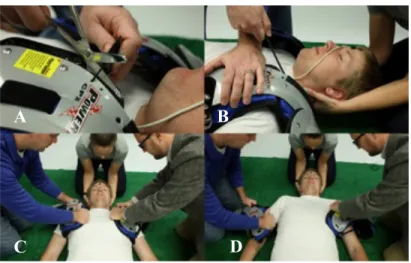
Riddell™ RipKord Shoulder Pads
The Riddell™ RipKord shoulder pads (Figure 4.1), designed in 2011, represent a
novel removal technique utilizing a stiff guided cable laced through the shoulder pads
connecting right and left sides posteriorly. On the right side of the shoulder pads there are
two posteriorly fastened loop-tabs that insert through two slits on the back of the left half
of the shoulder pads. The RipKord runs through the loops, thus securing both sides
together. After the anterior laces are cut, the attachment (zip tie) of the RipKord is cut,
and the RipKord pulled. Two rescuers are then able to slide each side of the shoulder
pads laterally from underneath the injured athlete (Kordecki et al., 2011).
Modified Borg CR10 Rating of Perceived Exertion
A modified Borg CR10 scale was administered to each participant following the
completion of each technique for evaluation of RPE. This scale was used to draw
conclusions about the perceived difficulty of each removal technique. Each participant