Review
The Role of Hypoxia-Inducible Factor
Post-Translational Modifications in Regulating its
Localisation, Stability and Activity
Adam Albanese 1, Leonard A. Daly 2, Daniela Mennerich 3, Thomas Kietzmann3, and Violaine See *1
1 Department of Molecular Physiology and Cell Signalling, Institute of Systems Molecular and Integrative
Biology; [email protected], [email protected]
2 Department of Biochemistry and System Biology; Institute of Systems Molecular and Integrative Biology;
3 Faculty of Biochemistry and Molecular Medicine, Biocenter Oulu, University of Oulu, Finland;
[email protected]; [email protected] * Correspondence: [email protected];
Abstract: The hypoxia signalling pathway enables adaptation of cells to decreased oxygen availability. When oxygen becomes limiting, the central transcription factors of the pathway, inducible factors (HIFs), are stabilised and activated to induce the expression of hypoxia-regulated genes, thereby maintaining cellular homeostasis. Whilst hydroxylation has been thoroughly described as the major and canonical modification of the HIF-α subunits, regulating both HIF stability and activity, a range of other post-translational modifications decorating the entire protein play also a crucial role in altering HIF localisation, stability, and activity. These modifications, their conservation throughout evolution and their effects on HIF-dependent signalling are discussed in this review.
Keywords: Hypoxia; HIF-1α; HIF-2α; Posttranslational modifications; Phosphorylation; cysteine phosphorylation, Methylation; Acetylation; Ubiquitination; Sumoylation; S-Nitrosylation; Signalling
1. Introduction
Many pathways in mammalian cells rely on molecular oxygen; especially during the final step of the mitochondrial respiratory chain. If oxygen levels drop below a cell-dependent critical level, cells experience hypoxia and cannot sustain aerobic respiration and subsequent ATP production. A switch to glycolysis, the less efficient but oxygen-independent pathway of producing ATP, is required to ensure cell survival. The sensing of cellular oxygen levels, the associated switch between modes of energy generation and ultimately the adaption to a low oxygen environment, is controlled by the hypoxia signalling pathway. Hypoxia-inducible factors (HIFs), of which the first was described as a nuclear factor that enhances transcription of the erythropoietin (EPO) gene under hypoxic conditions by binding to a 3' enhancer sequence element, are part of this pathway and maintain the adaptation at the transcriptional level [1,2]. HIFs consists of an oxygen-dependent α subunit that is destabilised in normoxia and a constitutively expressed β subunit (HIF-1β or ARNT) [3,4]. Three
HIF-α subunits (HIF-1α, HIF-2α, HIF-3α) have been described, of which HIF-1α and HIF-2α are the best understood and considered as the major activators of hypoxia-induced gene transcription [2,5,6]. HIF-α and HIF-1β heterodimerise via their basic helix-loop-helix (bHLH)/Per-ARNT-Sim (PAS) domains to form the active transcription factor dimer, which then binds to hypoxia response elements (HREs) within the DNA of target genes [7]. HIFs then recruit general co-activators such as CBP/p300 via the C-terminal transactivation domain (C-TAD), leading to the expression of more than ~300 genes [8,9].
While HIF-1α and HIF-2α appear to be able to bind the same HRE, they can occupy distinct genomic sites, which vary with cell types. They display different subnuclear localisation and intranuclear diffusion speed [10]. In addition, neither HIF-1α nor HIF-2α could substitute the lack of DNA binding caused by the absence of the one or the other HIF-α variant [11]. These findings support in part, the idea that HIF-1α accounts for acute and HIF-2α for chronic responses to hypoxia [12]. HIF-3α is less explored than HIF-1α or HIF-2α. HIF-3α mRNA is subject to alternative splicing in humans and in mice [13,14]. A specific mouse splice variant called inhibitory PAS domain protein (IPAS) was shown to interact directly with HIF-1α. The IPAS/HIF-1α complex was unable to bind to HREs and suggested to be a negative regulator of HIF-1α [15,16]. By contrast, the long human splice variant HIF-3α2 induces expression of various genes among them the EPO gene [17].
The canonical regulation of HIF-α protein stability and activity involves a series of molecular
interactions and reversible covalent modifications to specific amino acids, termed post-translational modifications (PTMs) [18-20]. A PTM can alter a protein's enzymatic activity, localisation, stability, and/or interaction with other proteins. Therefore, these non-genetically encoded modifications that are mainly carried out by enzymes, add to the complexity of the proteome as they can ascribe different functionalities to the same gene product. Common PTMs to a target protein include phosphorylation, acetylation, methylation, and alkylation as well as the covalent linkage of fatty acids, saccharides or small proteins such as ubiquitin and SUMO (small ubiquitin-related modifier) [21]. This review examines and discusses the current PTM landscape of HIF-α and their respective functional consequences on HIFs.
2. Canonical regulation of HIF-α, the role of ubiquitination
Whilst regulation of HIF mRNA levels by hypoxia plays a minor role in HIF abundance, regulation of protein stability, via PTMs, is essential for appropriate HIF accumulation during hypoxia [22]. The HIF-α proteins are destabilised in normoxia via the ubiquitin-proteasome pathway (Figure 1) [23,24]. To achieve this, post-translational hydroxylation of two conserved proline residues (Pro-402/Pro-564 in HIF-1α; Pro-405/ Pro-531 in HIF-2α; P492 in HIF-3α) residing within an oxygen-dependent-degradation (ODD) domain is required [18,19,25,26]. Prolyl hydroxylation is carried out by three mammalian HIF prolyl hydroxylases (PHD1, -2, and -3; also known as EglN2, EglN1, and EglN3, respectively). PHD2 acts as the main regulator of HIF-α degradation and has a key role in HIF- α intracellular dynamics [27,28]. Once hydroxylated, the HIF-α proteins are recognised by an
E3-ligase complex containing the von-Hippel-Lindau protein (pVHL), which acts as the substrate recognition unit and, together with Cullin-2 (Cul-2), Elongin-1, Elongin-2 and Ring-Box 1 (RBX1), polyubiquitinates K532, K538 or K567 on HIF-1α (and K497, K503 or K512 on HIF-2α) [29-32]. The polyubiquitylated HIF-α proteins are then degraded via the 26S proteasome [18]. When cells are deprived of oxygen, PHDs have less molecular oxygen available to act as a co-factor, hence decreasing their activity and subsequent HIF-α hydroxylation [33]. Consequently, HIF-α subunits
accumulate in the nucleus, form a heterodimer with the constitutively expressed HIF-1β and bind to HREs [7]. HIF-α hydroxylation is not exclusively mediated via PHDs, but also by the Factor-Inhibiting HIF (FIH). FIH is an asparaginyl hydroxylase and modifies HIF-1α and HIF-2α on their C-TAD residues N803 and N847, respectively [34,35]. During hypoxia, FIH activity is suppressed, allowing HIF-1α or -2α to complex with the CBP/p300 co-activators and increased transcriptional activation. HIF-α hydroxylation has long been considered to be an irreversible PTM, but it was recently shown, by mass spectrometry, that the FIH-mediated asparagine hydroxylation is indeed a reversible process [36].
Figure 1. The canonical oxygen-dependent degradation mechanism for HIF-α by pVHL-mediated 26S proteasomal degradation and its inhibition during hypoxia.
Polyubiquitination is a quintessential PTM in preventing unwanted HIF-α accumulation in
normoxia. Aside from ubiquitination, the action of deubiquitinating enzymes (termed DUBs) are well-known for their roles in regulating HIF-α. The action of DUBs on HIFs has been extensively and recently reviewed and will not be covered here [37,38].
2. Non-canonical PTMs regulating HIF-α subunits
Whilst there are many well-characterised binding partners and indirect regulators of HIF-1α and
HIF-2α, PTMs constitute an essential direct regulatory mechanism for the HIF transcription factors [39]. PTMs abundantly decorate the full-length of these oxygen-sensitive proteins to exert specific regulatory forces. Most of these covalent modifications are enzymatically driven, with some exceptions such as S-nitrosylation. For the past two decades, the PTM landscape of HIF-1α and HIF
-2α has been ever-expanding, showing the intrinsic complexity and crosstalk of diverse intracellular signalling pathways implicating HIF activity, stability and localisation (Figure 2).
PHD
pVHL
HIF-1β
Normoxia
Hypoxia
HIF-α OH # O2
O2
# O2
O2
Cyt
opl
asm
Nucleu
s
OH
HIF-α OH OH
pVHL
HIF-1β
Ub Ub
Ub Ub
PHD
pVHL
HIF-1β HIF-α
O2
O2
OH
HIF-α OH OH
pVHL
HIF-1β
Ub Ub
Ub Ub
O2
O2
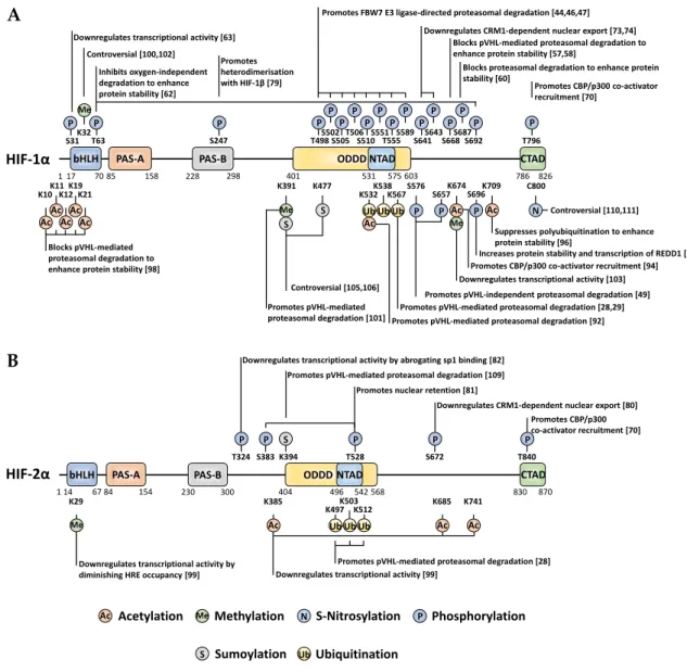
Figure 2. Localisation and function of non-canonical HIF-α PTMs mapped onto full-length HIF-1α (A) and HIF-2α (B). When a given PTM has multiple publications stating conflicting functional outcomes, then the functionality is denoted as 'controversial'.
3. Phosphorylation
Phosphorylation is an extremely common and well-studied PTM, involving the enzymatic addition of a phosphate group to serine, threonine, or tyrosine residues on a target protein (canonically within vertebrates). Moreover, a recent investigation highlighted the existence of additional 'non-canonical' phosphorylatable residues within human cells, including histidine, arginine, lysine, aspartate, glutamate and cysteine [40]. HIF-1α/HIF-2α modification by phosphorylation is abundant and has varied roles in regulating their stability, activity, subcellular localisation, and binding partner interactions (Figure 2). Whilst many of these direct phosphorylation events occur irrespective of oxygen tension, the modifications within the ODD domain appear to occur under normoxia exclusively.
3.1 Phosphorylation by GSK-3β
Glycogen Synthase Kinase-3 (GSK-3) is a serine/threonine kinase that was initially identified as a negative regulator of glycolysis. In mammals, two different isoforms have been identified: GSK-3α
and GSK-3β, and despite their homology of 98% in their catalytical domain, their roles in metabolism are different [41-43]. Given that GSK-3 phosphorylates various upstream and downstream targets of the PI3K/AKT/mTOR signalling pathways, its activity is subject to tight regulation. Given the fact
826 575
531
1 17 7085 158 228 298 401 603 786
PAS-A PAS-B ODDD K532K538K567
S551 T555S589 T498S502S505T506S510
S576 S657
S668S687S692 S641S643 S247 T63 T796 NTAD CTAD HIF-1α S696K709 K19 K21 K12 K11 K674 K32 K391 K477 870 542 496
1 14 6784 154 230 300 404 568 830
PAS-A PAS-B ODDD NTAD CTAD
HIF-2α
S383 T528
T324 S672 T840
K29 K385 K685 K741
K394 Blocks pVHL-mediated
proteasomal degradation to enhance protein stability [98]
Controversial [100,102] Inhibits oxygen-independent degradation to enhance protein stability [62]
Promotes FBW7 E3 ligase-directed proteasomal degradation [44,46,47] Downregulates CRM1-dependent nuclear export [73,74]
Blocks pVHL-mediated proteasomal degradation to enhance protein stability [57,58]
Blocks proteasomal degradation to enhance protein stability [60]
Promotes CBP/p300 co-activator recruitment [70]
Promotes heterodimerisation with HIF-1β [79]
Controversial [105,106] Promotes pVHL-mediated
proteasomal degradation [101] Promotes pVHL-mediated proteasomal degradation [92] Promotes pVHL-mediated proteasomal degradation [28,29]
Promotes pVHL-independent proteasomal degradation [49] Downregulates transcriptional activity [103]
Increases protein stability and transcription of REDD1 [68] Promotes CBP/p300 co-activator recruitment [94]
Suppresses polyubiquitination to enhance protein stability [96]
Downregulates CRM1-dependent nuclear export [80]
Promotes pVHL-mediated proteasomal degradation [28] Downregulates transcriptional activity [99]
Downregulates transcriptional activity by diminishing HRE occupancy [99]
Promotes CBP/p300 co-activator recruitment [70]
Promotes nuclear retention [81] Promotes pVHL-mediated proteasomal degradation [109] Downregulates transcriptional activity by abrogating sp1 binding [82]
C800
Controversial [110,111]
Acetylation Methylation S-Nitrosylation Phosphorylation Sumoylation Ubiquitination Me Ub S Ac K10 bHLH bHLH Ac Ac Ac Ac
P P P
P P P
P P
P P P
P P P
Me S Ac
Ub Ub P
P P Ac Me P Ac P Me
K497K503K512
Ub Ub Ub Ac
N
Ac Ac
P P S P P P
Ac Me N P
Ub S
S31 P
Downregulates transcriptional activity [63]
A
that early hypoxia increased PKB/Akt activity as well as HIF-1α protein levels, it was shown that downregulation of GSK-3 enhanced HIF-1α, whereas overexpression of GSK-3β decreased HIF-1α
protein levels, suggesting that HIF-1α is a direct target of GSK-3β [44,45]. Indeed, two independent investigations found distinct clusters of HIF-1α phosphorylation within its ODD domain and N-TAD, directly deposited by GSK-3β. One study identified S551, T555 and S589 as GSK-3β target sites based on kinase assays and experiments in hepatoma cells, while another study reported T498, S502, S505, T506 and S510 as GSK-3β sites in ovarian cancer cells [44,46]. Whilst this implies cell type specific aspects in the action of GSK-3, in both cases phosphorylation of HIF-1α by GSK-3β mediated the FBW7-E3 ubiquitin ligase-directed proteasomal degradation. This could be antagonised by the DUB ubiquitin-specific protease 28 (USP28) [46,47]. Interestingly, USP28 was found to be subject of a HIF-regulated positive feedback loop. Therein, the USP28-inactivating sumoylation of USP28 at K99 occurring under normoxia is reversed by direct interaction with SENP1 [48]. SENP1 itself is a transcriptional target of HIF-1. Hence, induced expression of SENP1 under hypoxia promotes desumoylation and activation of USP28, which then can contribute to further stabilisation of HIFs by their deubiquitynation activity (Figure 3). Taken together, this suggests that GSK-3β/USP28/SENP1 are highly coordinated to maintain an appropriate HIF response depending on oxygen availability in addition to the PHD-pVHL system.
3.2. Phosphorylation by PLK3
Polo-like kinase 3 (PLK3), a regulator of the cellular stress response and cell cycle progression, targets HIF-1α for degradation via direct phosphorylation of two sites, S576 and S657 [49]. This phosphorylation occurs during normoxia to target HIF-1α for proteasomal degradation in a pVHL -independent manner. Only one of these target sites, S576, is located within the ODD domain, while the other, S657, is immediately after the HIF-1α nuclear export signal (NES). The role of PLK3 in regulating cell survival and proliferation in vivo has been further demonstrated by the same group, via PLK3-dependent phosphorylation of PTEN to enhance PTEN protein stability [50]. More recently, evidence has been gathered suggesting that PLK3 is suppressed by both hypoxia and Ni(II) (which acts as a hypoxia mimetic by blocking PHDs) [51,52]. The latter studies proposed a signalling paradigm whereby, in normoxia, PLK3 destabilises both HIF-1α and the E3-ligase SIAH2. Among the SIAH2 targets are PLK3 and PHD1/3 [52-54]. Thus, under hypoxia, or in the presence of at least Ni(II)), SIAH2 protein levels increase and then induce ubiquitin-mediated proteasomal degradation of PLK3 as well as PHD1 and PHD3. Although USP28 appears to be capable of reversing PLK3 polyubiquitination to increase its stability, USP28 levels and activity were found to be suppressed by both hypoxia and Ni(II), which would contribute to PLK3 suppression (Figure 3). While the co-existence of both pVHL-dependent and -independent mechanisms for normoxia-specific HIF-1α
degradation might initially appear redundant, the presence of both systems may form a contingency to ensure appropriate HIF regulation in the event of pVHL becoming functionally compromised.
3.3 Phosphorylation by CDKs
Not all phosphorylations of HIFs decrease their stability, some do the opposite. Cyclin-dependent kinases (CDKs) belong to an important family of proteins involved in regulating and fine -tuning several stages of the cell cycle [55]. HIF-1α has been shown to act as a negative regulator of DNA replication, exherting control over cell cycle progression [56]. CDK1, a key cell cycle regulator conserved across all eukaryotes and promotes entry into the M-phase of mitosis, directly interacts with and phosphorylates HIF-1α at S668, in an oxygen-independent manner. Although this phosphorylation increased HIF's protein stability, the mechanisms appear to involve both the proteasome and/or lysosomal degradation of HIF-1α during the G1/S transition [57,58]. In addition, CDK2 positively regulates HIF-1α transactivity in cancer cells, and it was postulated that CDK2 could uncouple the transcriptional and non-transcriptional functions of HIF-1α in a manner analogous to
the well-characterised mechanism involving cMYC and SKP2 [57,59]. Furthermore, CDK5, which is distinct from the other CDKs as it does not involve a cyclin subunit for catalytic activation, modifies HIF-1α at S687 and increases its stability [60]. Overall, this highlights the importance of
oxygen-dependent and -inoxygen-dependent mechanisms merging at HIF-lα to determine cellular fate during growth and division.
3.4 Phosphorylation by PKA
The intracellular protein kinase A (PKA), an essential kinase activated by cAMP, has also been associated with increased HIF stability. Initially, PKA was shown to be involved in the HIF-lα
response to intermittent hypoxia in EAhy926 endothelial cells, yet the phosphorylation sites within HIF-1α remained unknown [61]. Later on, it was found that PKA phosphorylated 6 different sites spanning the full length of HIF-1α in vitro, with T63 and S692 phosphorylation promoting oxygen-independent stabilisation and increased transcriptional activity of HIF-1α [62]. Interestingly, of these 6 phosphorylation sites, S31 has recently been evidenced in a separate study, to abrogate HIF-1α -dependent transcription without impacting protein stability; opening up avenues of direct transcriptional regulation of HIF by PTM and potentially delineating the consensus that HIF-α
stability results in HIF transcriptional regulation [63]. A recent study identified a feedback mechanism where hypoxia activates PKA via HIF-1α mediated suppression of the PKA regulatory subunit 2B (PRKAR2B) transcription, by sequestering SP1 from the PRKAR2B promoter [64]. Overall, these aspects show that the potential of crosstalk between these key signalling pathways in altering the cellular response.
3.5 Phosphorylation by pATM
Following the detection of DNA damage, p53, a potent tumour suppressor transcription factor, is stabilised via phosphorylation by ataxia telangiectasia mutated protein (pATM) [65]. p53 and
HIF-1α display extensive crosstalk to balance survival and pro-apoptotic signalling, in response to severe hypoxia or anoxia [66,67]. pATM directly modifies HIF-1α at S696 in response to hypoxia, thereby
increasing its stability as well as its transcriptional activity [68]. In contradiction to this activatory role of pATM on HIF-1α, others have found that loss of pATM positively regulates the transcription and translation of HIF-1 (α and β) proteins through oxidative stress [69]. Together, this suggests a complex interrelation between pATM and HIF-1α involving direct and indirect regulatory aspects, ranging from transcriptional control via the regulation of translation and/or protein stability.
3.6 HIF-1α vs HIF-2α phosphorylation, similarities and differences
Given the high sequence homology between HIF-lα and HIF-2α, it is unsurprising to see equivalent phosphorylation-dependent regulation between the two HIF-α proteins. Recruitment of the CBP/p300 co-activators to the HIF-α C-TAD requires phosphorylation of a conserved threonine within the C-TAD at T796 and T844 of HIF-1α and mouse HIF-2α (human HIF-2α T840), respectively [70]. Despite unsuccessful identification of the specific kinase responsible for this phosphorylation, CKII-like kinase was suggested to play a role, yet direct evidence has not yet been obtained [70-72]. HIF-α transcriptional activity and its interaction with HIF-1β are dependent on reaching a
sufficient nuclear concentration. Nuclear localisation can be regulated by HIF-1α phosphorylation at S641/S643, within the NES motif, by p42/44 mitogen-activated protein kinase (MAPK, hereafter referred to as ERK1/2) [73,74]. This ERK-dependent phosphorylation promotes HIF-1α nuclear localisation by blocking exportin chromosomal maintenance 1 (CRM1)-dependent nuclear export. Moreover, ERK1/2 phosphorylation of HIF-1α also indirectly regulates gene-specific HIF-1α
transcriptional activation and enhances protein stability. Either S641 and S643 phosphorylation constitutes a PIN1 consensus motif (pSer-Pro), whereby PIN1, a peptidyl-prolyl cis/trans isomerase overexpressed in several human cancers, induces a conformational change in HIF-1α to increase protein stability and transcriptional activity in an ERK1/2-dependent manner [75,76]. Interestingly, an inverse association has been noted between cancer and Alzheimer's Disease (AD) concerning PIN1 regulation of HIF-1α [77]. While PIN1 overexpression in cancer has been correlated with increased HIF-1α stability, the inverse is suggested to occur with AD whereby PIN1 promotes HIF-1α
degradation via a GSK-3β-dependent mechanism [75,78]. Once HIF-1α localises to the nucleus, its interaction with HIF-1β can be disrupted by phosphorylation of S247 by casein kinase 1 δ (CK1δ) [79].
Much like HIF-1α, HIF-2α nuclear localisation is regulated by ERK1/2 phosphorylation at S672 [80]. Consequently, this masks the NES within HIF-2α and directly inhibits CRM1-dependent nuclear export of HIF-2α at an atypical NES. Despite in vitro findings for HIF-2α being capable of interacting with PIN1, no investigation has yet identified equivalent HIF-1/PIN1 regulation on HIF-2 [76]. Furthermore, HIF-2α nuclear localisation is also dependent on additional modification at S383 and
T528 by CK1δ, which assists indirectly to the CRM1-dependent mechanism, by facilitating nuclear retention likely via binding to immobile nuclear (or chromatin) components [81].
While the similarity of mechanisms regulating both HIF-α proteins is not surprising, it is the specific differences in their respective sequences that specialise them for some distinct roles. For instance, during hypoxia, HIF-1α suppresses key mismatch DNA-damage repair genes by competing with MYC for SP1 binding within target gene promoters, but, this does not occur with HIF-2α due to unique phosphorylation within its PAS-B domain on T324 by protein kinase D1 (PKD1) [82]. The T324 phosphorylation occurs within a motif requiring an obligatory upstream proline, which is not present in HIF-1α, hence abrogating SP1 binding.
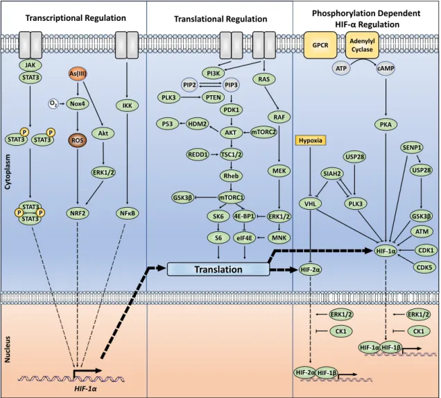
3.7 Beyond direct HIF protein phosphorylation; indirect kinase-dependent HIF-α regulation Kinases regulating HIF-α transcription, synthesis, or degradation by acting upstream or downstream on critical regulators of these processes were also shown to integrate different signalling pathways with the HIF response (Figure 3). Despite HIF-α transcriptional regulation not being the
focus of this review, it is nevertheless important to highlight how such signalling crosstalks including JAK/STAT3 and NF-kB signalling have been associated with upregulation of HIF-1α at the transcriptional level [83-87]. Furthermore, PI3K/AKT and ERK1/2 appear to affect at least HIF-1α
transcription in response to reactive oxygen species (ROS) generated by arsenite [88]. ROS involve binding of nuclear factor erythroid 2-related factor 2 (NRF2) to an antioxidant response element (ARE) located approximately 30 kilobases upstream of the HIF1A transcriptional start [88,89]. The participation of mTOR in the regulation of HIF-1α protein translation was also described [90]. Further, an mTOR signalling motif (FVMVL) immediately C-terminal of the PAS-A domain in
HIF-1α appeared to modulate the recruitment of CBP/p300 [91]. Altogether, those findings outline the additional control, via kinase-controlled pathways, of HIF- α production.
Figure 3. The intracellular protein-dependent cell signalling for the transcriptional, translational, and phosphorylation-dependent regulation of HIF-α.
4. Acetylation
Acetylation is a compelling class of modification in terms of HIF-α functional outcome, leading
to diverse effects. Since establishing the canonical consensus of HIF-α stabilisation, investigations
have highlighted that acetylation plays a crucial role in coordinating this fundamental response. Mouse arrest defective-1 (mARD1225) acetylation of the HIF-1α ODD domain, at K532, accelerates the
HIF-1α/pVHL interaction under normoxia, contributing to HIF-1α proteasomal degradation [92]. However, given that humans do not express mARD1225 it remains unclear as to whether this
regulation occurs in human cells. Some investigations have been unsuccessful in reproducing this observed increase in protein degradation when using either mARD1225 or human ARD1
(hARD1/NAA10) [93,94]. Recent evidence suggests that FIH is required to directly modify hARD1/NAA10, at W38, during normoxia (utilising molecular oxygen as a cofactor) to activate its lysine acetyltransferase activity, thereby facilitating HIF-1α acetylation [95]. This FIH-mediated activation could explain the previously suggested normoxia specific regulation imposed by hARD1/NAA10. The co-activator p300 also acetylates HIF-1α, at K709, to increase protein stability under hypoxia by suppressing its polyubiquitylation [96]. This acetylation is antagonised and removed by SIRT2, which interacts directly to enhance the HIF-1α-PHD2 interaction [97].
Acetylation has also been recognised to induce changes in HIF-α outside of the canonical pVHL-mediated pathway. In that respect, HDAC4 regulates HIF-1α deacetylation, which in turn enhances
STAT3 P PTEN PLK3 PDK1 AKT HDM2 P53 RAS RAF MEK ERK1/2 PI3K TSC1/2 Rheb mTORC1 REDD1 GSK3β 4E-BP1 SK6 eIF4E S6 MNK mTORC2 GPCR Adenylyl Cyclase ATP cAMP IKK
NFκB
VHL PLK3 GSK3β CDK5 CDK1 ATM HIF-2α HIF-1α SIAH2 PKA Hypoxia USP28
HIF-1αHIF-1β
HIF-2αHIF-1β
CK1 CK1
Translation
Transcriptional Regulation Translational Regulation Phosphorylation Dependent HIF-αRegulation
Cyt opl asm Nucleu s STAT3P PSTAT3 STAT3 STAT3P ERK1/2 ERK1/2 HIF-1α ROS As(III) PIP3 PIP2 Nox4 # O2 NRF2 Akt ERK1/2 USP28 SENP1 JAK
HIF-1α protein stability by blocking non-pVHL-mediated proteasomal degradation [98]. Additionally, HDAC4 increases HIF-1α transactivity. Although the precise localisation of the individual lysine residues was not determined, the authors postulated that a combination of 5 lysines (K10, K11, K12, K19 and K21) within the bHLH domain is involved. In terms of the acetylation machinery modifying HIF-α, HDAC4 is not the sole regulator. Under hypoxia, the CBP/p300-associated factor (PCAF) facilitates the CBP/p300 interaction with the HIF-1α CTAD via acetylation of K674 [94]. Given that K674 is conserved across other mammals containing the HIF-1α ortholog, it is likely that modification of this site plays an integral role in regulating transactivity. Interestingly, K674 acetylation can be antagonised by SIRT1, which abrogates the CBP/p300 interaction to inactivate HIF-1α. However, SIRT1 can itself be downregulated by hypoxia. SIRT1 also interacts with HIF-2α
to facilitate the deacetylation of residues K385, K685 and K741 [99]. While SIRT1 downregulates
HIF-1α transactivity, it does the opposite to HIF-2α leading to increased transcriptional activation.
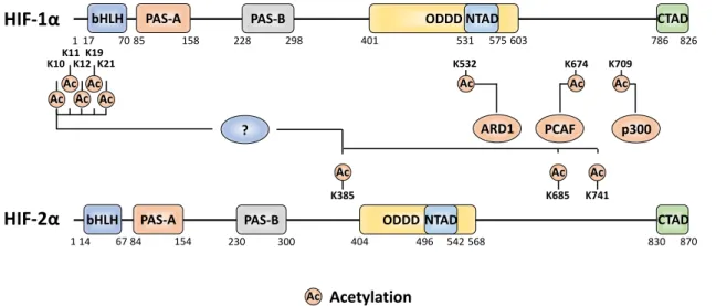
Figure 4. The amino acid sites of HIF-α subjected to acetylation and the acetyltransferase enzymes attributed to the relevant sites. A '?' represents an unknown regulator, not currently described in the literature.
5. Methylation
Most-commonly methylation of proteins occurs at arginine or lysine residues. Methylation has been widely studied in conjunction with histone proteins where they regulate DNA accessibility for transcription. In addition, methylation also regulates other proteins, including HIF-α, to induce
functional changes. SET7/9, a monomethyl transferase known for its role in gene activation via modification of histone H3, has been found to also interact with HIF-1α and to methylate multiple sites [100-102]. The SET7/9-mediated methylation at K391 on HIF-1α induces protein destabilisation via the canonical PHD/VHL pathway [101]. This could be antagonised by lysine-specific demethylase 1 (LSD1). LSD1 is a member of the nucleosome remodelling and deacetylase (NuRD) complex. It has been proposed that the LSD1 demethylating activity suppresses PHD2-mediated hydroxylation and promotes deacetylation of HIF-1α at K532 (supposed to be acetylated by ARD1) [92,101].
Other studies indicate that SET7/9 can methylate both HIF-1α and HIF-2α at the conserved sites
K32 and K29 within the bHLH domains, respectively [102]. Initially, K32 HIF-1α (and K29 HIF-2α) methylation was found to induce transcriptional inhibition independent of HIF-1α protein degradation, while a later investigation reported that SET7/9 methylated HIF-1α was degraded by
the 26S proteasome in a hydroxylation-independent manner [100,102]. Again, LSD1 was capable of reversing the methylation at K32 and of stabilising HIF-1α under hypoxic conditions. In line, LSD1 was then found to upregulate HIF-1α-dependent angiogenesis by increasing CBP, MTA1 and
HIF-1α binding to the VEGF promoter [100,101]. Interestingly, K32 resides near two residues found to be 826 575
531
1 17 7085 158 228 298 401 603 786
PAS-A PAS-B ODDD
K532
NTAD CTAD
HIF-1
α
K709 K19
K21 K12 K11
K674
870 542
496
1 14 6784 154 230 300 404 568 830
PAS-A PAS-B ODDD NTAD CTAD
HIF-2
α
K385 K685 K741
Ac K10
bHLH
bHLH
Ac Ac Ac
Ac Ac Ac Ac
PCAF
ARD1 p300
?
Ac Ac Ac
Acetylation
mutated in various human cancers, S28Y and R30Q, which alter the SET7/9 consensus site when mutated to prevent methylation, thus leading to increased HIF-1α stability [100]. Aside from SET7/9 mono-methylation, more recently, G9a/G9a-like protein (GLP) has been recognised to both mono- and di-methylate HIF-1α at K674 [103]. This is the same site that can be acetylated by PCAF (see above), but with here an opposite effect, by inhibiting transactivation [103]. Lysine acetylation and methylation have distinct effects on the physicochemical properties of a target residue, either maintaining a residues positive charge or neutrilising it, respectively, leading to distinct proteoforms. The mutual exclusivity of these lysine PTMs evidence the complex cross-regulation of HIF-1α.
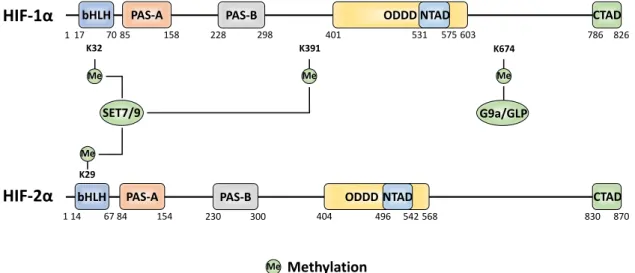
Figure 5. The amino acid sites of HIF-α methylation and the methyltransferase enzymes responsible for each site-specific modification.
6. SUMOylation
SUMO is comparable to ubiquitin in terms of its overall molecular structure and molecular weight yet can lead to distinct changes in a given protein regulation depending on the SUMO isoforms [104]. The functional outcome of HIF-1α SUMOylation remains unclear due to controversy between investigations. The first report of HIF-1α SUMOylation suggested that modification by SUMO-1 increased HIF-1α protein stability and transactivity [105]. SUMO-1 was proposed to compete with ubiquitin for linkage at K391 and K477 within HIF-1α's SUMO consensus sequences. Contrary to this, another study evidenced that SUMOylation of K391 and K477 induces a decrease in transcriptional activity, which appeared to be independent of altering HIF-1α's half-life under hypoxia [106]. Aside from the identification and characterisation of specific SUMOylatable residues, several investigations have identified enzymes capable of initiating SUMO conjugation to HIF-1α.
PIASy is an E3-ligase responsible for SUMOylating HIF-1α in two regions. One site residing within the ODD domain (between residues 331-698) and the other lying further upstream (between residues 211-330) [107]. PIASy-mediated SUMOylation negatively regulated HIF-1α transactivity and protein stability and reduced epithelial cell angiogenic activity. Further, the SUMO E3 ligase Cbx4 modifies HIF-1α at K391 and K477 to enhance HIF-1's transcriptional activity [108]. While both HIF-1α and
HIF-2α are targets for SUMOylation, fewer sites of modifications have been identified in HIF-2α. Despite HIF-2α containing two SUMO consensus sites, only one, K394, was found to be conjugatable by SUMO [109]. Interestingly, the enzymatic addition of 2 facilitated recognition by SUMO-targeted ubiquitin ligases (pVHL and RNF4) to rapidly degrade HIF-2α under hypoxia. Furthermore, this investigation also highlighted that the SUMO-protease SENP1, which has shown action against HIF-1α, is also capable to recognise and regulate HIF-2α.
826 575
531
1 17 7085 158 228 298 401 603 786
PAS-A PAS-B ODDD NTAD CTAD
HIF-1
α
870 542
496
1 14 6784 154 230 300 404 568 830
PAS-A PAS-B ODDD NTAD CTAD
HIF-2
α
bHLH
bHLH
Methylation
Me
SET7/9 G9a/GLP
K674 K391
Me Me
K32
Me
K29 Me
7. S-Nitrosylation
Not all PTMs are enzymatically driven, with one such example being nitrosylation. S-nitrosylation is yet another PTM with conflicting views regarding the functional outcome of its modification. Initially, C800 S-nitrosylation was identified as a critical modification for CBP/p300 recruitment and transcriptional activation [110]. However, a later investigation found that incorporation of the polar NO group potentially disrupted this interaction with the result that C800 S-nitrosylation suppressed CBP/p300 recruitment [111]. In addition, HIF-1α stability can be upregulated through NO-mediated S-nitrosylation under normoxic conditions, at C533 (mouse sequence) in the ODD domain. The stabilisation process appeared to be independent of the VHL degradation pathway [112]. Overall, S-nitrosylation seems to regulate at least HIF-1α stability and transactivity. No data are available for HIF-2α.
8. Uncharacterised PTMs 8.1 Glycosylation
While many classes of PTM have been discussed within this review, there are still many classes that are yet to be characterised as modifiers of HIF-α. Protein glycosylation, the addition of a sugar-moiety to proteins, ranges from simple monosaccharide modifications of nuclear transcription factors to highly complex branched polysaccharide additions to cell surface receptors. While several investigations suggest a link between glycosylation and hypoxia, it is still unclear whether and how HIF-α subunits are directly involved in these processes and whether they can themselves be targeted for glycosylation [113,114].
8.2 S-Glutathionylation
S-glutathionylation is the addition of the tripeptide glutathione (GSH) to cysteine residues of proteins. It is often stimulated by oxidative as well as nitrosative stress, yet also occurs in unstressed cells. It is involved in various cellular processes by modulating protein functions and preventing irreversible oxidation of protein thiols. Changes in oxidised glutathione can modulate HIF signalling via S-glutathionylation of target cysteines in human oral squamous cell carcinoma cells, and in C2C12 mouse myoblasts [115-117]. The latter study also identified, through a biotin switch assay and subsequent MS analysis, GSH adducts on cysteine 520 (C520) within the ODD domain of human
HIF-1α, which led to HIF-lα protein stabilisation [117]. Interestingly, C520 in human HIF-1α is equivalent
to C533 in the mouse sequence for which that S-nitrosylation prevented HIF-1α degradation [112].
8.3 Neddylation
Neddylation is a process by which a ubiquitin-like protein called neural precursor cell-expressed developmentally down-regulated 8 (NEDD8), is conjugated to its target proteins. As a result of the conjugation process subcellular localisation, protein stability, and activity of targeted proteins can be modified. While there is limited knowledge about direct neddylation of HIFs, we do know that
HIF-1α as well as HIF-2α are capable of covalent modification by NEDD8. NEDD8 stabilised HIF-1α
under both normoxia and hypoxia in VHL deficient cells, suggesting a VHL-independent process [118]. More recently it was reported that SerpinB3 (SB3), a hypoxia and HIF-2α-dependent cysteine-protease inhibitor, can directly neddylate and stabilise HIF-2α, which led to an upregulation of its target genes in liver cancer cells [119]. Overall, these reports underline the role of neddylation in HIF regulation, but none of the reports has yet shown the exact sites within the HIF proteins where the neddylation occurs.
9 Conclusion
While many different classes and sites of PTM have been discussed here, numerous others have been identified as part of high-throughput mass spectrometry studies and are yet to be functionally
investigated. Phosphositeplus is a mass spectrometry data repository for PTM data [120]. Searching Phosphositeplus identifies in excess of 50 PTMs (spanning phosphorylation, acetylation, sumoylation and ubiquitination) that have been confidently identified under different cellular conditions between HIF-1α and HIF-2α, that lack functional characterisation. Further to this, a very recent investigation, using a combination of immunoprecipitation and mass spectrometry, discovered multiple types of PTMs (and binding partners) of HIF-1α and HIF-2α and determined their O2 dependence [63]. A total
of 41 (32 novel) and 39 (34 novel) different PTMs on HIF-1α and HIF-2α, respectively, were identified, spanning 13 different types of PTM, including an array of different cysteine modifications together with non-canonical cysteine phosphorylation. Their relevance for additional mechanisms of oxygen-dependent regulation of HIF will require further investigations. We here provided an overview on how post-translational modifications are critical steps for regulating HIF-α activity and stability. Given that hypoxia plays a vital role in many pathophysiological aspects and is a prominent micro-environmental feature in many aspects of life, it is not surprising that HIF PTM dysregulation can result in dramatic changes in HIF functions and may contribute to diseases. To date, most data available contribute to HIF-1α regulation and therefore more knowledge about PTM of HIF-2α and
HIF-3α will be required to increase basic understanding of hypoxia signalling, its crosstalk with other signalling networks and improve the potential for therapeutic intervention.
10. Acknowledgments
We apologise to all researchers who excellently contributed to the field and whose work was not cited due to space limitations. AA is a recipient of a MRC DiMeN studentship. LD is supported by BBSRC. TK is supported by the Academy of Finland SA296027, the Jane and Aatos Erkko Foundation, the Finnish Cancer Foundation, the Sigrid Juselius Foundation, the University of Oulu, and Biocenter Oulu.
References
1. Semenza, G.L.; Nejfelt, M.K.; Chi, S.M.; Antonarakis, S.E. Hypoxia-inducible nuclear factors bind to an enhancer element located 3' to the human erythropoietin gene. Proc Natl Acad Sci U S A 1991, 88, 5680-5684, doi:10.1073/pnas.88.13.5680.
2. Semenza, G.L.; Wang, G.L. A nuclear factor induced by hypoxia via de novo protein synthesis binds to the human erythropoietin gene enhancer at a site required for transcriptional activation. Mol Cell Biol 1992, 12, 5447-5454, doi:10.1128/mcb.12.12.5447.
3. Wang, G.L.; Jiang, B.H.; Rue, E.A.; Semenza, G.L. Hypoxia-inducible factor 1 is a basic-helix-loop-helix-PAS heterodimer regulated by cellular O2 tension. Proc Natl Acad Sci U S A 1995, 92, 5510-5514, doi:10.1073/pnas.92.12.5510.
4. Wang, G.L.; Semenza, G.L. Purification and characterization of hypoxia-inducible factor 1. J Biol Chem 1995, 270, 1230-1237, doi:10.1074/jbc.270.3.1230.
5. Ema, M.; Taya, S.; Yokotani, N.; Sogawa, K.; Matsuda, Y.; Fujii-Kuriyama, Y. A novel bHLH-PAS factor with close sequence similarity to hypoxia-inducible factor 1alpha regulates the VEGF expression and is potentially involved in lung and vascular development. Proc Natl Acad Sci U S A 1997, 94, 4273-4278, doi:10.1073/pnas.94.9.4273.
6. Gu, Y.Z.; Moran, S.M.; Hogenesch, J.B.; Wartman, L.; Bradfield, C.A. Molecular characterization and chromosomal localization of a third alpha-class hypoxia inducible factor subunit, HIF3alpha. Gene Expr 1998, 7, 205-213.
7. Wenger, R.H.; Stiehl, D.P.; Camenisch, G. Integration of oxygen signaling at the consensus HRE. Sci STKE 2005, 2005, re12, doi:10.1126/stke.3062005re12.
8. Arany, Z.; Huang, L.E.; Eckner, R.; Bhattacharya, S.; Jiang, C.; Goldberg, M.A.; Bunn, H.F.; Livingston, D.M. An essential role for p300/CBP in the cellular response to hypoxia. Proc Natl Acad Sci U S A 1996, 93, 12969-12973, doi:10.1073/pnas.93.23.12969.
9. Manalo, D.J.; Rowan, A.; Lavoie, T.; Natarajan, L.; Kelly, B.D.; Ye, S.Q.; Garcia, J.G.; Semenza, G.L. Transcriptional regulation of vascular endothelial cell responses to hypoxia by HIF-1. Blood 2005, 105, 659-669, doi:10.1182/blood-2004-07-2958.
10. Taylor, S.E.; Bagnall, J.; Mason, D.; Levy, R.; Fernig, D.G.; See, V. Differential sub-nuclear distribution of hypoxia-inducible factors (HIF)-1 and -2 alpha impacts on their stability and mobility. Open Biol 2016, 6, doi:10.1098/rsob.160195.
11. Smythies, J.A.; Sun, M.; Masson, N.; Salama, R.; Simpson, P.D.; Murray, E.; Neumann, V.; Cockman, M.E.; Choudhry, H.; Ratcliffe, P.J., et al. Inherent DNA-binding specificities of the 1alpha and HIF-2alpha transcription factors in chromatin. EMBO Rep 2019, 20, doi:10.15252/embr.201846401.
12. Lofstedt, T.; Fredlund, E.; Holmquist-Mengelbier, L.; Pietras, A.; Ovenberger, M.; Poellinger, L.; Pahlman, S. Hypoxia inducible factor-2alpha in cancer. Cell Cycle 2007, 6, 919-926, doi:10.4161/cc.6.8.4133.
13. Makino, Y.; Kanopka, A.; Wilson, W.J.; Tanaka, H.; Poellinger, L. Inhibitory PAS domain protein (IPAS) is a hypoxia-inducible splicing variant of the hypoxia-inducible factor-3 alpha locus. Journal of Biological Chemistry 2002, 277, 32405-32408, doi:10.1074/jbc.C200328200.
14. Pasanen, A.; Heikkila, M.; Rautavuoma, K.; Hirsila, M.; Kivirikko, K.I.; Myllyharju, J. Hypoxia-inducible factor (HIF)-3a is subject to extensive alternative splicing in human tissues and cancer cells and is regulated by HIF-1 but not HIF-2. Int J Biochem Cell B 2010, 42, 1189-1200, doi:10.1016/j.biocel.2010.04.008.
15. Heikkila, M.; Pasanen, A.; Kivirikko, K.I.; Myllyharju, J. Roles of the human hypoxia-inducible factor (HIF)-3 alpha variants in the hypoxia response. Cell Mol Life Sci 2011, 68, 3885-3901, doi:10.1007/s00018-011-0679-5.
16. Makino, Y.; Uenishi, R.; Okamoto, K.; Isoe, T.; Hosono, O.; Tanaka, H.; Kanopka, A.; Poellinger, L.; Haneda, M.; Morimoto, C. Transcriptional up-regulation of inhibitory PAS domain protein gene expression by hypoxia-inducible factor 1 (HIF-1) - A negative feedback regulatory circuit in HIF-1-mediated signaling in hypoxic cells. Journal of Biological Chemistry 2007, 282, 14073-14082, doi:10.1074/jbc.M700732200.
17. Tolonen, J.P.; Heikkil?, M.; Malinen, M.; Lee, H.M.; Palvimo, J.J.; Wei, G.H.; Myllyharju, J. A long hypoxia-inducible factor 3 isoform 2 is a transcription activator that regulates erythropoietin. Cell Mol Life Sci 2020, 77, 3627-3642, doi:10.1007/s00018-019-03387-9.
18. Ivan, M.; Kondo, K.; Yang, H.; Kim, W.; Valiando, J.; Ohh, M.; Salic, A.; Asara, J.M.; Lane, W.S.; Kaelin, W.G., Jr. HIFalpha targeted for VHL-mediated destruction by proline hydroxylation: implications for O2 sensing. Science 2001, 292, 464-468, doi:10.1126/science.1059817.
19. Jaakkola, P.; Mole, D.R.; Tian, Y.M.; Wilson, M.I.; Gielbert, J.; Gaskell, S.J.; von Kriegsheim, A.; Hebestreit, H.F.; Mukherji, M.; Schofield, C.J., et al. Targeting of HIF-alpha to the von Hippel-Lindau ubiquitylation complex by O2-regulated prolyl hydroxylation. Science 2001, 292, 468-472, doi:10.1126/science.1059796.
20. Masson, N.; Willam, C.; Maxwell, P.H.; Pugh, C.W.; Ratcliffe, P.J. Independent function of two destruction domains in hypoxia-inducible factor-alpha chains activated by prolyl hydroxylation. EMBO J 2001, 20, 5197-5206, doi:10.1093/emboj/20.18.5197.
21. Ribet, D.; Cossart, P. Ubiquitin, SUMO, and NEDD8: Key Targets of Bacterial Pathogens. Trends Cell Biol 2018, 28, 926-940, doi:10.1016/j.tcb.2018.07.005.
22. Gorlach, A. Regulation of HIF-1alpha at the transcriptional level. Curr Pharm Des 2009, 15, 3844-3852, doi:10.2174/138161209789649420.
23. Huang, L.E.; Arany, Z.; Livingston, D.M.; Bunn, H.F. Activation of hypoxia-inducible transcription factor depends primarily upon redox-sensitive stabilization of its alpha subunit. J Biol Chem 1996, 271, 32253-32259, doi:10.1074/jbc.271.50.32253.
24. Salceda, S.; Caro, J. Hypoxia-inducible factor 1alpha (HIF-1alpha) protein is rapidly degraded by the ubiquitin-proteasome system under normoxic conditions. Its stabilization by hypoxia depends on redox-induced changes. J Biol Chem 1997, 272, 22642-22647, doi:10.1074/jbc.272.36.22642.
25. Huang, L.E.; Gu, J.; Schau, M.; Bunn, H.F. Regulation of hypoxia-inducible factor 1alpha is mediated by an O2-dependent degradation domain via the ubiquitin-proteasome pathway. Proc Natl Acad Sci U S A 1998, 95, 7987-7992, doi:10.1073/pnas.95.14.7987.
26. Maynard, M.A.; Qi, H.; Chung, J.; Lee, E.H.L.; Kondo, Y.; Hara, S.; Conaway, R.C.; Conaway, J.W.; Ohh, M. Multiple splice variants of the human HIF-3 alpha locus are targets of the von Hippel-Lindau E3 uhiquitin ligase complex. Journal of Biological Chemistry 2003, 278, 11032-11040, doi:10.1074/jbc.M208681200.
27. Berra, E.; Benizri, E.; Ginouves, A.; Volmat, V.; Roux, D.; Pouyssegur, J. HIF prolyl-hydroxylase 2 is the key oxygen sensor setting low steady-state levels of HIF-1 alpha in normoxia. Embo Journal 2003, 22, 4082-4090, doi:DOI 10.1093/emboj/cdg392.
28. Bagnall, J.; Leedale, J.; Taylor, S.E.; Spiller, D.G.; White, M.R.; Sharkey, K.J.; Bearon, R.N.; See, V. Tight control of hypoxia-inducible factor-alpha transient dynamics is essential for cell survival in hypoxia. J Biol Chem 2014, 289, 5549-5564, doi:10.1074/jbc.M113.500405.
29. Maxwell, P.H.; Wiesener, M.S.; Chang, G.W.; Clifford, S.C.; Vaux, E.C.; Cockman, M.E.; Wykoff, C.C.; Pugh, C.W.; Maher, E.R.; Ratcliffe, P.J. The tumour suppressor protein VHL targets hypoxia-inducible factors for oxygen-dependent proteolysis. Nature 1999, 399, 271-275, doi:10.1038/20459.
30. Ohh, M.; Park, C.W.; Ivan, M.; Hoffman, M.A.; Kim, T.Y.; Huang, L.E.; Pavletich, N.; Chau, V.; Kaelin, W.G. Ubiquitination of hypoxia-inducible factor requires direct binding to the beta-domain of the von Hippel-Lindau protein. Nat Cell Biol 2000, 2, 423-427, doi:10.1038/35017054.
31. Paltoglou, S.; Roberts, B.J. HIF-1alpha and EPAS ubiquitination mediated by the VHL tumour suppressor involves flexibility in the ubiquitination mechanism, similar to other RING E3 ligases. Oncogene 2007, 26, 604-609, doi:10.1038/sj.onc.1209818.
32. Tanimoto, K.; Makino, Y.; Pereira, T.; Poellinger, L. Mechanism of regulation of the hypoxia-inducible factor-1 alpha by the von Hippel-Lindau tumor suppressor protein. EMBO J 2000, 19, 4298-4309, doi:10.1093/emboj/19.16.4298.
33. Stiehl, D.P.; Wirthner, R.; Koditz, J.; Spielmann, P.; Camenisch, G.; Wenger, R.H. Increased prolyl 4-hydroxylase domain proteins compensate for decreased oxygen levels. Evidence for an autoregulatory oxygen-sensing system. J Biol Chem 2006, 281, 23482-23491, doi:10.1074/jbc.M601719200.
34. Lando, D.; Peet, D.J.; Gorman, J.J.; Whelan, D.A.; Whitelaw, M.L.; Bruick, R.K. FIH-1 is an asparaginyl hydroxylase enzyme that regulates the transcriptional activity of hypoxia-inducible factor. Genes Dev 2002, 16, 1466-1471, doi:10.1101/gad.991402.
35. Mahon, P.C.; Hirota, K.; Semenza, G.L. FIH-1: a novel protein that interacts with HIF-1alpha and VHL to mediate repression of HIF-1 transcriptional activity. Genes Dev 2001, 15, 2675-2686, doi:10.1101/gad.924501.
36. Rodriguez, J.; Haydinger, C.H.D.; Peet, D.J.; Nguyen, L.; von Kriegsheim, A. Asparagine hydroxylation is a reversible post-translational modification. Mol Cell Proteomics 2020, 10.1074/mcp.RA120.002189, doi:10.1074/mcp.RA120.002189.
37. Mennerich, D.; Kubaichuk, K.; Kietzmann, T. DUBs, Hypoxia, and Cancer. Trends Cancer 2019, 5, 632-653, doi:10.1016/j.trecan.2019.08.005.
38. Schober, A.S.; Berra, E. DUBs, New Members in the Hypoxia Signaling clUb. Front Oncol 2016, 6, 53, doi:10.3389/fonc.2016.00053.
39. Semenza, G.L. A compendium of proteins that interact with HIF-1 alpha. Exp Cell Res 2017, 356, 128-135, doi:10.1016/j.yexcr.2017.03.041.
40. Hardman, G.; Perkins, S.; Brownridge, P.J.; Clarke, C.J.; Byrne, D.P.; Campbell, A.E.; Kalyuzhnyy, A.; Myall, A.; Eyers, P.A.; Jones, A.R., et al. Strong anion exchange-mediated phosphoproteomics reveals extensive human non-canonical phosphorylation. EMBO J 2019, 38, e100847, doi:10.15252/embj.2018100847.
41. Woodgett, J.R. Molecular-Cloning and Expression of Glycogen-Synthase Kinase-3 Factor-A. Embo Journal 1990, 9, 2431-2438, doi:DOI 10.1002/j.1460-2075.1990.tb07419.x.
42. Hughes, K.; Ramakrishna, S.; Benjamin, W.B.; Woodgett, J.R. Identification of Multifunctional Atp-Citrate Lyase Kinase as the Alpha-Isoform of Glycogen-Synthase Kinase-3. Biochem J 1992, 288, 309-314, doi:DOI 10.1042/bj2880309.
43. Force, T.; Woodgett, J.R. Unique and overlapping functions of GSK-3 isoforms in cell differentiation and proliferation and cardiovascular development. J Biol Chem 2009, 284, 9643-9647, doi:10.1074/jbc.R800077200.
44. Flugel, D.; Gorlach, A.; Michiels, C.; Kietzmann, T. Glycogen synthase kinase 3 phosphorylates hypoxia-inducible factor 1alpha and mediates its destabilization in a VHL-independent manner. Mol Cell Biol 2007, 27, 3253-3265, doi:10.1128/MCB.00015-07.
45. Mottet, D.; Dumont, V.; Deccache, Y.; Demazy, C.; Ninane, N.; Raes, M.; Michiels, C. Regulation of hypoxia-inducible factor-1alpha protein level during hypoxic conditions by the phosphatidylinositol 3-kinase/Akt/glycogen synthase kinase 3beta pathway in HepG2 cells. J Biol Chem 2003, 278, 31277-31285, doi:10.1074/jbc.M300763200.
46. Cassavaugh, J.M.; Hale, S.A.; Wellman, T.L.; Howe, A.K.; Wong, C.; Lounsbury, K.M. Negative regulation of HIF-1alpha by an FBW7-mediated degradation pathway during hypoxia. J Cell Biochem 2011, 112, 3882-3890, doi:10.1002/jcb.23321.
47. Flugel, D.; Gorlach, A.; Kietzmann, T. GSK-3beta regulates cell growth, migration, and angiogenesis via Fbw7 and USP28-dependent degradation of HIF-1alpha. Blood 2012, 119, 1292-1301, doi:10.1182/blood-2011-08-375014.
48. Du, S.C.; Zhu, L.; Wang, Y.X.; Liu, J.; Zhang, D.; Chen, Y.L.; Peng, Q.; Liu, W.; Liu, B. SENP1-mediated deSUMOylation of USP28 regulated HIF-1alpha accumulation and activation during hypoxia response. Cancer Cell Int 2019, 19, 4, doi:10.1186/s12935-018-0722-9.
49. Xu, D.; Yao, Y.; Lu, L.; Costa, M.; Dai, W. Plk3 functions as an essential component of the hypoxia regulatory pathway by direct phosphorylation of HIF-1alpha. J Biol Chem 2010, 285, 38944-38950, doi:10.1074/jbc.M110.160325.
50. Xu, D.; Yao, Y.; Jiang, X.; Lu, L.; Dai, W. Regulation of PTEN stability and activity by Plk3. J Biol Chem 2010, 285, 39935-39942, doi:10.1074/jbc.M110.166462.
51. Kasprzak, K.S.; Sunderman, F.W., Jr.; Salnikow, K. Nickel carcinogenesis. Mutat Res 2003, 533, 67-97, doi:10.1016/j.mrfmmm.2003.08.021.
52. Li, C.; Park, S.; Zhang, X.; Dai, W.; Xu, D. Mutual regulation between Polo-like kinase 3 and SIAH2 E3 ubiquitin ligase defines a regulatory network that fine-tunes the cellular response to hypoxia and nickel. J Biol Chem 2017, 292, 11431-11444, doi:10.1074/jbc.M116.767178.
53. Nakayama, K.; Qi, J.; Ronai, Z. The ubiquitin ligase Siah2 and the hypoxia response. Mol Cancer Res 2009, 7, 443-451, doi:10.1158/1541-7786.MCR-08-0458.
54. Qi, J.; Kim, H.; Scortegagna, M.; Ronai, Z.A. Regulators and effectors of Siah ubiquitin ligases. Cell Biochem Biophys 2013, 67, 15-24, doi:10.1007/s12013-013-9636-2.
55. Asghar, U.; Witkiewicz, A.K.; Turner, N.C.; Knudsen, E.S. The history and future of targeting cyclin-dependent kinases in cancer therapy. Nat Rev Drug Discov 2015, 14, 130-146, doi:10.1038/nrd4504. 56. Hubbi, M.E.; Kshitiz; Gilkes, D.M.; Rey, S.; Wong, C.C.; Luo, W.; Kim, D.H.; Dang, C.V.; Levchenko, A.;
Semenza, G.L. A nontranscriptional role for HIF-1alpha as a direct inhibitor of DNA replication. Sci Signal 2013, 6, ra10, doi:10.1126/scisignal.2003417.
57. Hubbi, M.E.; Gilkes, D.M.; Hu, H.; Kshitiz; Ahmed, I.; Semenza, G.L. Cyclin-dependent kinases regulate lysosomal degradation of hypoxia-inducible factor 1alpha to promote cell-cycle progression. Proc Natl Acad Sci U S A 2014, 111, E3325-3334, doi:10.1073/pnas.1412840111.
58. Warfel, N.A.; Dolloff, N.G.; Dicker, D.T.; Malysz, J.; El-Deiry, W.S. CDK1 stabilizes HIF-1alpha via direct phosphorylation of Ser668 to promote tumor growth. Cell Cycle 2013, 12, 3689-3701, doi:10.4161/cc.26930.
59. Li, Q.; Kluz, T.; Sun, H.; Costa, M. Mechanisms of c-myc degradation by nickel compounds and hypoxia. PLoS One 2009, 4, e8531, doi:10.1371/journal.pone.0008531.
60. Herzog, J.; Ehrlich, S.M.; Pfitzer, L.; Liebl, J.; Frohlich, T.; Arnold, G.J.; Mikulits, W.; Haider, C.; Vollmar, A.M.; Zahler, S. Cyclin-dependent kinase 5 stabilizes hypoxia-inducible factor-1alpha: a novel approach for inhibiting angiogenesis in hepatocellular carcinoma. Oncotarget 2016, 7, 27108-27121, doi:10.18632/oncotarget.8342.
61. Toffoli, S.; Feron, O.; Raes, M.; Michiels, C. Intermittent hypoxia changes HIF-1 alpha phosphorylation pattern in endothelial cells: Unravelling of a new PKA-dependent regulation of HIF-1 alpha. Bba-Mol Cell Res 2007, 1773, 1558-1571, doi:10.1016/j.bbamcr.2007.06.002.
62. Bullen, J.W.; Tchernyshyov, I.; Holewinski, R.J.; DeVine, L.; Wu, F.; Venkatraman, V.; Kass, D.L.; Cole, R.N.; Van Eyk, J.; Semenza, G.L. Protein kinase A-dependent phosphorylation stimulates the transcriptional activity of hypoxia-inducible factor 1. Sci Signal 2016, 9, ra56, doi:10.1126/scisignal.aaf0583.
63. Daly, L.A.; Brownridge, P.J.; See, V.; Eyers, C.E. Oxygen-dependent changes in HIF binding partners and post-translational modifications regulate stability and transcriptional activity. bioRxiv 2020, 10.1101/2020.11.12.379768, 2020.2011.2012.379768, doi:10.1101/2020.11.12.379768.
64. Lucia, K.; Wu, Y.H.; Garcia, J.M.; Barlier, A.; Buchfelder, M.; Saeger, W.; Renner, U.; Stalla, G.K.; Theodoropoulou, M. Hypoxia and the hypoxia inducible factor 1 alpha activate protein kinase A by
repressing RII beta subunit transcription. Oncogene 2020, 10.1038/s41388-020-1223-6, doi:10.1038/s41388-020-1223-6.
65. Blackford, A.N.; Jackson, S.P. ATM, ATR, and DNA-PK: The Trinity at the Heart of the DNA Damage Response. Mol Cell 2017, 66, 801-817, doi:10.1016/j.molcel.2017.05.015.
66. Wang, P.; Guan, D.; Zhang, X.P.; Liu, F.; Wang, W. Modeling the regulation of p53 activation by HIF-1 upon hypoxia. FEBS Lett 2019, 593, 2596-2611, doi:10.1002/1873-3468.13525.
67. Zhou, C.H.; Zhang, X.P.; Liu, F.; Wang, W. Modeling the interplay between the HIF-1 and p53 pathways in hypoxia. Sci Rep 2015, 5, 13834, doi:10.1038/srep13834.
68. Cam, H.; Easton, J.B.; High, A.; Houghton, P.J. mTORC1 Signaling under Hypoxic Conditions Is Controlled by ATM-Dependent Phosphorylation of HIF-1 alpha. Mol Cell 2010, 40, 509-520, doi:10.1016/j.molcel.2010.10.030.
69. Ousset, M.; Bouquet, F.; Fallone, F.; Biard, D.; Dray, C.; Valet, P.; Salles, B.; Muller, C. Loss of ATM positively regulates the expression of hypoxia inducible factor 1 (HIF-1) through oxidative stress. Cell Cycle 2010, 9, 2814-2822, doi:10.4161/cc.9.14.12253.
70. Gradin, K.; Takasaki, C.; Fujii-Kuriyama, Y.; Sogawa, K. The transcriptional activation function of the HIF-like factor requires phosphorylation at a conserved threonine. Journal of Biological Chemistry 2002, 277, 23508-23514, doi:10.1074/jbc.M201307200.
71. Hubert, A.; Paris, S.; Piret, J.P.; Ninane, N.; Raes, M.; Michiels, C. Casein kinase 2 inhibition decreases hypoxia-inducible factor-1 activity under hypoxia through elevated p53 protein level. J Cell Sci 2006, 119, 3351-3362, doi:10.1242/jcs.03069.
72. Mottet, D.; Ruys, S.P.D.; Demazy, C.; Raes, M.; Michiels, C. Role for casein kinase 2 in the regulation of HIF-1 activity. Int J Cancer 2005, 117, 764-774, doi:10.1002/ijc.21268.
73. Mylonis, I.; Chachami, G.; Paraskeva, E.; Simos, G. Atypical CRM1-dependent nuclear export signal mediates regulation of hypoxia-inducible factor-1 alpha by MAPK. Journal of Biological Chemistry 2008, 283, 27620-27627, doi:10.1074/jbc.M803081200.
74. Mylonis, I.; Chachami, G.; Samiotaki, M.; Panayotou, G.; Paraskeva, E.; Kalousi, A.; Georgatsou, E.; Bonanou, S.; Simos, G. Identification of MAPK phosphorylation sites and their role in the localization and activity of hypoxia-inducible factor-1 alpha. Journal of Biological Chemistry 2006, 281, 33095-33106, doi:10.1074/jbc.M605058200.
75. Han, H.J.; Kwon, N.; Choi, M.A.; Jung, K.O.; Piao, J.Y.; Ngo, H.K.C.; Kim, S.J.; Kim, D.H.; Chung, J.K.; Cha, Y.N., et al. Peptidyl Prolyl Isomerase PIN1 Directly Binds to and Stabilizes Hypoxia-Inducible Factor-1 alpha. Plos One 2016, 11, doi:ARTN e014703810.1371/journal.pone.0147038.
76. Jalouli, M.; Dery, M.A.C.; Lafleur, V.N.; Lamalice, L.; Zhou, X.Z.; Lu, K.P.; Richard, D.E. The prolyl isomerase Pin1 regulates hypoxia-inducible transcription factor (HIF) activity. Cell Signal 2014, 26, 1649-1656, doi:10.1016/j.cellsig.2014.04.005.
77. Driver, J.A.; Zhou, X.Z.; Lu, K.P. Pin1 dysregulation helps to explain the inverse association between cancer and Alzheimer's disease. Bba-Gen Subjects 2015, 1850, 2069-2076, doi:10.1016/j.bbagen.2014.12.025.
78. Lonati, E.; Brambilla, A.; Milani, C.; Masserini, M.; Palestini, P.; Bulbarelli, A. Pin1, a new player in the fate of HIF-1 alpha degradation: an hypothetical mechanism inside vascular damage as Alzheimer's disease risk factor. Front Cell Neurosci 2014, 8, doi:ARTN 110.3389/fncel.2014.00001.
79. Kalousi, A.; Mylonis, I.; Politou, A.S.; Chachami, G.; Paraskeva, E.; Simos, G. Casein kinase 1 regulates human hypoxia-inducible factor HIF-1. J Cell Sci 2010, 123, 2976-2986, doi:10.1242/jcs.068122.
80. Gkotinakou, I.M.; Befani, C.; Simos, G.; Liakos, P. ERK1/2 phosphorylates HIF-2 alpha and regulates its activity by controlling its CRM1-dependent nuclear shuttling. J Cell Sci 2019, 132, doi:UNSP jcs22569810.1242/jcs.225698.
81. Pangou, E.; Befani, C.; Mylonis, I.; Samiotaki, M.; Panayotou, G.; Simos, G.; Liakos, P. HIF-2alpha phosphorylation by CK1delta promotes erythropoietin secretion in liver cancer cells under hypoxia. J Cell Sci 2016, 129, 4213-4226, doi:10.1242/jcs.191395.
82. To, K.K.; Sedelnikova, O.A.; Samons, M.; Bonner, W.M.; Huang, L.E. The phosphorylation status of PAS-B distinguishes HIF-1alpha from HIF-2alpha in NBS1 repression. EMBO J 2006, 25, 4784-4794, doi:10.1038/sj.emboj.7601369.
83. BelAiba, R.S.; Bonello, S.; Zahringer, C.; Schmidt, S.; Hess, J.; Kietzmann, T.; Gorlach, A. Hypoxia up-regulates hypoxia-inducible factor-1 alpha transcription by involving phosphatidylinositol 3-kinase and nuclear factor kappa B in pulmonary artery smooth muscle cells. Mol Biol Cell 2007, 18, 4691-4697, doi:10.1091/mbc.E07-04-0391.
84. D'Ignazio, L.; Bandarra, D.; Rocha, S. NF-kappaB and HIF crosstalk in immune responses. FEBS J 2016, 283, 413-424, doi:10.1111/febs.13578.
85. Obacz, J.; Pastorekova, S.; Vojtesek, B.; Hrstka, R. Cross-talk between HIF and p53 as mediators of molecular responses to physiological and genotoxic stresses. Mol Cancer 2013, 12, doi:Artn 9310.1186/1476-4598-12-93.
86. Taylor, C.T. Interdependent roles for hypoxia inducible factor and nuclear factor-kappaB in hypoxic inflammation. J Physiol 2008, 586, 4055-4059, doi:10.1113/jphysiol.2008.157669.
87. Vollmer, S.; Kappler, V.; Kaczor, J.; Flugel, D.; Rolvering, C.; Kato, N.; Kietzmann, T.; Behrmann, I.; Haan, C. Hypoxia-inducible factor 1 alpha is upregulated by oncostatin M and participates in oncostatin M signalling. Eur J Cell Biol 2010, 89, 6-7.
88. al Taleb, Z.; Petry, A.; Chi, T.F.; Mennerich, D.; Gorlach, A.; Dimova, E.Y.; Kietzmann, T. Differential transcriptional regulation of hypoxia-inducible factor-1 alpha by arsenite under normoxia and hypoxia: involvement of Nrf2. J Mol Med 2016, 94, 1153-1166, doi:10.1007/s00109-016-1439-7.
89. Lacher, S.E.; Levings, D.C.; Freeman, S.; Slattery, M. Identification of a functional antioxidant response element at the HIF1A locus. Redox Biology 2018, 19, 401-411, doi:10.1016/j.redox.2018.08.014.
90. Laughner, E.; Taghavi, P.; Chiles, K.; Mahon, P.C.; Semenza, G.L. HER2 (neu) signaling increases the rate of hypoxia-inducible factor 1 alpha (HIF-1 alpha) synthesis: Novel mechanism for HIF-1-mediated vascular endothelial growth factor expression. Molecular and Cellular Biology 2001, 21, 3995-4004, doi:Doi 10.1128/Mcb.21.12.3995-4004.2001.
91. Land, S.C.; Tee, A.R. Hypoxia-inducible factor 1alpha is regulated by the mammalian target of rapamycin (mTOR) via an mTOR signaling motif. J Biol Chem 2007, 282, 20534-20543, doi:10.1074/jbc.M611782200.
92. Jeong, J.W.; Bae, M.K.; Ahn, M.Y.; Kim, S.H.; Sohn, T.K.; Bae, M.H.; Yoo, M.A.; Song, E.J.; Lee, K.J.; Kim, K.W. Regulation and destabilization of HIF-1alpha by ARD1-mediated acetylation. Cell 2002, 111, 709-720, doi:10.1016/s0092-8674(02)01085-1.
93. Arnesen, T.; Kong, X.; Evjenth, R.; Gromyko, D.; Varhaug, J.E.; Lin, Z.; Sang, N.; Caro, J.; Lillehaug, J.R. Interaction between HIF-1 alpha (ODD) and hARD1 does not induce acetylation and destabilization of HIF-1 alpha. FEBS Lett 2005, 579, 6428-6432, doi:10.1016/j.febslet.2005.10.036.
94. Lim, J.H.; Lee, Y.M.; Chun, Y.S.; Chen, J.; Kim, J.E.; Park, J.W. Sirtuin 1 modulates cellular responses to hypoxia by deacetylating hypoxia-inducible factor 1alpha. Mol Cell 2010, 38, 864-878, doi:10.1016/j.molcel.2010.05.023.
95. Kang, J.; Chun, Y.S.; Huh, J.; Park, J.W. FIH permits NAA10 to catalyze the oxygen-dependent lysyl-acetylation of HIF-1alpha. Redox Biol 2018, 19, 364-374, doi:10.1016/j.redox.2018.09.002.
96. Geng, H.; Liu, Q.; Xue, C.; David, L.L.; Beer, T.M.; Thomas, G.V.; Dai, M.S.; Qian, D.Z. HIF1alpha protein stability is increased by acetylation at lysine 709. J Biol Chem 2012, 287, 35496-35505, doi:10.1074/jbc.M112.400697.
97. Seo, K.S.; Park, J.H.; Heo, J.Y.; Jing, K.; Han, J.; Min, K.N.; Kim, C.; Koh, G.Y.; Lim, K.; Kang, G.Y., et al. SIRT2 regulates tumour hypoxia response by promoting HIF-1alpha hydroxylation. Oncogene 2015, 34, 1354-1362, doi:10.1038/onc.2014.76.
98. Geng, H.; Harvey, C.T.; Pittsenbarger, J.; Liu, Q.; Beer, T.M.; Xue, C.; Qian, D.Z. HDAC4 protein regulates HIF1alpha protein lysine acetylation and cancer cell response to hypoxia. J Biol Chem 2011, 286, 38095-38102, doi:10.1074/jbc.M111.257055.
99. Dioum, E.M.; Chen, R.; Alexander, M.S.; Zhang, Q.; Hogg, R.T.; Gerard, R.D.; Garcia, J.A. Regulation of hypoxia-inducible factor 2alpha signaling by the stress-responsive deacetylase sirtuin 1. Science 2009, 324, 1289-1293, doi:10.1126/science.1169956.
100. Kim, Y.; Nam, H.J.; Lee, J.; Park, D.Y.; Kim, C.; Yu, Y.S.; Kim, D.; Park, S.W.; Bhin, J.; Hwang, D., et al. Methylation-dependent regulation of HIF-1 alpha stability restricts retinal and tumour angiogenesis. Nature Communications 2016, 7, doi:ARTN 1034710.1038/ncomms10347.
101. Lee, J.Y.; Park, J.H.; Choi, H.J.; Won, H.Y.; Joo, H.S.; Shin, D.H.; Park, M.K.; Han, B.; Kim, K.P.; Lee, T.J., et al. LSD1 demethylates HIF1 alpha to inhibit hydroxylation and ubiquitin-mediated degradation in tumor angiogenesis. Oncogene 2017, 36, 5512-5521, doi:10.1038/onc.2017.158.
102. Liu, X.; Chen, Z.; Xu, C.; Leng, X.; Cao, H.; Ouyang, G.; Xiao, W. Repression of hypoxia-inducible factor alpha signaling by Set7-mediated methylation. Nucleic Acids Res 2015, 43, 5081-5098, doi:10.1093/nar/gkv379.
103. Bao, L.; Chen, Y.; Lai, H.T.; Wu, S.Y.; Wang, J.E.; Hatanpaa, K.J.; Raisanen, J.M.; Fontenot, M.; Lega, B.; Chiang, C.M., et al. Methylation of hypoxia-inducible factor (HIF)-1alpha by G9a/GLP inhibits HIF-1 transcriptional activity and cell migration. Nucleic Acids Res 2018, 46, 6576-6591, doi:10.1093/nar/gky449. 104. Han, Z.J.; Feng, Y.H.; Gu, B.H.; Li, Y.M.; Chen, H. The post-translational modification, SUMOylation,
and cancer (Review). Int J Oncol 2018, 52, 1081-1094, doi:10.3892/ijo.2018.4280.
105. Bae, S.H.; Jeong, J.W.; Park, J.A.; Kim, S.H.; Bae, M.K.; Choi, S.J.; Kim, K.W. Sumoylation increases HIF-1alpha stability and its transcriptional activity. Biochem Biophys Res Commun 2004, 324, 394-400, doi:10.1016/j.bbrc.2004.09.068.
106. Berta, M.A.; Mazure, N.; Hattab, M.; Pouyssegur, J.; Brahimi-Horn, M.C. SUMOylation of hypoxia-inducible factor-1alpha reduces its transcriptional activity. Biochem Biophys Res Commun 2007, 360, 646-652, doi:10.1016/j.bbrc.2007.06.103.
107. Kang, X.; Li, J.; Zou, Y.; Yi, J.; Zhang, H.; Cao, M.; Yeh, E.T.; Cheng, J. PIASy stimulates HIF1alpha SUMOylation and negatively regulates HIF1alpha activity in response to hypoxia. Oncogene 2010, 29, 5568-5578, doi:10.1038/onc.2010.297.
108. Li, J.; Xu, Y.; Long, X.D.; Wang, W.; Jiao, H.K.; Mei, Z.; Yin, Q.Q.; Ma, L.N.; Zhou, A.W.; Wang, L.S., et al. Cbx4 governs HIF-1alpha to potentiate angiogenesis of hepatocellular carcinoma by its SUMO E3 ligase activity. Cancer Cell 2014, 25, 118-131, doi:10.1016/j.ccr.2013.12.008.