The Evaluation of Vancomycin Microspheres on
Intracellular Staphylococcus aureus and the Effect of
Bacteria on Eukaryotic Cell Wall Permeability
Henry Nettey1,2, Grace Lovia Allotey-Babington2, Martin J. D’Souza1
1Department of Pharmaceutical Sciences, Mercer University, Atlanta, USA; 2Department of Pharmaceutics and Microbiology,
School of Pharmacy, University of Ghana, Legon, Ghana. Email: [email protected]
Received April 17th, 2013; revised May 25th, 2013; accepted June 10th, 2013
Copyright © 2013 Henry Nettey et al. This is an open access article distributed under the Creative Commons Attribution License, which permits unrestricted use, distribution, and reproduction in any medium, provided the original work is properly cited.
ABSTRACT
Sepsis is a host’s response to an intravascular infection; however, in most patients the disease recurs after a seemingly effective treatment. The reappearance of bacteria in the systemic circulation has been attributed to their ability to enter and hide within host endothelial cells. This study shows that internalized S. aureus is released into circulation by a pos- sible mechanism of exocytosis through actin polymerization. Bacterial cell wall components (permeation enhancers) were significantly more effective in altering endothelial cell monolayer integrity than controls. Vancomycin has been determined to be effective in the treatment of S. aureus infections; however, the microencapsulated formulation of van- comycin was significantly more effective in reducing plasma and intra-tissue S. aureus than the conventional solution formulation. Microencapsulation of vancomycin, using albumin as a matrix, did not alter the bioactivity of the drug.
Keywords: Microencapsulation; Endothelial Cells; Permeation Enhancers; Albumin
1. Introduction
Death due to sepsis and peritonitis continues to be a problem in the intensive care unit. Despite aggressive approaches by modern day medicine, the mortality rate from sepsis is still very high. The pathogenesis of perito- nitis and sepsis has been attributed mainly to the pres- ence of microorganisms and the release of endotoxin into systemic circulation [1]. These invading microorganisms and their products tend to trigger extensive host defense mechanisms which include the release of cytokines and the activation of neutrophils and macrophages [2]. It has been shown that circulating microorganisms take refuge within professional as well as non-professional phago- cytic cells [3,4]. These intracellular bacteria multiply and eventually find their way out of the cells and re-infect other cells. The mechanism by which intracellular bacte- ria exit host cells is still unclear, however, it has been hypothesized that the presence of bacteria and the cellu- lar components in systemic circulation compromises the integrity of the endothelium [5]. This eventually leads to vascular leakage, multiple organ failure and death. It is the goal of this research to show that intra-endothelial
S. aureus are released by exocytosis and that actin po-
lymerization is a possible mechanism by which this oc-curs.
Vancomycin, a tricyclic antibiotic, has been a key player in the antimicrobial war and is often used as the antibiotic of last resort in treating most resistant S. aureus
the therapeutic efficacy of the drug.
The goal in the treatment of peritonitis and sepsis should be an early eradication of the causative agent and a prolonged presence of the drug (within the therapeutic range) to prevent recurrence of the disease. In this study, the uptake and exocytosis of S. aureus by endothelial cells was evaluated. Also the effect of bacterial cell wall components on endothelial integrity was evaluated. Fi- nally, the effect of encapsulated vancomycin in a perito- nitis rat model was assessed.
2. Materials and Methods
Human microvascular endothelial cells (HMEC’s) and culture media (MCDB131, supplemented EBM media) were obtained from the Centers for Disease Control (CDC). Lyophilized powder of S. aureus strain ATCC6538 was purchased from ATCC, Bethesda, Maryland. Tripti- case soy agar, tripticase soy broth, culture flasks, petri dishes, transwell inserts (Costar #4750), 24-well tissue culture plates, and pipettes were purchased from Fi- sher Scientific Company, Norcross, Georgia. Antibi- otics and other drug powders were purchased from Sig- ma Chemical Company (St. Louis, MO). Male Sprague Dawley rats were purchased from Harlan (Indianapolis, IN).
2.1. Preparation of Microspheres
In a previous study, vancomycin loaded albumin micro- spheres were prepared by the spray-dry method. Drug loading in the microspheres was approximately 18.6% with an encapsulation efficiency of 89% [12]. The mean microsphere size was 5 ± 1.6 µm. The micro- spheres were used in all the experiments described be- low.
2.2. Exocytosis of Intracellular S. aureus from HMEC’s
HMEC’s were grown to confluency in 24-well plates and exposed to S. aureus in the ratio of 100:1/S. aureus: HMEC’s. After 1 hour of exposure, cells were washed twice with sterile phosphate buffered saline (PBS) and adhered extracellular bacteria were killed using gen- tamicin solution (100 µg/ml) for 1 hour. In a preliminary study, the infected cells were incubated at 37˚C and at various time intervals serial dilutions of the extracellular media were plated to determine the number of exocyto- sed bacteria, if any [10]. Results obtained from this study were used to determine the incubation time of infected cells prior to treatment with cytochalasin D. Cytochalasin D, a potent inhibitor of actin polymerization, was used to determine whether actin polymerization was involved in the release of bacteria from cells and hence in exocytosis.
After the gentamicin wash, HMEC’s were incubated in fresh media for three hours after which they were divided into two groups. One group was treated with 1 µg/ml of cytochalasin D (in MCDB131, supplemented EBM me- dia) for two hours while the other group had no cyto- chalasin D treatment. The cells were washed twice with PBS and incubated in HMEC media (EBM) until ready for analysis. At various time intervals, serial dilutions of the media were plated on agar and bacterial cell count determined. HMEC’s were washed twice with PBS, and adhered extracellular bacteria were killed using gen- tamicin solution. The cells were then lysed for 1 hour with 1% triton-X 100 solution. Serial dilutions of cell lysates were also plated on agar to determine the intra- cellular bacterial count at each time point.
2.3. Effect of S. aureus and Its Cell Wall Products on HMEC Permeability
For this group of experiments HMEC’s were grown to confluency on transwell inserts (Costar #4750). The in- serts were pre-treated with 20 µl collagen, and allowed to dry in a sterile hood for two hours. 2 × 10⁵ cells/insert
(0.15 ml) were plated for 72 hours, which was the time required for the cells to reach confluency. 600 µl of me- dia was added to each of the bottom wells. At the end of the incubation period, the media was removed from both the inserts and the bottom wells, cells were washed with PBS, and sterile Hanks Balanced Salt Solution (1% HBSS) was added to both the inserts and the wells. The cells which had to be challenged with S. aureus were exposed to the bacteria for one hour (to allow uptake) after which they were washed off with HBSS. For each plate, the cells were divided into five groups, and each group was treated with 100 µl of one of the following cell wall components or bacteria: Control (1% HBSS); Lipo-teichoic acid (LTA) 100 µg/ml; Peptidoglycan (PepG) 100 µg/ml; LTA + Pep G (100 µg/ml); and S. aureus
2.4. Efficacy of Vancomycin Microspheres and Solution in Rats
The efficacy of vancomycin microspheres as compared to solution was determined in septic shock rat models. Three scenarios were evaluated: Prophylactic treatment, simultaneous treatment, and delayed treatment. In each study, the rats were divided into three groups: 1) Control group: In which rats (n = 2, one rat died before samples could be taken) were injected with blank albumin micro- spheres; 2) Vancomycin solution group: In which rats (n = 3) were treated with vancomycin solution; 3) Vanco- mycin microsphere group: In which rats (n = 3) were treated with encapsulated vancomycin.
2.5. Prophylactic Vancomycin Treatment
In the prophylactic treatment study, rats were injected intraperitonially (IP), with drug solution, microspheres, or blank (15 mg/kg). Four hours after treatment, the rats were anesthetized; IP, with ketamine (100 mg/kg) and xylazine (10 mg/kg) and tail vein blood samples (400 µl) were obtained. The rats were then injected, IP, with 0.5 ml of a suspension of S. aureus (1.0 × 10⁸ cfu/ml). Tail
vein blood samples were obtained at 4, 24, 48, 72, and 96 hrs post infection. All the rats received their respective treatments of twice a day for three days. Serial dilutions of blood samples were plated on agar, and bacteremia count was obtained 24 hours after incubation at 37˚C. The rats were euthanized on day five and the liver, spleen, and lungs were excised. Each organ was immediately placed in a 1% Hanks Balanced Salt solution (HBSS) with 100 µg/ml of gentamicin for 30 minutes.
The organs were washed twice with HBSS, weighed, and homogenized in 10 ml of 1% Triton-X 100 solution using a hand-held homogenizer. The homogenate was kept at 4˚C for 1 hour, after which 50 µl of serial dilutions were plated on tripticase soy agar (TSA). Bacterial count per organ was determined after 24 hours of incubation at 37˚C.
2.6. Simultaneous Vancomycin Treatment
In the simultaneous treatment study, the rats were weigh- ed and anesthetized intraperitoneally (IP) with Ketamine (100 mg/kg) and xylazine (10 mg/kg) after which tail vein blood samples (400 µl) were obtained. The rats were injected, IP, with 0.5 ml of S. aureus (1.0 × 10⁸ cfu/ml).
Immediately after bacterial injection, the rats were given subcutaneous (around the neck area) injections of drug treatment or control blank microspheres. Treatment was continued, IP, twice daily for three days thereafter. Blood samples were obtained at 4, 24, 48, 72, 96 hr post infec-tion, serially diluted, and plated overnight at 37˚C. On day five, the rats were euthanized, their organs removed and homogenates plated on agar.
2.7. Delayed Vancomycin Treatment
In a real case scenario, a patient is normally infected by a microorganism before treatment is initiated. The de- layed treatment of infected rats mimics a real case situation. In this study, the rats were injected, IP, with 0.5 ml of S. aureus (1.0 × 10⁸ cfu/ml). Four hours after
infection, the rats were anesthetized, and tail vein blood samples were obtained. These were serially di- luted and plated on agar. The rats were then given IP injections of drug microspheres, solution, or control. Tail vein blood samples were obtained at 8, 24, 48, 72, and 96 hr post infection, serially diluted, and plated overnight at 37˚C.All the rats received their respective treatments twice a day for three days. On day five, the rats were euthanized, their organs removed and ho- mogenates plated on agar.
2.8. Statistical Analysis
All data were analyzed using a two-tailed t-test.
3. Results and Discussions
3.1. Exocytosis of Intracellular S. aureus from HMEC’s
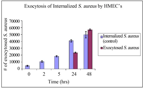
The number of internalized bacteria increased steadily with time, however no bacteria was exocytosed in the first 5 hours of incubation (Figure 1). There was no
[image:3.595.307.537.549.690.2]analysis of the extracellular media between 5 and 24 hours, however after 24 hours of incubation, exocytosed bacteria was detected in the media. The number of exo- cytosed bacteria increased thereafter with time. These results were used to determine the incubation time of infected HMEC’s prior to treatment with cytochalasin D. The number of intracellular S. aureus increased with time in the cytochalasin D untreated cells, whereas the number of intracellular bacteria decreased in the treated cells.
There was a significantly higher number of intracellular bacteria in the cytochalasin D untreated cells at 9 and 24 hours of incubation than in treated cells, p < 0.05. The number of exocytosed bacteria from cytochalasin D un- treated cells was significantly higher than those released from cytochalasin D treated cells at 7 and 24 hours of incubation (Figure 2). These results indicate that cyto-
chalasin D was effective in inhibiting both uptake and exocytosis of S. aureus, hence actin polymerization is a possible mechanism of exocytosis. Our results confirm what others have found: That the actin cytoskeleton is actively involved in exocytosis [13].
3.2. Effect of S. aureus and Its Cell Wall Products on HMEC permeability
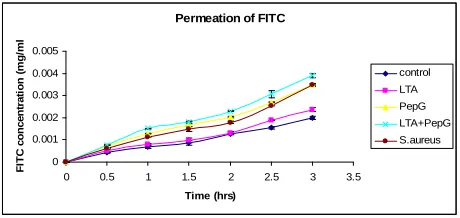
The effect of Lipoteichoic acid (LTA), peptidoglycan (Pep G), and S. aureus on the permeability of various molecular weight compounds through an endothelial cell monolayer was evaluated. Previous studies have shown that LTA can cause moderate hypotension in rats, but is unable to cause multiple organ failure or death by itself. [14] However, both LTA and pepG act in synergy to re- lease TNF- and gamma-interferon (IFN-γ), and to cause shock and multiple organ failure in anesthetized rats [15]. The first compound analyzed was FITC. PepG, PepG + LTA, and S. aureus were all significantly more effective than the control (1% HBSS) in increasing the permeation of FITC, p < 0.05 (Figure 3). The permeation of FITC-
Dextran through the HMEC monolayer showed a similar result as that seen in the case of FITC. FITC-Dextran,
Figure 2. S. aureus infected HMEC’s were divided into two groups. One group was treated with cytochalasin D (1 µg/ml) for 2 hours. Cytochalasin D was washed off and the cells were incubated at 37˚C and at various time points serial dilutions of the extracellular media were plated to deter- mine number of exocytosed bacteria. Number of internal- ized bacteria was also determined. There was a significantly higher number of internalized bacteria by cells not treated with cytochalasin D (−cyt D) than treated cells at 9 and 24 hours p < 0.05. The number of exocytosed S. aureus from HMEC’s was significantly higher from cytochalasin D un- treated cells than from the treated cells at 7 and 24 hours of incubation p < 0.05.
Permeation of FITC
0 0.001 0.002 0.003 0.004 0.005
0 0.5 1 1.5 2 2.5 3 3.5
Time (hrs)
F
ITC
c
o
nc
e
n
tr
a
ti
o
n (
m
g
/ml
control
LTA
PepG LTA+PepG
S.aureus
Figure 3. The effect of lipoteichoic acid (LTA), Peptidogly- can (PepG), and S. aureus (SA) on the permeation of FITC through HMEC monolayer. All the compounds used were more effective than the control (1% HBSS) in increasing the permeation of FITC, however, only PepG, PepG + LTA, and S. aureus produced significantly higher effect than the control, p < 0.05.
being a higher molecular weight molecule than FITC alone, showed a rather slower rate and smaller amount of permeation through the monolayer. All the compounds used were more effective than the control (1% HBSS) in increasing the permeation of FITC-Dextran, however, there was a significantly higher permeation effect only after 1 hour of incubation, p < 0.05 (Figure 4). In the
case of FITC-HSA permeation, no permeation of HSA was observed until after one hour of exposure, and this effect was observed after exposure to the combination of PepG + LTA. The combination of PepG + LTA was most effective in altering the monolayer integrity after three hours of exposure (Table 1). Table 2 shows the effect of
lipoteichoic acid (LTA), Peptidoglycan (PepG), LTA + PepG, and S. aureus (SA) on the permeation of S. aureus
through HMEC monolayer. Only LTA and LTA + PepG were effective in significantly increasing the permeation
of S. aureus at all time points p < 0.05.
3.3. Prophylactic Vancomycin Treatment
[image:4.595.308.538.96.205.2] [image:4.595.58.290.474.594.2]-0.0005 0 0.0005 0.001 0.0015 0.002 0.0025 0.003 0.0035
0 1 2 3 4
Co n c e n tr a ti o n (m g /m l) Time (hrs) FITC-Dextran Permeation Control LTA PepG LTA+PepG S. aureus
[image:5.595.57.288.94.187.2]Figure 4. The effect of lipoteichoic acid (LTA), Peptidogly- can (PepG), and S. aureus (SA) on the permeation of FITC- Dextran through HMEC monolayer. All the compounds used were more effective than the control (1% HBSS) in in- creasing the permeation of FITC-Dextran, however, there was a significantly higher permeation effect only after 1 hour of incubation, p < 0.05.
Table 1. Effect of LTA, PepG, LTA + PepG and S. aureus on FITC-HSA permeation through endothelial monolayer.
FITC-HSA Concentration (×10−3 mg/ml). Values are means ± SD (n = 3) Time
(hr) Control LTA Pep G LTA + PepG SA
0 0.00 ± 0.00 0.00 ± 0.00 0.00 ± 0.00 0.00 ± 0.00 0.00 ± 0.00
0.5 0.00 ± 0.00 0.00 ± 0.00 0.00 ± 0.00 0.00 ± 0.00 0.00 ± 0.00
1.0 0.00 ± 0.00 0.00 ± 0.00 0.00 ± 0.00 0.21 ± 0.30 0.00 ± 0.00
1.5 0.00 ± 0.00 0.00 ± 0.00 0.20 ± 0.10 *2.00 ± 0.40 0.00 ± 0.00
2.0 0.00 ± 0.00 0.00 ± 0.00 *1.14 ± 0.40 *2.07 ± 0.40 0.00 ± 0.00
2.5 0.50 ± 0.37 0.09 ± 0.10 *2.20 ± 0.13 *2.87 ± 0.61 0.74 ± 0.70
3.0 0.95± 0.37 1.50 ± 0.99 *3.17 ± 0.51 *3.96 ± 0.41 1.56 ± 1.20
[image:5.595.57.287.306.469.2]*Statistically significant difference at p < 0.05 compared with control (me- dia).
Table 2. Effect of LTA, PepG, LTA+PepG and S. aureus on S. aureus permeation through endothelial monolayer.
S. aureus colony forming units/ml (×10−3 cfu/ml) Values are means ± SD (n = 3) Time
(hr) Control LTA Pep G LTA + PepG SA
0 0.00 ± 0.00 0.00 ± 0.00 0.00 ± 0.00 0.00 ± 0.00 0.00 ± 0.00
1.0 165 ± 35 *225 ± 22 200 ± 29 *314 ± 6 144 ± 51
2.0 199 ± 27 *299 ± 13 230 ± 43 *396 ± 16 197 ± 68
3.0 205 ± 22 *352 ± 30 257 ± 49 *454 ± 4 232 ± 60
*Statistically significant difference at p < 0.05 compared with control (me-
dia).
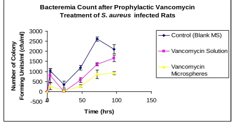
in bacterial load after that could be due to exocytosis of internalized bacteria (Figure 5). Tissue analysis after five
days of infection also confirmed the bacteremia results: Vancomycin microspheres were significantly more effec- tive in reducing tissue bacteria load than either solution
or blank microspheres (Figure 6). In all cases the bacte-
rial load in the whole of the liver was higher than in ei- ther the spleen or the lung, however, the number of bac- teria per gram of liver was less than in either organ.
3.4. Simultaneous Vancomycin Treatment
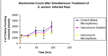
Simultaneous treatment of S. aureus infected rats pro- duced similar results as the prophylactic treatment, al- though not as significant a difference. Vancomycin mi- crospheres reduced the bacteremia levels more than ei- ther blank microspheres or vancomycin solution (Figure 7); however a significant difference in bacterial levels
was seen only at 48 and 96 hours post infection. The presence of bacteria in the systemic circulation despite treatment could be due to either low systemic drug con- centration or continued exocytosis of intracellular bacte- ria. It is possible that bacterial multiplication exceeded drug action.
Tissue analysis after five days of infection also show- ed that vancomycin microspheres were more effective in reducing intracellular bacterial load than either van-
Bacteremia Count after Prophylactic Vancomycin Treatment of S. aureus infected Rats
-500 0 500 1000 1500 2000 2500 3000
0 50 100 150
Time (hrs) N u m b er of C o lony F o rm in g U n it s /m l ( c fu /m
l) Control (Blank MS)
Vancomycin Solution
[image:5.595.306.537.366.487.2]Vancomycin Microspheres
Figure 5. Effect of vancomycin microspheres and solution on circulating S. aureus. Treatment was initiated prior to bacterial infection. The microsphere formulation was more effective in reducing bacterial load than the solution form or control after 24 hours of infection. p < 0.05.
Bacterial Levels in Organs after 5 days of Infection 0 200 400 600 800 1000 1200 1400 1600 1800 Co ntrol (blan k M
S)
VCN solu
tion
VC N m
icros phere s # of C o lon y Fo rm in g U ni ts /gm of Ti s s u e (c fu /g m ) Liver Spleen Lung
[image:5.595.58.288.530.644.2] [image:5.595.310.536.560.685.2]comycin solution or blank microspheres (Figure 8). In all
cases the blank microsphere group had the largest num-ber of bacteria in the tissue.
3.5. Delayed Vancomycin Treatment
In the delayed treatment of S. aureus infected rats it was observed that the solution form was better than the mi- crosphere form in reducing the plasma bacterial load after 8 hours of infection. It is expected that by the time treat-ment is initiated, the systemic and intracellular bacterial load would be very high due to bacterial multiplication. The dose of drug used was unchanged; hence the bac-teremia count was high due to insufficient drug action. A possible explanation for the higher efficacy of vanco- mycin solution after eight hours of infection is that, the drug was readily available in the solution form. Drug release from the microsphere formulation increased with time thereafter and that was the reason for the decreased bacterial load at later time points. Vancomycin micro- spheres reduced the bacteremia load significantly more than either the solution or the blank microspheres at 48
Bacteremia Count after Simultaneous Treatment of
S. aureus Infected Rats
0 1000 2000 3000 4000 5000
0 50 100 150
Time (hrs) # of C o lony For m ing U n its /m l (c fu/ m
l) Control (Blank
Microspheres)
Vancomycin Solution
[image:6.595.307.537.346.467.2]Vancomycin Microspheres
Figure 7. Effect of vancomycin microspheres and solution on circulating S. aureus. Treatment was initiated immedi- ately after bacterial infection. The difference in effects was not significant (p > 0.05).
Bacterial Levels in Organs after 5 days of Infection 0 200 400 600 800 1000 1200 1400 1600 Co ntrol (
blan k M S) VCN so lution VC N mi
[image:6.595.58.288.372.491.2]cros phere s # of C ol ony For m ing U ni ts /gm of Ti s s ue ( c fu /g m ) Liver Spleen Lung
Figure 8. Bacterial levels in the liver, spleen and lung after 5 days of infection. Rats were treated with drug microspheres, solution or blank media immediately after bacterial infec- tion.
and 72 hours post infection (Figure 9).
Analysis of tissue samples excised after five days of infection showed that vancomycin microsperes were more effective than either solution or blank microspheres in reducing the bacterial load in the liver and spleen (Fig- ure 10). There was no significant difference between the
formulations in reducing the bacterial load in the lungs (p > 0.05).
4. Conclusions
It can be concluded from these results that S. aureus is exocytosed after it is taken up by endothelial cells. Exo- cytosis and phagocytosis of bacteria occur simultane- ously, however, initially, the rate of phagocytosis far ex-ceeds exocytosis. With increasing intracellular bacterial load, the exocytosis rate as well as extracellular bacterial multiplication exceeds phagocytosis. The partial inhibit- tion of exocytosis by cytochalasin D and the decreasing intracellular bacterial load shows that actin po-
Bacteremia Count after delayed treatment of
S. aureus infected Rats
0 1000 2000 3000 4000 5000 6000 7000
0 50 100 150
Time (hrs) # of C o lony For m ing U n it s / m l (c fu
/ml) Control(Blankmicrospheres)
Vancomycin Solution
Vancomycin Microspheres
Figure 9. Effect of vancomycin microspheres and solution on circulating S. aureus. Treatment was initiated 4 hours after bacterial infection. Vancomycin microspheres were significantly more effective in reducing systemic bacterial load than the solution form or control at 48 and 72 hours post infection (p < 0.05).
Bacterial Levels in Organs after 5 days of Infection 0 2000 4000 6000 8000 10000 Cont rol (
blank MS ) VCN solut ion VCN mic rosp heres # o f C o lo ny F o rm in g U n it s /gm of Ti s s ue ( c fu /g m ) Liver Spleen Lung
[image:6.595.308.538.553.685.2] [image:6.595.58.287.557.686.2]lymerization is a possible mechanism of exocytosis as well as phagocytosis. The profound effect that the com- bination of LTA and PepG had on the endothelial cell monolayer integrity shows that both components of the gram positive cell wall are required for vascular leakage to occur considerably.
In vivo challenge studies of peritonitis rat models sup-
ported previous in vitro resultsthat vancomycin micro- spheres were more effective in killing intracellular bacte- ria [10]. In all three scenarios, namely prophylactic, si- mul-taneous, and delayed treatment, the microencapsu- lated form of vancomycin was more effective in reducing the systemic bacterial load than either blank micro- spheres or free vancomycin. Similar results were seen in the tissue analysis of bacteria levels.
The comparable distribution of both microspheres and bacteria in the spleen and liver will ensure effective kill- ing of microbes (by encapsulated drugs) which infect these organs. In cases where the lungs are the infected organs, microspheres of larger sizes (>7 µm) would be more effective. In sepsis, bacteria within endothelial cells are the main target; hence treatment would be more ef- fective if professional phagocytes are initially saturated with blank microspheres, before treatment is initiated.
5. Acknowledgements
This study was supported by the Dialysis Clinic, Atlanta and Mercer University Southern School of Pharmacy.
REFERENCES
[1] R. G. Holzheimer, “The Significance of Endotoxin Re- lease in Experimental and Clinical Sepsis in Surgical Pa- tients—Evidence for Antibiotic—Induced Endotoxin Re- lease?” Infection, Vol. 26, No. 2, 1998, pp. 77-84. doi:10.1007/BF02767765
[2] E. C. Hack, L. A. Aarden and L. G. Thijs, “Role of Cyto- Kines in Sepsis,” Advances in Immunology, Vol. 66, 1997, pp. 101-195. doi:10.1016/S0065-2776(08)60597-0 [3] R. J. Hamill, J. M. Vann and R. A. Proctor, “Phagocytosis
of Staphylococcus aureus by Cultured Aortic Endothelial Cells: Model for Post-Adherence Events in Endovascular Infections,” Infection and Immunity, Vol. 54, 1986, pp. 833-836.
[4] H. Beekhuizen, J. S. Van de Gevel, B. Olsson, I. J. Van Benten and R. Van Furth, “Infection of Human Vascular Endothelial Cells with Staphylococcus aureus Induces Hyperadhesiveness for Human Monocytes and Granulo- cytes,” The Journal of Immunology, Vol. 158, No. 2, 1997, pp. 774-782.
[5] E. C. Hack and M. D. Zeerleder, “The Endothelium in Sepsis: Source of and a Target for Inflammation,” Criti- cal Care Medicine, Vol. 29, No. 7, 2001, pp. S21-S27. doi:10.1097/00003246-200107001-00011
[6] M. D’Souza, C. W. Oettinger and G. V. Milton, “Micro- spheres Containing Neutralizing Antibodies to Tumor Necrosis Factor-α and Interleukin-1β Protect Rats from Staphylococcus aureus-Induced Peritonitis,” Journal of Interferon & Cytokine Research, Vol. 20, No. 10, 2000, pp. 907-913. doi:10.1089/10799900050163280
[7] P. K. Gupta, C. T. Hung and D. G. Perrier, “Albumin Microspheres I: Release Characteristics of Adriamycin,” International Journal of Pharmaceutics, Vol. 33, No. 1-3, 1986, pp. 137-146. doi:10.1016/0378-5173(86)90047-5 [8] M. D’Souza and P. DeSouza, “Preparation and Testing of
Cyclosporine Microsphere and Solution Formulations in the Treatment of Polyarthritis in Rats,” Drug Develop- ments in Industrial Pharmacy, Vol. 24, No. 9, 1998, pp. 841-852. doi:10.3109/03639049809088529
[9] M. D’Souza and R. Pourfazib, “Improved Efficacy of a Microencapsulated Macrophage Colony-Stimulating Fac- tor and Methotrexate in Melanoma,” Drug Developments in Industrial Pharmacy, Vol. 25, No. 5, 1999, pp. 583- 590. doi:10.1081/DDC-100102212
[10] H. Nettey, D. Haswani, C. Oettinger and M. J. D’Souza, “In Vitro Antimicrobial Effect of Encapsulated Vanco- mycin on Internalized Staphylococcus aureus within En- dothelial Cells,” Drug Development and Industrial Phar- macy, Vol. 33, No. 2, 2007, pp. 133-139.
doi:10.1080/03639040600867989
[11] V. Schafer, H. Briesen, H. Rubsamen-Waigman, A. M. Steffan, C. Royer and J. Kreuter, “Phagocytosis and Deg- radation of Human Serum Albumin Microspheres and Nanoparticles in Human Macrophages,” Journal of Mi- croencapsulation, Vol. 11, No. 3, 1994, pp. 261-269. doi:10.3109/02652049409040455
[12] H. Nettey, D. Haswani, C. Oettinger and M. J. D’Souza, “Formulation and Testing of Vancomycin Loaded Albu- min Microspheres Prepared by Spray-Drying,” Journal of Microencapsulation, Vol. 23, No. 6, 2006, pp. 632-642. doi:10.1080/02652040600776564
[13] S. Gasman, S. Chasserot-Golaz, N. Vitale and M. F. Ba- der, “Bacterial Toxins: Useful for Studying G-Proteins Implicated in the Mechanism of Exocytosis in Neuroen- docrine Cells,” Journal de la Société de Biologie, Vol. 193, No. 6, 1999, pp. 451-456.
[14] S. J. De Kimpe, M. L. Hunter and C. F. Bryant, “Delayed Circulatory Failure Due to the Induction of Nitric Oxide Synthase by Lipoteichoic Acid from Staphylococcus aureus in Anaesthetized Rats,” British Journal of Phar- macology, Vol. 114, No. 6, 1995, pp. 1317-1323.
doi:10.1111/j.1476-5381.1995.tb13349.x
[15] S. J. De Kimpe, M. Kengatharan, C. Thimermann and J. R. Vane, “The Cell Wall Components Peptidoglycan and Lipoteichoic Acid from Staphylococcus aureus Act in Synergy to Cause Shock and Multiple Organ Failure,” Proceedings of the National Academy of Sciences, Vol. 92, No. 22, 1995, pp. 10359-10363.