Implies a Clonal Population Structure
Benoît Chassain,aLudovic Lemée,aJennifer Didi,aJean-Michel Thiberge,bSylvain Brisse,bJean-Louis Pons,cand Martine Pestel-Carona
University of Rouen, Rouen University Hospital, GRAM EA 2656, Rouen, Francea; Institut Pasteur, Genotyping of Pathogens and Public Health, Paris, Franceb; and University Paris Descartes, EA 4065, Paris, Francec
Staphylococcus lugdunensisis recognized as one of the major pathogenic species within the genusStaphylococcus, even though it
belongs to the coagulase-negative group. A multilocus sequence typing (MLST) scheme was developed to study the genetic rela-tionships and population structure of 87S. lugdunensisisolates from various clinical and geographic sources by DNA sequence analysis of seven housekeeping genes (aroE,dat,ddl,gmk,ldh,recA, andyqiL). The number of alleles ranged from four (gmkand ldh) to nine (yqiL). Allelic profiles allowed the definition of 20 different sequence types (STs) and five clonal complexes. The 20 STs lacked correlation with geographic source. Isolates recovered from hematogenic infections (blood or osteoarticular isolates) or from skin and soft tissue infections did not cluster in separate lineages. Penicillin-resistant isolates clustered mainly in one clonal complex, unlike glycopeptide-tolerant isolates, which did not constitute a distinct subpopulation withinS. lugdunensis. Phylogenies from the sequences of the seven individual housekeeping genes were congruent, indicating a predominantly muta-tional evolution of these genes. Quantitative analysis of the linkages between alleles from the seven loci revealed a significant linkage disequilibrium, thus confirming a clonal population structure forS. lugdunensis. This first MLST scheme forS.
lug-dunensisprovides a new tool for investigating the macroepidemiology and phylogeny of this unusually virulent
coagulase-nega-tiveStaphylococcus.
S
ince its first description in 1988,Staphylococcus lugdunensis(15) has rapidly been recognized as one of the major
patho-genic species within the genusStaphylococcus, despite belonging to
the coagulase-negative group.S. lugdunensisis recovered from the
normal skin flora (1), and numerous infections have been related
to inguinal-area carriage (37). Cutaneous infections account for
more than 50% ofS. lugdunensisinfections (38), but tissue
infec-tions with various abscess localizainfec-tions are reported, with tissue
damage or poor clinical outcome (13). Bacteremia is frequently
associated with endocarditis (36), which develops more
fre-quently on native valve than on prosthetic material (28) and has a
clinical course resemblingStaphylococcus aureusinfection (36).
Hematogenic infections, such as prosthetic joint infections or
os-teomyelitis, are also considered as aggressive as withS. aureus(30).
The pathogenic potential ofS. lugdunensis, which is thus closer to
that ofS. aureusthan those of other coagulase-negative
staphylo-cocci, may be related to virulence factors such as a
fibrinogen-binding protein (25), a von Willebrand factor-binding protein
(26), or synergistic hemolysins (7). In addition, the genome ofS.
lugdunensisN920143 contains anisdlocus (19), which encodes
proteins involved in iron acquisition inS. lugdunensis(18) or in
cutaneous colonization inS. aureus(6).
UnlikeS. aureusor numerous coagulase-negative
staphylo-cocci,S. lugdunensisremains largely susceptible to
antistaphylo-coccal antibiotics. Penicillinase production is observed for less
than 50% of isolates (14), and methicillin resistance is still rare
(30,38). However, a particular feature ofS. lugdunensisis
vanco-mycin and/or teicoplanin tolerance (4,14). We found, by
analyz-ing seven coagulase-negative staphylococcal species, that this
gly-copeptide tolerance phenomenon was restricted toS. lugdunensis.
Indeed, all theS. lugdunensisisolates investigated displayed strict
glycopeptide tolerance or at least decreased and slower
suscepti-bility to glycopeptide bactericidal activity (4).
Phylogenetic analyses within pathogenic species provide
pre-cious knowledge about their genetic population structure and their relation to patterns of virulence and/or antimicrobial sus-ceptibility. Multilocus sequence typing (MLST), which character-izes bacterial multilocus genotypes by using intragenic sequences of a set of housekeeping genes, was initially proposed for
popula-tion genetics analysis ofNeisseria meningitidis(12). It allowed the
characterization of recombinant population structures for N.
meningitidisorStreptococcus pneumoniae(10,12) and, conversely,
clonal population structure forS. aureus(8) orClostridium difficile
(21), whose mutational evolution generates deeper recognizable
phylogenetic lineages. MLST has proved essential for analyzing
the evolution of methicillin-resistantS. aureusclones and their
filiation from ancestral methicillin-susceptible clones (9). MLST
also offers the possibility to open shared sequence databases on the World Wide Web that provide an extensive and continuously up-dated view about the long-term epidemiology and evolution of a
given species (22).
Although MLST analyses have been promoted forS. aureus(8)
andStaphylococcus epidermidis (24), there is still a need for an
MLST scheme devoted to S. lugdunensis, whose phylogenetic
structure remains unknown. The aim of the present work was to
develop an MLST scheme forS. lugdunensisand to analyze the
mode of evolution of this pathogenicStaphylococcus species in
relation to antimicrobial susceptibility.
Received2 May 2012Returned for modification29 May 2012
Accepted2 July 2012
Published ahead of print11 July 2012
Address correspondence to Martine Pestel-Caron, martine.pestel-caron@univ -rouen.fr.
Copyright © 2012, American Society for Microbiology. All Rights Reserved.
doi:10.1128/JCM.00988-12
on May 16, 2020 by guest
http://jcm.asm.org/
MATERIALS AND METHODS
Bacterial isolates.A total of 87S. lugdunensisisolates were studied:S. lugdunensisATCC 49576, S. lugdunensisATCC 43809,S. lugdunensis ATCC 700328, and 84 human clinical isolates (skin or soft tissue isolates, osteoarticular isolates, blood isolates, and material device isolates) col-lected from 1991 to 2011 from 8 different geographic sources in France,
Belgium, and Slovenia. Isolates were identified asS. lugdunensisby Gram
stain, colony morphology, biochemical profile from the Phoenix auto-mated microbiology system (Becton Dickinson), and detection of pyrro-lidonyl-arylamidase (Oxoid biochemical identification system [O.B.I.S.]; Oxoid). For some isolates, the species identification was confirmed by
real-time PCR amplification of an internal fragment of thefblgene (5)
usingfbl forward(5=-GTAAATAGCGAGGCACAAGC-3=) andfbl reverse
(5=-GGTAAATCGTATCTGCCGCT-3=) primers.-Lactamine
suscepti-bility was determined by chromogenic detection of penicillinase produc-tion (Cefinase; bioMérieux) and by the agar disk diffusion method ac-cording to the recommendations of the Comité de l’Antibiogramme de la Société Française de Microbiologie (2) for oxacillin and moxalactam (de-tection of methicillin resistance). The bactericidal activities of vancomy-cin and teicoplanin were determined previously for 13 strains (4).
PFGE.Pulsed-field gel electrophoresis (PFGE) typing of SmaI (New England BioLabs)-digested DNA was performed as previously described (23). Briefly, genomic DNA incorporated in agarose plugs was digested at 25°C overnight, and large restriction fragments were separated in a 1% agarose gel at 14°C for 16 h by using the Gene Path system (Bio-Rad). The patterns were digitized with Molecular Analyst Fingerprinting software (Bio-Rad), and PFGE patterns were interpreted as recommended by Tenover et al. (33).
MLST.Seven housekeeping loci were selected for the characterization ofS. lugdunensisisolates by MLST (Table 1):aroE(shikimate
dehydroge-nase),dat(D-amino acid aminotransferase),ddl(D-alanine:D-alanine
li-gase),gmk(guanylate kinase),ldh(L-lactate dehydrogenase),recA
(re-combinase), andyqiL(acetyl-coenzyme A acetyltransferase). The choice
of these housekeeping genes was based on their use in MLST schemes ofS.
aureus(8),S. epidermidis(34),Listeria monocytogenes(29), orC. difficile (16,21) and on the availability of sequence data from S. lugdunensis HKU09-01 (35). The seven loci were present in single copies and
physi-cally scattered on theS. lugdunensisHKU09-01 genome.
Each DNA sample for PCR amplification was extracted from a single bacterial colony from a tryptic soy agar culture using InstaGene DNA (Bio-Rad) according to the manufacturer’s recommendations. PCRs were performed on a GeneAmp PCR system 9700 thermal cycler (Applied
Bio-systems) in a final volume of 25l containing 0.50M each primer, 12.5
l of Reddymix (Thermo Fisher Scientific), and 5l of extracted DNA.
The PCR mixtures were heated for 4 min at 95°C, and then a touch-down procedure was performed, consisting of 30 s at 95°C, annealing for 30 s at temperatures decreasing from 60°C to 50°C during the first 11 cycles (with 1°C decremental steps in cycles 1 to 11), and ending with an extension step at 72°C for 30 s. A total of 50 cycles were performed.
PCR products were then purified with a Nucleospin extract II kit (Ma-cherey-Nagel) and sequenced with the same primers as for PCR by using the DTCS sequencing kit (Beckman) on a GenomeLab GeXP analyzer (Beckman Coulter). Different sequences of a given locus were given allele numbers, and each unique combination of alleles (multilocus allelic pro-file) was assigned a sequence type (ST). Single-point polymorphisms were assessed by sequencing both DNA strands from two separate PCR exper-iments.
Computer analysis of MLST data.Clustering of the 87 isolates (and
sequence data from S. lugdunensisHKU09-01) was performed using
BioEdit Sequence Alignment Editor (http://www.mbio.ncsu.edu/BioEdit /bioedit.html) for alignment of the nucleotide sequences, and gene trees were constructed from sequence data using the neighbor-joining method and bootstrapping algorithms contained in MEGA 4.0 software (32). The average numbers of nucleotide differences between alleles and the ratios
of nonsynonymous to synonymous substitutions (dN/dS) were calculated TABLE
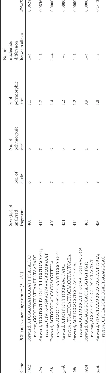
1 Genetic polymorphism of the seven housekeeping genes analyzed by MLST Gene PCR and sequencing primers (5 = ¡ 3 = ) Size (bp) of analyzed fragments No. of alleles No. of polymorphic sites %o f polymorphic sites No. of
nucleotide differences between
alleles dN / dS a aroE Forward, ATCGGAGATCCGATTTCACATTC; reverse, GGCGTTGTATTAATTATAATATC 460 6 5 1.1 1–3 0.0628 dat Forward, TCGTGGTTATGTTTTTGGTGACGGT; reverse, CTATGAGAAGTAAAGCCAGGAAT 412 8 7 1.7 1–4 0.0836 ddl Forward, AGTGCGGAGCACGACGTTTCA; reverse, ACACTTGATCCCAAATTCGCCGGT 420 7 6 1.4 1–4 0.0000 gmk Forward, ATAGTTCTTTCCGGACCATC; reverse, TCATTGACTACAACGTAATCATA 431 4 5 1.2 1–5 0.0000 ldh Forward, ACTTGCAGGTGCCACGTCGA; reverse, GCTACGCATTTGCAATGGTAACGCA 414 4 5 1.2 1–4 0.0000 recA Forward, GCACGGCCACCAGGTGTTGT; reverse, AGGCCGTCGCGTATCTAGTGT 463 5 4 0.9 1–3 0.0000 yqiL Forward, GTGCTAAACGCACACCAATTGGA; reverse, CTTCAGCATCGATTAGAGGCAC 450 9 8 1.8 1–5 0.2412 adN / dS ,ratio of nonsynonymous to synonymous substitution.
on May 16, 2020 by guest
http://jcm.asm.org/
[image:2.585.328.516.71.725.2]to test the degree of selection operating on a locus using the START pro-gram (http://www.mlst.net).
The index of association (Ia) between alleles (31) was used to test for
linkage disequilibrium between alleles of the seven loci analyzed (http: //www.mlst.net). The observed variance in the distribution of allelic mis-matches in all pairwise comparisons of the allelic profiles was compared to that expected in a freely recombining population (linkage equilibrium). The significance of the difference in the observed and expected variances was evaluated by computing the maximum variance in the distribution of allelic mismatches obtained using 100 randomizations of the data set.
The BURST program (http://www.mlst.net) was used to define clonal complexes (CCs; groups in which every isolate shares at least five identical alleles with at least one other isolate) and to characterize ancestral geno-types and their corresponding single-locus variants (SLVs, isolates that differ at only one of the seven loci) within these clonal complexes.
RESULTS
Allelic variation inS. lugdunensis.Data reporting the allelic
vari-ation of the seven housekeeping genes are summarized inTable 1.
The number of individual alleles for each of the seven
housekeep-ing genes ranged from 4 forgmkandldhto 9 foryqiL. The number
of polymorphic sites on a given locus varied from 4 (forrecA) to 8
(foryqiL), and the maximum number of nucleotide differences
between alleles of a given locus ranged from 3 foraroEandrecAto
5 foryqiLandgmk. The variations in the sequences extended over
the whole stretch of each of the seven genes investigated (http:
//www.pasteur.fr/mlst). Most polymorphisms resulted in ymous substitutions, with the ratios of nonsynonymous to
synon-ymous substitutions (dN/dS) varying from 0 (forddl,gmk,ldh,
andrecA) to 0.2412 (foryqiL). These low ratios indicate a lack or a
very limited contribution of environmental selection to the se-quence variation in the seven housekeeping genes analyzed, which are thus assumed to be suitable for a population genetic study.
MLST analysis ofS. lugdunensisisolates.MLST analysis of
the 87S. lugdunensisisolates generated a total of 20 different STs
(Table 2). For each ST shared by at least two isolates, PFGE anal-ysis displayed identical or closely related profiles, further arguing for probable clonal relationships between these isolates. An MLST database specific for this species and containing allelic and ST data has been made available on the website of the Pasteur Institute of
Paris (http://www.pasteur.fr/mlst).
The results of clustering from concatenated sequence data are
shown inFig. 1. Half of the STs were represented by a single
iso-late. Among STs shared by several isolates, the most frequently encountered were ST3 (18 isolates), ST2 (15 isolates), and ST1 (11 isolates). Five clonal complexes (CCs) could be characterized by
using the method and the criteria described by Feil et al. (11)
(Table 2). The ancestral genotypes and their single-locus variants (probable derived genotypes) could be identified for CC1, CC2,
and CC3 (Fig. 2). Of note, the sl96 strain, corresponding to the
first published complete genome ofS. lugdunensis, belongs to CC1
but harbors a unique ST (ST11).
Dendrograms based on sequence data of each housekeeping gene. Dendrograms from the sequences of the seven separate housekeeping genes were found to be congruent overall, indicat-ing a predominantly mutational evolution of these genes. No re-combinational event could be detected by comparing monolocus trees (data not shown).
Estimation of the relative contributions of recombination and mutation to genomic evolution ofS. lugdunensis.Since the congruence of dendrograms based on allelic variation of each housekeeping gene suggested a low frequency of recombination, a quantitative analysis of the linkage between alleles from the seven
[image:3.585.40.548.78.311.2]loci was performed by calculating the index of association (Ia)
TABLE 2Characteristics of the 20 allelic profiles (STs) and clonal complexes
STa
Allelic profile
Clonal complex
No. of
isolates Geographic origin(s)b
aroE dat ddl gmk ldh recA yqiL
1 1 1 1 1 1 1 1 CC1 11 A, B, C, D, F
2 3 3 2 2 1 2 3 CC2c 15 A, B, D, E, G
3 4 3 4 2 3 4 6 CC3c 18 A, B, C, D, E, F
4 6 4 3 1 1 3 3 CC4 3 A, C
5 5 6 5 3 4 4 7 CC5 4 A, B, C
6 1 1 1 1 1 1 2 CC1c 8 A, B
7 1 1 1 1 1 1 9 CC1 1 D
8 2 3 2 2 1 2 3 CC2 1 A
9 1 4 3 1 1 3 4 CC3 1 A
10 3 5 2 2 2 3 5 8 A, B, C, F
11 1 1 1 1 1 6 1 CC1 1 —d
12 1 1 7 1 1 1 2 CC1 4 A, C
13 3 3 6 4 1 5 3 3 A, G
14 3 3 2 2 1 2 5 CC2 1 B
15 1 2 1 1 1 1 2 CC1 4 A, D, E
16 5 3 4 2 3 1 6 CC4 1 A
17 6 4 6 1 1 3 3 CC3 1 D
18 5 6 5 3 4 4 8 CC5 1 D
19 3 7 2 2 1 2 3 CC2 1 C
20 4 8 4 2 3 4 6 CC4 1 C
aST, sequence type.
b
Geographic origins are as follows: A, Rouen, France; B, Nantes, France; C, Nancy, France; D, Bordeaux, France; E, Montpellier, France; F, Versailles, France; G, Louvain, Belgium; H, Maribor, Slovenia.
c
Ancestral genotype within the clonal complex.
dS. lugdunensisHKU09-01, unknown geographic source (35).
on May 16, 2020 by guest
http://jcm.asm.org/
on May 16, 2020 by guest
http://jcm.asm.org/
(31). When all the isolates were included in the analysis, a
signif-icant linkage disequilibrium was detected (Iaof 2.91). At the level
of STs (one isolate from each ST, to avoid bias due to a possible
epidemic population structure),Iawas calculated at 1.88,
con-firming the significant linkage disequilibrium between alleles and, thus, a clonal structure of the population studied.
Clustering of S. lugdunensis isolates. No correlation was found between genotype and geographic origin; for example, ST3, the ancestral genotype of CC3, was shared by isolates from 6 geo-graphic origins. Similarly, ST1 and ST2 were each shared by iso-lates from 5 different geographic origins. Isoiso-lates recovered from hematogenic infections (blood or osteoarticular isolates) or from skin and soft tissue infections did not cluster in separate lineages.
Twenty-seven S. lugdunensisisolates (31% of isolates) were
found to be resistant to penicillin G by penicillinase production; no isolate exhibited methicillin resistance. Three STs belonging to CC1 (ST6, the ancestral genotype, and ST12 and ST15) contained 13 of the 27 penicillin-resistant isolates and contained only 4 pen-icillin-susceptible isolates, indicating that penicillin-resistant iso-lates are associated with this phylogenetic lineage. Conversely, ST1 and ST7, which were also inferred to derive from the ancestral genotype ST6 in CC1, contain only penicillin-susceptible isolates, indicating the possible loss of the penicillinase-encoding plasmid. Alternatively, they may represent ancestral genotypes predating the clonal expansion of ST6 after the acquisition of resistance.
The six isolates that we previously characterized as exhibiting
glycopeptide (vancomycin and/or teicoplanin) tolerance (4)
be-longed to five different STs (ST6, ST15, ST8, ST9, and ST10) and to three clonal complexes (CC1, CC2, and CC4) and, thus, did not define a specific lineage. In addition, two STs (ST6 and ST10) included both nontolerant isolates and glycopeptide-tolerant isolates, and ST5 included a nontolerant isolate together with a vancomycin-tolerant isolate. Overall, these data suggest that glycopeptide-tolerant isolates do not constitute a distinct
sub-population withinS. lugdunensis, implying multiple
indepen-dent evolution of the genetic elements involved in this pheno-type by isolates from different genetic backgrounds.
DISCUSSION
The aim of the present study was to design an MLST scheme, based on the allelic polymorphism of seven housekeeping genes, which should be suitable for population genetics and intraspecific
phylogeny analysis ofS. lugdunensis. Only macrorestriction PFGE
(37) had been used previously for molecular typing of this
organ-ism, but this approach would not, henceforward, constitute the reference method for these purposes.
We analyzed 87S. lugdunensisisolates from various clinical
infections and geographic sources. The allelic polymorphism (number of distinct STs among the isolates and number of
poly-morphic sites per locus) appears lower than that ofS. aureusorS.
epidermidis, which themselves display a moderate genetic
variabil-ity, with few well-conserved phylogenetic lineages (17,24). The
low apparent rate of recombination results in a clonal population
structure forS. lugdunensis, as illustrated by the index of
associa-tion between alleles of the different loci, which reveals linkage
disequilibrium (31), the congruence of dendrograms of individual
housekeeping genes, and the characterization of clonal complexes within the population studied. Thus, if homologous
recombina-tion does exist, it rarely contributes to the evolurecombina-tion ofS.
lug-dunensis. Sequence data analysis revealed that only synonymous
substitutions were detected in genesddl,gmk,ldh, andrecA,
sug-gesting that most nonsilent mutations are eliminated through pu-rifying selection.
PFGE typing allowed us to check for the lack of an epidemio-logical link between isolates sharing the same ST. Since PFGE was unable to further resolve these MLST clusters, this method did not demonstrate a clear improvement in the discrimination of isolates compared to MLST; thus, MLST should be proposed as an
effi-cient alternative approach for molecular typing ofS. lugdunensis,
with the additional advantage of providing unambiguous and portable sequence data suited for large-scale epidemiology.
Although S. lugdunensis is recognized as one of the major
pathogenic species within the genusStaphylococcus, little is known
about the virulence determinants implicated in invasive tions. In the present study, isolates from skin and soft tissue infec-tions or from hematogenic infecinfec-tions (blood or osteoarticular iso-lates) were found to be randomly distributed in the main lineages depicted from the MLST dendrogram. It should be informative to further analyze allelic polymorphism of genes encoding
fibrino-gen-binding protein (25), von Willebrand factor-binding protein
(26), or synergistic hemolysins (7), which have been proposed as
virulence determinants ofS. lugdunensis, and to investigate how
this variation is related to MLST data. This could contribute to the description of putative hypervirulent lineages within the species.
S. lugdunensisremains largely susceptible to antibiotics and, surprisingly, displays a low frequency of methicillin resistance,
unlike other coagulase-negative staphylococci orS. aureus. The
FIG 1Dendrogram showing genetic relationships of the 87S. lugdunensisisolates (and sequence data fromS. lugdunensisHKU09-01) based on the composite
sequence of the seven housekeeping genes (aroE,dat,ddl,gmk,ldh,recA, andyqiL). Clonal complexes (CC) are represented with brackets. Rooting of the
dendrogram was established using homologous sequence data fromStaphylococcus haemolyticusJCSC1435. Origin: A, Rouen, France; B, Nantes, France; C,
Nancy, France; D, Bordeaux, France; E, Montpellier, France; F, Versailles, France; G, Louvain, Belgium; and H, Maribor, Slovenia. Clinical source: OA,
osteoarticular; SST, skin and soft tissue; M, material device; B, blood. Penicillin: R, resistant; S, susceptible. *, ref1 (reference sequence 1),S. lugdunensisATCC
700328; ref2,S. lugdunensisATCC 43809; ref3,S. lugdunensisATCC 49576; **, sl96 isS. lugdunensisHKU09-01.
FIG 2Clonal complexes with ancestral genotypes identified. (A) Clonal
com-plex 1, including ST6 (ancestral genotype), ST1, ST7, ST12, and ST15 (de-scended from single-locus variant genotypes), and ST11 (de(de-scended from double-locus variant genotype). (B) Clonal complex 2, including ST2 (ances-tral genotype) and ST8, ST14, and ST19 (descended from single-locus variant genotypes). (C) CC3 including ST3 (ancestral genotype) and ST16 and ST20 (descended from single-locus variant genotypes).
on May 16, 2020 by guest
http://jcm.asm.org/
[image:5.585.43.282.70.169.2]present collection of clinical isolates is concordant with these data, with 31% of isolates producing penicillinase and a lack of methi-cillin-resistant isolates. Half of the penicillinase-producing iso-lates belong to a single clonal complex (CC1), reflecting vertical transmission within this lineage. Of note, ST1 and ST7, which are single-locus variants of the ST6 ancestral genotype of CC1, con-tain penicillin-susceptible isolates, suggesting a probable loss of
the penicillinase-encoding plasmid or transposon (19, 20).
Be-sides this clonal complex, penicillinase-producing isolates are widely distributed, either as a result of horizontal transmission or multiple independent acquisitions. Concerning methicillin
resis-tance, which is still uncommon inS. lugdunensis, collecting MLST
data from methicillin-resistant isolates could allow the study of their filiation from ancestral methicillin-susceptible clones, as was
successfully deduced from MLST data forS. aureus(9,27).
Vancomycin and/or teicoplanin tolerance (lack of bactericidal activity of these presumed bactericidal antibiotics) appears as a
particular feature ofS. lugdunensis(4,14). We previously
investi-gated a total of 13 isolates for glycopeptide tolerance and
charac-terized six isolates tolerant to vancomycin and/or teicoplanin (4).
These six tolerant isolates are distributed in different MLST lin-eages in the present study, and some STs (ST6, ST10, and ST5) include both nontolerant and glycopeptide-tolerant isolates. Thus, although glycopeptide tolerance was not investigated in the whole population analyzed by MLST, it may be assumed that this phenomenon is not restricted to a distinct subpopulation within
S. lugdunensis. Further studies are currently in progress to inves-tigate the implication of the major peptidoglycan hydrolase AtlL
(3) in this impaired bactericidal activity of vancomycin and/or
teicoplanin.
This work provides the first phylogenetic analysis ofS.
lug-dunensisand gives strong arguments for a clonal population struc-ture and mutational evolution of this pathogen. MLST combines the advantages of (i) a sequence-based typing method, which makes the data unambiguous and readily comparable between different laboratories, and (ii) a phylogenetic approach to genetic diversity, since it is based on sequence polymorphism of
house-keeping genes apart from selective pressure. TheS. lugdunensis
MLST database, which is hosted at the website of the Institut
Pas-teur Paris (http://www.pasteur.fr/mlst), should be enriched from
sequence data from worldwide and clinically diverse isolates, to follow the global epidemiology and evolution within the species. In addition, MLST data could be used as typing markers, possibly in combination with virulence gene data, to increase the discrim-inatory power and to investigate the short-term epidemiology of
S. lugdunensis, which remains until now poorly investigated.
ACKNOWLEDGMENTS
We thank M. Delmée, F. Doucet-Populaire, V. Dubois, G. Grise, H. Marchandin, A. Morel, F. Mory, A. Reynaud, and M. Rupnik for supply-ing strains.
REFERENCES
1.Bieber L, Kahlmeter G.2010.Staphylococcus lugdunensisin several niches
of the normal skin flora. Clin. Microbiol. Infect.16:385–388.
2.Bonnet R, et al.2011. Comité de l’Antibiogramme de la Société Française
de Microbiologie. Recommandations 2011. Société Française de Microbi-ologie, Paris, France.
3.Bourgeois I, et al.2009. Characterization of AtlL, a bifunctional autolysin
ofStaphylococcus lugdunensiswith acetylglucosaminidase and
N-acetylmuramoyl-l-alanine amidase activities. FEMS Microbiol. Lett.290:
105–113.
4.Bourgeois I, Pestel-Caron M, Lemeland JF, Pons JL, Caron F.2007.
Tolerance to the glycopeptides vancomycin and teicoplanin in
coagulase-negativestaphylococci.Antimicrob. Agents Chemother.51:740 –743.
5.Chatzigeorgiou KS, Siafakas N, Petinaki E, Zerva L.2010.fblgene as a
species-specific target forStaphylococcus lugdunensisidentification. J.
Clin. Lab. Anal.24:119 –122.
6.Clarke SR, et al.2007. TheStaphylococcus aureussurface protein IsdA
mediates resistance to innate defenses of human skin. Cell Host Microbe
1:199 –212.
7.Donvito B, et al.1997. Synergistic hemolytic activity ofStaphylococcus
lugdunensisis mediated by three peptides encoded by a non-agrgenetic
locus. Infect. Immun.65:95–100.
8.Enright MC, Day NP, Davies CE, Peacock SJ, Spratt BG.2000.
Multi-locus sequence typing for characterization of methicillin-resistant and
methicillin-susceptible clones ofStaphylococcus aureus.J. Clin. Microbiol.
38:1008 –1015.
9.Enright MC, et al.2002. The evolutionary history of methicillin-resistant
Staphylococcus aureus(MRSA). Proc. Natl. Acad. Sci. U. S. A.99:7687– 7692.
10. Enright MC, Spratt BG.1998. A multilocus sequence typing scheme for
Streptococcus pneumoniae: identification of clones associated with serious
invasive disease. Microbiology144(Pt 11):3049 –3060.
11. Feil EJ, Li BC, Aanensen DM, Hanage WP, Spratt BG.2004. eBURST:
inferring patterns of evolutionary descent among clusters of related
bac-terial genotypes from multilocus sequence typing data. J. Bacteriol.186:
1518 –1530.
12. Feil EJ, Maiden MC, Achtman M, Spratt BG.1999. The relative
contri-butions of recombination and mutation to the divergence of clones of Neisseria meningitidis.Mol. Biol. Evol.16:1496 –1502.
13. Frank KL, Del Pozo JL, Patel R.2008. From clinical microbiology to
infection pathogenesis: how daring to be different works for
Staphylococ-cus lugdunensis.Clin. Microbiol. Rev.21:111–133.
14. Frank KL, Reichert EJ, Piper KE, Patel R. 2007. In vitro effects of
antimicrobial agents on planktonic and biofilm forms ofStaphylococcus
lugdunensisclinical isolates. Antimicrob. Agents Chemother.51:888 – 895.
15. Freney J, et al.1988.Staphylococcus lugdunensissp. nov. and
Staphylococ-cus schleiferisp. nov. Two species from human clinical specimens. Int. J.
Sys. Bacteriol.38:168 –172.
16. Griffiths D, et al.2010. Multilocus sequence typing ofClostridium
diffi-cile.J. Clin. Microbiol.48:770 –778.
17. Grundmann H, et al.2002. Determining the genetic structure of the
natural population ofStaphylococcus aureus: a comparison of multilocus
sequence typing with pulsed-field gel electrophoresis, randomly amplified
polymorphic DNA analysis, and phage typing. J. Clin. Microbiol.40:
4544 – 4546.
18. Haley KP, Janson EM, Heilbronner S, Foster TJ, Skaar EP. 2011.
Staphylococcus lugdunensisIsdG liberates iron from host heme. J.
Bacte-riol.193:4749 – 4757.
19. Heilbronner S, et al.2011. Genome sequence ofStaphylococcus
lugdunen-sisN920143 allows identification of putative colonization and virulence
factors. FEMS Microbiol. Lett.322:60 – 67.
20. Kotsakis SD, Tzouvelekis LS, Zerva L, Liakopoulos A, and Petinaki E.
2012.Staphylococcus lugdunensisstrain with a modified PBP1A/1B
ex-pressing resistance to beta-lactams. Eur. J. Clin. Microbiol. Infect. Dis.
31:169 –172.
21. Lemee L, Dhalluin A, Pestel-Caron M, Lemeland JF, Pons JL.2004.
Multilocus sequence typing analysis of human and animalClostridium
difficileisolates of various toxigenic types. J. Clin. Microbiol.42:2609 – 2617.
22. Lemee L, Pons JL.2010. Multilocus sequence typing forClostridium
difficile.Methods Mol. Biol.646:77–90.
23. Maslow JN, Slutsky AM, Arbeit RD.1993. Application of pulsed-field gel
electrophoresis to molecular epidemiology, p 563–572.InPersing DH,
Smith TF, Tenover FC, White TJ (ed), Diagnostic molecular microbiol-ogy: principles and applications. American Society for Microbiology, Washington, DC.
24. Miragaia M, Thomas JC, Couto I, Enright MC, de Lencastre H.2007.
Inferring a population structure forStaphylococcus epidermidisfrom
mul-tilocus sequence typing data. J. Bacteriol.189:2540 –2552.
25. Nilsson M, Bjerketorp J, Guss B, Frykberg L.2004. A fibrinogen-binding
protein ofStaphylococcus lugdunensis.FEMS Microbiol. Lett.241:87–93.
26. Nilsson M, et al.2004. A von Willebrand factor-binding protein from
Staphylococcus lugdunensis.FEMS Microbiol. Lett.234:155–161.
on May 16, 2020 by guest
http://jcm.asm.org/
27. Oliveira DC, Tomasz A, de Lencastre H.2002. Secrets of success of a human pathogen: molecular evolution of pandemic clones of
meticillin-resistantStaphylococcus aureus.Lancet Infect. Dis.2:180 –189.
28. Patel R, et al.2000. Frequency of isolation ofStaphylococcus lugdunensis
among staphylococcal isolates causing endocarditis: a 20-year experience.
J. Clin. Microbiol.38:4262– 4263.
29. Ragon M, et al.2008. A new perspective onListeria monocytogenes
evo-lution. PLoS Pathog.4:e1000146. doi:10.1371/journal.ppat.1000146.
30. Shah NB, et al.2010. Laboratory and clinical characteristics of
Staphylo-coccus lugdunensisprosthetic joint infections. J. Clin. Microbiol.48:1600 – 1603.
31. Smith JM, Smith NH, O’Rourke M, Spratt BG.1993. How clonal are
bacteria? Proc. Natl. Acad. Sci. U. S. A.90:4384 – 4388.
32. Tamura K, Dudley J, Nei M, Kumar S.2007. MEGA4: Molecular
Evo-lutionary Genetics Analysis (MEGA) software version 4.0. Mol. Biol. Evol.
24:1596 –1599.
33. Tenover FC, et al. 1995. Interpreting chromosomal DNA restriction
patterns produced by pulsed-field gel electrophoresis: criteria for bacterial
strain typing. J. Clin. Microbiol.33:2233–2239.
34. Thomas JC, et al.2007. Improved multilocus sequence typing scheme for
Staphylococcus epidermidis.J. Clin. Microbiol.45:616 – 619.
35. Tse H, et al.2010. Complete genome sequence of Staphylococcus
lug-dunensisstrain HKU09-01. J. Bacteriol.192:1471–1472.
36. Vandenesch F, Etienne J, Reverdy ME, Eykyn SJ.1993. Endocarditis due
toStaphylococcus lugdunensis: report of 11 cases and review. Clin. Infect.
Dis.17:871– 876.
37. van der Mee-Marquet N, et al.2003.Staphylococcus lugdunensis
infec-tions: high frequency of inguinal area carriage. J. Clin. Microbiol.41:
1404 –1409.
38. Wu AB, et al.2011. Clinical and microbiological characteristics of
com-munity-acquiredStaphylococcus lugdunensisinfections in Southern
Tai-wan. J. Clin. Microbiol.49:3015–3018.