High-affinity T helper epitope induces
complementary helper and APC polarization,
increased CTL, and protection against viral
infection
Jeffrey D. Ahlers, … , Elaine K. Thomas, Jay A. Berzofsky
J Clin Invest.
2001;
108(11)
:1677-1685.
https://doi.org/10.1172/JCI13463
.
Natural viral proteins do not always make optimal vaccines. We have found that sequence
modification to increase epitope affinity for class II MHC molecules (epitope enhancement)
can improve immunogenicity. Here we show first that a higher-affinity helper
epitope-enhanced HIV vaccine not only induces more cytotoxic T lymphocytes (CTLs), but also
skews helper cells toward Th1 cytokine production and protects against HIV-1 recombinant
vaccinia viral challenge. Furthermore, we elucidate a novel mechanism in which the
higher-affinity vaccine induces dramatically more effective helper cells with a higher level of
CD40L per helper cell and more positive cells, which in turn more effectively conditions
dendritic cells (DCs) for CTL activation in a second culture. The improved helper cells also
induce much greater IL-12 production by DCs, accounting for the reciprocal T helper
polarization to Th1, and increase costimulatory molecule expression. Thus, increasing
affinity for class II MHC results in a complementary interaction in which T helper and
antigen-presenting cells polarize each other, as well as increase CTL, and provide greater
vaccine efficacy against viral infection.
Article
Find the latest version:
Introduction
Viral proteins have not evolved to be optimal vaccines. We have demonstrated previously proof of principle for the approach of “epitope enhancement,” whereby mod-ifying the amino acid sequence of a Th epitope in the HIV-1 envelope to enhance binding to a class II MHC molecule can increase immunogenicity (1, 2). Attach-ing this modified Th epitope to a cytotoxic T lympho-cyte (CTL) epitope enhanced CTL induction in vivo (2). Genetic mapping showed that the enhanced CD8+CTL
response was due to an improved CD4 +cell response.
We (3) and others (4–6) have shown similar epitope enhancement for peptides binding to class I MHC mol-ecules. However, the molecular mechanisms by which increased affinity for class II MHC translates into increased efficacy remain unclear. Is the enhanced pep-tide simply active at lower concentrations? Is the ference only quantitative, or are there qualitative dif-ferences in the response? One might have expected a peptide with higher affinity for MHC to be more immunogenic, but not necessarily to induce a qualita-tively different response.
Here we focus on the mechanism of epitope enhance-ment for class II MHC–restricted helper T cells in aug-menting a CTL response. We and others have found an essential role for CD4+T helper cells in induction of
CTL responses to peptide vaccines (7–9). The mecha-nism of CD4+cell help for CTL induction appears to
involve an intermediate step requiring conditioning of the APC via CD40 ligation by CD40L (10–13), but the downstream effects of this ligation have not been deter-mined. Although an upregulation of essential costim-ulatory molecules on the antigen-presenting cells (APCs) has been suggested as a possible mechanism of help for CTL induction, the identity of the specific cos-timulatory molecules involved and the mechanism enhancing CD8+CTL activation remain unclear. Also,
it was not known how the modified peptide increases help; we show here it acts by increasing CD40L expres-sion. Furthermore, it was not known whether an increase in APC conditioning by augmentation of help would simply result in more effective antigen presen-tation or would qualitatively alter the APC to induce a more polarized response.
All of these questions are addressed in the current study. We find that modification of a Th cell epitope to increase binding to MHC causes quantitatively more T cells to undergo upregulation of CD40L and to express higher levels of CD40L per cell, which interacts with CD40 to condition the APC for CTL induction. The pri-mary measurable effect of the improved APC condition-ing by the higher-affinity peptide is upregulation of IL-12 production, along with some difference in B7-1 and B7-2 expression. Thus, unexpectedly, the APC becomes more polarizing toward a Th1 response. Fur-thermore, the conditioned APCs, removed from the Th
High-affinity T helper epitope induces complementary
helper and APC polarization, increased CTL,
and protection against viral infection
Jeffrey D. Ahlers,
1Igor M. Belyakov,
1Elaine K. Thomas,
2and Jay A. Berzofsky
11Molecular Immunogenetics and Vaccine Research Section, Metabolism Branch, National Cancer Institute,
National Institutes of Health, Bethesda, Maryland, USA
2Immunex Corporation, Seattle, Washington, USA
Address correspondence to: Jay A. Berzofsky, Molecular Immunogenetics and Vaccine Research Section,
Metabolism Branch, National Cancer Institute, Building 10, Room 6B-12 (MSC#1578), National Institutes of Health, Bethesda, Maryland 20892-1578, USA. Phone: (301) 496-6874; Fax: (301) 480-0681; E-mail: [email protected].
Received for publication June 7, 2001, and accepted in revised form October 1, 2001.
Natural viral proteins do not always make optimal vaccines. We have found that sequence modifica-tion to increase epitope affinity for class II MHC molecules (epitope enhancement) can improve immunogenicity. Here we show first that a higher-affinity helper epitope-enhanced HIV vaccine not only induces more cytotoxic T lymphocytes (CTLs), but also skews helper cells toward Th1 cytokine production and protects against HIV-1 recombinant vaccinia viral challenge. Furthermore, we eluci-date a novel mechanism in which the higher-affinity vaccine induces dramatically more effective helper cells with a higher level of CD40L per helper cell and more positive cells, which in turn more effectively conditions dendritic cells (DCs) for CTL activation in a second culture. The improved helper cells also induce much greater IL-12 production by DCs, accounting for the reciprocal T helper polarization to Th1, and increase costimulatory molecule expression. Thus, increasing affinity for class II MHC results in a complementary interaction in which T helper and antigen-presenting cells polarize each other, as well as increase CTL, and provide greater vaccine efficacy against viral infection.
cell, are more potent stimulators of CTL in vitro. Thus, epitope enhancement to increase peptide affinity for MHC class II results in a qualitative as well as quantita-tive change in help, through complementary interac-tions in which increased CD40L on the helper cells induces more polarizing APCs that act back on the T helper cell to produce a Th1-polarized response, as well as higher levels of CTL and protection against viral infec-tion. Corollaries are that one mechanism to polarize APCs is to increase the stimulation through CD40 by CD40L and that peptide affinity for class II MHC can be used to mediate this effect for more effective vaccines.
Methods
Animals. BALB.A10 and A.AL mice were bred in our ani-mal facility (BioCon Inc., Rockville, Maryland, USA) and used between 6 and 10 weeks of age. Both strains possess the class II molecule Ek, which bind Th
pep-tides T1 and T1(A) and also express the class I Dd
mol-ecule, which presents the HIV-1 CTL epitope P18IIIB.
Bone marrow–derived dendritic cells. Dendritic cells (DCs) were derived from bone marrow cultures according to the procedure used previously (14), with slight modifi-cation. Bone marrow cells were plated in Nunc flasks at 2 × 10⁶per 100 mm/10 ml in complete medium (2) plus
GM-CSF (30 ng/ml; Peprotech Inc., Rocky Hill, New Jer-sey, USA) ± IL-4 (200 U/ml; Life Technologies Inc., Grand Island, New York, USA). Day 7–9 cells were deplet-ed of B cells, granulocytes, T cells, and NK cells using an excess of anti-B220 coated (Dynal Inc., Lake Success, New York, USA) and biotinylated anti-CD3, R6B, and pan NK(DX5)-streptavidin–coated magnetic beads. Buoyant cells after centrifugation on Lympholyte M (CedarLane Laboratories Ltd., Hornsby, Ontario,
Cana-da) were about 80% CD11c+. In some experiments
CD11c+cells were positively selected using magnetic
beads (Miltenyi Biotech, Auburn, California, USA).
Antigen. Multideterminant cluster peptides (15) containing overlapping T helper epitopes were colin-early synthesized with P18IIIB, an immunodominant CTL epitope in mice (16) to form a synthetic peptide vaccine construct (17). The constructs used for immunization were PCLUS 3-18IIIB,
KQIIN-MWQEVGKAMYAPPISGQIRRIQRGPGRAFVTIG,
composed of HIV-1 IIIB envelope amino acid residues 421–444 according to Los Alamos data base number-ing (18), linked to P18IIIB shown in bold (amino acid residues 308–322), and the corresponding con-struct with an epitope-enhanced helper portion, PCLUS 3(A)-18IIIB, KQIINMWQAVGKAMYAPPIS-GQIRRIQRGPGRAFVTIGK. The helper peptide PCLUS 3 contains the native Th helper epitope, T1 KQIINMWQEVGKAMYA, whereas PCLUS 3(A) con-tains the modified Th epitope, T1A,
KQIIN-MWQAVGKAMYA, created by replacing Glu with
Ala. T1(A) has higher affinity for the I-Ek(1). Peptides
(synthesized by Multiple Peptide Systems, San Diego, California, USA) were single peaks as seen by HPLC and by mass spectrometry.
Viruses. Recombinant vaccinia virus vPE16 expressing HIV-IIIB gp 160 was a kind gift of Bernard Moss and Patricia Earl, National Institute of Allergy and Infectious Diseases, NIH (19). Virus was injected intraperitoneally into female mice in protection experiments, and 5 days later, ovaries were homogenized and sonicated and virus titered by plaque assay on BSC-1 cells (20).
Generation of T helper line. Spleen cells from BALB.A10
congenic mice (H-2Ek) immunized twice
subcuta-neously with PCLUS 3-18IIIB in Montanide ISA-51 (a kind gift of Jean-Pierre Planchot, Seppic Inc., Fairfield, New Jersey, USA) were stimulated twice with T1(A) under Th1-polarizing conditions: IL-12 (10 ng/ml) and anti–IL-4 (10 µg/ml). CD4+cells were positively
select-ed with anti-CD4 beads (Dynal Inc.) and clonselect-ed by lim-iting dilution. Th1 clones expressing IL-2 and IFN-γ, but not IL-4, were selected. Proliferation was assayed by [3H] thymidine incorporation of 3 ×10⁴to 5 ×10⁴Th
cells stimulated with T1(A) and 5 × 10⁵irradiated
syn-geneic spleen cells in 96-well plates for 96 hours.
Cytotoxicity assay. A.AL (I-Ek, Dd) and BALB.A10 (I-Ek,
Dd) mice were immunized subcutaneously with vaccines
(20 nmol) in Montanide ISA-51. Spleen cells (107) were
stimulated with 107peptide-pulsed (0.5 µM P18IIIB for
1.5 hours) naive irradiated (30 Gy) spleen cells in 12-well plates (Corning Costar Corp., Acton, Massachusetts, USA). Twelve hours later Rat T Stim (Collaborative Bio-medical Research, Bedford, Massachusetts, USA) was added as a source of IL-2 (final concentration 7.5%). Nonadherent cells 4 days later were centrifuged over mouse Lympholyte M (CedarLane Laboratories Ltd.), replated with 2.5 U/ml rmIL-2, and assayed on day 7–8 in triplicate in 96-well round-bottom plates on 5,000 tar-get cells, either 18Neo (3T3 fibroblasts neoR) or P815
P18IIIB pulsed or unpulsed in a 5-hour assay. Sponta-neous lysis was less than or equal to 10%. CTL data were normally distributed based on the Shapiro-Wilk test, and means are plotted as arithmetic means plus or minus SEM. Error bars smaller than the symbols could not be shown. Lysis without peptide was less than 10%, except where indicated.
RT-PCR. Total RNA was isolated from CD4+T cells
12–20 hours after in vitro restimulation, mRNA was reversed transcribed as described (21), and PCR was performed under nonsaturating conditions using primers described previously (21) for the housekeeping gene HPRT and murine cytokines and controls: IL-2, IL-4, IL-10, and IFN-γ(CLONTECH Laboratories, Palo Alto, California, USA).
ELISA. Duplicate samples of 100 µl of 48- to72-hour culture supernatants from 5 ×10⁵CD4+cells
stimulat-ed in vitro with peptide and 5 ×10⁵DCs, were tested in
an IFN-γELISA assay (Life Technologies Inc.). Forty-eight–hour culture supernatants were tested for IL-12 with an IL-12 (p40 and p70) set (OPTEIA; PharMingen, San Diego, California, USA).
DC conditioning cultures. Peptide T1, or T1(A), or no peptide was added to 5 ×10⁵DCs and 5 ×10⁴
for 15–18 hours. T cell–DC conjugates were disrupted by washing with HBSS with 1% BSA, 25 mM HEPES, and 15 mM EDTA. CD4+T cells were removed with an
excess of anti–CD4-coated magnetic beads (Dynal Inc.), and the remaining conditioned APCs were pulsed with the CTL epitope P18IIIB (0.5 µM) for 1 hour. Pulsed DCs were irradiated and replated at 5 ×10⁵per well in 12-well plates with 5 ×10⁶CD8+
cells negatively selected from spleen cells from animals primed in vivo more than 1 month earlier with PCLUS 3-18IIIB. Control DCs were incubated with GM-CSF only, or Th cells without peptide, or with murine
trimeric CD40L as a positive control (3 µg/ml;
Immunex Corp., Seattle, Washington, USA). In block-ing experiments, Ab’s were added to DC-CD8+cell
cul-tures at the initiation of culture and on day 3.5.
Block-ing mouse Ab’s used at 10–15 µg/ml — anti–B7-1
(16-10A1), anti–B7-2 (GL1), ICAM (3E2), anti-CD44H (TM-1), anti–IL-12 (C17-8) — were obtained as no azide/low endotoxin preparations from PharMin-gen or dialyzed overnight at 4°C against PBS; murine anti-41BBL was a kind gift from Immunex Corp.
FACS analysis. T1-Th1 cells (3 × 10⁴) were incubated in
96-well round-bottom plates with 3 × 10⁵DCs or
puri-fied naive splenic B cells (B220+) and T1 or T1(A)
pep-tide. Phycoerythrin-conjugated (PE-conjugated) rat anti-mouse CD40L was added initially and again 20 hours later. FITC-labeled rat anti-mouse CD4+cells
were examined for CD40L expression by FACScan (Bec-ton Dickinson Immunocytometry Systems, San Jose, California, USA). Cultures of DCs unconditioned or conditioned by CD4+ T1-specific Th cell clones or
CD40L (Immunex Corp.) for 24–36 hours were stained with PE-CD11c+and FITC class I (Dd), FITC anti–class
II (Ek, 14.4.4), FITC B7-1, B7-2, ICAM, CD44H, CD11b,
CCR5, and CD40 (PharMingen), and anti-41BBL (Immunex Corp.). PE-conjugated H-2Dd/I-10 peptide
tetramer staining was performed as described previous-ly (22) in the presence of Fc block (anti-CD16/CD32).
Statistical analysis. Means were plotted as geometric or arithmetic based on the distribution of data deter-mined by the Shapiro-Wilk test. ∆means represent the experimental minus no-antigen control mean of tripli-cate samples. A two-way ANOVA was applied to CTL data, thereby averaging the respective values for each curve over all the effector/target (E/T) ratios. Means were compared using Tukey’s multiple comparison test as described (23), and significance was determined at α = 0.05. IFN-γand IL-12 ELISA data were treated sim-ilarly or by the Student ttest at P= 0.01.
Results
Modification of Th epitope in Th-CTL vaccine construct elicits enhanced CD8+CTL response. To determine the effect on
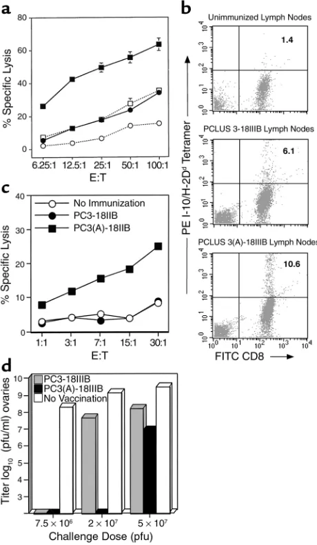
[image:4.576.307.537.338.733.2]the CTL response of improving the helper epitope with-out altering the CTL epitope, we immunized BALB.A10 mice with PCLUS 3-18IIIB and the construct containing the modified helper epitope, PCLUS 3(A)-18IIIB, and measured CTL responses after in vitro stimulation. Immunization with PCLUS 3(A)-18IIIB resulted in greater CTL induction with an eightfold horizontal shift in E/T ratio after a single immunization (Figure 1a). This
Figure 1
Epitope enhancement of Th epitope in Th-CTL peptide vaccine con-struct elicits enhanced CTL response. (a) BALB/c mice were immu-nized subcutaneously with 20 nmol native vaccine construct PCLUS 3-18IIIB or construct with the modified helper epitope PCLUS 3(A)-18IIIB. Spleen cells were restimulated in vitro 14 days later with irra-diated P18IIIB-pulsed syngeneic spleen cells, and 7 days later tested in a 51Cr-release assay on P815 targets pulsed with 0.5 µM P18IIIB.
Open symbols represent nonspecific lysis of target cells without pep-tide. Curves are significantly different at α = 0.001 by Tukey multiple comparison test. (b) Draining lymph node cells from immunized ani-mals were stimulated 4 days with P18IIIB and stained with anti-CD8 and P18-I10/H-2Ddtetramer. (c) Draining lymph node cells from
increase in CTL is confirmed by a doubling of the spe-cific CD8+cells binding P18-I10/H-2Ddtetramers in
draining lymph nodes after 4 days of stimulation with P18 peptide, from 4.7% to 9.2% after immunizing with PCLUS 3-18IIIB versus PCLUS 3(A)-18IIIB, respectively, with nonspecific background 1.4% in unimmunized mice subtracted (Figure 1b).
In a separate experiment, when the CTL activity was measured ex vivo without restimulation using draining lymph node cells 3 days after immunization, a signifi-cant CTL response was already evident in animals nized with PCLUS 3(A)-18IIIB, but not in those immu-nized with PCLUS 3-18IIIB (Figure 1c). Thus, the modified Th epitope with higher affinity for MHC class II resulted in enhanced help for class I–restricted CTL induction as determined by greater numbers and activi-ty and induction of CD8+CTL without in vitro
stimula-tion. Thus, help was indeed limiting for CTL inducstimula-tion. To determine if this enhanced CTL response was more protective in vivo, we challenged immunized ani-mals intraperitoneally with increasing doses of virus. We have found this model to be strictly dependent on the presence of activated CD8+CTL at the site of viral
challenge. Complete protection was achieved with both vaccine constructs at low-dose viral challenge; howev-er, at the intermediate dose complete protection was seen only in animals immunized with the enhanced (T1A)-vaccine construct (Figure 1d). Furthermore, at the highest challenge dose, virus was still inhibited by greater than 99% (100-fold) compared with 95% (20-fold) with the unmodified vaccine. Thus, greater induc-tion of CD8+ CTL by immunization with the modified
helper epitope-CTL vaccine construct correlated with improved protection from viral challenge.
Epitope modification polarizes the Th response to Th1 pheno-type. To determine the nature of enhanced helper
func-tion for inducfunc-tion of CD8+CTL and
to determine if the difference was purely quantitative or also qualita-tive, we measured cytokine mRNA in
CD4+ cells from draining lymph
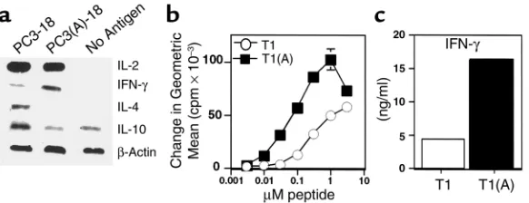
nodes 3 days after immunization. Unexpectedly, a clear difference in cytokines was observed. The native Th epitope, T1, polarized CD4+ cells
toward a Th2 phenotype as early as day 3, whereas the construct con-taining the higher-affinity binding Th peptide T1(A) skewed the response toward Th1 (Figure 2a). In three separate experiments, IFN-γ mRNA was at least 2.5-fold greater in animals immunized with the enhanced construct. In addition, IL-4 expression was either absent or decreased approximately threefold
in CD 4+ cells immunized with
T1(A), and IL-10 expression was also reduced (Figure 2a). In contrast, we did not observe a significant difference in proliferation or IL-2 expression (Figure 2a and data not shown).
The difference between the two peptides in vivo was also reflected in vitro in an established T1-specific Th1 clone for which the proliferative dose-response curve was shifted 30-fold, and IFN-γproduction significant-ly increased when stimulated with the higher-affinity peptide T1(A) (Figure 2, b and c). Because we have observed recently that selection of IFN-γ–producing CD4+ cells require a higher threshold of signaling
through the TCR (24), the increased strength of signal delivered by the higher-affinity peptide could, in part, explain these results. We hypothesized that the higher-affinity peptide T1(A) may produce a stronger CD40L signal to activate DCs to produce IL-12, producing a more polarizing APC. Subsequent experiments were designed to test this hypothesis.
Differential upregulation of CD40L on CD4+T cells
stimu-lated with high-affinity peptide T1(A). To examine the mechanism of improved help for CTL, we stimulated a CD4+Th1 cell clone specific for peptide T1 with either
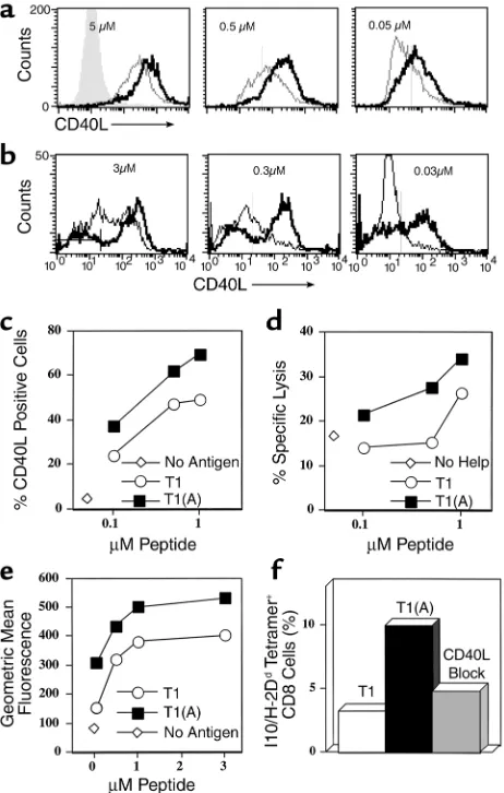
T1 or T1(A) and examined expression of CD40L over a range of 2 logs antigen dose (Figure 3a). At the highest dose tested (5 µM), all CD4+cells expressed CD40L
regardless of stimulating peptide used; however, levels of expression were markedly higher on T cells stimu-lated with the higher-affinity peptide, T1(A). In addi-tion, at decreasing doses of antigen, more CD4+cells
[image:5.576.60.351.53.165.2]were activated to express CD40L (at 0.05 µM peptide 68% versus 26%), and these expressed more CD40L per cell when stimulated with T1(A). The dose for inducing CD40L expression was reduced by greater than or equal to 1 log when T1(A) was used. Furthermore, this increase in CD40L expression was found when using either DCs (Figure 3a) or purified splenic B cells (Fig-ure 3b) as APCs. These results were highly reproducible Figure 2
Higher-affinity binding peptide T1(A) skews T cells toward a Th1 response. (a) Inguinal and periaortic lymph node cells were harvested from animals immunized with PCLUS 3-18IIIB (PC3-18) or PCLUS 3(A)-18IIIB [PC3(A)-18] and stimulated overnight with the immuniz-ing peptide at 3 µM. Total RNA was isolated from positively selected CD4+T cells 20 hours
after in vitro stimulation and semiquantitative RT-PCR performed under nonsaturating con-ditions to compare relative cytokine mRNA levels. These results were repeated with similar results in two additional experiments. (b) Stimulation of a T1 CD4+T cell line with peptide
in four additional experiments. Thus, we show unex-pectedly that increasing the affinity of the helper epi-tope for the class II MHC molecule results not only in increased numbers of activated helper T cells, but also in qualitatively different help, with more CD40L per cell and polarized to Th1 cytokines.
Enhanced helper function through conditioning and polar-izing of APCs. To determine if enhanced T helper CD40L expression was responsible for the enhanced CTL induction in vivo, we did a two-step mixing experiment in which peptide-specific CD4+Th1 cells were
stimu-lated overnight with different concentrations of pep-tide and DCs, and then the resulting DCs were used to stimulate CTL. The first stage, CD40L expression on gated CD4+cells 20 hours after stimulation with T1
and T1(A), is shown in Figure 3c. Stimulation with the modified Th epitope T1(A) elicited more total CD40L+
T cells at all antigen concentrations (e.g., 72% vs. 50% at 1 µM peptide). There was a clear dose-response effect. Parallel cultures of the same cell preparation were then depleted of CD4+ T cells to recover the DCs, which were
then pulsed with the CTL epitope peptide P18IIIB, irra-diated, and mixed with CD8+T cells enriched from
spleens of animals immunized with PCLUS 3-18IIIB. The level of CTL activity induced by these conditioned
DCs (Figure 3d) closely paralleled the level of CD40L expression induced on the Th cells used to condition the DCs (Figure 3c). In a separate experiment using less-rested Th cells already 93% CD40L positive, the level of CD40L per cell was increased significantly more by T1(A) than by T1 (Figure 3e), and the resulting DCs induced twice the percentage of CD8+cells that were
P18-I10/H-2Ddtetramer-positive (Figure 3f).
Further-more, blocking CD40L on the Th cell stimulated with peptide T1(A) abrogated IL-12 production by DCs (Fig-ure 4) and reduced the CTL response (Fig(Fig-ure 3f and see Figure 6a), formally establishing a link between the enhanced helper function and CTL induction. Thus we conclude that the epitope-enhanced helper peptide is more effective at inducing CTL because it better upreg-ulates CD40L on the Th cell, which then more effec-tively conditions APCs to better activate CTL after the Th cells have been removed.
CD40L expression on CD4+cells stimulated with
[image:6.576.60.291.56.419.2]higher-affinity binding peptide T1(A) correlates with enhanced IL-12 production by DCs, resulting in polarizing DCs. To deter-mine the molecular differences in the DCs that made those conditioned by T1(A) more effective at inducing CTLs, we examined cytokine production and surface expression of costimulatory molecules. T1-specific Figure 3
Qualitative and quantitative upregulation of CD40L on CD4+T cells
stimulated with T1(A) and concordant increased activation of DCs to stimulate CTL. (a) CD40L expression on T1-specific CD4+T cells
incubated overnight with different concentrations of T1 (dashed line) or T1(A) (solid line) and DCs, or without peptide (shaded area). (b) CD40L expression on gated CD4+T1-specific T cells stimulated 36
hours with indicated concentrations of T1 or T1(A) peptide and naïve A.AL splenic B cells (B220+selected). The bar in the left panel
indicates background CD40L without peptide. (c) Percentage CD4+/
CD40L+cells plotted versus peptide concentration for Th1 (5 × 10⁴
CD4+) cells incubated 24 hours with 5 ×10⁵BALB.A10 DCs and T1
or T1(A) peptide, or no peptide (open diamond). (d) 5 × 10⁶DCs of
the same preparation as in cwere incubated with different concen-trations of peptide and 5 ×10⁵of the same CD4+cells, but in 12 well
plates. CD4+cells were depleted by magnetic beads 24 hours later.
Remaining DCs were pulsed with 0.5 µM CTL epitope P18IIIB, irra-diated, and 5 × 10⁶added to 5 × 10⁶purified CD8+cells from spleens
of 3 mice immunized 8 weeks earlier with PCLUS3-18IIIB, and rmIL-2 (10 U/ml) was added on day 0. Specific lysis 7 days later on P18IIIB-pulsed P815 targets at a 50:1 E:T ratio is plotted versus Th peptide concentration used in conditioning cultures, or without Th peptide (open diamonds). Similar results were obtained in 2 other experiments. (e) Level of CD40L expression on the Th cells as a func-tion of peptide concentrafunc-tion, using less rested Th cells which were 93% CD40L+even without stimulation. (f) P18-I10/H-2Dd tetramer
staining of CD8+cells stimulated with 1 µM P18-pulsed DC
CD4+Th cells were stimulated in vitro with DCs and
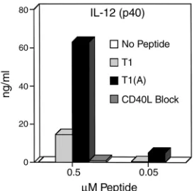
either T1 or T1(A) and IL-12 production determined 48 hours later by ELISA for IL-12 (p40 subunit; with sim-ilar results for IL-12 p70 heterodimer, not shown). IL-12 production was increased more than threefold in cells stimulated with T1(A) compared
with T1 at the optimum proliferative dose (0.5 µM peptide) (Figure 4) and sig-nificantly increased at a log lower dose (0.05 µM peptide). This effect was com-pletely blocked by the addition of anti-CD40L Ab. Therefore, the induction of IL-12 production by DCs is dependent upon CD40L-CD40 interaction, and epitope enhancement leads to recipro-cal cross talk between the T cell and DC, increasing CD40L expression on the CD4+Th cell and IL-12 production by
the DC, thus skewing Th cell phenotype toward Th1. A further implication is that one mechanism to generate polar-izing APCs skewed toward Th1 induc-tion is to increase the helper signal with a higher-affinity peptide.
Differential upregulation of costimulation on DCs by higher-affinity Th epitope T1(A). We also wanted to determine if enhanced CD40L expression con-tributed to an upregulation of any cos-timulatory molecules on the APCs. Bone marrow–derived DCs were incu-bated with peptide T1 or T1(A) and the T1-specific Th clone for 24 hours. Cells were gated on class II or CD11c+and
examined for costimulatory molecules, B7-1, B7-2, ICAM, CD44H, 41BBL, CCR5, and CD40. In contrast to class II expression, B7-1, B7-2, and CD40, were
upregulated in cells conditioned with Th and either peptide, but B7-1 expression was greater with the high-er-affinity peptide T1(A) (Figure 5a). In a subsequent
experiment (Figure 5b), when we gated on CD11c+
cells, we found a marked increase in the level and num-bers of cells expressing both B7-1 and B7-2 in DCs conditioned with the Th stimulated with T1(A). Although we always observed an upregulation of both ICAM and CD44H in cultures incubated with peptide compared with those without, increases were similar with either peptide, as was true for 41BBL or CCR5 (data not shown).
We performed blocking experiments to determine whether these molecules were necessary for the enhanced CTL induction. DCs conditioned overnight with T1(A) and T1-specific Th cells were depleted of CD4+cells, pulsed with P18IIIB, and added to purified
CD8+cells from mice immunized with PC3-18IIIB. DCs
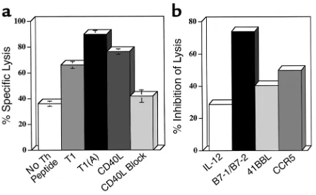
[image:7.576.90.227.54.189.2]conditioned with the T1(A) or CD40L elicited signifi-cantly higher levels of lysis than those conditioned with T1 or with Th-cells only, without peptide (Figure 6a). Furthermore, the enhanced CTL response could be blocked by the addition of anti-CD40L (10 µg/ml) dur-ing the initiation of culture, down to the level found with DCs preincubated with Th cells only, without pep-tide. (The high background with DCs that were not Figure 4
Induction of IL-12 production by DCs is dramatically increased by Th cells stimulated with higher-affinity peptide and is blocked by anti-CD40L. Triplicate cultures of 5 × 10⁴ T1-specific CD4+Th cell clone
were stimulated with 5 × 10⁵DCs, and 50 hours later culture
[image:7.576.253.534.379.565.2]super-natants were assayed for IL-12 by ELISA. These results were highly reproducible in three separate experiments.
Figure 5
Differential expression of costimulatory molecules. Eight-day A.AL DC cultures (5 × 10⁵
cells/well of 96-well round-bottom plate) grown with GM-CSF (10 ng/ml) were con-ditioned for 20 hours by incubation with 5 × 10⁴CD4+Th cells and 0.5 µM peptide
T1 or T1(A). (a) Gated class II+cells were examined for expression of CD40 and
cos-timulatory molecules B7-1 and B7-2 after stimulation with T1(A) (solid heavy line) compared with T1 (light line). DC cultures incubated with the Th cell without peptide are shown as the shaded area and are similar to cultures incubated without Th cells (not shown). (b) Eight-day BALB.A10 DCs were conditioned similarly for 36 hours. Gated CD11c+ shown in the first panel as the shaded area compared with PE-isotype
conditioned by CD4+Th cells and peptide may be due
to in vitro maturation of DCs). The addition of both anti-B7-1/B7-2 significantly decreased CTL induction (Figure 6b), where anti–IL-12, anti–41BBL, or anti–CCR5, partially reduced CTL. In six separate block-ing experiments we could reproducibly inhibit CTL induction with the combination anti–B7-1/B7-2. We could not inhibit CTL with Ab to B7-1 or B7-2 alone, ICAM, CD44H (alone or in combination), or repro-ducibly inhibit CTL by greater than 40% with anti–IL-12, anti-41BBL, or anti-CCR5 (Figure 6b and data not shown). Thus, the major effect of the CD4+Th cell in
conditioning DCs for enhanced CTL induction occurs through CD40L-CD40 interaction and is dependent on upregulation of both B7-1 and B7-2. Upregulation of IL-12 production appears to be another major effect of conditioning, although blocking by anti–IL-12 was only partial. The increased IL-12 expression at least accounts for the DC’s becoming more Th1 polarizing, but may be in sufficient excess for CTL induction that Ab block-ade was incomplete. Thus, we conclude that the enhanced peptide with higher affinity for MHC class II acts by upregulating CD40L on the T helper cell, which in turn acts through CD40 on the DC to upregulate both B7-1/B7-2 and IL-12 production, thus making a more potent DC for CTL induction and also a more polarizing one for Th1 induction (Figure 7).
Discussion
These experiments define a mechanism of enhanced CTL induction by modification of a CD4+T helper cell
epitope. We have found not only quantitative but also qualitative improvement in the immune response through epitope enhancement and have dissected each step of the process (Figure 7). The improved peptide increases T helper activation by augmenting CD40L expression, which in turn increases activation of DCs and alters the DCs qualitatively, making them more Th1 polarizing through increased IL-12 production. The conditioned DCs, through higher IL-12 and B7-1 and B7-2, more effectively activate CTL and induce better protection against viral infection, but also feed back on the helper T cells to polarize them more toward Th1 phenotype.
We have shown that CD4+T cells are essential for
induction of CD8+CTL responses to peptide-based
vac-cines (2, 7, 23), and other groups have shown a similar requirement for CD4+help to many cell-based antigens
(9, 13, 25, 26). However, it was not expected that CD4 help would be the limiting factor in improving a CD8+
CTL response, such that altering the helper epitope affinity for class II MHC, without modifying the CTL epitope presented by class I MHC, would greatly aug-ment CTL induction. It was also not known whether such increased help would increase protection against viral infection, as we now show. These findings have important implications for vaccine design.
The mechanism of CD4+T cell help for CTL has
been clearly shown to involve activation of immature DCs to a fully mature competent state through CD40L-CD40 interaction (10–12), but the effect of the CD40L on the conditioned DC that results in increased activity has not been determined. Upregula-tion of costimulatory molecules has been assumed, but not yet demonstrated. In this study, we find that increasing the affinity of a Th epitope for class II MHC
[image:8.576.60.290.54.196.2]more efficiently upregulates CD40L on CD4+Th 1
Figure 6
Blocking both B7-1 and B7-2 on DCs inhibits the CTL response. BALB.A10 DC cultures (2.5 ×10⁶) conditioned for 24 hours with Th
cells and peptide, T1, or T1(A) (0.5 µM), or no peptide, were deplet-ed of CD4+cells, pulsed for 1 hour with the CTL epitope P18IIIB (0.5
µM), irradiated, and added to 5 ×10⁶negatively selected splenic
CD8+cells from BALB.A10 mice primed 1 month earlier
subcuta-neously with PCLUS 3-18IIIB (20 nmol). (a) DCs conditioned with Th cell and T1(A) elicited higher levels of CTL than DCs conditioned with either Th cells and T1 or no peptide, or overnight treatment with trimeric murine CD40L (3 µg/ml). Lytic activity was determined on day 7 in a 5-hour 51Cr assay on 0.5 µM P18IIIB-pulsed P815 target
cells (E/T = 10:1). (b) DCs were conditioned with Th cells and T1(A) (0.5 µM) for 24 hours and CD4+cells removed. Cells were pulsed for
1.5 hours with P18IIIB, irradiated, and used to stimulate immune CD8+cells. Anti–B7-1/B7-2 (15 µg/ml each), anti–IL-12 (15 µg/ml),
[image:8.576.305.538.528.666.2]anti-CCR5 (10 µg/ml), or anti-41BBL (10 µg/ml) were added to pulsed DCs at the initiation of CTL culture and on day 3. Similar results were obtained in two additional experiments.
Figure 7
cells both quantitatively and qualitatively; i.e., both the number of CD40L+cells and the level per cell.
Fur-thermore, the level of CD40L expression was shown to correlate directly to induction of IL-12 by DCs and blocking CD40L on the Th-cell abrogated IL-12 pro-duction. Although it was known that signals delivered to DCs through CD40 can induce IL-12 production (27, 28), it was unexpected that augmentation of help through increasing the density of CD40L on helper T cells would generate polarizing DCs that would in turn skew the helper response to Th1. This may be a general approach to produce such polarizing APCs. The biological significance is supported by a recent study showing that protective anti–tumor immunity required CD40-mediated activation of DCs (29).
Thus, a key effect of enhanced Th epitope affinity is increased IL-12 production. IL-12 acts in driving the selective expansion of Th1 cells (23, 30, 31), enhances CTL proliferation (32), and may further upregulate cos-timulation (33). In fact, we observed that the higher-affinity peptide clearly skewed the Th phenotype to Th1. Thus, the strength of TCR signal determines levels of CD40L, which in turn determines IL-12 production by APCs and selection of a Th1 phenotype. In a two-step culture, the upregulation of CD40L on the T helper cells in the first culture directly correlated with the ability of DCs conditioned in this culture to stimulate CD8+CTL
activity in a second culture. Blockade of increased CTL induction in the second culture by treatment of the helper cells in the first culture with anti-CD40L (Figure 6a) suggests that this correlation represents cause and effect. These results complement and help to explain the mechanism by which altered peptide ligands have been shown to skew Th phenotype in the opposite direc-tion, toward Th2 (34–38). Thus, both complementary results strongly support the conclusion that strength of signal contributes to T helper polarization.
DCs activated by CD4+ help stimulated with the
higher-affinity peptide T1(A) induced somewhat high-er levels of expression of B7-1 and B7-2, but not othhigh-er costimulatory molecules, ICAM, CD44H, or 41BBL (39), and the resulting increase in CTL was inhibited by blocking both B7-1 and B7-2. However, the increase in B7-1 and B7-2 was modest compared with that of IL-12 production. Conditioning thus appears to repre-sent predominantly an increase in IL-12 production and secondarily an increase in B7-1 and B7-2. IL-15 production by activated DCs may also play a role in CTL proliferation (40, 41), which we can investigate as reagents for detecting murine IL-15 become available. The ability of DCs to stimulate antigen-primed CD8+
CTL independent of CD4 cells is dependent on the state of activation and maturation of DCs (42). Besides CD40L-CD40 interaction, inflammatory stimuli can also activate DCs for induction of CTL (13, 43, 44). Thus, DCs may be activated and CTL elicited in both CD4–dependent and CD4-independent pathways (45). A recent study (46) showed that although CD8+
CTL-mediated resolution of acute lymphocyte
choriomenin-gitis virus infection occurred in CD40L-deficient mice, chronic infection was not controlled, suggesting a need for CD40L-CD40 interaction and a polarized CD4+Th
cell response in resolving chronic viral infections. In summary, while exploring the mechanisms behind the epitope enhancement effect, we discovered several novel basic mechanisms by which T helper cells con-tribute to CTL induction and can generate polarizing APCs. Epitope enhancement to increase the affinity of a class II MHC binding peptide can dramatically enhance the efficacy of a Th-CTL peptide vaccine con-struct for CTL induction and protection against viral infection, both quantitatively and qualitatively. The enhanced peptide shifts the quality of the response toward Th1 by greater upregulation of CD40L on the helper T cells, which then more effectively condition and polarize the DCs by upregulation of B7-1 and B7-2 expression and IL-12 production, which improves CTL induction and skews the cytokine profile. Better understanding of the mechanism of epitope enhance-ment may lead to the rational design of more effective second-generation vaccines.
Acknowledgments
We thank Alfred Singer and Alan Sher for critical read-ing of the manuscript and helpful discussions; Patricia Earl and Bernard Moss for the recombinant vaccinia virus; Jean-Pierre Planchot for the Montanide ISA 51; and Immunex Corp. for the CD40L.
1. Boehncke, W.-H., et al. 1993. The importance of dominant negative effects of amino acids side chain substitution in peptide-MHC molecule interactions and T cell recognition. J. Immunol.150:331–341. 2. Ahlers, J.D., Takeshita, T., Pendleton, C.D., and Berzofsky, J.A. 1997.
Enhanced immunogenicity of HIV-1 vaccine construct by modification of the native peptide sequence. Proc. Natl. Acad. Sci. USA.
94:10856–10861.
3. Sarobe, P., et al. 1998. Enhanced in vitro potency and in vivo immuno-genicity of a CTL epitope from hepatitis C virus core protein following amino acid replacement at secondary HLA-A2.1 binding positions. J. Clin. Invest.102:1239–1248.
4. Pogue, R.R., Eron, J., Frelinger, J.A., and Matsui, M. 1995. Amino-termi-nal alteration of the HLA-A*0201-restricted human immunodeficiency virus pol peptide increases complex stability and in vitro immuno-genicity. Proc. Natl. Acad. Sci. USA.92:8166–8170.
5. Parkhurst, M.R., et al. 1996. Improved induction of melanoma-reactive CTL with peptides from the melanoma antigen gp100 modified at HLA-A*0201-binding residues. J. Immunol.157:2539–2548.
6. Slansky, J.E., et al. 2000. Enhanced antigen-specific antitumor immuni-ty with altered peptide ligands that stabilize the MHC-peptide-TCR complex. Immunity.13:529–538.
7. Shirai, M., et al. 1994. Helper-CTL determinant linkage required for priming of anti-HIV CD8+ CTL in vivo with peptide vaccine constructs.
J. Immunol.152:549–556.
8. Ahlers, J.D., et al. 1996. Candidate HIV type 1 multideterminant cluster peptide-P18MN vaccine constructs elicit type 1 helper T cells, cytotoxic T cells, and neutralizing antibody, all using the same adjuvant immu-nization. AIDS Res. Hum. Retroviruses.12:259–272.
9. Bennett, S.R.M., Carbone, F.R., Karamalis, F., Miller, J.F.A.P., and Heath, W.R. 1997. Induction of a CD8+ cytotoxic T lymphocytes response by cross-priming requires cognate CD4+ T cell help. J. Exp. Med.186:65–70. 10. Ridge, J.P., Di Rosa, F., and Matzinger, P. 1998. A conditioned dendritic cell can be a temporal bridge between a CD4+ T-helper and a T-killer cell.
Nature.393:474–478.
11. Bennett, S.R.M., et al. 1998. Help for cytotoxic-T-cell responses is medi-ated by CD40 signalling. Nature. 393:478–480.
12. Schoenberger, S.P., Toes, R.E.M., van der Voort, E.I.H., Offringa, R., and Melief, C.J.M. 1998. T-cell help for cytotoxic T lymphocytes is mediated by CD40-CD40L interactions. Nature.393:480–483.
cytotoxic T lymphocyte priming capacity upon activation by T helper cell-independent or -dependent stimuli. J. Exp. Med.192:145–150. 14. Lutz, M.B., et al. 1999. An advanced culture method for generating large
quantities of highly pure dendritic cells from mouse bone marrow. J. Immunol. Method.s223:77–92.
15. Berzofsky, J.A., et al. 1991. Construction of peptides encompassing mul-tideterminant clusters of HIV envelope to induce in vitro T-cell respons-es in mice and humans of multiple MHC typrespons-es. J. Clin. Invest.88:876–884. 16. Takahashi, H., et al. 1988. An immunodominant epitope of the HIV gp160 envelope glycoprotein recognized by class I MHC molecule-restricted murine cytotoxic T lymphocytes. Proc. Natl. Acad. Sci. USA.
85:3105–3109.
17. Ahlers, J.D., et al. 1993. Construction of an HIV-1 peptide vaccine con-taining a multideterminant helper peptide linked to a V3 loop peptide 18 inducing strong neutralizing antibody responses in mice of multiple MHC haplotypes after two immunizations. J. Immunol.150:5647–5665. 18. Kuiken, C., et al., 2000. HIV sequence compendium 2000. Los Alamos
National Laboratory. Los Alamos, New Mexico, USA. 592. pp. 19. Earl, P.L., Hugin, A.W., and Moss, B. 1990. Removal of cryptic poxvirus
transcription termination signals from the human immunodeficiency virus type 1 envelope gene enhances expression and immunogenicity of a recombinant vaccinia virus. J. Virol.64:2448–2451.
20. Alexander-Miller, M.A., Leggatt, G.R., and Berzofsky, J.A. 1996. Selective expansion of high or low avidity cytotoxic T lymphocytes and efficacy for adoptive immunotherapy. Proc. Natl. Acad. Sci. USA.93:4102–4107. 21. Ahlers, J.D., Belyakov, I.M., Matsui, S., and Berzofsky, J.A. 2001. Mecha-nisms of cytokine synergy essential for vaccine protection against viral challenge. Int. Immunol.13:897–908.
22. Belyakov, I.M., et al. 2001. Activating CTL precursors to reveal CTL func-tion without skewing the repertoire by in vitro expansion. Eur. J. Immunol. In press.
23. Ahlers, J.D., Dunlop, N., Alling, D.W., Nara, P.L., and Berzofsky, J.A. 1997. Cytokine-in-adjuvant steering of the immune response phenotype to HIV-1 vaccine constructs: GM-CSF and TNF-alpha synergize with IL-12 to enhance induction of CTL. J. Immunol.158:3947–3958.
24. Ahlers, J.D., Belyakov, I.M., Matsui, S., and Berzofsky, J.A. 2001. Signals delivered through TCR instruct IL-12R expression: IL-12 and TNFα syn-ergize for IL-12R expression at low antigen dose. Int. Immunol.
13:1433–1442.
25. Takahashi, H., Nakagawa, Y., Yokomuro, K., and Berzofsky, J.A. 1993. Induction of CD8+ CTL by immunization with syngeneic irradiated HIV-1 envelope derived peptide-pulsed dendritic cells. Int. Immunol.
5:849–857.
26. Husmann, L.A., and Bevan, M.J. 1988. Cooperation between helper T cells and cytotoxic T lymphocyte precursors. Ann. NY Acad. Sci.
532:158–169.
27. Cella, M., et al. 1996. Ligation of CD40 on dendritic cells triggers pro-duction of high levels of interleukin-12 and enhances T cell stimulato-ry capacity: T-T help via APC activation. J. Exp. Med.184:747–752. 28. Ding, L., and Shevach, E.M. 1998. Differential effects of CD28
engage-ment and IL-12 on T cell activation by altered peptide ligands. J. Immunol.161:6614–6621.
29. Mackey, M.F., et al. 1998. Dendritic cells require maturation via CD40 to generate protective antitumor immunity. J. Immunol.161:2094–2098. 30. Macatonia, S.E., Hsieh, C.-S., Murphy, K.M., and O’Garra, A. 1993. Den-dritic cells and macrophages are required for Th1 development of CD4+ T cells from ab-TCR transgenic mice: IL-12 substitution for macrophages to stimulate IFN-g production is IFN-g-dependent. Int. Immunol.5:1119–1128.
31. Macatonia, S.E., et al. 1995. Dendritic cells produce IL-12 and direct the development of Th1 cells from naive CD4+ T cells. J. Immunol.
154:5071–5079.
32. Trinchieri, G. 1998. Proinflammatory and immunoregulatory functions of interleukin-12. Intern. Rev. Immunol.16:365–396.
33. Kelleher, P., and Knight, S.C. 1998. IL-12 increases CD80 expression and the stimulatory capacity of bone marrow-derived dendritic cells. Int. Immunol.10:749–755.
34. Pfeiffer, C., et al. 1995. Altered peptide ligands can control CD4 T lym-phocyte differentiation in vivo. J. Exp. Med.181:1569–1574.
35. Tao, X., Grant, C., Constant, S., and Bottomly, K. 1997. Induction of IL-4-producing CD4+ T cells by antigenic peptides altered for TCR bind-ing. J. Immunol.158:4237–4244.
36. Chaturvedi, P., Yu, Q., Southwood, S., Sette, A., and Singh, B. 1996. Pep-tide analogs with different affinities for MHC alter the cytokine profile of T helper cells. Int. Immunol.8:745–755.
37. Rogers, P.R., and Croft, M. 1999. Peptide dose, affinity, and time of dif-ferentiation can contribute to the Th1/Th2 cytokine balance. J. Immunol.
163:1205–1213.
38. Constant, S.L., and Bottomly, K. 1997. Induction of Th1 and Th2 CD4+ T cell responses: the alternative approaches. Annu. Rev. Immunol.
15:297–322.
39. Tan, J.T., Whitmire, J.K., Ahmed, R., Pearson, T.C., and Larsen, C.P. 1999. 4-1BB ligand, a member of the TNF family, is important for the genera-tion of antiviral CD8 T cell responses. J. Immunol.163:4859–4868. 40. Kuniyoshi, J.S., et al. 1999. Dentritic cell secretion of IL-15 is induced by
recombinant huCD40LT and augments the stimulation of antigen-spe-cific cytolytic T cells. Cell. Immunol.193:48–58.
41. Kanai, T., Thomas, E.K., Yasutomi, Y., and Letvin, N.L. 1996. IL-15 stim-ulates the expansion of aids virus-specific CTL1. J. Immunol.
157:3681–3687.
42. Young, J.W., and Steinman, R.M. 1990. Dendritic cells stimulate primary human cytolytic lymphocyte responses in the absence of CD4+ helper T cells. J. Exp. Med.171:1315–1332.
43. Ruedl, C., Kopf, M., and Bachmann, M.F. 1999. CD8+ T cells mediate CD40-independent maturation of dendritic cells in vivo. J. Exp. Med.
189:1875–1883.
44. Cella, M., Engering, A., Pinet, V., Pieters, J., and Lanzavecchia, A. 1997. Inflammatory stimuli induce accumulation of MHC class II complexes on dendritic cells. Nature.388:782–787.
45. Lu, Z., et al. 2000. CD40-independent pathways of T cell help for prim-ing of CD8(+) cytotoxic T lymphocytes. J. Exp. Med.191:541–550. 46. Whitmire, J.K., et al. 1999. CD40-CD40 ligand costimulation is required