Characterization of protein kinase C beta
isoform activation on the gene expression of
transforming growth factor-beta, extracellular
matrix components, and prostanoids in the
glomeruli of diabetic rats.
D Koya, … , K Kuboki, G L King
J Clin Invest.
1997;
100(1)
:115-126.
https://doi.org/10.1172/JCI119503
.
Induction of protein kinase C (PKC) pathway in the vascular tissues by hyperglycemia has
been associated with many of the cellular changes observed in the complications of
diabetes. Recently, we have reported that the use of a novel, orally effective specific
inhibitor of PKC beta isoform (LY333531) normalized many of the early retinal and renal
hemodynamics in rat models of diabetes. In the present study, we have characterized a
spectrum of biochemical and molecular abnormalities associated with chronic changes
induced by glucose or diabetes in the cultured mesangial cells and renal glomeruli that can
be prevented by LY333531. Hyperglycemia increased diacylglycerol (DAG) level in cultured
mesangial cells exposed to high concentrations of glucose and activated PKC alpha and
beta1 isoforms in the renal glomeruli of diabetic rats. The addition of PKC beta selective
inhibitor (LY333531) to cultured mesangial cells inhibited activated PKC activities by high
glucose without lowering DAG levels and LY333531 given orally in diabetic rats specifically
inhibited the activation of PKC beta1 isoform without decreasing PKC alpha isoform
activation. Glucose-induced increases in arachidonic acid release, prostaglandin E2
production, and inhibition of Na+-K+ ATPase activities in the cultured mesangial cells were
completely prevented by the addition of LY333531. Oral feeding of LY333531 prevented the
increased mRNA expression of TGF-beta1 and extracellular matrix components such as
fibronectin and alpha1(IV) collagen […]
Research Article
Find the latest version:
J. Clin. Invest.
© The American Society for Clinical Investigation, Inc. 0021-9738/97/07/0115/12 $2.00
Volume 100, Number 1, July 1997, 115–126
Characterization of Protein Kinase C
b
Isoform Activation on the Gene Expression
of Transforming Growth Factor-
b
, Extracellular Matrix Components, and
Prostanoids in the Glomeruli of Diabetic Rats
Daisuke Koya,* Michael R. Jirousek,‡ You-Wei Lin,* Hidehiro Ishii,* Koji Kuboki,* and George L. King*
*Research Division, Joslin Diabetes Center and Department of Medicine, Brigham and Women’s Hospital and Harvard Medical School, Boston, Massachusetts 02215; and ‡Lilly Research Laboratories, Indianapolis, Indiana 46285
Abstract
Induction of protein kinase C (PKC) pathway in the vascu-lar tissues by hyperglycemia has been associated with many of the cellular changes observed in the complications of diabetes. Recently, we have reported that the use of a novel, orally effective specific inhibitor of PKC b isoform (LY333531) normalized many of the early retinal and renal hemodynamics in rat models of diabetes. In the present study, we have characterized a spectrum of biochemical and molecular abnormalities associated with chronic changes in-duced by glucose or diabetes in the cultured mesangial cells and renal glomeruli that can be prevented by LY333531. Hyperglycemia increased diacylglycerol (DAG) level in cul-tured mesangial cells exposed to high concentrations of glucose and activated PKC a and b1 isoforms in the renal glomeruli of diabetic rats. The addition of PKC b selective inhibitor (LY333531) to cultured mesangial cells inhibited activated PKC activities by high glucose without lowering DAG levels and LY333531 given orally in diabetic rats spe-cifically inhibited the activation of PKC b1 isoform without decreasing PKC a isoform activation. Glucose-induced in-creases in arachidonic acid release, prostaglandin E2 pro-duction, and inhibition of Na1-K1 ATPase activities in the
cultured mesangial cells were completely prevented by the addition of LY333531. Oral feeding of LY333531 prevented the increased mRNA expression of TGF-b1 and extracellu-lar matrix components such as fibronectin and a1(IV) col-lagen in the glomeruli of diabetic rats in parallel with inhibi-tion of glomerular PKC activity. These results suggest that the activation of PKC, predominately the b isoform by hy-perglycemia in the mesangial cells and glomeruli can partly contribute to early renal dysfunctions by alteration of pros-taglandin production and Na1-K1 ATPase activity as well
as the chronic pathological changes by the overexpression of TGF-b1 and extracellular matrix components genes. (J. Clin. Invest. 1997. 100:115–126.) Key words: arachidonic
acid release • PGE2• TGF-b1 • a1(IV) collagen • fibronectin
Introduction
Diabetes now accounts for 35% of all new cases of end-stage renal disease in the United States (1). The causal factors for its development are most likely metabolic since the results of Dia-betes Control and Complication Trial (DCCT) have clearly es-tablished that the strict maintenance of euglycemia by inten-sive insulin treatment can delay the onset and progression of diabetic nephropathy and other microvascular diseases (2). Amongst the various metabolic changes induced by diabetes, hyperglycemia is likely the single most important causal factor for the development of vascular dysfunctions in the microves-sels (2–7).
Multiple cellular consequences of glucose-induced activa-tion in cultured vascular cells have been described including increases in TGF-b, activation of cytosolic phospholipase A2
(cPLA2),1 and inhibition of Na1-K1 ATPase activities (3, 4,
8–10). Multiple biochemical mechanisms have been proposed to explain the adverse effects of hyperglycemia (3–7). We and others have reported that a consistent effect of hyperglycemia is the activation of the diacylglycerol (DAG)–protein kinase C (PKC) pathway which have been associated with the regula-tion of many vascular funcregula-tions (11–18). Activaregula-tion of DAG– PKC pathways in the vascular tissues has been associated with early abnormal hemodynamic functions in diabetic rats (11, 13). Amongst the many isoforms of PKC, PKC b2 isoform was predominately activated by hyperglycemia chronically in the retina, aorta, and heart of diabetic rats (12, 13).
However, direct consequences of DAG–PKC activation es-pecially the b isoform have been difficult to establish due to the lack of a PKC b isoform selective inhibitor for in vivo stud-ies. We have recently described a macrocylic bisindolylmale-mide compound (LY333531) which can inhibit PKC b isoform at concentrations 50 times less than for many other PKC iso-forms and 1,000 times less than many serine–threonine and ty-rosine kinases (11, 19). Oral treatment with LY333531 in rats with streptozotocin (STZ)-induced diabetes prevented the on-set of early abnormalities in retinal and renal hemodynamics including the decrease in retinal blood flow, increase in GFR, and urinary albumin excretion rate (11).
In the present study, we have used this PKC b isoform se-lective inhibitor LY333531 to determine the role of PKC b
isoform activation in causing alterations of biochemical param-eters and gene expressions associated with chronic and patho-logical changes in diabetic glomerulopathy. These parameters are prostanoid production, Na1-K1ATPase activities, gene
ex-Address correspondence to George L. King, Joslin Diabetes Center, One Joslin Place, Boston, MA 02215. Phone: 617-732-2622; FAX: 617-732-2637; E-mail: [email protected]
Received for publication 13 December 1996 and accepted in re-vised form 8 April 1997.
1. Abbreviations used in this paper: cPLA2, cytosolic phospholipase
A2; DAG, diacylglycerol; ECM, extracellular matrix; PA,
pressions of extracellular matrix components and TGF-b1 in cultured mesangial cells and renal glomeruli.
Methods
Animals. Male Sprague-Dawley rats (200 grams; Taconic Farms, Germantown, NY) were placed in one of four groups: control, control treated with LY333531 (10 mg/kg body wt), 12 wk of diabetes, and 12 wk of diabetes treated with LY333531 (10 mg/kg body wt). For 4-wk study, only diabetic group was treated with 10 mg/kg body wt LY333531. Control group received 1 ml/kg body wt of sterile 20 mM citrate buffer (pH 4.5) by intraperitoneal injection. Diabetes was in-duced by a single intraperitoneal injection of sterile STZ (65 mg/kg body wt, Sigma Chemical Co., St. Louis, MO) in citrate buffer. The diabetic state was confirmed 48 h after STZ injections by blood glu-cose levels exceeding 300 mg/dl. Control and diabetic rats were ran-domly divided into four groups and LY333531 were given in the diet at a concentration of 10 mg/kg body wt. Blood glucose concentration was determined weekly in all animals. All experiments were con-ducted in accord with the National Institutes of Health (NIH) Guide for the Care and Use of Laboratory Animals.
Isolation of glomeruli from rats and the culture of mesangial cells.
Rat mesangial cells were obtained from kidneys of male Sprague-Dawley rats (100–150 grams) as previously described with a few mod-ifications (20). Kidneys were dissected from killed rats under aseptic conditions. After removing the capsules, the entire kidneys were ho-mogenized in ice-cold DME and passed through sieves of various sizes to isolate the glomeruli. Glomeruli were then washed twice with DME and plated onto 10-cm tissue culture dish (Costar, Cambridge, MA) in 10 ml of DME containing 5 mg/ml each of insulin, transferrin, and selenium (Gibco BRL, Grand Island, NY), 5.5 mM glucose, 100 U/ml penicillin, and 100 mg/ml streptomycin with 20% fetal bovine serum (Gibco BRL). Mesangial cells from human glomeruli were ob-tained by the same method except the kidneys were provided by Na-tional Disease Research Interchange (NDRI, Philadelphia, PA). Identification of mesangial cells was performed as previously de-scribed (21). Mesangial cells of passage 2–6 were used for cell studies. For the assays of PKC activity, 86Rb uptake, immunoblot analysis,
arachidonic acid release, PGE2 production, and in situ 32P
orthophos-phate labeling, glomeruli from each group of rats were isolated after perfusion of kidneys with 20 ml of DME containing 20 mM Hepes, pH 7.4.
Assay of total DAG contents. Subconfluent rat mesangial cells in 6-well culture dishes (Costar) were exposed to 5.5 or 22 mM glucose for 4 d with daily changes of media. The cells were washed twice and pre-incubated for 1 h in serum-free DME containing various concen-trations of LY333531. After washing the cells, total DAG contents were measured with a radioenzymatic assay kit (Amersham Corp., Arlington Heights, IL) using DAG kinase (Calbiochem Corp., San Diego, CA) that quantitatively converts DAG to [32P]phosphatidic
acid (PA) in the presence of [g-32P]-ATP (New England Nuclear,
Boston, MA). In brief, total cellular lipids were extracted twice ac-cording to the methods of Bligh and Dyer (22) and total DAG was measured as described by manufacturer’s instructions. The resulting [32P]PA was separated by silica gel G thin layer plates (EM
Separa-tions, Gibbstown, NJ) in a chamber containing chloroform/acetone/ methanol/acetic acid/water (10:4:3:2:1) according to the method of Priess et al. (23). PA was visualized by autoradiography and identi-fied by comigration with radio-labeled PA derived from DAG stan-dard from 31 to 2,000 pmol. The spots were scraped and radioactivity was counted by liquid scintillation counter (CS6500; Beckman Instru-ments, Fullerton, CA). The values of total DAG contents were nor-malized by cellular proteins measured as described by Bradford (24).
Assay of protein kinase C activity. Subconfluent mesangial cells in 12-well culture dishes (Costar) were exposed to 5.5 or 22 mM glu-cose for 4 d with daily changes of media. The cells were washed twice and preincubated for 1 h in serum-free DME in the presence or
ab-sence of LY333531. PKC activity in mesangial cells was determined with a modified method described by Heasley and Johonson (25). Briefly, cells were rinsed twice with 2 ml of DME containing 20 mM Hepes (pH 7.4) and then with 2 ml of a salt solution (137 mM NaCl, 5.4 mM KCl, 0.3 mM sodium phosphate, 0.4 mM potassium phos-phate, 5.5 mM glucose, 10 mM MgCl2, 25 mM b-glycerophosphate,
5 mM EGTA, 2.5 mM CaCl2, and 20 mM Hepes, pH 7.4). The cells
were preincubated with the salt solution for 10 min at 378C and incu-bated for another 15 min in the presence or absence of 100 mM PKC-specific peptide substrate, RKRTLRRL (26) with 50 mg/ml digitonin and 100 mM ATP mixed with [g-32P]ATP (, 1,500 cpm/pmol; New
England Nuclear, Boston, MA). The reaction was terminated with 5% TCA (final concentration). Aliquots of the reaction mixture were spotted on 3 3 3 cm phosphocellulose papers (P-81; Whatman Instru-ments, Maidstone, United Kingdom) and washed with three changes of 75 mM phosphoric acid and one change of 75 mM sodium phos-phate (pH 7.5). The radioactivity of phosphorylated substrate was de-termined by liquid scintillation counting. Protein contents were mea-sured by Bradford’s method (24).
AA release and assay of prostaglandin E2. Subconfluent
mesan-gial cells in 12-well plates were maintained in media containing 5.5 or 22 mM of glucose for 4 d, and then were labeled with 0.5 mCi/ml [3H]arachidonic acid (221 Ci/mmol; New England Nuclear) for 20 h at
378C. After the removal of the radioactive medium, the cells were washed four times with DME containing 0.1% BSA and preincu-bated in the presence or absence of LY333531 for 1 h. For the deter-mination of glomerular AA release, isolated glomeruli were labeled with 5 mCi/ml [3H]arachidonic acid for 3 h at 378C and then washed
three times with the same buffer after centrifugation at 800 g for 5 min. The medium was replaced with 0.5 ml of fresh medium and incu-bated for an additional 1 h. The media was then collected and the amount of [3H] arachidonic acid released by cells or glomeruli for 1 h
was determined by scintillation counting. The cells or glomeruli were washed again and solubilized with 0.5 ml of 1 M NaOH. A 100-ml ali-quot was retained for protein determination and the remaining was subjected to scintillation counting to determine the [3H]arachidonic
acid content of the cells or glomeruli. The data of [3H]arachidonic
acid release were presented as percentage of total counts (radioactiv-ity in the released fraction 1 those in the solubilized cells) released for 1 h. There was no difference in the amount of incorporation of [3H]arachidonic acid between the cells exposed to 5.5 and 22 mM
glu-cose for 4 d, and between glomeruli isolated from control rats and those from diabetic rats.
The amount of PGE2 produced by mesangial cells and renal
glomeruli was measured in nonextracted cellular and glomerular su-pernatants using ELISA kit (Amersham Corp.) following the manu-facturer’s instructions. The data were normalized by protein concen-trations as described.
Assay of Na1-K1 ATPase activity. Na1-K1 ATPase activity was
estimated by 86Rb uptake as previously described (8). Mesangial cells
were grown in 12-well dishes and exposed to medium containing 5.5 or 22 mM glucose for 4 d. The cells were washed twice and preincu-bated for 1 h in serum-free DME in the presence or absence of the stated concentrations of LY333531. The cells were then washed with DME and incubated with 86Rb (67.8 mCi/mmol, New England
Nu-clear) at a final concentration of 1 mCi/ml with or without 2 mM oua-bain (Sigma Chemical Co.) for 10 min at 378C. 86Rb uptake was
termi-nated by aspirating the medium and rapidly washing four times with ice-cold 100 mM MgCl2. The cells were extracted with 0.5 ml of 10%
TCA to measure radioactivity incorporated into the cells by scintilla-tion counting. The ouabain-sensitive fracscintilla-tion of 86Rb uptake was
cal-culated by subtracting the fraction of the cellular uptake observed in the presence of ouabain from total 86Rb uptake. The 86Rb uptake into
glomeruli isolated from each group was also measured as described here. The data were normalized by cellular protein.
(Molecu-lar Research Center, Cincinnati, OH) (27). Total RNA (20 mg) was loaded on a 1% agarose gel with 9% formaldehyde, which was sepa-rated in Mops buffer (20 mM Mops, 5 mM Na acetate, 0.5 mM EDTA, pH 7.0). The RNA was transferred on nylon membrane (ICN, Aurora, OH). After UV-crosslinking, the membranes were prehybridized and hybridized to 32P-labeled 0.3 kb EcoRI fragment
of pCRII TGF-b1 cDNA for TGF-b1 kindly provided by Dr. Ziyadeh
(28), 1.8 kb EcoRI and SalI fragment of clone PE 123 for mouse
a1(IV) collagen by Dr. Kurkinen (29), and 582-bp PCR fragment for rat fibronectin (30) by Dr. Lorenzi, and Dr. Willert in 0.1 M Pipes, 0.2 M NaPO4, 0.1 M NaCl, 1 mM EDTA, 5% SDS, and 30 mg/ml
salmon sperm DNA at 658C. Stringent washing was done in 50% of 13 SSC, 5% SDS at 658C for over 1 h. The expression of mRNA was quantified with a PhosphorImager (Molecular Dynamics, Sunnyvale, CA) and normalized using 36B4 as the standard cDNA probe (31).
Immunoblot analysis of PKC a and b1. Rat glomerular mesan-gial cells or isolated glomeruli were washed three times with PBS, pH 7.4 and once with the buffer (20 mM Tris-HCl, pH 7.5, 2 mM EDTA, 0.5 mM EGTA, 1 mM PMSF, 1 mM DTT, 0.3 M sucrose, and 25 mg/ml leupeptin). PKC proteins in the membranous and cytosolic fractions were partially purified using DEAE cellulose (12, 13) and then pro-tein concentrations were determined according to the method of Bradford (24). Partially purified PKC fractions (30–50 mg protein/ lane for PKC a and 70–100 mg/lane for PKC b1) were separated on 8% SDS-PAGE gel under reducing conditions, transferred to PVDF membranes (Schleicher & Schuell, Keene, NH) and blocked over-night with PBS containing 0.1% Tween-20 and 3% BSA. The mem-brane was incubated with anti–PKC a antibody which was raised against the peptide corresponding to amino acids 313–326 or b1 anti-body which was raised against the peptide corresponding to amino acids 661–671 (Gibco BRL) overnight at 48C. The immunoreactive bands were visualized by enhanced chemiluminescence (Amersham Corp.) according to the manufacturer’s instructions. Protein expres-sions were quantified with a phosphor image densitometer (Molecu-lar Dynamics).
In situ 32P orthophosphate labeling of PKC a and b1 in
glomer-uli. Isolated glomeruli from each group were labeled with 1 mCi/ml of 32P orthophosphate (New England Nuclear) in Hepes-buffered
phosphate-free DME for 3 h at 378C. After centrifugation at 800 g, the medium was removed. The labeled glomeruli were washed three times with ice-cold phosphate free DME and lysed with 1 ml of RIPA buffer; 20 mM Tris-HCl, pH 7.4, 1% Triton X-100, 2 mM EDTA, 0.5 mM EGTA, 2 mM PMSF, 25 mg/ml leupeptin, 25 mg/ml aprotinin, 40 mM NaF, 2 mM Na3VO4, 1 mM DTT. The solubilized glomeruli
were sonicated and centrifuged at 14,000 g for 20 min at 48C. Protein concentrations of supernatants were measured (24) and the superna-tants were then used to immunoprecipitate PKC’s with either anti– PKC a or b1 antibodies (Gibco BRL) overnight at 48C. Immunopre-cipitates were captured on protein A sepharose (Pharmacia, Uppsala, Sweden), washed five times with RIPA buffer and analyzed by
SDS-Figure 1. (A) Effect of PKC b inhibitor, LY333531 on high glucose-induced total DAG contents in rat glomerular mesangial cells. After 4 days exposure to 5.5 or 22 mM glucose, total lipids were extracted and total DAG was determined by an enzymatic assay with DAG ki-nase. The results are derived from six separate experiments and each experiment was performed in triplicate. *P, 0.01 vs 5.5 mM. (B) Ef-fect of PKC b inhibitor, LY333531 on high glucose-induced PKC ac-tivity in rat glomerular mesangial cells. After 4 d exposure to 5.5 or 22 mM glucose, PKC activity was measured by in situ PKC assay that used a specific PKC substrate, RKRTLRRL in digitonin-permeabi-lized cells. The results are derived from six separate experiments and each experiment was performed in triplicate. *P, 0.01 vs 5.5 mM glucose. **P, 0.01 vs 5.5 mM glucose without PKC b inhibitor. #P,
[image:4.612.62.295.142.585.2]0.01 vs 22 mM glucose without PKC b inhibitor.
Table I. Effect of PKC b Inhibitor LY333531 on PKC Activity and AA Release in Cultured Human Mesangial Cells
Culture conditions In situ PKC activity AA release (%)
pmol/mg protein, min
5.5 mM glucose 103.664.5 1.860.1 5.5 mM glucose 1
20 nM PKC b inhibitor 66.261.9* 1.660.1 5.5 mM glucose 1
200 nM PKC b inhibitor 28.260.8 1.660.1 22 mM glucose 168.262.8 2.660.1‡
22 mM glucose 1
20 nM PKC b inhibitor 75.863.5 1.660.1 22 mM glucose 1
200 nM PKC b inhibitor 32.461.2 1.560.1
Data were shown by mean6SEM, n5 5 for PKC activity, n5 4 for AA release. *P, 0.01 vs other groups. ‡P, 0.01 vs 5.5 mM glucose.
[image:4.612.314.555.537.693.2]PAGE followed by autoradiography. The 32P labeled PKC’s were
vi-sualized and quantitated by PhosphorImager (Molecular Dynamics).
Statistical analysis. Results were expressed as mean6SEM. Com-parison among each group was performed by one-way ANOVA fol-lowed by Neuman-Keuls test to evaluate statistical significance be-tween two groups. P values of , 0.05 were defined as statistically significant.
Results
Effect of PKC b isoform selective inhibitor LY333531 on glu-cose-induced activation of DAG levels and PKC.The effect of
PKC b selective inhibitor LY333531 on glucose-induced in-crease in DAG and PKC levels was determined in culture mes-angial cells exposed to either 5.5 or 22 mM of glucose. The concentrations of LY333531 used were 20 and 200 nM. We have previously shown that 20 nM of LY333531 can selectively inhibit protein kinase C b but not PKC a in aortic smooth muscle cells overexpressing PKC b (11). At 200 nM, PKC b se-lective inhibitor LY333531 inhibited the activation of both PKC a and PKC b1, b2 isoforms (11, 19). Total DAG levels were increased in the cultured rat mesangial cells from 7.960.4 to 12.361.2 nM/mg protein when the glucose concentration in the media was increased from 5.5 to 22 mM (Fig. 1 A). The
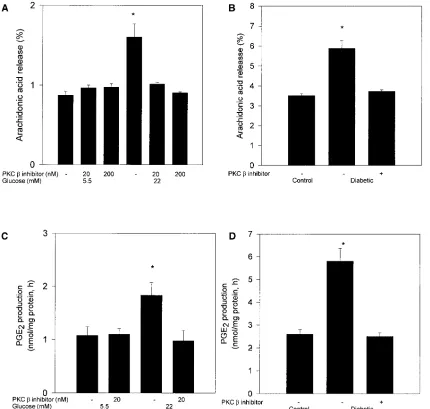
ad-Figure 2. (A) Effect of PKC b inhibitor, LY333531 on high glucose-induced arachidonic acid release. Rat glomerular mesangial cells were la-beled with [3H]arachidonic acid after 3 d exposure to 5.5 or 22 mM glucose. The cells were treated with or without LY333531 for 1 h before
measurement of [3H]arachidonic acid release. The results were derived from three separate experiments and each experiment was performed in
triplicate. *P, 0.05 vs other columns. (B) Effect of PKC b inhibitor, LY333531 treatment (10 mg/kg body wt) on 4-wk diabetes-induced arachi-donic acid release. Isolated glomeruli labeled with [3H]arachidonic were incubated for 1 h before measurement of [3H]arachidonic acid release.
The results were derived from four separate rats. *P, 0.05 vs other columns. (C) Effect of PKC b inhibitor, LY333531 on high glucose-induced PGE2 production in rat glomerular mesangial cells. Rat glomerular mesangial cells were incubated with 5.5 or 22 mM glucose for 4 d and treated
with or without LY333531 for 1 h before the measurement of PGE2 production. The results were derived from three separate experiments and
each experiment was performed in duplicate. *P, 0.05 vs other columns. (D) Effect of PKC b inhibitor, LY333531 treatment (10 mg/kg body wt) on 4-wk diabetes-induced PGE2 production in rat glomeruli. After incubating isolated glomeruli for 1 h, the supernatants were used to
[image:5.612.57.487.209.618.2]dition of PKC b isoform inhibitor LY333531 at 20 or 200 nM did not change the levels of total DAG in the rat mesangial cell (Fig. 1 A). The addition of LY333531 for 1 h did not change the total protein concentrations compared with those cultured in the absence of LY333531.
Increasing glucose concentration in the cultured rat mesan-gial cells from 5.5 to 22 mM also increased the specific in situ PKC activity from 24.562.5 to 34.062.5 pmol/mg protein, min in parallel with increases in DAG levels (Fig. 1 B). The addi-tion of 20 nM of PKC b isoform selective inhibitor LY333531 did not significantly lower PKC activity at 5.5 mM of glucose but significantly decreased the PKC activity in cells cultured under 22 mM of glucose to 21.161.8 pmol/mg protein per min (Fig. 1 B) reaching levels comparable with that in cells exposed to 5.5 mm glucose alone. The addition of 200 nM of PKC b in-hibitor LY333531 significantly decreased the in situ PKC activ-ities in rat mesangial cells cultured either at 5.5 or 22 mM of glucose by 52 and 54%, respectively. Similar to the results of rat mesangial cells, 22 mM of glucose also significantly in-creased PKC activity in human mesangial cells by 62% com-pared with 5.5 mM of glucose (Table I). Interestingly, the addi-tion of PKC b inhibitor LY333531 at the concentrations of either 20 nM or 200 nM decreased the levels of in situ PKC activities in both cells cultured under 5.5 and 22 mM of glucose (Table I).
Effect of PKC b isoform selective inhibitor LY333531 on glucose-induced increases of AA release and PGE2 produc-tion. Since the above results showed that PKC b inhibitor LY333531 can inhibit specifically the glucose-induced PKC ac-tivity, we have characterized the effect of LY333531 to prevent the increases in release of AA and the production of PGE2 in
the mesangial cells induced by glucose. Fig. 2 A showed that el-evating glucose level from 5.5 to 22 mM in the media increased AA release from rat mesangial cells by 82%. The addition of LY333531 at either 20 or 200 nM did not alter AA release in cells cultured at 5.5 mM of glucose but prevented the increase observed at 22 mM of glucose (Fig. 2 A). As shown in Table I, increasing glucose level from 5.5 to 22 mM in human mesan-gial cells also increased AA release by 47%, but the addition of 20 nM PKC b inhibitor LY333531 normalized the increased AA release in human mesangial cells exposed to 22 mM of glu-cose. The glomerular AA release was also measured in con-trol, diabetic, and diabetic rats treated with LY333531. Treat-ment with LY333531 prevented the increase in AA release induced by diabetes (Fig. 2 B).
PGE2 production by rat mesangial cells was also increased
from 1.060.1 to 1.860.1 nmol/mg protein per h when glucose concentration in the media was elevated from 5.5 to 22 mM (Fig. 2 C). When the activation of PKC was inhibited by the addition of 20 nM of LY333531, the increased PGE2
produc-tion in cells cultured under 22 mM glucose was prevented (Fig. 2 C). However, LY333531 did not change the basal PGE2
production in mesangial cells cultured under 5 mM of glucose (1.160.1 nmol/mg protein per h) compared to basal values of control cells cultured under 5.5 mM glucose. In addition, glomerular PGE2 production in the diabetic rats was also
in-creased compared with control rats (Fig. 2 D). Treatment with LY333531 prevented the increase in PGE2 production in
dia-betic rats (Fig. 2 D).
Effect of PKC b isoform selective inhibitor LY333531 on glucose-induced inhibition of Na1-K1 ATPase activity. Since we
have previously reported that hyperglycemia-induced activa-tion of cytosolic-phospholipase A2 (cPLA2) in aortic smooth
muscle cells can result in the inhibition of Na1-K1 ATPase via
the overproduction of AA release and PGE2 (8), the effect of
LY333531 to regulate Na1-K1 ATPase activity in rat
mesan-gial cells exposed to either 5.5 or 22 mM of glucose was exam-ined. After 4 d of incubation, the ouabain-sensitive 86Rb
up-take, a physiological marker of Na1-K1 ATPase activity in rat
mesangial cells was decreased by 28% in cells exposed to 22 mM glucose as compared to 5.5 mM of glucose (Fig. 3). The addi-tion of either 20 or 200 nM of PKC b inhibitor LY333531 in-creased and normalized the ouabain-sensitive 86Rb uptake by
rat mesangial cells cultured under 22 mM of glucose (Fig. 3). The addition of PKC b inhibitor LY333531 at 20 or 200 nM did not alter the Na1-K1 ATPase activities in cells cultured under
[image:6.612.318.557.55.263.2]5.5 mM of glucose.
Figure 3. Effect of PKC b inhibitor on glucose-induced inhibition of Na1-K1 ATPase in rat glomerular mesangial cells. After 4 d exposure
to 5.5 or 22 mM glucose, the cells were treated with or without LY333531 for 1 h. 86Rb uptake was corrected for ouabain sensitivity
and determined after 10 min incubation as described in Methods. The results were derived from three separate experiments and each ex-periment was performed in triplicate. *P, 0.05 vs 5.5 mM glucose and 22 mM glucose with 20 or 200 nM PKC b inhibitor.
Table II. Physiological Characteristics of Control and Diabetic Rats with or without 10 mg/kg Body Wt PKC b Inhibitor, LY333531 Treatment for 12 wk
Animals Number Body wt
Blood glucose
Kidney wt/ body wt
treatment grams mg/dl gram/100 grams‡
Control 35 500.5618.2 102.567.5 0.6260.01 Control 1
PKC b inhibitor 25 497.5610.3 98.466.2 0.6460.01 Diabetic 35 266.5628.9* 434.5614.9* 1.3960.10* Diabetic 1
PKC b inhibitor 25 252.3619.1* 464.5619.7* 1.3160.07*
*P, 0.01 vs control and control 1 PKC b inhibitor; ‡Data were shown
[image:6.612.314.556.594.710.2]Effect of PKC b isoform selective inhibitor LY333531 treat-ment on in vivo physiological parameters.Diabetes in the rats was confirmed with glucose measurements of 434.5614.9 mg/dl in diabetic vs 102.567.5 mg/dl in control rats. Treatment of di-abetic rats with 10 mg/kg body wt of PKC b selective inhibitor LY333531 in rat chow did not prevent hyperglycemia, changes in weight gain, and increases in kidney wt/body wt ratio in the diabetic animals (Table II). Blood pressure and pulse rate did not change with diabetes or treatment with LY333531 for up to 12 wk (data not shown). The similar trends were observed in the rats with 4 wk of diabetes with or without LY333531 treat-ment (Table III).
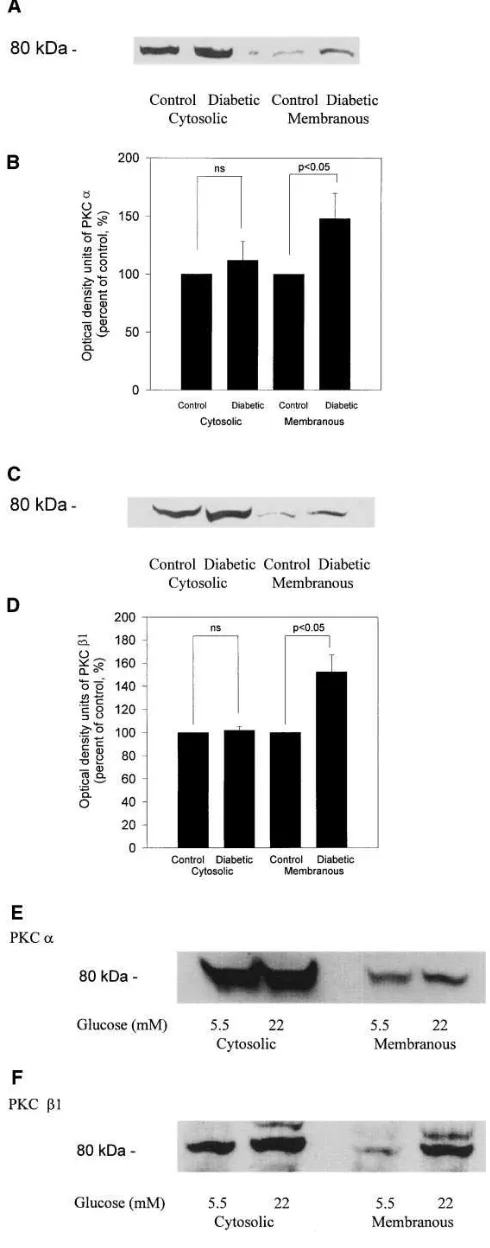
The in vivo effect of diabetes and PKC b selective inhibitor LY333531 on PKC isoform expressions in the glomeruli.We and others have reported that PKC activities in the glomeruli were increased by diabetes and can be inhibited by oral feeding of PKC b inhibitor LY333531 at 10 mg/kg body weight (11, 17). In the present study, we characterized the changes in various PKC isoforms in the glomeruli induced by diabetes by two methods (11, 17). First, the activation of specific PKC isoforms was defined by an increase in the protein expression of specific PKC isoforms in the membranous fraction, an indicator of PKC activation (32). Using immunoblot analysis, PKC a and
b1 isoforms were observed to be increased in the membrane fraction (Fig. 4, A–D) without any alterations in the d or e iso-forms (data not shown). Diabetes increased the amount of PKC a and b1 by 50% in the membranous fractions isolated from glomeruli as compared to control rat (Fig. 4, A and B for
[image:7.612.312.557.62.686.2]a; Fig. 4, C and D for b1). PKC b2 isoform did not appear to have changed although its content in the glomeruli was barely detectable and was much lower than PKC b1. The amounts of PKC a and b1 isoforms in the cytosolic and membranous frac-tions of rat mesangial cells were also assessed by immunoblot
Table III. Physiological Characteristics of Control Rats and Diabetic Rats with or without 10 mg/kg Body Wt PKC b Inhibitor, LY333531 Treatment for 4 wk‡
Animals Number Body wt Blood glucose
treatment grams mg/dl
Control 8 368.869.1 102.562.1 Diabetic 8 281.3612.2* 394.4614.3* Diabetic 1
PKC b inhibitor 8 287.568.4* 400.4618.2*
*P, 0.01 vs control rats. ‡These rats were used for the determination of
[image:7.612.57.302.93.183.2]glomerular AA release and PGE2 production.
Figure 4. Immunoblot analysis of glomeruli from control and 12-wk diabetic rats, and rat glomerular mesangial cells exposed to 5 or 22 mM glucose for 4 d with anti–PKC a and b1 antibody. (A, C, E, and F). PKC in the membranous and cytosolic fractions was partially purified from isolated glomeruli from three rats of control and diabetic rats or rat mesangial cells using DEAE cellulose. 30–50 mg protein for PKC
a and 70–100 mg for PKC b1were loaded in each lane and membranes were blotted with anti–PKC a or b1 antibodies. Representative re-sults were shown in A for PKC a, in C for PKC b1 in renal glomeruli, in E for PKC a, and in F for PKC b1 in rat mesangial cells. (B and D).
analysis showing that incubation of mesangial cells with 22 mM glucose increased the amounts of both PKC a and b1 isoforms in the membranous fraction (Fig. 4 E for PKC a; and Fig. 4 F
for PKC b1).
The second method used to evaluate the activation of PKC
a and b1 by diabetes was by determining the amount of phos-phorylation of PKC a and b1 in the glomeruli from control and diabetic rats. Increases in phosphorylation of PKC iso-forms have been shown to correspond to their state of activa-tion (33–37). For this study, we isolated glomeruli from control and diabetic rats with or without treatment with PKC b
inhibi-tor LY333531 and labeled them with 32P orthophosphate. The
glomeruli were then processed and immunoprecipitated with either PKC a or b1 antibodies as described in the Methods section. The results as shown in Fig. 5, A and B indicated that diabetes increased phosphorylation of both PKC a and b1 by 60 and 75%, respectively. Oral treatment of rats with LY333531 at 10 mg/kg body wt prevented the increase in phos-phorylation only of PKC b1 isoform but not PKC a isoform, providing the first in vivo evidence that LY333531 can specifi-cally inhibit PKC b1 isoform in glomeruli from diabetic animals.
Effect of diabetes and PKC b selective inhibitor LY333531 on Na1-K1 ATPase activity in glomeruli of diabetic rats. As
described in the previous section, elevating glucose concentra-tion in the media from 5.5 to 22 mM inhibited Na1-K1 ATPase
activity as measured by 86Rb uptake into rat and human
mes-angial cells which was prevented by LY333531, indicating that glucose was mediating its effect via PKC b isoform activation. To substantiate this finding in vivo, ouabain-sensitive 86Rb
up-take was quantitated using isolated glomeruli from control and diabetic rats. After having diabetes for 12 wk, ouabain-sensi-tive 86Rb uptake in the glomeruli of diabetic rats was
de-creased by . 50% compared with control rats (Fig. 6). Oral feeding of PKC b selective inhibitor LY333531 to nondiabetic controls did not significantly change the rate of 86Rb uptake. In
contrast, when diabetic rats were fed with LY333531, ouabain-sensitive 86Rb uptake returned to normal at 104% of control
(Fig. 6).
Effect of PKC b selective inhibitor LY333531 on the mRNA expression of TGF-b1 and extracellular matrix components in glomeruli of control and diabetic rats. Increases in the expres-sion of TGF-b1 and extracellular matrix components in the glomeruli of diabetes are associated with the development of basement membrane thickening and mesangial expansion, characteristic of chronic and pathological findings in the
glom-Figure 5. Effect of PKC b inhibitor, LY333531 (10 mg/kg body wt) on diabetes-induced phosphorylation of PKC a and b1. Isolated glomeruli from three rats (six kidneys) of each group were labeled with 32P orthophosphate in phosphate free DME. After lysing
glom-eruli with lysis buffer, the samples were immunoprecipitated with anti–PKC a and b1 antibody and analyzed by SDS-PAGE followed by autoradiography. The representative results from three separate experiments and mean6SEM of three experiments were shown in A
[image:8.612.315.538.58.259.2]for PKC a and in B for PKC b1 isoforms. *P, 0.05 vs other groups.
Figure 6. Effect of PKC b inhibitor LY333531 (10 mg/kg body wt) on 12-wk diabetes-induced Na1-K1 ATPase in rat glomeruli. Using
glomeruli from four rats of each group, 86Rb uptake was corrected for
[image:8.612.58.244.59.494.2]eruli of diabetic nephropathy (38, 39). The effects of diabetes and treatment with LY333531 at 10 mg/kg body wt on the gene expressions of TGF-b1, fibronectin, and a1(IV) collagen were examined in the glomeruli of control and diabetic rats. Glom-erular TGF-b1 mRNA level was increased by 88626% in dia-betic rats compared with that in control rats (Fig. 7). Oral treatment with PKC b selective inhibitor LY333531 in control rats did not alter TGF-b1 mRNA expression significantly. However, when LY333531 was given to diabetic rats for 12 wk, the increases in TGF-b1 mRNA level were prevented (Fig. 7).
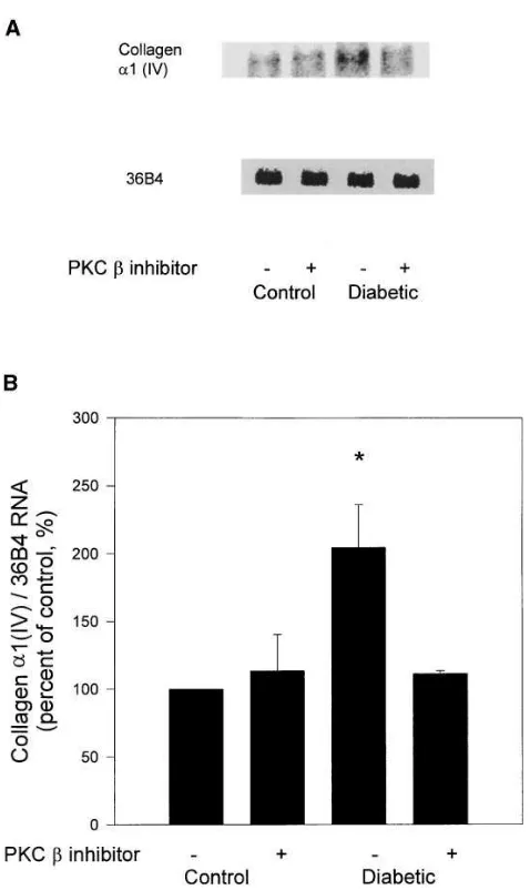
The mRNA expressions of extracellular matrix compo-nents, fibronectin, and a1(IV) collagen were also quantitated in the glomeruli. Similar to the expression of TGF-b1, both fi-bronectin and a1(IV) collagen mRNA expressions were in-creased in diabetic rats by 85 and 104%, respectively (Figs. 8 and 9). Again treatment with PKC b inhibitor LY333531 orally
did not change their expression in the control animals whereas oral application of LY333531 in the chow at 10 mg/kg body wt prevented the increases in mRNA expression of both fibronec-tin and a1(IV) collagen in diabetic rats (Figs. 8 and 9).
Discussion
[image:9.612.72.421.59.444.2]The mechanisms by which hyperglycemia cause adverse ef-fects in the vasculature of diabetic patients are probably multi-ple since alterations of glucose metabolism and its metabolites can affect the flux and regulation of numerous cellular meta-bolic pathways (3–7). One such important cellular pathway which has been shown to be consistently activated is the DAG–PKC signal transduction system (7). Consistent with previous reports from our laboratory and others, the levels of total DAG and activated fraction of PKC were increased by
Figure 7. Effect of PKC b inhibitor, LY333531 treatment (10 mg/kg body wt) on 12-wk diabetes-induced fibronectin mRNA expression in glomeruli. (A) A representative Northern blot of glomerular RNA probed with PCR fragment for rat fibronectin in upper panel and 36B4 RNA in lower panel. (B) Summary of quantification of fi-bronectin/36B4 message ratios. The results were derived from six rats of each group. Four kidneys were processed for each experiment. *P, 0.05 vs control and diabetic with PKC b inhibitor treatment.
[image:9.612.246.545.62.444.2]elevating glucose levels in the cultured mesangial cells and in the renal glomeruli of diabetic rats (11, 14, 17, 18). The activa-tion of the various PKC isoforms was evaluated by their phos-phorylation levels and by the changes of their protein levels in the membranous fractions. The use of the PKC protein ratio of membranous to cytosolic fraction by itself may not be ade-quate since it is possible that some of the activated PKC could have been dissociated from the membrane fraction during the isolation procedures (25). Increases in serine and threonine phosphorylation of PKC isoforms have been shown to occur after PKC has been activated by DAG and Ca21 (33–36). Thus,
quantitation of the phosphorylation levels of PKC isoform provided a complementary method to determine whether the various specific isoforms are activated (33–36). The
assess-ments of isoform activation and inhibition were performed on the renal glomeruli and on cultured mesangial cells to evaluate the in vivo and in vitro actions of PKC b isoform inhibitor LY333531 in the renal glomerular cells which was not done previously (11). Both the immunoblotting and phosphoryla-tion assays suggested that PKC isoform a and b1 were acti-vated. The lack of decrease in the PKC band from the cytosolic fractions for these isoforms could be due to the insensitivity of the assay to detect the small changes (Fig. 5). The finding of both a and b1 PKC isoforms to be activated in the glomeruli are similar to the findings in the retina where multiple PKC isoforms including PKC a and b2 were activated by diabetes (13). However, the finding of PKC b1 to be activated in the glomeruli differed from the retina, heart, or the aorta which exhibited predominately PKC b2 isoform activations (12, 13). One possible explanation for this difference between vascular tissues and glomeruli is that the expression of PKC b1 appears to be much greater than PKC b2 in the renal glomeruli. This possibility was supported by the results of immunoblot analysis which also showed increased expression of PKC a and b1 iso-forms in the membranous fraction of rat mesangial cells ex-posed to elevating glucose concentrations.
The use of 32P orthophosphate-labeling study provided the
direct in vivo evidence that PKC isoform a and b1 were acti-vated in the glomeruli of diabetic animals. This method al-lowed us to determine for the first time whether the inhibitory effect of LY333531 was specific to PKC b isoforms in vivo. In a previous report, we have reported that the inhibitory effect of LY333531 was reversible and did not prevent the translocation of PKC, and was a competitive inhibitor at the ATP binding site in in vitro studies (11, 19). Therefore, the use of transloca-tion assay would not be able to detect the activatransloca-tion states of PKC or the effectiveness of LY333531 since translocation in-duced by DAG preceded the binding of ATP in the activation sequence of PKC (37). In contrast, quantitation of the phos-phorylation level of the PKC isoforms will directly determine the activation status of the various PKC isoforms (33–36). Our present results clearly indicated that LY333531 used at 10 mg/ kg body wt specifically inhibited PKC b1 isoform activations, thus providing in vivo evidence that LY333531 can selectively inhibit PKC b1 similar to its in vitro inhibitory effect on PKC activity in addition to our previous report showing its inhibi-tory effect on PKC b2 (11, 19). The use of LY333531 affected only PKC activity but not DAG levels in mesangial cells, sug-gesting that PKC activity may not affect phospholipase C (PLC) activity. Alternatively, the source of the increased DAG is due to de novo synthesis and not due to activation of PLC activity as shown previously (9, 17). It is also interesting to note that the inhibition of PKC b1 isoform phosphorylation by LY333531 has consistently caused a parallel increase in basal PKC a phosphorylation level. The mechanism of this in-crease in PKC a phosphorylation is not clear. Further studies are needed to characterize the cellular consequences of this change in PKC a isoform phosphorylation by LY333531.
[image:10.612.56.295.52.453.2]Functionally, our previous report suggested that oral treat-ment of LY333531 can ameliorate the early hemodynamic dys-functions in the glomeruli of diabetic rats suggesting that PKC activation may potentially have a pathophysiologic role in the development of diabetic vascular complications (11). How-ever, the hemodynamic changes were very early parameters which may not translate into chronic and pathological findings of diabetic nephropathy (11). In the present study, we have
Figure 9. Effect of PKC b inhibitor, LY333531 treatment (10 mg/kg body wt) on 12-wk diabetes-induced type IV collagen mRNA expres-sion in glomeruli. (A) A representative Northern blot of glomerular RNA probed with cDNA for mouse a1(IV) collagen in upper panel and 36B4 RNA in lower panel. (B) Summary of quantification of
characterized the effect of PKC b inhibitor LY333531 on a host of changes both in cultured mesangial cells and in glomer-uli of diabetic rats. Hyperglycemia’s effect to activate cPLA2
has been reported previously by us in the aortic smooth muscle cells exposed to elevated level of glucose (8). Furthermore, PLA2 appeared to be activated in the renal glomeruli of
dia-betic rats as reported by Craven et al. (9). The result of the ac-tivation of PLA2 can lead to an increase in the amount of
PGE2 and arachidonic acid release which have been reported
to be increased in vivo in the glomeruli of diabetic rats (9, 40, 41). These findings suggest that activation of PLA2 may be
re-sponsible for the increased production of the prostanoids in the glomeruli of diabetic rats.
The possible consequence of increases in the prostanoids could be the vasodilatation of afferent arterioles that leads to increased glomerular filtration rate of diabetic animals (11). These effects of hyperglycemia were also studied in cultured human mesangial cells since results derived from human may not be similar to those from rodent cells. The results of human mesangial cells were very consistent with our previous findings that hyperglycemia can increase PKC activity and PLA2
activ-ity in a similar manner as those in smooth muscle cells (8) and rat mesangial cells (10). These results have provided the first direct comparative study of rodent and human mesangial cells with respect to glucose-induced activation of PKC and its pos-sible cellular effects.
The consequences of PKC activation specifically the b1 iso-form were evaluated by measuring PLA2 activity and the
inhi-bition of Na1-K1 ATPase in the renal glomeruli. Although the
inhibition of Na1-K1 ATPase by hyperglycemia or diabetes
has been reported in multiple vascular and neuronal tissues (4, 8), our present finding in the renal glomeruli and cultured mes-angial cells provided direct evidence that the activation of PLA2 and the inhibition of Na1-K1 ATPase both in vitro and
in vivo was due to activation of PKC b isoforms. The possible consequences of Na1-K1 ATPase inhibition in the renal
glom-eruli could be multiple including the maintenance of the cellu-lar integrity, vascucellu-lar and epithelial cell permeability, and so-dium/hydrogen transport (42, 43). Abnormalities in many of these cellular functions have been reported in the diabetic vas-cular complications.
One of the hallmarks of diabetic nephropathy is character-ized by progressive mesangial expansion, eventually leading to a decrease in filtration surface area and a reduction in glomer-ular filtration rate (38, 39). Mesangial expansion is due to an accumulation of ECM components, caused by either increased production or impaired degradation. A substantial amount of data suggest that the increased ECM synthesis by mesangial cells and other vascular cells was induced by elevated levels of glucose (44–48). Studies have suggested that TGF-b expres-sion stimulated by hyperglycemia may contribute to the ex-pansion of the mesangium since TGF-b is known to stimulate ECM production (3, 49–53). In this study, we have shown that the activation of PKC b correlated with an increased mRNA expression of TGF-b1 and ECM components in glomeruli of diabetic rats. Moreover, treatment with PKC b inhibitor LY333531 normalized the gene expression of TGF-b1, fibro-nectin and a1(IV) collagen. Our results did not demonstrate that increased TGF-b mRNA expression directly caused en-hanced gene expression of fibronectin and a1(IV) collagen. However, others have reported that TGF-b stimulated ECM accumulation such as fibronectin, laminin, and type IV
col-lagen in renal cells (52, 54). In addition, neutralization of TGF-b
by anti–TGF-b antibody attenuated the increase in mRNA levels for fibronectin and a1(IV) collagen in kidney of diabetic mice and cultured mesangial cells exposed to high glucose (53, 55), suggesting that TGF-b expression was directly responsible in the mesangial matrix expansion.
Using PKC b isoform specific inhibitor (LY333531), we have provided evidence that the activation of PKC b isoform by hyperglycemia could be involved in the mediation of abnor-mal mesangial and glomerular functions such as mRNA over-expression of TGF-b and ECM components and overproduc-tion of prostanoids. Our previous studies have shown that PKC b2 isoform was preferentially activated by the increase in de novo DAG synthesis in cultured vascular cells exposed to elevated levels of glucose and various vascular tissues from di-abetic animals (12, 13). In retina, aorta, and the heart, PKC b
isoform was predominately increased although PKC a was also increased in the retina to a lesser extent (13). Thus, PKC activation in the glomeruli differed from other vascular tissues in two ways. First, PKC b1 isoform was activated and not PKC
b2 isoform. Second, membranous protein levels of both PKC a
and b1 were equally increased. The selectivity of PKC b1 over
b2 was likely due to a greater expression of PKC b1 isoform in the glomeruli than other tissues. It is not surprising that PKC a
isoform was detected to be activated since Kikkawa et al. has made similar observations in rat mesangial cells exposed to high concentrations of glucose (16). Since PKC b isoform in-hibitor LY333531 (10 mg/kg body wt) was able to prevent dia-betes-induced changes in Na1-K1 ATPase activity, and gene
expressions of TGF-b1 and ECM, it is likely that only PKC b
isoform was responsible. This conclusion was substantiated by the measurements of the phosphorylation levels of PKC a and
b1 in the renal glomeruli directly. In addition, at an oral dose of 10 mg/kg body wt, the plasma level of LY333531 was 19 nM which was threefold greater than the IC50 for PKC b1 at 6 nM
but 10-fold less than IC50 of PKC a at 360 nM (11, 19).
In summary, these results provided substantial evidence that the activation of PKC particularly PKC b1 isoform by hy-perglycemia and diabetes can alter glomerular cell functions and gene expressions that are related to the chronic pathologi-cal changes of diabetic nephropathy. The availability of an orally effective PKC b selective inhibitor has provided an im-portant insight into the specific biological actions of PKC b
isoform and suggested the usefulness of PKC b specific inhibi-tor in the treatment of diabetic complications.
Acknowledgments
The authors are grateful to Dr. Fuad N. Ziyadeh for mouse TGF-b1 cDNA, to Dr. Markku Kurkinen for mouse a1(IV) collagen cDNA, and to Dr. Mara Lorenzi and Dr. Susanne Willert for rat fibronectin PCR product. We also thank Ms. Joan C. Taylor for her assistance in the preparation of the manuscript.
This study was supported by National Institutes of Health (NIH)/ National Institute of Diabetes and Digestive and Kidney Diseases DK36836-10 and NIH/National Eye Institute EY5110-12.
References
1. U.S. Renal Data System, USRDS 1994 Annual Data Report. National Institutes of Health, National Institute of Diabetes and Digestive and Kidney Disease, Bethesda, MD, July 1994.
long-term complications in insulin-dependent diabetes mellitus. N. Engl. J. Med. 329:977–986.
3. Sharma, K., and F.N. Ziyadeh. 1995. Hyperglycemia and diabetic kidney disease. The case for transforming growth factor-b as a key mediator. Diabetes.
44:1139–1146.
4. Greene, D.A., S.A. Lattimer, and A.F. Sima. 1987. Sorbitol, phospho-inositides, and sodium-potassium-ATPase in the pathogenesis of diabetic com-plications. N. Engl. J. Med. 316:599–606.
5. Brownlee, M., H. Vlassara, and A. Cerami. 1984. Nonenzymatic glycosy-lation and pathogenesis of diabetes complication. Ann. Intern. Med. 101:527–537.
6. Williamson, J.R., K. Chang, M. Frangos, K.S. Hasan, Y. Ido, T. Kawa-mura, J.R. Nyengaard, M.V.D. Enden, C. Kilo, and R.G. Tilton. 1993. Hyper-glycemic pseudohypoxia and diabetic complications. Diabetes. 42:801–813.
7. DeRubertis, F.R., and P.A. Craven. 1994. Activation of protein kinase C in glomerular cells in diabetes. Mechanisms and potential links to the pathogen-esis of diabetic glomerulopathy. Diabetes. 43:1–8.
8. Xia, P., R.M. Kramer, and G.L. King. 1995. Identification of the mecha-nism for the inhibition of Na1, K1- adenosine triphosphatase by hyperglycemia
involving activation of protein kinase C and cytosolic phospholipase A2. J. Clin. Invest. 96:733–740.
9. Craven, P.A., M.C. Patterson, and F.R. DeRubertis. 1988. Role of en-hanced arachidonate availability through phospholipase A2 pathway in media-tion of increased prostaglandin synthesis by glomeruli from diabetic rats. Dia-betes. 37:429–435.
10. Williams, B., and R.W. Schreir. 1993. Glucose-induced protein kinase C activity regulates arachidonic acid release and eicosanoid production by cul-tured glomerular mesangial cells. J. Clin. Invest. 92:2889–2896.
11. Ishii, H., M.R. Jirousek, D. Koya, C. Takagi, P. Xia, A. Clermont, S.-E. Bursell, T.S. Kern, L.M. Ballas, W.F. Heath, et al. 1996. Amelioration of vascu-lar dysfunctions in diabetic rats by an oral PKC b inhibitor. Science (Wash. DC). 272:728–731.
12. Inoguchi, T., R. Battan, E. Handler, J.R. Sportsman, W. Heath, and G.L. King. 1992. Preferential elevation of protein kinase C isoform bII and dia-cylglycerol levels in the aorta and heart of diabetic rats: differential reversibility to glycemic control by islet cell transplantation. Proc. Natl. Acad. Sci. USA. 89: 11059–11063.
13. Shiba, T., T. Inoguchi, J.R. Sportsman, W.F. Heath, S. Bursell, and G.L. King. 1993. Correlation of diacylglycerol level and protein kinase C activity in rat retina to retinal circulation. Am. J. Physiol. 265:E783–E793.
14. Ayo, S.H., R. Radnik, J.A. Garoni, D.A. Troyer, and J.I. Kreisberg. 1991. High glucose increases diacylglycerol mass and activate protein kinase C in mesangial cell culture. Am. J. Physiol. 261:F571–F577.
15. Williams, B., and R.W. Schreier. 1992. Characterization of glucose-induced in situ protein kinase C activity in cultured vascular smooth muscle cells. Diabetes. 41:1464–1472.
16. Kikkawa, R., M. Haneda, T. Uzu, D. Koya, T. Sugimoto, and Y. Shigeta. 1994. Translocation of protein kinase a and z in rat glomerular mesangial cells cultured under high glucose conditions. Diabetologia. 37:838–841.
17. Craven, P.A., and F.R. DeRubertis. 1989. Protein kinase C is activated in glomeruli from streptozotocin diabetic rats. Possible mediation by glucose. J. Clin. Invest. 83:1667–1675.
18. Craven, P.A., C.M. Davidson, and F.R. DeRubertis. 1990. Increase in diacylglycerol mass in isolated glomeruli by glucose from de novo synthesis of glycerolipids. Diabetes. 39:667–674.
19. Jirousek, M.R., J.R. Gilling, C.M. Gonzalez, W.F. Heath, J.H. Mc-Donald III, D.A. Neel, C.J. Rito, U. Singh, L.E. Stramm, A. Melikian-Badalian, et al. 1996. (S)-13-[(Dimethylamino)methyl]-10,11,14,15-tetrahydro-4,9:16,21- dimetheno-1H,13H-dibenzo[e,k]pyrrolo[3,4-h][1,4,13]oxadiazacyclohexadecene-1,2(2H)-dione (LY333531) and related analogues: isozyme selective inhibitors of protein kinase Cb. J. Med. Chem. 39:2664–2671.
20. Koya, D., R. Kikkawa, M. Haneda, T. Uzu, T. Sawada, N. Kajiwara, K. Sakamoto, T. Sugimoto, and Y. Shigeta. 1993. Nipradilol inhibits rat mesangial cell mitogenesis through the activation of soluble guanylate cyclase. Eur. J. Pharmacol. 245:79–82.
21. Kikkawa, R., K. Umemura, M. Haneda, T. Arimura, K. Ebata, and Y. Shigeta. 1987. Evidence for existence of polyol pathway in cultured rat mesan-gial cells. Diabetes. 36:240–243.
22. Bligh, E.G., and W.J. Dyer. 1959. A rapid method of total lipid extrac-tion and purificaextrac-tion. Can. J. Biochem. Physiol. 37:911–917.
23. Preiss, E.G., L.R. Loomis, R.M. Bell, and J.E. Niedel. 1984. Quantita-tive measurement of sn-1,2,diacylglycerol. Methods Enzymol. 141:294–300.
24. Bradford, M.M. 1976. A rapid and sensitive method for the quantitation of microgram quantities of protein utilizing the principle of protein-dye bind-ing. Anal. Biochem. 72:248–254.
25. Heasley, L.E., and G.L. Johonson. 1989. Regulation of protein kinase C by nerve growth factor, epidermal growth factor, and phorbol ester in PC 12 pheochromocytoma cells. J. Biol. Chem. 264:8646–8652.
26. Yasuda, I., A. Kishimoto, S.-I. Tanaka, M. Tominaga, A. Sakurai, and Y. Nishizuka. 1990. A synthetic peptide substrate for selectivity assay of protein kinase C. Biochem. Biophys. Res. Commun. 166:1220–1227.
27. Chomczynski, P., and N. Sacchi. 1987. Single step method of RNA isola-tion by acid guanidinium thiocyanate-phenol-chroloform extracisola-tion. Anal.
Bio-chem. 162:156–159.
28. Sharma, K., and F.N. Ziyadeh. 1994. Renal hypertrophy is associated with upregulation of TGF-b1 gene expression in diabetic BB and NOD mouse.
Am. J. Physiol. 267:F1094–F1101.
29. Kurkinen, M., M.R. Condon, B. Blumberg, D.P. Barlow, S. Quinoens, J. Saus, and T. Pihlajaniemi. 1987. Extensive homology between the carboxyl-ter-minal peptides of mouse a1(IV) and a2(IV) collagen. J. Biol. Chem. 262:8496– 8499.
30. Schwarzbauer, J.E., R.S. Patel, D. Fonda, and R.O. Heynes. 1987. Mul-tiple sites of alternative splicing of the rat fibronectin gee transcript. EMBO (Eur. Mol. Biol. Org.) J. 6:573–580.
31. Aiello, L.P., G.S. Robinson, Y.W. Lin, Y. Nishio, and G.L. King. 1994. Identification of multiple genes in bovine retinal pericytes altered by exposure to elevated levels of glucose by using mRNA differential display. Proc. Natl. Acad. Sci. USA. 91:6231–6235.
32. Kraft, A.S., and W.B. Anderson. 1983. Phorbol ester increases the amounts of calcium/phospholipid dependent kinase associated with the plasma membrane. Nature (Lond.). 301:621–623.
33. Newton, A.C. 1995. Protein kinase C: structure, function, and regula-tion. J. Biol. Chem. 270:28495–28498.
34. Zhang, J., L. Wang, J. Pertrin, W.R. Bishop, and R.W. Bond. 1993. Characterization of site-specific mutants altered at protein kinase C b1 isozyme autophosphorylation sites. Proc. Natl. Acad. Sci. USA. 90:6130–6134.
35. Ohno, S., Y. Konno, Y. Akita, A. Yano, and K. Suzuki. 1990. A point mutation at the putative ATP-binding site of protein kinase Ca abolishes the kinase activity and renders it down-regulation-insensitive. A molecular link be-tween autophosphorylation and down-regulation. J. Biol. Chem. 265:6296– 6300.
36. Mitchell, F.E., R.M. Marais, and P.J. Parker. 1989. The phosphorylation of protein kinase C as a potential measure of activation. Biochem. J. 261:131–136. 37. Bell, R.M., and D.J. Burns. 1991. Lipid activation of protein kinase C. J. Biol. Chem. 266:4661–4664.
38. Mauer, S.M., M.W. Steffes, E.N. Ellis, D.E.R. Southerland, D.M. Brown, and F.C. Goetz. 1984. Structural–functional relationships in diabetic nephropathy. J. Clin. Invest. 74:1143–1155.
39. Steffes, M.W., R.W. Bilous, D.E.R. Southerland, and M. Mauer. 1992. Cell and matrix components of the glomerular mesangium in type 1 diabetes.
Diabetes. 41:679–684.
40. Craven, P.A., F.R. Caines, and F.R. DeRubertis. 1987. Sequential alter-ations in glomerular prostaglandin and thromboxane synthesis in diabetic rats: relationship to the hyperfiltration of early diabetes. Metab. Clin. Exp. 36:95–103.
41. Schambelan, M., S. Blake, M. Sraer, M. Bens, M.-P. Nivez, and F. Wahbe. 1985. Increased prostaglandin production by glomeruli isolated from rats with streptozotocin-induced diabetes mellitus. J. Clin. Invest. 75:404–412.
42. Vasilets, L.A., and W. Schwarz. 1993. Structure-function relationship of cation binding in the Na1/K1-ATPase. Biochem. Biophys. Acta. 1154:201–222.
43. Ewart, H.S., and A. Klip. 1995. Hormonal regulation of the Na1-K1
ATPase: Mechanisms underlying rapid and sustained changes in pump activity.
Am. J. Physiol. 269:C295–C311.
44. Wakisaka, M., M.J. Spiro, and R.G. Spiro. 1994. Synthesis of type VI collagen by cultured glomerular cells and comparison of its regulation by glu-cose and other factors with that of type IV collagen. Diabetes. 43:95–103.
45. Danne, J., M.J. Spiro, and R.J. Spiro. 1993. Effect of high glucose on type IV collagen production by cultured glomerular epithelial, endothelial, and mesangial cells. Diabetes. 42:170–177.
46. Ayo, S.H., R.A. Radnik, W.F. Glass II, J.A. Garoni, E.R. Rampt, D.R. Appling, and J.I. Kreisberg. 1991. Increased extracellular matrix synthesis and mRNA in mesangial cells grown in high-glucose medium. Am. J. Physiol. 260: F185–F191.
47. Pugliese, G., F. Pricci, F. Dugliese, P. Mene, L. Lenti, D. Andreani, G. Galli, A.A. Casini, S. Bianchi, C.M. Rotella, and V.D. Mario. 1994. Mecha-nisms of glucose-enhanced extracellular matrix accumulation in rat glomerular mesangial cells. Diabetes. 43:478–490.
48. Studer, R.K., P.A. Craven, and F.R. DeRubertis. 1993. Role of protein kinase C in the mediation of increased fibronectin accumulation by mesangial cells grown in high-glucose medium. Diabetes. 42:118–126.
49. Yamamoto, T., T. Nakamura, N.A. Noble, E. Ruoslahti, and W.A. Bor-der. 1993. Expression of transforming growth factor b is elevated in human and experimental diabetic nephropathy. Proc. Natl. Acad. Sci. USA. 90:1814–1818.
50. Shankland, S.J., and J.W. Scholey, with the technical assistance of H. Ly, and K. Thai. 1994. Expression of transforming growth factor-b1 during diabetic renal hypertrophy. Kidney Int. 46:430–442.
51. Ziyadeh, F.N., and K. Sharma. 1995. Role of transforming growth fac-tor-b in diabetic glomerulosclerosis and renal hypertrophy. Kidney Int. 48 (Suppl.):34–36.
52. Bollineni, J.S., and A.S. Reddi. 1993. Transforming growth factor-b1 en-hances glomerular collagen synthesis in diabetic rats. Diabetes. 42:1673–1677.
53. Ziyadeh, F.N., K. Sharma, M. Ericksen, and G. Wolf. 1994. Stimulation of collagen gene expression and protein synthesis in murine mesangial cells by high glucose is mediated by activation of transforming growth factor-b. 1994. J. Clin. Invest. 93:536–542.
for transforming growth factor b in thromboxane-induced increases in mesan-gial cell fibronectin synthesis. Diabetes. 44:335–339.
55. Sharma, K., Y. Jin, J. Guo, and F.N. Ziyadeh. 1996. Neutralization of TGF-b by anti-TGF-b antibody attenuates kidney hypertrophy and the en-hanced extracellular matrix gene expression in STZ-induced diabetic mice. Di-abetes. 45:522–530.
56. Nishizuka, Y. 1988. The molecular heterogeneity of protein kinase C and its implications of cellular regulation. Nature (Lond.). 334:661–665.