© 2016, IRJET ISO 9001:2008 Certified Journal Page 1149
Automatic Detection of Mass In Mammogrammic Images Using Dual-Tree
Wavelet Transform
Heartlin Maria.H
1, Evangelin Mercy Agatha.P
2, Dr. P. Kavipriya
31,2
Department of ECE, Sathyabama University, Chennai – 119
3
Assistant Professor, Sathyabama University Chennai - 119
---***---ABSTRACT:
In this paper we have proposed an improvised method in which classification of Mammogrammic images using neural network is involved. The proposed method involves segmentation of the Mammogrammic images using dual tree wavelet transform for medical analysis. The specialty of this method is that it enables the detection of cancer at early stages. This can be done by row and column segregation of the Mammogrammic image, which is called as bi clustering. At the end of this analysis abnormal and normal images will be sorted out. These sorted images will be then used for Computer Aided Diagnosis. The wavelet transform that is used in this method decomposes the Mammogrammic image to get edge details on horizontal and vertical directions, which helps in early detection of breast cancer.Keywords: Segmentation, Neural network Computer-Aided Detection (CAD), Feature extraction
1.INTRODUCTON
Breast cancer is a cancer that gets greater, stronger, and more complete from chest tissue. Signs of breast cancer may involve the size of an egg in the chest, a change in chest form, dimpling of the skin, liquid (or gas) coming from the pointed part, or a red scaly material put over damaged place of skin. In critical stages of the disease, there may be bone pain, swollen lymph, hard growths, shortness of breath, or yellow skin. Cancers undergoing growth from the ducts are within one's knowledge and are known as ductal carcinomas, while those undergoing growth from lobules are within one's knowledge and are known as lobular carcinomas. In addition, there are more than 18 other sub-types of Breast cancer. Some cancers undergo growth from pre-invasive lesions such as ductal carcinoma in situ. The diagnosis of Breast cancer is made likely by taking a biopsy of the about mass, bit. Once the diagnosis is made, further tests are done to come to a decision about if the cancer has put out on top beyond the chest and which treatments it may be acting in answer to. The balance of benefits against causes damage of Breast cancer going-over is open to argument. A 2013 Cochrane paper stated that it is unclear if mammographic going-over does more good or cause damage. Organization suggests going-over every 2 years in women 50 to 74 years old. The medical substances tamoxifen or raloxifene may be used in a hard work to put a stop to Breast cancer in those who are at high danger of undergoing growth it. Sorts of surgery (make, become, be) different from breast-conserving surgery to mastectomy. Chest remake may take place at the time of surgery or at a later day. In those in whom the cancer has put out on top to other parts of the body, treatments 1are mostly directed at getting (making) better quality of living and comfort. Outcomes for Breast cancer (make, become, be) different depending on the cancer letters used for printing, size, range and degree of disease. In undergoing growth countries still living rates are poorer. Everywhere on earth, Breast cancer is the leading sort of cancer in women, accounting for 25% of all examples. In 2012 it resulted in 1.68 million 24 cases and 522,000 deaths.
© 2016, IRJET ISO 9001:2008 Certified Journal Page 1150 Fig(1) Normal case
Fig(2) Benign case
It is not cancer and does not spread
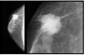
Fig (3) malignant case
[image:2.612.247.397.400.498.2]© 2016, IRJET ISO 9001:2008 Certified Journal Page 1151
2. SURVEY ON THE PREVIOUS WORK:
Overtime there has been a lot of researches on mammogram images. Various algorithms have been used for automatic computer aided diagnosis out of which a few are as follows:
Initially, the K-means clustering was applied for dynamically and automatically generated seed points and it determined the threshold values for each region. The region growing algorithm was used with the existing input parameters which was said to divide the mammogram into homogeneous regions based the intensity of the pixel. This method was used to automatically segment and detect the boundary of different disjoint breast tissues. Hence, using a computer-aided detection/diagnosis (CAD/CADx) system as supplement to the radiologists’ assessment has an important role. Mammographic Image Analysis Society (MIAS) database was used. The obtained results show the efficiency of this method. The disadvantage of the k-means algorithm is that the user as an input parameter must determine the number of clusters. Therefore, taking a fixed number of regions and the same regions for all images of database is irrelevant. The drawback of SRG algorithm is that the automating seed generation and dependency of output on order sorting of pixel is difficult. But in image segmentation mammograms this discontinuity is an advantage and a highlight of the kmeans algorithm.
Being able to elucidate 3D information from stereo mammograms was an important precursor to conducting 3D digital analysis of data from this promising new modality. The problem was generally much harder than the classic stereo matching problem on visible light since almost all of the 3D structural information of interest existed as complex network of multilayered, heavily occluded curvilinear structures. Taking this problem into count, a method has been formulated where a new stereo model can minimize global energy function to densely estimate disparity on stereo mammogram images, by introducing a new singularity index as a constraint. Curvilinear structures, such as vasculature and spicules, are salient structures in the breast, and accurately positioning them in 3D was a great task. Experiments on synthetic images with known ground truth and on real stereo mammograms clearly enhances the advantages of the proposed stereo model over the canonical stereo one. Experimental results on synthetic images with known ground truth data and on real stereo mammograms show the advantages of the proposed model over the canonical model for our application. The drawback in this method is that the loss of 3D information due to the projection of the breast onto a 2D image plane. The 3D to 2D projection process results in what is commonly referred to as anatomical noise due to overlapping out of plane tissue structures. A drawback of the two baseline stereo models is that neither model is designed to promote curvilinear masses in disparity space to better preserve fine-scale curvilinear structures.
2D/3D registration method, which was based on biomechanical modeling of the breast, was implemented. Initially the Mammograms were segmented into fatty, glandular and tumorous tissue and then the process was carried on. For each tissue, the average sound speed and attenuation of its corresponding USCT images were calculated and noted. Tumorous tissues could be separated from fatty and glandular tissue using a fixed absolute sound speed threshold. Through combining sound speed and attenuation, the extraction of fatty tissue from glandular tissue was improvised. From sound, speed and attenuation information’s that were obtained using the Mammogrammic images, required information could be obtained for diagnosis. This may benefit early breast cancer detection in future.
© 2016, IRJET ISO 9001:2008 Certified Journal Page 1152
3. EXISTING METHOD
:This CADe system for mammographic mass detection comprises four major stages: pre-processing, detection of suspicious mass region, feature extraction, and classification. Fig(4) shows an overview of the proposed mass detection scheme, and the following section presents each component in detail
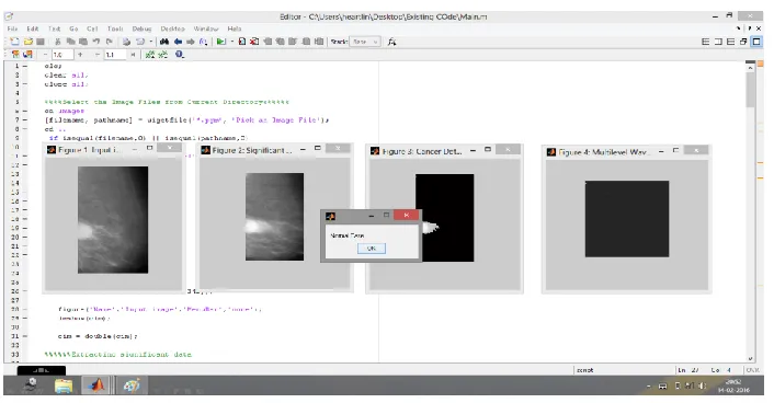
3.1 Screenshot of the output
Fig (4)
3.2 Discussion and conclusion
[image:4.612.131.484.215.399.2]© 2016, IRJET ISO 9001:2008 Certified Journal Page 1153
4. PROPOSED METHOD
• Energy : p(x , y) is the GLCM
• Contrast :
• Entropy :
• Correlation Coefficient :
sum(sum((x- μx)(y-μy)p(x , y)/σxσy))
• Homogeneity : sum(sum(p(x , y)/(1 + [x-y])))
Gray Level Cooccurrence Features
Fig (5) shows the block diagram of the proposed method
4.2Explanation
Dual-Tree wavelet transform
Dual-tree discrete wavelet transform (DWT) provides advantages over the critically sampled DWT for signal
and image processing. The dual-tree DWT is implemented as two separate two-channel filter banks. To gain the advantages described in this example, you cannot arbitrarily choose the scaling and wavelet filters used in the two tree.
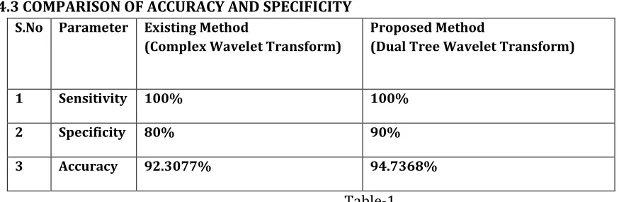
4.3 COMPARISON OF ACCURACY AND SPECIFICITY
S.No Parameter Existing Method(Complex Wavelet Transform)
Proposed Method
(Dual Tree Wavelet Transform)
1 Sensitivity 100% 100%
2 Specificity 80% 90%
3 Accuracy 92.3077% 94.7368%
[image:5.612.45.482.470.613.2]© 2016, IRJET ISO 9001:2008 Certified Journal Page 1154
4.4 GUI output screenshot
Fig(6)
4.5 Screenshot of accuracy, sensitivity and specificity of proposed method
© 2016, IRJET ISO 9001:2008 Certified Journal Page 1155
5. CONCLUSION:
This project implemented an automatic breast cancer image classification using texture features and it will be classified effectively based on artificial neural network. Here, probabilistic neural network was used for classification based on unsupervised leaning using wavelet statistical features and target vectors. The threshold was estimated from smoothing details of images accurately for effective breast cancer segmentation. In addition with, the statistical features are extracted from co-occurrence matrix of detailed coefficients of segmented images. These features are useful to train a neural network for an automatic classification process. Finally this system is very useful to diagnose the diseases from mammographic images for early detection of cancer.6. REFERENCE:
[1] Shen-Chuan Tai, Zih-Siou Chen and Wei-Ting Tsai, “An Automatic Mass Detection System in Mammograms Based on Complex Texture Features “, IEEE Journal of Biomedical and Health Informatics, Vol.18, No. 2, March 2014
[2] AbdelaliElmoufidi, Khalid El Fahssi, Said Jai-Andaloussi, AbderrahimSekkaki,“Automatically Density Based Breast Segmentation for Mammograms by using Dynamic K-means Algorithm and Seed Based Region Growing”, Instrumentation and Measurement Technology Conference (I2MTC), 2015 IEEE International.
[3] Gautam S. Muralidhar, Alan C. Bovik and Mia K. Markey, “Disparity Estimation on Stereo Mammograms”. IEEE Transactions On Image Processing, Vol. 24, No. 9, September 2015
[4] TorstenHopp, Torsten Hopp, Aurelien Stromboni, Neb Duric, Nicole V. Ruiter , “Evaluation of breast tissue characterization by ultrasound computer tomography using a 2D/3D image registration with mammograms”,Ultrasonics Symposium (IUS), 2013 IEEE International.
[5]Pratham Arora and Mandeep Singh, “New Intensity Based Features for Classification of Mammograms”, Electrical and Computer Engineering (CCECE), 2014 IEEE 27th Canadian Conference on May 2014.
[6] Peter C. Tay, “A Micro calcification Enhancement Method for Mammogram Images”, Image Processing (ICIP), 20th IEEE International Conference, September 2013.
[7] Sujoy Kumar Biswas and Dipti Prasad Mukherjee, “Recognizing Architectural Distortion in Mammogram: A Multiscale Texture Modeling Approach with GMM”. IEEE Transaction On Biomedical Engineering, Vol. 58, No. 7, Page No. 2023, July 2011.
[8] SalimLahmiri and MounirBoukadoum, “Hybrid Cosine and Radon Transform-based processing for Digital Mammogram Feature Extraction and Classification with SVM”. 33rd Annual International Conference of the IEEE EMBS Boston, Massachusetts USA, August 30 - September 3, 2011.