the epidermal barrier
Andrzej Slominski
J Clin Invest.
2007;
117(11)
:3166-3169.
https://doi.org/10.1172/JCI33508
.
In this issue of the
JCI
, Aberg and colleagues report that psychological stress disrupts the
skin’s antimicrobial barrier and increases the severity of cutaneous infections (see the
related article beginning on page 3339). This effect is mediated by endogenous
glucocorticoids produced secondarily to stress-related activation of the
hypothalamic-pituitary-adrenal axis. Thus, this study provides what I believe to be the first mechanistic link
between psychological stress and increased susceptibility to microbial infection.
Commentary
Find the latest version:
the N terminus is also important to limit proteolysis. After proteasomal digestion, MHC class I peptides often have a defini- tive C terminal residue but not a final N ter-minus (10). The latter will be produced by aminopeptidases present in the cytoplasm and/or the ER, such as ERAP (11). The sec-ond important function of the termini of a peptide is for its translocation from the cytoplasm to the ER and preferential bind-ing to the TAP transporter. Indeed, the three N terminal positions and the C termi- nal residue of cytoplasmic peptides are criti-cal in the selection of substrates by TAP and their efficient transport (12). The final role of the termini of a peptide is in the process of its binding to MHC class I molecules. At 8, 9, and even 10 residues in length, peptide binding to MHC class I can be optimal and satisfy anchoring to pockets (13). Beyond this length, MHC does not exclude binding altogether, but binding becomes subopti-mal by allowing only the extremities of the peptide to be associated with the MHC mol-ecule. The peptide–MHC class I complexes that are produced by 12-, 13-, or even 14-mer peptides (14) are structurally unique with a central bulge that will be recognized and flattened by the T cell receptor upon binding (15). Thus, the flanking residues of an MHC class I epitope can influence pro-cessing, transport, MHC binding, and T cell recognition. These considerations are criti-cal for the design of peptide-based vaccines,
but their prediction is often difficult and requires in vivo models to be confirmed.
It is clear from the studies by Wahlström et al. (3) and Le Gall et al. (7) that appear in this issue of the JCI that the lessons we can learn from antigen presentation are some of the most challenging to translate into therapeutic approaches, but nobody should be afraid of a challenge that might bring huge returns.
Acknowledgments
L. Teyton is supported by the NIH. Address correspondence to: Luc Teyton, Department of Immunology, The Scripps Research Institute, 10550 North Torrey Pines Road, La Jolla, California 92037, USA. Phone: (858) 784-2728; Fax: (858) 784-8805; E-mail: [email protected].
1. Bjorkman, P.J., et al. 1987. Structure of the human class I histocompatibility antigen, HLA-A2. Nature.
329:506–512.
2. Townsend, A.R., et al. 1986. The epitopes of influ-enza nucleoprotein recognized by cytotoxic T lymphocytes can be defined with short synthetic peptides. Cell. 44:959–968.
3. Wahlström, J., et al. 2007. Identification of HLA-DR– bound peptides presented by human bronchoal-veolar lavage cells in sarcoidosis. J. Clin. Invest.
117:3576–3582. doi:10.1172/JCI32401.
4. Berlin, M., Fogdell-Hahn, A., Olerup, O., Eklund, A., and Grunewald, J. 1997. HLA-DR predicts the prognosis in Scandinavian patients with pul-monary sarcoidosis. Am. J. Respir. Crit. Care Med.
156:1601–1605.
5. Katchar, K., Wahlstrom, J., Eklund, A., and
Grunewald, J. 2001. Highly activated T-cell recep-tor AV2S3(+) CD4(+) lung T-cell expansions in pulmonary sarcoidosis. Am. J. Respir. Crit. Care Med.
163:1540–1545.
6. Hunt, D.F., et al. 1992. Characterization of pep-tides bound to the class I MHC molecule HLA-A2.1 by mass spectrometry. Science. 255:1261–1263. 7. Le Gall, S., Stamegna, P., and Walker, B.D. 2007.
Portable flanking sequences modulate CTL epi-tope processing. J. Clin. Invest. 117:3563–3575. doi:10.1172/JCI32047.
8. Takahashi, H., et al. 1989. Structural requirements for class I MHC molecule-mediated antigen pre-sentation and cytotoxic T cell recognition of an immunodominant determinant of the human immunodeficiency virus envelope protein. J. Exp. Med. 170:2023–2035.
9. Kaul, R., et al. 2001. CD8+ lymphocytes respond to
different HIV epitopes in seronegative and infected subjects. J. Clin. Invest. 107:1303–1310.
10. Cascio, P., Hilton, C., Kisselev, A.F., Rock, K.L., and Goldberg, A.L. 2001. 26S proteasomes and immunoproteasomes produce mainly N-extend-ed versions of an antigenic peptide. EMBO J.
20:2357–2366.
11. Yan, J., et al. 2006. In vivo role of ER-associated pep-tidase activity in tailoring peptides for presentation by MHC class Ia and class Ib molecules. J. Exp. Med.
203:647–659.
12. Uebel, S., et al. 1995. Requirements for peptide binding to the human transporter associated with antigen processing revealed by peptide scans and complex peptide libraries. J. Biol. Chem.
270:18512–18516.
13. Fremont, D.H., Matsumura, M., Stura, E.A., Peter-son, P.A., and Wilson, I.A. 1992. Crystal structures of two viral peptides in complex with murine MHC class I H-2Kb. Science. 257:919–927.
14. Probst-Kepper, M., et al. 2004. Conformational restraints and flexibility of 14-meric peptides in com-plex with HLA-B*3501. J. Immunol. 173:5610–5616. 15. Tynan, F.E., et al. 2007. A T cell receptor flattens
a bulged antigenic peptide presented by a major histocompatibility complex class I molecule. Nat. Immunol. 8:268–276.
A nervous breakdown in the skin:
stress and the epidermal barrier
Andrzej Slominski
Department of Pathology and Laboratory Medicine, Health Science Center, University of Tennessee, Memphis, Tennessee, USA.
In this issue of the
JCI
, Aberg and colleagues report that psychological stress
disrupts the skin’s antimicrobial barrier and increases the severity of
cutane-ous infections (see the related article beginning on page 3339). This effect
is mediated by endogenous glucocorticoids produced secondarily to
stress-related activation of the hypothalamic-pituitary-adrenal axis. Thus, this study
provides what I believe to be the first mechanistic link between psychological
stress and increased susceptibility to microbial infection.
Skin changes during systemic stress
The main function of the epidermis is to regulate epidermal permeability and to act as a physical, chemical, and antimicrobial defense system via the actions of the outer- most layer of the epidermis, the stratum cor-neum (1). The natural antimicrobial defenses
of the skin also involve elements of the innate immune response such as the production of antimicrobial peptides, lipids, Toll-like recep- tors, proinflammatory cytokines, and che-mokines (1). During periods of psychological stress the cutaneous homeostatic permeabil-ity barrier is disturbed, as is the integrity and protective function of the stratum corneum (1). Furthermore, a large number of skin dis- eases, including atopic dermatitis and psoria-sis, appear to be precipitated or exacerbated by psychological stress (2). Nevertheless, the underlying mechanism of a specific patho-genic role for psychological stress in skin pathology has remained unclear.
Nonstandard abbreviations used: CRF, corticotro-pin-releasing factor; CRF1, CRF receptor type 1; HPA, hypothalamic-pituitary-adrenal; MSH, melanocyte-stimulating hormone; POMC, proopiomelanocortin.
Conflict of interest: The author has declared that no conflict of interest exists.
Hypothalamic-pituitary-adrenal axis
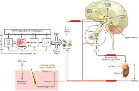
The main adaptive response to sustained systemic stress is mediated by the hypo-thalamic-pituitary-adrenal (HPA) axis (Figure 1). This response begins with the stress-induced hypothalamic production of corticotropin-releasing factor (CRF), which, in the anterior pituitary, activates CRF receptor type 1 (CRF1) and induces the production and release of the proopi-omelanocortin-derived (POMC-derived) adrenocorticotropic hormone (ACTH) (3–5). Psychological stress can also acti-vate the HPA axis and many of the adverse effects of psychological stress on the
body’s functions are secondary to sus-tained HPA activity (3, 4). The expression of CRF and POMC can also be stimulated by proinflammatory cytokines (6), thus involving the immune system in the cen- tral regulation of the HPA axis. Pituitary-derived ACTH stimulates adrenocortical production and secretion of glucocorti- coids such as the steroid hormone cor-tisol (in humans) and corticosterone (in rodents) (4). These glucocorticoids coun-teract the effects of stressors, suppress the immune system, and attenuate the func-tional activity of the HPA axis via feedback inhibition of CRF and POMC expression.
Psychological stress disrupts the natural antimicrobial defenses of the epidermis
[image:3.585.47.531.84.402.2]In this context, the study reported by Aberg and colleagues in this issue of the JCI (7) rep- resents a timely contribution to our under- standing of the deleterious effect of psycho-logical stress on epidermal function. These authors demonstrate psychological stress– induced alterations in the structure and function of the stratum corneum, causing the downregulation of epidermal and adnex-al expression of antimicrobial peptides and an increased severity of cutaneous infection in mice. These effects were accompanied by
Figure 1
increased glucocorticoid production, could be mimicked by systemic glucocorticoid administration, and attenuated by adminis-tration of the CRF1 antagonist antalarmin and also by administration of the glucocor- ticoid receptor antagonist RU-486. Further- more, antalarmin, RU-486, and adrenalecto-my were shown to enhance the otherwise low constitutive expression of antimicrobial pep-tides in these animals. Because homeostasis of the permeability barrier was restored by the use of CRF1 or glucocorticoid receptor antagonists, the authors concluded that psy-chological stress inhibits the antimicrobial action of the epidermal and adnexal com-partments via glucocorticoid-induced HPA axis activation. They also propose what they believe to be a novel therapeutic approach to the treatment of psychological stress disrup-tion of the cutaneous and extracutaneous epithelial barrier defense mechanisms: phar- macological blockade of CRF1 or glucocorti- coid receptors in order to attenuate the activ-ity of the regulatory and/or functional arms of the HPA axis. The major strength of the current work is represented by its conceptual and clini-cal implications (7). However, data on the functional parameters of the HPA axis are limited, since neither plasma nor skin cor-ticosterone, POMC, ACTH, or CRF levels were measured or correlated with circadian rhythm (under physiological conditions, plasma ACTH and glucocorticoid levels fluctuate in a circadian fashion). Future studies, including experiments in geneti-cally modified mice in which the genes encoding CRF1, POMC, or glucocorticoid-regulating enzymes and their receptors are altered, will hopefully provide proof of con-cept of this proposed mechanism. It must also be acknowledged that the use of animal models (rodents) to test the proposed con-nection between protective barrier function, psychological stress, and HPA axis activity is limited because of the fundamental dif-ferences in skin physiology and pathology between rodents and humans. For example, mice are nocturnal species, while human skin is continuously exposed to solar radia- tion during the daytime (8). These differ-ences are strikingly associated with specific anatomic and histologic characteristics of epidermal and adnexal compartments and their biological activities. For instance, dramatic changes in skin morphology and physiologic function have been previously shown to synchronize with alterations in the hair follicle cycle in mice, an effect not seen in human skin (8, 9). Thus, the dynam-ic interactions between the brain and skin (by way of psychological stress) should opti-mally be confirmed in humans.
The cutaneous HPA system
Throughout evolution, the skin — being continuously exposed to hostile biological, chemical, or physical (i.e., solar or thermal energy) factors — had to develop efficient sensory and signaling capabilities to dif-ferentially react to changes in the external environment in order to protect, restore, or maintain the local and global homeostasis that is critical for survival. We have previ- ously proposed that the precise coordina-tion and execution of local homeostatic responses are mediated by a cutaneous neu-roendocrine system, able to reset adaptation mechanisms through rapid (neural) or slow (humoral) pathways acting at the local or systemic level (8). The impact of psychologi-cal stress must be considered in the context of this bidirectional pathway of communi-cation between the brain and skin (10). Human skin indeed expresses elements of the HPA axis including CRF, CRF1, and POMC as well as the processing machinery that generates ACTH, β-endorphin, and melanocyte-stimulating hormone (MSH) peptides (in a regulated and spatiotemporal- ly restricted manner) and their correspond-ing receptors (11, 12). Skin cells also express the enzymatic machinery involved in the sequential metabolism of cholesterol to the steroid hormone pregnenolone and to corti-costeroids, and produce endogenous cortisol and corticosterone (8, 13), albeit at very low levels (14). These elements may be organized in functional, cell type–specific regulatory loops within the structural hierarchy of the HPA axis, (e.g., CRF→CRF1→POMC→
ACTH→cortisol and/or corticosterone), in which CRF-induced production of cortico-steroids is dependent on both CRF1 activity and expression of POMC (8, 12, 13). Environ-mental stressors such as ultraviolet radiation stimulate expression of CRF, POMC, and their corresponding receptors (11, 12, 15, 16), while negative feedback regulation of cutane-ous CRF expression by cortisol has also been demonstrated (17). Thus, differential, CRF1-driven responses in the skin reproduce key features of the central HPA axis at both the whole-tissue and single-cell levels (12, 13).
Merging of the central and peripheral stress response systems
Because the HPA axis coordinates an adap-tive response to counteract the actions of stressors, stabilize organ function, and
restore general homeostasis, one is faced with an intellectual dilemma. How can a system designed to protect whole-body homeostasis (via the HPA axis) endanger the organism’s survival through disruption of epidermal barrier function on multiple levels? I pro-pose that during evolution the fundamental mechanism of the stress response, the HPA axis, developed first in the integument to pro-mote species survival and was then adapted and perfected by the CNS and endocrine sys-tem (Figure 1). This primordial HPA in the integument was composed of CRF, POMC, and steroid signaling pathways that act in concert with the innate immune system in order to create the optimal response against pathogens and other stressors and protect internal homeostasis. In this system, CRF, acting directly through CRF1, stimulates skin immune responses through the stimula-tion of NF-κB to increase production of IL-1, IL-6, and TNF-α (12). This system can also be activated by pathogens in the form of lipo-polysaccharides and bacterial antigens. CRF production is induced, which in turn stimu-lates proinflammatory cytokines through activation of CRF1 (18). This self-amplifying innate immune response is then interrupted by an inhibitory loop mediated by CRF1-acti-vated production of POMC peptides (ACTH,
α-MSH, and β -endorphin, which have anti-inflammatory properties; ref. 19) and finally via corticosteroids induced by ACTH (Figure 1). In the context of epidermal barrier forma-tion, it is significant that activation of CRF1 stimulates keratinocyte survival (perhaps through activation of NF-κB) and increases keratinocyte differentiation (through Ca2+
clearly illustrated by the redundancy in orga-nizational structure of the stress response systems in the CNS and the skin (8, 13). This redundancy has its functional consequences when psychological stress–mediated activa-tion of central HPA axis signaling negatively affects protective and antimicrobial skin barrier function, as is demonstrated in this issue by Aberg et al. (7). These findings will hopefully have clinical impact by stimulat-ing the development of systemic and topical selective receptor antagonists for peptide and steroidal messengers of the HPA axis, as well as topical use of stimulators of cor-tisol metabolic inactivation, and the use of inhibitors of steroidogenesis in order to improve the antimicrobial and protective barrier activity of the skin. This may also be the dawn of a new clinical approach for the treatment of psychological stress–induced inflammatory dermatoses via the use of sys-temic or topical CRF1 antagonists.
Acknowledgments
Comments from Jacobo Wortsman and Curtis Dohan as well as NIH support (grant AR052190) are acknowledged.
Address correspondence to: Andrzej Slo-minski, Department of Pathology and
Laboratory Medicine, Health Science Cen-ter, University of Tennessee, 930 Madison Avenue, Memphis, Tennessee 38163, USA. Phone: (901) 3741; Fax: (901) 448-6979; E-mail: [email protected].
1. Elias, P.M. 2005. Stratum corneum defensive functions: an integrated view. J. Invest. Dermatol.
125:183–200.
2. Arck, P.C., Slominski, A., Theoharides, T.C., Peters, E.M., and Paus, R. 2006. Neuroimmunology of stress: skin takes center stage. J. Invest. Dermatol.
126:1697–1704.
3. McEwen, B.S. 2007. Physiology and neurobiology of stress and adaptation: central role of the brain.
Physiol. Rev. 87:873–904.
4. Chrousos, G.P. 1995. The hypothalamic-pituitary-adrenal axis and immune-mediated inflammation.
N. Engl. J. Med. 332:1351–1362.
5. Vale, W., Spiess, J., Rivier, C., and Rivier, J. 1981. Characterization of a 41-residue ovine hypothalam-ic peptide that stimulates secretion of corticotropin and beta-endorphin. Science. 213:1394–1397. 6. Turnbull, A.V., and Rivier, C. 1995. Regulation
of the HPA axis by cytokines. Brain Behav. Immun.
9:253–275.
7. Aberg, K.M., et al. 2007. Psychological stress down- regulates epidermal antimicrobial peptide expres-sion and increases severity of cutaneous infections in mice. J. Clin. Invest. 117:3339–3349. doi:10.1172/ JCI31726.
8. Slominski, A., and Wortsman, J. 2000. Neuroendo-crinology of the skin. Endocr. Rev. 21:457–487.
9. Stenn, K.S., and Paus, R. 2001. Controls of hair fol-licle cycling. Physiol. Rev. 81:449–494.
10. Paus, R., Theoharides, T.C., and Arck, P.C. 2006.
Neuroimmunoendocrine circuitry of the ‘brain-skin connection’. Trends Immunol. 27:32–39. 11. Slominski, A., Wortsman, J., Luger, T., Paus, R.,
and Solomon, S. 2000. Corticotropin releasing hormone and proopiomelanocortin involvement in the cutaneous response to stress. Physiol. Rev.
80:979–1020.
12. Slominski, A., et al. 2006. Corticotropin releasing hormone and the skin. Front. Biosci. 11:2230–2248. 13. Slominski, A., Wortsman, J., Tuckey, R.C., and Paus,
R. 2007. Differential expression of HPA axis homolog in the skin. Mol. Cell. Endocrinol. 265–266:143–149. 14. Slominski, A., et al. 2004. A novel pathway for
sequential transformation of 7-dehydrocholesterol and expression of the P450scc system in mamma-lian skin. Eur. J. Biochem. 271:4178–4188.
15. Slominski, A., Tobin, D.J., Shibahara, S., and Worts- man, J. 2004. Melanin pigmentation in mamma-lian skin and its hormonal regulation. Physiol. Rev.
84:1155–1228.
16. Zbytek, B., Wortsman, J., and Slominski, A. 2006. Characterization of a ultraviolet B-induced corti- cotropin-releasing hormone-proopiomelanocor-tin system in human melanocytes. Mol. Endocrinol.
20:2539–2547.
17. Ito, N., et al. 2005. Human hair follicles display a functional equivalent of the hypothalamic-pitu-itary-adrenal axis and synthesize cortisol. FASEB J.
19:1332–1334.
18. Zbytek, B., and Slominski, A.T. 2007. CRH medi-ates inflammation induced by lipopolysaccharide in human adult epidermal keratinocytes. J. Invest. Dermatol. 127:730–732.
19. Luger, T., Paus, R., Lipton, J., and Slominski, A. 1999. Cutaneous neuromodulation: the proopiomelano-cortin System. Ann. N. Y. Acad. Sci. 885:1–479.
20. Bohm, M., Luger, T.A., Tobin, D.J., and Garcia-Bor-ron, J.C. 2006. Melanocortin receptor ligands: new horizons for skin biology and clinical dermatology.
J. Invest. Dermatol. 126:1966–1975.
OX40 signaling directly triggers
the antitumor effects of NKT cells
Dapeng Zhou
Department of Melanoma Medical Oncology, Division of Cancer Medicine, University of Texas MD Anderson Cancer Center, Houston, Texas, USA.
Pathways involving the costimulatory molecule OX40 and OX40 ligand
(OX40L) enhance tumor rejection. It was presumed that this effect was
medi-ated by changes in DCs and/or T cells. In this issue of the
JCI
, Zaini et al.
report that, in mice, intratumoral injection of DCs genetically modified to
express OX40L suppressed the growth of a preexisting melanoma by directly
triggering an antitumor NKT cell response (see the related article beginning
on page 3330). This work suggests that the intratumoral NKT cell
popula-tion may be harnessed for cancer immunotherapy and that OX40
costimula-tion may be used as a unique trigger of the antitumor activity of these cells.
The effective T cell response to a tumor antigen requires not only stimulation by
Nonstandard abbreviations used: α-GalCer, α -galactosylceramide; OX40L, OX40 ligand; OX40L-DC, OX40L gene–transduced DC.
Conflict of interest: The author has declared that no conflict of interest exists.
Citation for this article: J. Clin. Invest. 117:3169–3172 (2007). doi:10.1172/JCI33976.
the tumor antigen itself via the TCR signal- ing pathway, but also costimulatory mol- ecules (1). In the early stage of T cell stimu-lation, the costimulatory molecule CD28 is required for T cell proliferation and sur-vival through recognition of B7 molecules expressed by DCs. “Second-wave” costim-ulatory molecules, represented by TNF receptor family members, are required by
activated T cells for completion of their dif-ferentiation programs and exertion of their antitumor functions such as the release of cytokines and cytotoxic granules. The TNF receptor family of costimulatory molecules continues to be of interest in an effort to identify candidate targets for the develop-ment of immunotherapeutics. Activation of many of these signaling pathways has caused tumor rejection in preclinical and clinical studies (2).
OX40 and OX40 ligand in antitumor immunity
The OX40 costimulatory molecule is a member of the TNF receptor family and is expressed on activated CD4+ T cells and
CD8+ T cells (1). OX40 ligand (OX40L)