EXTRACTION OF RETINAL BLOOD VESSEL USING
ARTIFICIAL BEE-COLONY OPTIMIZATION
1KAVYA K,2DECHAMMA M.G,3SANTHOSH KUMAR B.J
1Assistant Professor, Department of Computer Science, Amrita Vishwa Vidyapeetham, Amrita University,
Mysuru, India
2, 3P G Scholars, Department of Computer Science, Amrita Vishwa Vidyapeetham, Amrita University,
Mysuru, India
E-mail: 1[email protected],2[email protected],3[email protected]
ABSTRACT
Retinal blood vessel Extraction in retinal images allows early diagnosis of disease and is useful in detecting ocular disorders and helps in laser surgery. Automating this process provides several benefits including minimizing subjectivity and eliminating a painstaking. This paper proposes an automated retinal blood vessel segmentation approach based on Fuzzy C-Means (FCM) clustering and then performed extraction using Artificial Bee-colony (ABC) to improve the accuracy of segmented image. FCM allocate the values of membership to the pixels instead of separating the pixels as in hard clustering problem and the clustering is optimized using ABC swarm based optimization algorithm, finally the system classify the images according to the level of damage in blood vessel using support vector machine (SVM). The performance was evaluated on DRIVE database and an accuracy of 96.35% was obtained.
Keywords:Fundus Camera, Clustering, Fuzzy C-Means, Artificial Bee-Colony, Support Vector Machine.
1. INTRODUCTION
Retinal blood vessel damage can be caused by many diseases, which include Diabetic retinopathy, retinal vein occlusion, and glaucoma. In some cases retinal diseases can even leads to permanent blindness. Until it becomes too critical the patient might not notice. Early diagnosis of such diseases is necessary in detecting disorders. Current methods to detect such vessel damage require manual intervention and trained ophthalmologists. Retinal blood vessel morphology will be an important indicator of such diseases, which includes segmentation of blood vessels from the retinal images obtained from Fundus camera. The low-contrast images at the retina results in narrow blood vessels of the retina which are difficult to extract, hence accurate segmentation of blood vessel is necessary. Segmentation aids the screening process of retinopathy by reducing the number of incorrect decisions taken by the doctors in micro aneurysm detection. Detecting abnormalities in the early stages of the disease is critical because they are the indicators of sight-threatening retinopathy [1].
There are mainly three automated based approaches for blood vessel segmentation [2]: they are thresholding method, machine trained classifiers and tracking method. In the first method, many different operators are used to increase the contrast between vessel and background, such as Laplacian operators, Sobel operators, Gaussian filters modeling the gray cross-section of retinal blood vessel. Then the gray threshold is selected to determine the vessel and it is crucial, because small threshold make more noise and gray threshold causes loss of some fine vessels, adaptive or local threshold is used. Blood vessel tracking is another technique for blood vessel segmentation, whereby vessel Center locations are automatically sought along the blood vessel longitudinal axis from a starting point to the ending point.
algorithms based on bio-inspired methods have been established. One such bio-inspired method is the ABC algorithm, which is a population-based optimization algorithm [3]. Once the blood vessels are extracted it is important to classify them, the Support Vector Machine (SVM) algorithm classifies a data which is a common task in machine learning. A SVM constructs a hyperplanes in a high or infinite dimensional space, which can be utilized for classification. Larger the distance between hyperplane and nearest training- data point of any class higher the separation rate, this method is used to classify the image on the basis of the level of extent of vessel damage.
2. LITERATURE SURVEY
Numerous attempts are described in the literature for Segmentation of blood vessel. Some of the approaches that are in relevance with the proposed work are discussed in brief.
R. Radha et.al [4] proposed Retinal image examination through efficient detection of exudates and recognizes whether the retina is normal or abnormal. The contrast image is enhanced by curvelet transform. Hence, morphology operators are applied to the enhanced image in order to find out the retinal image ridges. A simple thresholding method along with opening and closing operation specify the remained ridges belonging to blood vessels. The clustering method is used for successful detection of exudates of eye. Here the threshold value is assumed which are not always the criteria.
J. Benadict Raja et.al [5] proposed parallel method which has an infrastructure (network of computer) to segment a high resolution image and the data classification: line operator and features based on Gabor wavelet. At each pixel of retinal image we build a partition scheme which reduces the idle time of the participating nodes and allows the parallel computation; therefore the speed of vessel segmentation is increased. The use of enhancement/threshold segmentation based algorithm further enhances the accuracy of segmentation.
Reza Kharghanian et.al [6] have proposed a method for segmenting blood vessels from retinal images and extract two sets of features for image feature vector consisting of the pixel intensity, two features from orthogonal line operators and four features from Gabor wavelet transform in different scales. Comparing the result of classification using two classifiers-SVM and Bayesian.
Mendonça et.al [7] have proposed vessels segmentation algorithm using blood vessel
centerlines followed by the blood vessel filtering process. Multi scale morphological enhancement technique was used to refine the contrast of the blood vessels. Wilcoxon matched pairs testing algorithm was improved to prove the accuracy of their results.
Manoj et al. [8] have used feed forward back propagation neural network classifier for segmentation of blood vessel. The features like morphological transformation vectors, gradient vector regions and line strength vectors were extracted from the retinal images.
Fraz et al. [9] used ensemble classifier to segment the vessels. The Gabor transform and gradient vector field were developed and used as the feature in ensemble classifier. The authors achieved 72.62% sensitivity, 97.64% specificity.
Marín et al. [10] have proposed grey level and moment feature based supervised classifier for blood vessel segmentation. The background homogenization was applied to improve the contrast of the retinal blood vessels. The authors achieved 94.52% of accuracy in DRIVE dataset and 69.44% of sensitivity.
Soares et al. [11] have proposed pixel-processing-based methods and tracking-based methods. The vessel segmentation is performed by the pixel-processing-based approach in a two-pass operation. First, using detection processes such as morphological preprocessing techniques and adaptive filtering the appearance of the vessel is enhanced. The second operation is the identification of the vessel structure using thinning or branch-point operations to classify a pixel as a vessel or background (BG) to improve the accuracy of the vessel Segmentation.
3. PROPOSED METHODOLOGY
Input Fundus Image
Classification
Fig.1. Proposed Blood Vessel Extraction System
Perform Pre-processing
Enhanced Vessel Segmentation
3.1 Pre-Processing of Fundus Image
The retinal images in the data set are often noisy and badly illuminated because of unknown noise and camera settings. Also there is an extensive variation of color of retina from patient to patient. Thus the various preprocessing steps are applied to Fundus images, which include filtering and green channel extraction. In order to highlight blood vessel the presence of optic disc (OD) region should be found and removed. To remove optic disc we create a mask and mask the optic disc region.
Once this is done the image is filtered to reduce noise using median filtering and green channel of the image is extracted because the green-channel image provides maximum local contrast between the background and foreground and the image is converted to gray scale for segmentation.
3.2 Image Segmentation Using FCM
The goal of segmentation is to simplify or change the representation of an image into something that is more meaningful and easier to inspect. The features involved are intensity histogram, mean, variance, energy, texture etc. Segmentation method depends on the level of detail that the application requires. Here we are using Fuzzy C-Means (FCM) clustering for blood vessel segmentation.
FCM is a method of clustering which enables one piece of data to belong to two or more clusters and it is commonly used in pattern recognition. Basically this algorithm works by allocating a membership to each data point corresponding to each cluster centre on the basis of distance between the cluster and the data point. More the data is close to the cluster centre more is its membership towards the particular cluster centre. The iterative unsupervised FCM algorithm is the most extensively used clustering algorithm for image segmentation. The membership matrix is defined by equation (1),
=
∑ ( )
( )
Here value of Uij is between [0, 1], dij is the Euclidean distance between ith centre and jth data point and value of m is between [1, ∞] is a weighting exponent.
3.3 Extraction Using Artificial Bee-Colony Despite the quality of FCM, it is delicate to initial states and gets stuck in local optima solutions. In addition, a FCM clustering problem is
a combinatorial optimization problem. In order to address these issues extraction is performed using Artificial Bee-Colony optimization algorithm.
The Artificial Bee-Colony optimization algorithm is inspired from the intelligent foraging behavior of honey bee swarms. Its power is its quality/intelligibility and its robustness. In the ABC algorithm, each food source position represents a solution to a particular problem and the amount of nectar in a food source represents the objective function (the fitness) of the solution. In order to calculate the fitness values of the solutions the following equation Fitiis employed,
+ ( , ) ( , ) = ∣∣ − , ∣∣
……… (2)
Probability value Probifor the solution by means of their fitness values is employed using equation (3),
= ( )
∑ ( )
Here NP is a total number of populations.
3.4 Classification Using SVM
Support Vector Machine (SVM) performs the robust nonlinear classification and is defined by a separating hyperplane. SVM is independent of the dimensionality of the feature space and that results obtained are very accurate. It performs better than other classifiers even with small numbers of available training samples. In this project we perform classification on the segmented image depending on the extent of blood-vessel damage. Extracted features of clustering algorithms are given as input to the SVM classifier and classify as critically damaged class and moderately damage class [12].
Algorithm flow of Proposed Methodology:
1. Initialize the real number RN, number of cluster CN, the size of the population NP, the value of limit, the value of MR and the maximum cycles number MN.
2. Create initial population Si using the below equation:
, = + Ø, × ( − )
3. Compute the membership matrix Uij
4. Evaluate the population using equation(2)
5. For each employed bee
6. Produce new solution.
7. Compute the membership matrix Ui,k
8. Calculate the fitness using equation(2)
9. Apply the greedy selection process
10. Compute the probablity value Probifor the solutions.
11. Select a solution Si depending on Probi
12. Repeat step 6, 7, 8 and apply the greedy selection process.
13. If there is unused solution then replaces that solution with a new randomly produced solution for the scout using equation 3.
14. Assign cycle = cycle + 1
15. Memorize the best solution (best cluster centers) achieved yet
16. Until cycle = Maximum cycles Number
17. Do the segmentation by allocating each pixel to the cluster for which the membership value is higher.
18. Once the segmentation is done we extract the feature with normal image, critically damage and moderately damage image.
19. These features are stored in a template form.
20. The next image is extracted and SVM is used to match the features and classify the segmented image on the basis of the features.
4. RESULT AND ANALYSIS
The Image processing is simulated in the MATLAB and the test of proposed technique are
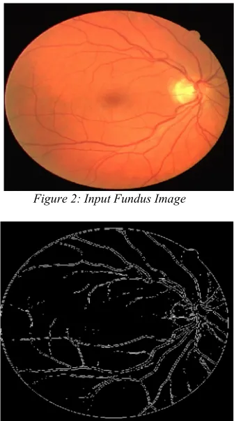
[image:4.612.332.500.195.497.2]performed with respect to the vessel extraction accuracy using publicly available DRIVE database and the images obtained from eye hospitals. Figure.3 shows the Blood vessel segmentation using Fuzzy C-Means and figure.4 shows the extract features of blood vessel using Artificial Bee-Colony [ABC] Optimization and classification is done successfully using SVM algorithm.
Figure 2: Input Fundus Image
[image:4.612.333.502.516.670.2]Figure 3: Vessel Segmentation using FCM
Figure 4: Vessel Extraction using ABC
Sensitivity and Specificity as a measurement of Accuracy using the following equation.
= ( )
.
= ( )
= ( )
True positive [TP] = Number of abnormal pixels correctly detected.
False Positive [FP] = Number of normal pixels which are detected erroneously as abnormal pixels. False Negative [FN] = Number of abnormal pixels that are not detected correctly.
True Negative [TN] = Number of normal pixels which are correctly identified as normal pixels.
Table1: Comparison of our method with some different Extraction method
Algorithm Accuracy
Gabor 0.9377
Minimal Path 0.9432
Artificial Bee-Colony 0.9642
5. CONCLUSION
The Extraction of retinal blood vessel has a significant role in early diagnosis of disease. In this paper, we have examined the application of Artificial Bee Colony algorithm to obtain better quality of fuzzy clustering result and the algorithm is used to extract the segmented blood vessels from the retinal image, also we classify the extracted image using SVM classifier.
Modification of the existing algorithm can be done to improve the accuracy of the method and to improve the exploitation power of the ABC algorithm. The overall Sensitivity, Specificity and Accuracy are 96.35%, 89.50%, 96.42%.
REFERENCES
[1] X. Xu, Niemeijer M, Song Q, Sonka M, Garvin M.K. “Vessel boundary delineation on Fundus images using graph-based approach,” IEEE Transactions on Medical Imaging, vol. 30, no. 6, June 2011. [2] S. Chaudhuri, S. Chatterjee, N. Katz, M.
Nelson, and M. Goldbaum. “Detection of blood vessels in retinal images using two-dimensional matched filters,” IEEE
Transactions on Medical Imaging. Vol. 8 no.3:263269, September 1989.
[3] D. Karaboga, An idea based on honey bee swarm for numerical optimization, technical Report TR06, Erciyes University Engineering Faculty, Department of Computer Engineering, 2005.
[4] R. Radha and Bijee Lakshma, “Retinal image analysis using morphological process and clustering technique”, Signal & Image Processing: An International Journal (SIPIJ) Vol.4, No.6, December 2013.
[5] J. Benadict Raja, C.G.Ravichandran, on “Blood Vessel Segmentation for High Resolution Retinal Images”, IJCSI International Journal of Computer Science Issues, Vol. 8, Issue 6, No 2, November 2011.
[6] Reza Kharghanian and Alireza Ahma dyfard, “Retinal Vessel Segmentation Using Gabor Wavelet and Line Operator”, International Journal of Machine Learning and Computing, Vol. 2, No. 5, October 2012.
[7] A. M. Mendonça and A. Campilho, “Segmentation of Retinal blood vessels by combining the detection of centerlines and morphological reconstruction”, IEEE Transactions on Medical Imaging, vol. 25, no. 9, pp. 1200–1213, 2006.
[8] S. Manoj, Muralidharan, “Neural network based classifier for retinal blood vessel segmentation”, International Journal of Recent Trends in Electrical & Electronics Engineering, vol. 3, no. 1, pp. 44–53, December 2013.
[9] M. M. Fraz, P. Remagnino, “An ensemble classification-based approach applied to retinal blood vessel segmentation,” IEEE Transactions on Biomedical Engineering, vol. 59, no. 9, pp. 2538–2548, 2012. [10] D. Marín, A. Aquino, M. E.
Transactions on Medical Imaging, vol. 30, no. 1, pp. 146–158, 2011.
[11] J. V. B. Soares, J. J. G. Leandro, “Retinal blood vessel segmentation using the 2-D Gabor wavelet and supervised classification,” IEEE Transactions on Medical Imaging, vol. 25, no. 9, pp. 1214– 1222, 2006.
[12] Athira R V, Ferlin Deva Shahila D, “Detection of Retinal Hemorrhage Using Splat Feature Classification Technique,” Journal of Engineering Research and Applications ISSN : 2248-9622, Vol.4, Issue 1(Version 3), January 2014, pp.327-330.
[13] Miss. Vaibhavi B. Patil1 and Mr. Vivek Rai, “blood vessel segmentation from color retinal images using unsupervised texture classification,” International Journal of Technical Research and Applications e-ISSN: 2320-8163, www.ijtra.com Volume 4, Issue 1, (January-February, 2016).