INTELLIGENT SEGMENTATION OF MR BRAIN
STRUCTURES USING SUPERVISED CONTEXTUAL
CLUSTERING
1S.P. RENJITH, 2DR. N. KESAVAN NAIR
1
Research scholar, Anna University, India
2
Professor, CSI Institute of Technology, India
ABSTRACT
Image segmentation plays a vital role in medical imaging applications. Developing a robust and efficient algorithm for medical image segmentation has been a demanding area of growing research of interest during the last two decades. Image processing techniques provide a good tool for improving the manual screening of samples of Brain. This study presents a novel intelligent approach for the segmentation of anatomical brain structures in Magnetic Resonance Images (MRI) using supervised contextual clustering method. Matlab software ‘region props’ function has been used as one of the criteria to show the performance of the proposed segmentation method. The CC segmentation shows more segmented regions with less discontinuity within the objects present in the Brain image. The segmented results are compared with the conventional algorithms
Keywords: Image Segmentation, Medical Imaging, Brain Image, CC Segmentation, MRI
1. INTRODUCTION
Segmentation of tissues present in the medical images is an initial step in many medical imaging applications which includes three dimensional volume visualization, the detection of pathology and structure-functions present in it inorder to map both the scientific and clinical investigation. Accurate segmentation of magnetic resonance images (MRI) is essential in many neuroimaging applications [1]. Quantitative analysis of anatomical brain tissue such as white matter (WM), gray matter (GM) and cerebrospinal fluid (CSF) is important for clinical diagnosis, therapy of neurological diseases and for visualization and analysis [2]. Because of the advantages of MRI over other diagnosis of images, many researches in medical image segmentation applied its use for MR images. The different types of MR images obtained from the different excitation sequences, also called multispectral images, can provide different image intensity information for a given anatomical region and subject [7]. As we know, manual segmentation of MR brain images by an expert for all types of MR modalities involved in studies is not only exceedingly time consuming, but also exhausting for experts, which can lead to human errors. Therefore, an automatic segmentation method is necessary. Various methods are available for MRI image segmentation which are present in the literature [2-7].
A novel method for segmenting the region of interest present in the MRI brain images was proposed by Jun Kong et al in [8]. In their method, initially they reduced the noise present in it using a versatile wavelet-based filter. Then a watershed algorithm is applied to the brain tissues as an initial segmenting method in which the white matter along with some outer tissues are segmented. Inorder to reduce the outer tissues, further, they have used fuzzy clustering algorithm which removes the over segmentation.
In [11], the intensity of each and every tissue present in the brain image is modeled by a Parzen density fitted in the voxels which were selected from an affine-registered atlas. It is important stress that each and every atlas does not exist for the data at hand. Example of two such cases are brain data with pathologies or brain data obtained from young infants without any pathologies. In addition, conventional method improves the segmentation smoothness inorder to model the neighboring voxels using a Markov random field (MRF) based statistical spatial model [12]. These MRF-based algorithms are computationally intensive in nature requiring critical settings in its parameter. It is possible to use MRF with predefined settings, which is computationally faster but possibly less in accurate.
This paper is organized as follows. Section II explains an overview of the materials and methods present in this image segmentation process. Section III presents the proposed methodology in detail. Section IV provides the experimental results, and Section V contains the conclusion on this work and suggests some possible future enhancements.
2. MATERIALS AND METHODS
The primary objective of this work is to develop a Computer Aided System for Segmentation of white matter from the Magnetic Resonance images (MRI). Our proposed method does not affects the objects present in the lung image. The main advantage of this approach is that we can get the holes and objects present in the original image after segmentation without any change in its size and shape. This proposed approach has two main sections namely 1) Segmentation based on Contextual clustering and 2) Objects detection and estimation based on Matlab region properties.
2.1 Contextual Clustering Based Segmentation
Image segmentation plays an important role in image analysis and computer vision and it is considered as one of the major obstruction in the development of image processing technology. Recently, there has been considerable interest among researchers in
statistical clustering techniques in image segmentation was inspired by the statistical based methods. These methods were developed to study the equilibrium properties of large, lattice based systems consisting of interacting components as identical. In a clustering technique for image segmentation, each pixel is associated with one of the finite number of categories to form disjoint regions.
The contextual clustering based algorithms are assumed to be drawn from standard normal distribution. It segments a data into category 1 (ω0) and category 2 (ω1).
The following are the steps adopted for implementing the contextual clustering algorithm for segmenting the white matter from the Brain MRI images.
(i) Define decision parameter Tcc (positive)
and weight of neighborhood information β (positive). Let Nn be the total number of
data in the neighborhood. Let Zi be the
data itself, i.
(ii) Classify data with zi>Tα to ω1 and data to
ω0. Store the classification to C0 and C1.
(iii) For each data i, count the number of data ui, belonging to class ω1 in the
neighborhood of data i. Assume that the data outside the range belong to ω0.
(iv) Classify data with
to ω1 and other data to ω0. Store the
classification to variable C2.
(v) If C2 ≠C1 and C2 ≠ C0, copy C1 to C0, C2
to C1 and return to step iii, otherwise stop
and return to C2.
The contextual clustering implementation is as follows:
Step 1: Read a Pattern (Brain image feature).
Step 2: Sort the values of the Pattern.
Step 3: Find the Median of the Pattern Cm.
Step 4: Find the number of values greater than
the Median Values, Um.
Step 5: Calculate CC using Cm + (beta/Tcc) *
(Um – (bs/2)).
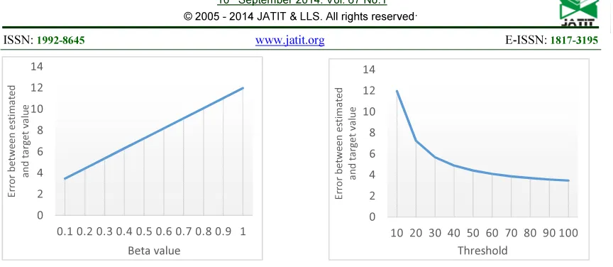
Fig. 1 Impact Of Beta Value In CC Estimation Fig. 2 Impact Of Threshold Value In CC Estimation
Figure 1 shows a steady increase in the error as the beta value changes from 0.1 to 1. Hence, lower beta value is preferred for better estimation by CC. Figure 2 shows a steady decrease in the error as the threshold value changes from 10 to 100. Hence, higher threshold value is preferred for better estimation by CC.
3. EXPERIMENTAL RESULTS AND DISCUSSION
[image:3.595.83.515.489.657.2]Brain MRI images have been considered in this presentation. Magnetic resonance images of different patients have been considered. The CC segmented results along with its original images has been presented in Table 1
Table 1 Segmented Results Of Different Brain Datasets
No Brain Image Segmented result
1
0 2 4 6 8 10 12 14
0.1 0.2 0.3 0.4 0.5 0.6 0.7 0.8 0.9 1
Er
ro
r b
etween
e
st
ima
ted
a
n
d
t
a
rg
et v
a
lu
e
Beta value
0 2 4 6 8 10 12 14
10 20 30 40 50 60 70 80 90 100
Er
ro
r
b
et
ween
es
ti
m
a
ted
a
n
d
ta
rg
e
t v
a
lu
e
2
3
4
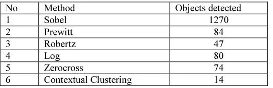
Except CC method, in all other segmentation methods, the number of objects are more and there are some objects segmented are not clear. Matlab ‘bwlabel’ function has been used and the number objects for each method is shown in
[image:5.595.133.463.177.353.2]Table 1. In addition to bwlabel’, the ‘Region props’ command has been used to find out correct number of segmented objects.
Figure 3: Comparison Of Number Of Objects In Each Segmentation Methods
Earlier researchers had used different metrics to evaluate the segmentation accuracy. In this paper, we have used ‘bwlabel’ and ‘Region props’ of Matlab to evaluate the accuracy of segmentation and from the experimental results,
it has been found that Contextual Clustering based segmentation gives much better segmentation accuracy when compared to that of remaining segmentation methods mentioned in this work.
Figure 4. Comparison Of Total Area Of Pixels In Each Segmentation Methods Table 1 Segmentation Accuracy For Different Methods
No Method Objects detected
1 Sobel 1270
2 Prewitt 84 3 Robertz 47
4 Log 80
5 Zerocross 74 6 Contextual Clustering 14
0 1000 2000 3000 4000 5000 6000
Original Sobel Prewitt Roberts Log Zerocross CC
N
o
o
f
o
bjec
ts
Segmentation methods
0 5000 10000 15000 20000 25000 30000
Original Sobel Prewitt Roberts Log Zerocross CC
To
tal
A
reao
f al
l o
bj
ec
ts
, pi
xel
s
[image:5.595.133.465.445.609.2] [image:5.595.167.434.647.733.2]4. CONCLUSION
In this paper we have proposed a new method for segmenting brain MR images based on supervised contextual clustering method. The main purpose of proposing this contextual clustering based segmentation method is to improve the segmentation accuracy by reducing the false segmentation. The method was validated both on simulated and real brain datasets, and the results were compared with state-of-the-art algorithms. We also represented that using the proposed framework, segmentation of the normal tissues is not degraded by the presence of abnormal tissues. Our proposed approach has been found more efficient when compared with other conventional segmentation algorithms. Matlab region properties are used for the estimation of the white matter segmentation accuracy.
REFERENCES
[1]. R. Larsen, M. Nielsen, and J. Sporring, “Atlas Guided Identification of Brain Structures by Combining 3D Segmentation and SVM Classification”MICCAI-2006, pp. 209–216, 2006
[2]. Pham, D., Xu, C., Prince, Current methods in medical image segmentation. Annual Review of Biomedical Engineering 2
(2000) 315–337
[3] Alan Wee-Chung Liew, Hong Yan: An Adaptive Spatial Fuzzy Clustering Algorithm for 3-D MR Image Segmentation, IEEE Transaction on Medical Imaging, vol 22, No 9 (2003). [4] Chris A. Cocosco, Alex P. Zijdenbos, Alan
C. Evans: A fully automatic and robust brain MRI tissue classification method. IEEE Transaction on Medical Image Analysis, vol.7, (2003) 513-527.
[5] K.V. Leemput, F. Maes, D. Vandermeulen, P. Suetens, A unifying framework for partial volume segmentation of brain MR images, IEEE Trans. Med. Imaging 22 (2003) 105–119.
[6] V. Barra, J. Boire, Automatic segmentation of subcortical brain structures in MR images using information fusion, IEEE Trans. Med. Imaging 20 (2001) 549–558. [7] Weibei Dou, Su Ruan, Yanping Chen ,
Daniel Bloyet , Jean-Marc Constans, “framework of fuzzy information fusion for the segmentation of brain tumor tissues on
MR images”, Image and Vision Computing, vol 25, pp- 164–171, 2007. [8] Jun Kong, Jianzhong Wang, Yinghua Lu, “A
Novel Approach for Segmentation of MRI Brain Images”, IEEE MELECON 2006, pp- 525-528.
[9] Arnaldo Mayer* and Hayit Greenspan, “An Adaptive Mean-Shift Framework for MRI Brain Segmentation”, IEEE TRANSACTIONS ON MEDICAL IMAGING, VOL. 28, NO. 8, 2009, pp-1238-1250.
[10] P. Anbeek, K. L. Vincken, G. S. van Bochove, M. J. P. van Osch, and J. van der Grond, “Probabilistic segmentation of brain tissue in MR imaging,” NeuroImage, vol. 27, pp. 795–804, 2005.
[11] S. K. Warfield, M. Kaus, F. A. Jolesz, and R. Kikinis, “Adaptive, template moderated, spatially varying statistical classification,”
Med.Image Anal., vol. 4, pp. 43–55, 2000. [12] Y. Zhang, M. Brady, and S. Smith,